Your new post is loading...

|
Scooped by
Gilbert C FAURE
August 8, 10:37 AM
|
What Fan et al. discovered
This paper shows something even more surprising.
Macrophages can obtain cytosolic proteins from living cells
without
-> killing the cell
-> inducing apoptosis
-> engulfing the cell
-> relying on classical phagocytosis
Instead, they remove extremely tiny vesicles from living cells.
While
-> The donor cell remains alive.
Antigen uptake: Breaking convention Subscribe to RSS feed
Yazmina Quintana-Perez; Livia Lacerda Mariano; Ana-Maria Lennon-Duménil
Trends in Immunology, Copyright © 2026 Elsevier Ltd
Article in Press : Corrected Proof
Original paper: Fan, A.C. et al. (2026) Submicrometre sampling of living cells by macrophages. Nature 654, 495–503
|
|
Scooped by
Gilbert C FAURE
March 25, 2:30 PM
|
🛡️ IMMUNE SYSTEM – COMPLETE ANATOMY
🔹 What is Immune System?
The immune system is the body’s defense system that protects against:
Bacteria 🦠
Viruses 🧬
Fungi 🍄
Parasites 🐛
👉 It identifies “self” vs “non-self” and destroys harmful invaders.
🧠 1. MAIN COMPONENTS
🔬 A. Primary Lymphoid Organs
Where immune cells are formed & matured:
✅ Bone Marrow
Produces all blood cells
B-cells mature here
✅ Thymus
T-cells mature here
Active mainly in childhood
🧪 B. Secondary Lymphoid Organs
Where immune response happens:
Lymph nodes
Spleen
Tonsils
Peyer’s patches (intestine)
🧬 2. TYPES OF IMMUNITY
🔹 1. Innate Immunity (Natural)
Present from birth
Fast response
Examples:
Skin barrier
Stomach acid
White blood cells
🔹 2. Adaptive Immunity (Acquired)
Develops over time
Specific + memory-based
Types:
Active (infection/vaccine)
Passive (mother antibodies)
🧪 3. IMMUNE CELLS
🧫 White Blood Cells (WBCs)
🔥 Neutrophils
First responders
Kill bacteria
🧬 Lymphocytes
B-cells → produce antibodies
T-cells → kill infected cells
🧹 Macrophages
Eat pathogens (phagocytosis)
⚔️ Natural Killer (NK) Cells
Kill virus-infected cells
🧴 4. ANTIBODIES (IMMUNOGLOBULINS)
Produced by B-cells
IgG → long-term protection
IgA → mucosal surfaces
IgM → first response
IgE → allergies
IgD → B-cell function
🔥 5. IMMUNE RESPONSE (STEP-BY-STEP)
Pathogen enters body
Recognized as foreign
WBCs activated
Antibodies produced
Pathogen destroyed
Memory cells formed
👉 Next time → faster response ⚡
⚙️ 6. IMPORTANT FUNCTIONS
Protects from infections
Removes dead cells
Fights cancer cells
Creates memory for future protection
🧪 7. COMMON DISORDERS
🔴 Autoimmune Diseases
Body attacks itself
Example: Rheumatoid arthritis
🔴 Immunodeficiency
Weak immune system
Example: HIV/AIDS
🔴 Allergies
Overreaction to harmless substances
🧾 QUICK SUMMARY
FeatureImmune SystemFunctionDefenseCellsWBCsOrgansBone marrow, thymus, lymph nodesTypesInnate & Adaptive
👉 Interested in Medical Coding training?
DM me for complete details. I’ll guide you step-by-step. Feel free to ask your doubts. 9052201814
#MedicalCoding
#HealthcareCareers
#MedicalCoder
#CodingTraining
#CareerGrowth
#OnlineTraining
#JobOpportunity
#WorkFromHome
|
|
Scooped by
Gilbert C FAURE
January 1, 4:38 AM
|
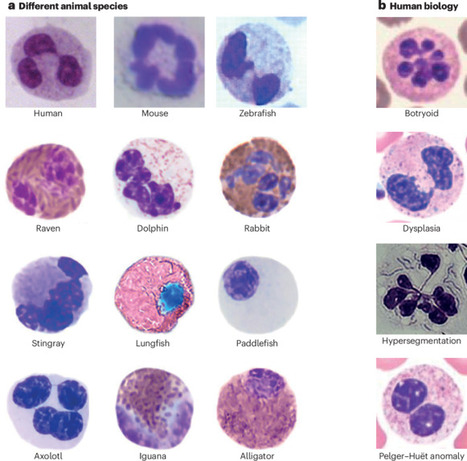
Since the discovery of the segmented shape of the neutrophil nucleus, scientists have pondered its physiological relevance. Yet, to this day, neither the functional relevance nor the molecular mechanisms underlying the segmentation of the neutrophil nucleus are fully understood. Some experimental evidence supports a role of nuclear segmentation in efficient neutrophil migration but its impact on key neutrophil functions, such as phagocytosis, degranulation or production of reactive oxygen species, remains unclear. Nonetheless, the role of nuclear shape in remodelling chromatin and regulating gene expression has high potential for biological and translational relevance. This Review aims to compile and connect the current studies on the neutrophil nucleus, while also discussing studies in other cell types that could inform us about the relevance or mechanisms of nuclear segmentation. In this Review, the authors consider a long-standing immunological conundrum — why do neutrophils have a segmented nucleus? They discuss the mechanisms that may underlie segmentation of the neutrophil nucleus and explain how nuclear segmentation may affect neutrophil functions, including migration and phagocytosis.
|
|
Scooped by
Gilbert C FAURE
December 23, 2024 1:06 PM
|
|
|
Scooped by
Gilbert C FAURE
June 6, 2024 2:22 AM
|
Phagocytosis is the process by which myeloid phagocytes bind to and internalize potentially dangerous microorganisms1. During phagocytosis, innate immune receptors and associated signalling proteins are localized to the maturing phagosome compartment, forming an immune information processing hub brimming with microorganism-sensing features2–8. Here we developed proximity labelling of phagosomal contents (PhagoPL) to identify proteins localizing to phagosomes containing model yeast and bacteria. By comparing the protein composition of phagosomes containing evolutionarily and biochemically distinct microorganisms, we unexpectedly identified programmed death-ligand 1 (PD-L1) as a protein that specifically enriches in phagosomes containing yeast. We found that PD-L1 directly binds to yeast upon processing in phagosomes. By surface display library screening, we identified the ribosomal protein Rpl20b as a fungal protein ligand for PD-L1. Using an auxin-inducible depletion system, we found that detection of Rpl20b by macrophages cross-regulates production of distinct cytokines including interleukin-10 (IL-10) induced by the activation of other innate immune receptors. Thus, this study establishes PhagoPL as a useful approach to quantifying the collection of proteins enriched in phagosomes during host–microorganism interactions, exemplified by identifying PD-L1 as a receptor that binds to fungi. Proximity labelling of phagosomal contents is used to identify proteins that localize to phagosomes in host–microorganism interactions.
|
|
Scooped by
Gilbert C FAURE
April 5, 2023 4:20 AM
|
bioRxiv - the preprint server for biology, operated by Cold Spring Harbor Laboratory, a research and educational institution
Tuberculosis (TB) is a major global health problem and the only currently-licensed vaccine, BCG, is inadequate. Many TB vaccine candidates are designed to be given as a boost to BCG; an understanding of the BCG-induced immune response is therefore critical, and the opportunity to relate this to circumstances where BCG does confer protection may direct the design of more efficacious vaccines. While the T cell response to BCG vaccination has been well-characterized, there is a paucity of literature on the humoral response. We demonstrate BCG vaccine-mediated induction of specific antibodies in different human populations and macaque species which represent important preclinical models for TB vaccine development. We observe a strong correlation between antibody titers in serum versus plasma with modestly higher titers in serum. We also report for the first time the rapid and transient induction of antibody-secreting plasmablasts following BCG vaccination, together with a robust and durable memory B cell response in humans. Finally, we demonstrate a functional role for BCG vaccine-induced specific antibodies in opsonizing mycobacteria and enhancing macrophage phagocytosis in vitro, which may contribute to the BCG vaccine-mediated control of mycobacterial growth observed. Taken together, our findings indicate that the humoral immune response in the context of BCG vaccination merits further attention to determine whether TB vaccine candidates could benefit from the induction o
|
|
Scooped by
Gilbert C FAURE
February 10, 2022 6:51 AM
|
Neutrophils are the most abundant leukocytes in human blood and the first cells responding to infection and injury. Due to their limited ex vivo lifespan and the impossibility to cryopreserve or expand them in vitro, neutrophils need to be purified from fresh blood for immediate use in experiments. Importantly, neutrophil purification methods may artificially modify the phenotype and functional characteristics of the isolated cells. The aim of this study was to expose the effects of ‘classical’ density-gradient purification versus the more expensive but faster immunomagnetic isolation on neutrophil phenotype and functionality. We found that in the absence of inflammatory stimuli, density-gradient-derived neutrophils showed increased polarization responses as well as enhanced release of reactive oxygen species (ROS), neutrophil extracellular traps (NETs) and granular proteins compared to cells derived from immunomagnetic isolation, which yields mostly quiescent neutrophils. Upon exposure to pro-inflammatory mediators, immunomagnetic isolation-derived neutrophils were significantly more responsive in polarization, ROS production, phagocytosis, NETosis and degranulation assays, in comparison to density-gradient-derived cells. We found no difference in chemotactic response in Multiscreen and under-agarose migration assays, but Boyden assays showed reduced chemotaxis of immunomagnetic isolation-derived neutrophils. Finally, we confirmed that density-gradient purificatio
Presentation Innate immunity is a new branch of immunology, confirmed by three Nobel Prize winners in 2011. It is the first line of defense against pathogens and is in a way the preliminary step of adaptive
immunity which occurs later, and only present in vertebrates.
This book examines the way in which innate immunity was discovered in invertebrates. As a starting point, it looks at the work of Louis Pasteur on silkworm disease and the findings of Ilya Metchnikov, discoverer of phagocytosis. It also investigates André Paillot, who in 1920 demonstrated the existence of humoral immunity in insects, unrelated to the type of immunity that was initially thought to be present in all vertebrates. Finally, Innate Immunity shows how the group directed by Jules Hoffmann found strong similarities between the innate immunity response of insects and mammals. The discovery of a receptor protein in Drosophila, which is also found in humans, was what led to Jules Hoffmann being awarded the Nobel Prize in 2011. The author Yves Carton, immunology graduate of the Pasteur Institute, worked as a geneticist at the EGCE CNRS laboratory (Gif-sur-Yvette and University Paris-Saclay, France) and is currently Emeritus Research Director at CNRS. Since few years, he has dedicated himself to the history of science, in particular the fields of entomology, biological control, Darwinism and immunology. more informations HERE.
Via Life Sciences UPSaclay
|
|
Scooped by
Gilbert C FAURE
January 26, 2020 1:11 PM
|
Microphthalmia/TFE (MiT) transcription factors (TFs), such as transcription factor
EB (TFEB) and transcription factor E3 (TFE3), are emerging as key regulators of innate
immunity and inflammation. Rapid progress in the field requires a focused update on
the latest advances. Recent studies show that TFEB and TFE3 function in innate immune
cells to regulate antibacterial and antiviral responses downstream of phagocytosis,
interferon (IFN)-γ, lipopolysaccharide (LPS), and adenosine receptors. Moreover, overexpression
of TFEB or TFE3 can drive inflammation in vivo, such as in atherosclerosis, while
in other scenarios they can perform anti-inflammatory functions.
|
|
Scooped by
Gilbert C FAURE
July 23, 2019 8:55 AM
|
The clearance of apoptotic cells is essential for proper development, homeostasis and the termination of immune responses in multicellular organisms. Thus, the cellular and molecular players taking part in the sequential events of this process are of great interest. Research in the last 20 years has indicated that specific ligands and receptors take part in the attraction of immune cells toward apoptotic targets and in the interactions between apoptotic cells and professional as well as non-professional phagocytes that engulf them. Moreover, the phagocytosis of apoptotic cells (efferocytosis) leads to significant phenotypic changes in the engulfing cells that suggest it is a major fate-determining event for phagocytes. Particularly, efferocytosis has an important impact on the inflammation-resolution axis as well as embryonic development and tissue morphogenesis. Deficiencies in these processes can result in health threats, such as autoimmunity, atherosclerosis, bone loss, obesity, infertility, neurodegeneration, fibrosis and cancer. This Research Topic issue will cover a wide range of subjects in apoptotic cell clearance: 1. Apoptotic cell clearance in embryonic development and tissue morphogenesis.2. Apoptotic cell clearance in the maintenance of homeostatic physiology. 3. Apoptotic cell clearance in inflammation, developmental immunology and autoimmunity. 4. Apoptotic cell clearance in pathologies.5. Molecular pathways involved in apoptotic cell clearance and relate

|
Suggested by
Société Francaise d'Immunologie
June 11, 2019 1:02 PM
|
Research ArticleInfectious disease Free access | 10.1172/jci.insight.126207 TLR9/MyD88/TRIF signaling activates host immune inhibitory CD200 in Leishmania infection Ismael P. Sauter,1 Katerine G. Madrid,1 Josiane B. de Assis,2 Anderson Sá-Nunes,2 Ana C. Torrecilhas,3 Daniela I. Staquicini,4 Renata Pasqualini,4 Wadih Arap,5 and Mauro Cortez1 First published May 16, 2019 - More info Abstract Virulent protozoans named Leishmania in tropical and subtropical areas produce devastating diseases by exploiting host immune responses. Amastigotes of Leishmania amazonensis stimulate macrophages to express CD200, an immunomodulatory ligand, which binds to its cognate receptor (CD200R) and inhibits the inducible nitric oxide synthase and nitric oxide (iNOS/NO) signaling pathways, thereby promoting intracellular survival. However, the mechanisms underlying CD200 induction in macrophages remain largely unknown. Here, we show that phagocytosis-mediated internalization of L. amazonensis amastigotes following activation of endosomal TLR9/MyD88/TRIF signaling is critical for inducing CD200 in infected macrophages. We also demonstrate that Leishmania microvesicles containing DNA fragments activate TLR9-dependent CD200 expression, which inhibits the iNOS/NO pathway and modulates the course of L. amazonensis infection in vivo. These findings demonstrate that Leishmania exploits TLR-signaling pathways not only to inhibit macrophage microbicidal function, but also to evade host systemic immune responses, which has many implications in the severity of the disease. Graphical Abstract Introduction Macrophages initiate the innate immune response against parasitic infections. After detecting parasites, signaling pathways triggered by the interaction of pathogen-associated molecular patterns (PAMPs) with Toll-like receptors (TLRs) are activated in macrophages, leading to pathogen elimination and recruitment of immune cells to restrain the remaining infection and inflammation (1, 2). Throughout the infection period, a delicate balance between activating and controlling the immune system preserves the integrity of the host to restore homeostasis. Yet, many pathogens have evolved to exploit these control mechanisms to escape host defenses and establish infection. The protozoan parasite Leishmania, which causes various clinical forms of leishmaniasis, constitutes a prime example of a pathogen that successfully adapted to live inside macrophages, thereby escaping their inherent cellular antimicrobial attributes (3, 4). Several clinically relevant models are available (5) including inbred and deficient mice (6) that allow studying all steps of the disease, from infection to the progression of lesions and ultimately death. Leishmaniasis comprises a range of tropical diseases that cause cutaneous, mucocutaneous, and visceral pathologies in humans and other animals (7, 8). Leishmania parasites are transmitted by infected hematophagous female sandflies (9). Parasite-host interactions are multifaceted, involving several pathogenicity factors for host immune responses and different species of Leishmania. In Brazil, Leishmania amazonensis is known to be the causative agent of cutaneous and diffuse cutaneous leishmaniasis (10). It was demonstrated that these parasites inhibit innate immune responses and promote their virulence by inducing expression of CD200 (also known as the OX-2 membrane glycoprotein), an immunoregulatory molecule that downregulates the leishmanicidal response of macrophages (11). CD200 and its cognate receptor (CD200R) are cell surface glycoprotein members of the immunoglobulin superfamily. CD200 is broadly expressed in many cell types, including thymocytes, B cells, activated T cells, neurons, and endothelial cells (12–14). On the other hand, CD200R expression is limited to myeloid cells and specific T cell populations (15–17). Activation of CD200R by CD200 suppresses the inflammatory response of macrophages and cytotoxic T cells, which induces tolerance and activates regulatory T cells (18, 19). During L. amazonensis infection, expression of CD200 in murine macrophages supports the growth of intracellular amastigotes by downregulating nitric oxide (NO) production by inducible nitric oxide synthase (iNOS), the main mechanism of parasite control (11, 20, 21). However, these reports did not reveal the molecular machinery that triggers these responses. We hypothesized that L. amazonensis activates TLR-mediated signaling to increase parasite virulence in experimental models. Results CD200 expression requires phagocytosis-mediated internalization. Amastigotes of L. amazonensis induce expression of CD200 in bone marrow macrophages (BMMs) at early stages of the host cell–parasite interaction (11). Notably, axenic amastigotes, which are obtained from axenic culture, and lesion amastigotes obtained from cutaneous lesions of infected mice, induced similar levels of CD200 expression in infected BMMs (Figure 1A), as shown by tandem immunoprecipitation (IP) followed by Western blot analysis with a specific anti-CD200 antibody. To investigate whether parasite attachment or internalization triggers intracellular inhibitory signaling pathways mediated by CD200 expression, we used BMMs derived from C57BL/6 mice treated or not with cytochalasin D (Cyto-D), a drug that affects actin polymerization and inhibits phagocytosis (22). We noted a marked inhibition of CD200 expression in BMMs treated with Cyto-D and infected with L. amazonensis amastigotes. We observed high CD200 expression in infected and nontreated BMMs (Figure 1B). In contrast, noninfected BMMs and noninfected BMMs treated with Cyto-D showed only basal levels of CD200 expression. To evaluate the effect of actin polymerization on cell invasion, we determined the number of intracellular parasites per infected cell compared to the number of parasites attached but not internalized. We identified attached parasites from internalized parasites under immunofluorescence (IF) analysis in nonpermeabilized infected cells. In this assay, attached parasites were stained with a polyclonal anti-Leishmania antibody followed by a secondary fluorophore-conjugated antibody (green), while only the nuclei and kinetoplasts of internalized parasites showed propidium iodide staining (red), as seen in Figure 1C. Treatment with Cyto-D reduced the number of internalized amastigotes by approximately 40% in BMMs, whereas it increased the number of attached parasites (Figure 1D), showing no effect on the total number of parasites associated with BMMs (Figure 1E). Because phagocytosis is an active process in macrophages that promotes internalization of parasites either live or dead, we reasoned that parasite viability would require inducing CD200 expression in BMMs. To address this possibility, we next incubated BMMs with live or dead amastigotes of L. amazonensis. We employed 2 different methods to kill the parasites: fixing with paraformaldehyde (PFA) or heating at 65°C degrees for 45 minutes. Parasite viability after treatment is shown in Supplemental Figure 1; supplemental material available online with this article; https://doi.org/10.1172/jci.insight.126207DS1 We only observed phagocytosis-induced CD200 expression in BMMs infected with live parasites (Figure 1F), despite the similar percentages of phagocytosed live and dead parasites (Figure 1, G and H). These results indicate that phagocytosis of live amastigotes is essential for activating intracellular pathway(s) that ultimately induce CD200 expression in BMMs infected by L. amazonensis. Figure 1 Macrophage CD200 expression induced by Leishmania amazonensis requires phagocytosis of viable parasites. (A) BMMs infected with axenic (Axe) or lesion (Les) amastigotes of L. amazonensis for 1 hour. (B) BMMs pretreated with cytochalasin D (Cyto-D) and infected (+) or not (–) with lesion amastigotes for 1 hour. CD200 protein levels analyzed by IP/Western blot. Actin from IP-input sample served as loading control. (C) Representative immunofluorescence images of the amastigote in vitro infection, showing attached (blue arrowheads) or internalized (white arrowheads) parasites. Amastigotes were stained sequentially with anti-Leishmania antibodies and anti–rabbit IgG Alexa Fluor 488 (green). Nuclei were stained with propidium iodide (red). (D) Percentage of the attached (white bars) or internalized (black bars) amastigote infection after Cyto-D treatment in BMMs. Results correspond to the mean ± SD of 3 independent experiments. **P < 0.01 (2-way ANOVA). (E) Total number of amastigotes in infected BMMs pretreated or not with cytochalasin. The data correspond to the mean ± SD of triplicates. Results expressed as amastigotes per 100 infected BMMs representative of 3 independent experiments. (F) IP/Western blot of CD200 in samples from BMMs incubated with live or dead amastigotes (either by PFA or heat treatment). (G) Representative images of BMMs incubated with live or dead amastigotes and processed as in C. Scale bars: 10 μm (C and G). (H) Percentage of infection for attached (white bars) or internalized (black bars) amastigotes for the in vitro assay shown in E. At least 300 cells per coverslip were counted for each assay. Results correspond to the mean ± SD of 3 independent experiments. Endosomal TLR9/MyD88/TRIF activation induces CD200 expression. Among the TLRs involved in innate immune responses against Leishmania spp., the most studied are TLR2 and TLR4 localized on the cellular plasma membrane and TLR3 and TLR9 localized on the membrane of endosomal compartments (23). To investigate whether induced CD200 expression is associated with downstream signaling pathways activated by TLRs, we treated BMMs with specific agonists: gram-positive bacterial peptidoglycan (PGN, a TLR2 agonist), polyinosinic-polycytidylic acid (poly I:C, a TLR3 agonist), gram-negative bacterial lipopolysaccharide (LPS, a TLR4 agonist), and unmethylated CpG motifs (CpG, a TLR9 agonist). We observed a marked and specific increase in the levels of CD200 expression only after treatment with the TLR9 agonist CpG (Figure 2A). This effect is confirmed by densitometric analysis of the bands (CD200/actin ratio). Indeed, the CpG-induced CD200 expression in BMMs occurred as early as 60 minutes after incubation, which corresponded to BMMs infected with amastigotes of L. amazonensis (11). Figure 2 Leishmania amazonensis–induced CD200 expression requires TLR9, MyD88, and TRIF signaling pathways in macrophages infected with amastigotes. (A) A TLR9 agonist induces significant upregulation of CD200. IP followed by Western blot (WB) of CD200 performed in BMMs stimulated for 1 hour with different TLR agonists: TLR2 (peptidoglycan), TLR3 (poly I:C), TLR4 (LPS), and TLR9 (CpG). WB with an actin mAb was used as a sample input control. Densitometric analysis of CD200/actin input ratio is presented in every WB. (B) BMMs from WT and TLR-deficient mice were infected for 1 hour with amastigotes, lysed, and subjected to CD200 IP/Western blot. (C) The number of amastigotes in infected BMMs from WT compared with BMMs from TLR–/– mice or (D) MyD88–/– and TRIF–/– mice. The data correspond to the mean ± SD of triplicates. Results expressed as amastigotes per 100 infected BMMs representative of 3 independent experiments and at least 300 cells per coverslip were counted for each assay. (E) Level of CD200 expression analyzed as in B in infected BMMs from WT and MyD88–/– and TRIF–/– mice. Densitometric analysis of CD200/actin input ratio is presented on the right. While TLRs expressed on the plasma membrane recognize surface PAMPs, endosomal TLRs recognize primarily microbial nucleic acids (24, 25). To evaluate whether L. amazonensis amastigotes induce CD200 expression through TLR activation, we used BMMs derived from TLR2-null, TLR3-null, TLR4-null, and TLR9-null mice. We detected high CD200 expression in infected BMMs derived from wild-type (WT), TLR2–/–, TLR3–/–, and TLR4–/– mice, whereas we observed no induction of CD200 expression in TLR9–/–-derived BMMs (Figure 2B). Among the tested BMMs, we found no significant differences in the percentage of infection (Supplemental Figure 2) or in the number of parasites per infected cell after 1 hour (Figure 2C). The TLR-associated signaling cascade is dependent on recruitment of 2 major adaptor proteins: MyD88 and TRIF (26). Although TLR9 activation promotes recruitment and signaling outcomes through recruiting MyD88, TRIF can serve as an alternative signaling route via TLR9 (27). To evaluate these TLR adaptor proteins in L. amazonensis–induced CD200 expression, we used BMMs derived from WT, MyD88-null (MyD88–/–), or TRIF-null (TRIF–/–) mice in infection assays, which showed no difference in the number of intracellular parasites after 1 hour of infection (Figure 2D). We only detected CD200 expression in infected BMMs from WT mice, not in BMMs from MyD88–/– or TRIF–/– mice (Figure 2E). These data show that parasite internalization is not affected by the absence of MyD88 or TRIF and suggest that TLR9-induced CD200 expression requires both adaptor proteins, MyD88 and TRIF. Taken together, the data indicate that intracellular parasites engage TLR9 signaling pathways through MyD88 and TRIF to induce expression of CD200 in BMMs upon infection with L. amazonensis amastigotes. TLR9-dependent CD200 expression inhibits iNOS/NO microbicidal responses. TLR9 recognizes unmethylated CpG DNA sequences (28, 29). To investigate TLR9 activation by L. amazonensis, we incubated BMMs from WT mice with different concentrations of total DNA purified from amastigotes and analyzed CD200 expression. We observed elevated expression of CD200 in macrophages incubated with 1 μg/ml of total parasite DNA (Figure 3A). Since the signaling pathways triggered by TLR9 induce expression of CD200, we hypothesized that TLR9 activation by L. amazonensis DNA would inhibit iNOS expression and NO production. Therefore, we primed BMMs with LPS and then incubated them with either the parasite (L.a) or purified parasite DNA (L.a DNA). We then analyzed the levels of iNOS and NO. As a control, we used recombinant CD200-Fc, which inhibits iNOS expression (11, 30). Purified L. amazonensis DNA inhibited iNOS expression in LPS-activated macrophages (Figure 3B) and NO production (Figure 3C) to similar levels after exposure to intracellular parasites or recombinant CD200-Fc. To correlate the involvement of TLR9 signaling and the iNOS/NO mechanism, we analyzed the levels of iNOS transcripts at initial time points of Leishmania infection in macrophages from WT and TLR9–/– mice by quantitative PCR (qPCR). Interestingly, an opposite response of iNOS transcripts was observed between WT and TLR9–/– infected macrophages. While iNOS transcripts decreased from 1 hour to 3 hours of infection in macrophages from WT mice, the levels of these transcripts were significantly higher after 3 hours in TLR9–/– infected macrophages, suggesting that without TLR9, induction of CD200 expression is inhibited, and consequently the levels of iNOS increase, favoring parasite elimination (Figure 3D). To verify that TLR9–/– BMMs are responsive to iNOS, we verified the production of nitrite in WT and TLR9–/– macrophages activated by LPS or CpG agonists. As expected, WT and TLR9–/– macrophages produced nitric oxide when stimulated with both agonists, with slightly higher production in TLR9–/– macrophages stimulated with LPS (Figure 3E). However, this effect is drastically inhibited in the presence of recombinant CD200-Fc (Figure 3F), indicating that TLR9 is directly associated with the expression of CD200, but independent of the iNOS/NO inhibition induced by this ligand. Because CpG induced CD200 expression in macrophages (Figure 2A), we expected a similar inhibitory effect on nitric oxide production. However, CpG increased the levels of nitrite, suggesting that parasite DNA and CpG induce signaling pathways through TLR9 that results in CD200 expression. Importantly, CD200 induced by CpG is not involved in macrophage iNOS/NO inhibition. These results suggest that DNA of L. amazonensis amastigotes specifically inhibits the iNOS/NO pathway by inducing TLR9-dependent CD200 expression in BMMs. Figure 3 Parasite DNA induces host CD200 and downregulates iNOS/NO. (A) CD200 protein levels in BMMs stimulated for 1 hour with different concentrations of Leishmania amazonensis DNA (0.1, 0.5, and 1 μg/ml) and densitometric analysis using CD200/actin input ratio. (B) Expression of iNOS in the protein extract from untreated or LPS-treated BMMs and infected with amastigotes (L.a) or treated with CD200-Fc (2 μg/ml) or with amastigotes-DNA (1 μg/ml). Densitometric analysis of iNOS/actin ratio is presented on the right. (C) NO levels in the supernatant of BMMs treated and processed as in B. Results correspond to the mean ± SD of triplicates. ***P < 0.001 (2-way ANOVA). (D) iNOS transcript levels determined by qPCR in BMMs from WT and TLR9–/– mice infected for 1 or 3 hours with axenic amastigotes of L. amazonensis. The data correspond to the mean ± SD of triplicates. ***P < 0.0002 (Student’s t test). (E) NO levels in the supernatant of BMMs from WT and TLR9–/– mice treated with LPS or CpG (1 μg/ml) and incubated for 24 hours. Results correspond to the mean ± SD of triplicates. WT: (–) vs. LPS, **P = 0.0017; (–) vs. CpG, *P = 0.0175; TLR9–/–: (–) vs. LPS, ****P < 0.0001; (–) vs. CpG, ***P = 0.0006 (2-way ANOVA). (F) NO levels in the supernatant of LPS-treated BMMs and treated with CD200-Fc (2 μg/ml) or CpG (1 μg/ml) and incubated for 24 hours. Results correspond to the mean ± SD of triplicates. Untreated: (–) vs. CpG, ***P < 0.0001; +LPS: (–) vs. CD200Fc, *P = 0.0394; (–) vs. CpG, ****P < 0.0001 (2-way ANOVA). L. amazonensis amastigote–secreted EVs induce host CD200. Several reports have demonstrated secretion of extracellular vesicles (EVs) in different species of Leishmania (31–33). These vesicles can be classified into microvesicles (mVEs: 100–1000 nm) or exosomes (40–100 nm) according to their size and density (34, 35). Parasite-derived EVs carrying active proteins and nucleic acids can have modulatory effects in macrophage signaling and function (33, 36, 37). Given the similarities in EV contents in vivo and in vitro (38), we investigated TLR9 activation by EVs secreted by axenic amastigotes of L. amazonensis. To evaluate EV shedding, we incubated parasites in culture media for 2 hours followed by scanning and transmission electron microscopy (SEM and TEM, respectively). We analyzed the supernatant containing secreted EVs by nanoparticle tracking analysis (NTA). As predicted, we observed numerous amastigote-associated EVs (arrows) budding from different locations on the surface of the parasite plasma membrane by SEM (Figure 4A) and TEM (Figure 4B). By NTA analysis, we observed that EVs recovered from the supernatant were mostly composed of mVEs varying from 94 nm to 216 nm in diameter (Figure 4C). Next, we loaded L. amazonensis amastigotes with carboxytetramethylrhodamine (TAMRA), a rhodamine-based fluorescent dye, and performed time-lapse mVE release experiments during BMM infection. By using confocal microscopy, we visualized punctate fluorescence corresponding to released mVEs (white arrowhead) at early time points of parasite contact with the macrophages (Figure 4, D and E). This time period corresponds to the rapid induction of CD200 in infected BMMs. Figure 4 Leishmaniaamazonensis amastigotes release DNA-containing EVs for CD200 induction. (A) Scanning electron (SEM) and (B) transmission electron microscopy (TEM) images showing the mVEs (arrowheads) generated on the membrane of L. amazonensis amastigotes. Scale bars: 10 μm (A) and 200 nm (B). A cropped image is presented for best visualization. (C) Nanoparticle tracking analysis (NTA) shows the concentration (particles per ml) and the size (particle size in nm) of L. amazonensis mVEs. Representative image of 2 different mEVs visualized by SEM are shown (inset). (D) The time-lapse release of mEV-TAMRA from L. amazonensis amastigotes in infected BMMs recorded for 60 minutes and examined by confocal microscopy. Representative DIC/TAMRA fluorescence (red) image at 20 minutes of infection. (E) Right images show 4 merged pictures from the time-lapse video recorded for 60 minutes (see Supplemental Video 1). The white arrowhead shows an mVE released by amastigotes. Scale bars: 5 μm (D and E). (F) CD200 levels in BMMs incubated for 1 hour with parasites (L.a), parasite mVEs, or purified mVE-DNA. CpG was used as a positive control to induce CD200 expression. IP-input samples were verified by using actin levels as a loading control, which was used in the densitometric analysis (CD200/actin input ratio). We then evaluated this vesicle budding mechanism in TLR9-induced CD200 expression. We purified the DNA content from mVEs (DNA-mVEs) from cultured amastigotes and added it to BMMs from WT mice. We used intact mVEs and CpG as controls. DNA-mVEs and intact mVEs induced CD200 expression to levels seen with the TLR9 agonist CpG by Western blot and confirmed with densitometric analysis (Figure 4F). These data show that DNA-containing mVEs secreted by amastigotes of L. amazonensis activate TLR9, which in turn induces CD200 expression and inhibits iNOS and NO production in macrophages. Together, these results provide mechanistic evidence for intracellular activation of TLR9 by L. amazonensis amastigote–secreted mVEs. TLR9 activation modulates L. amazonensis virulence in vivo. To evaluate the induced CD200 expression by TLR9 activation in vivo, we followed the progression of cutaneous lesions in the footpads of WT and TLR9–/– mice infected with L. amazonensis. We have previously demonstrated that CD200–/– mice are resistant to L. amazonensis infection and develop small cutaneous lesions containing fewer parasites than control mice (11). As early as the third week of infection, infected TLR9–/– mice developed much smaller lesions, containing a reduced number of parasites recovered at the end of the experiment (Figure 5, A–C, and Supplemental Figure 3A). As expected, WT mice showed rapidly growing and nonhealing lesions typical of L. amazonensis infection (Figure 5, A and B) that had an increased parasite load in the infected tissue (Figure 5C) relative to mild infection observed in TLR9–/– mice. Because of the remarkable difference in the progression of lesions observed between WT and TLR9–/– mice, we sought to compare the inflammatory response in the footpad lesions by determining the cytokine profile in each animal group. A tendency towards an inflammatory profile was observed in samples from TLR9–/– mice, with at least 3-fold more IL-12p70 and significantly low levels of the antiinflammatory IL-4 and IL-10 cytokines in comparison with WT mice (Figure 5D). Other cytokines such as IL-1β, TNF-α, IL-12p40, IL-6, and IFN-γ were either not significantly altered or undetectable (Supplemental Figure 3B). Figure 5 Low virulence of Leishmania amazonensis in TLR9–/– mice is associated with reduced CD11b+F4/80+CD200+ cells and proinflammatory cytokines in the lesion. (A) Lesion size during the course of WT and TLR9–/– mouse infection with L. amazonensis. Results correspond to the mean ± SD (n = 5). **P < 0.01 (Student’s t test). (B) Representative image of the lesions generated at the left footpad in WT and TLR9-/- mice in the L. amazonensis infection experiment shown in A. (C) Parasite load in the footpads of WT and TLR9–/– mice infected with L. amazonensis at the third week of infection. Results correspond to mean ± SD (n = 4). ***P < 0.001 (Student’s t test). (D) Cytokine profile of draining cells from footpad homogenates quantified by ELISA. Results correspond to the mean ± SD (n = 4). IL-12p70, *P = 0.0106; IL-4, ***P < 0.001; IL-10, *P < 0.0228 (Student’s t test). (E) By flow cytometry, CD45+ and CD45– cells from lesions were analyzed for CD200 expression in nonimmune (F) and immune cells (G). *P < 0.01 (Student’s t test). (H) Total macrophages at the lesion site in WT and TLR9–/– mice infected with L. amazonensis and analyzed by flow cytometry. Results correspond to the mean ± SD (n = 4). *P = 0.0106 (Student’s t test). (I) Representative histograms used to calculate the percentage of CD200+ macrophages and the absolute number of CD11b+F4/80+CD200+ cells at the lesion site of WT and TLR9–/– mice infected with L. amazonensis. Results correspond to the mean ± SD (n = 4). *P = 0.0114 (Student’s t test). One might suggest that such results have occurred due to the early phase of L. amazonensis infection. We did detect an antiinflammatory-like immune response (IL-12lo, IL-4hi, IL-10hi), which correlates with the TLR9-mediated CD200 phenotype. This cytokine profile was previously reported in L. amazonensis infection (39). At the fifth week of Leishmania infection, lesion progression intriguingly resumed in TLR9–/– mice, which maintained a low number of parasites compared with the parasite load from WT mouse lesions (Supplemental Figure 3C). In this context, we speculate that an exacerbated compensatory inflammatory reaction is the main cause for tissue damage and edema rather than parasite replication itself. To further investigate the role of TLR9 in the induction of CD200 expression in vivo, we removed the tissue lesions from mouse footpads and analyzed CD200 expression by flow cytometry. Total expression of CD200 was first analyzed in CD45+ and CD45– cell populations obtained from WT and TLR9–/– mice (Figure 5E and Supplemental Figure 4, A–D). Double-positive (CD45+CD200+) cells were significantly higher in WT than in TLR9–/– cells recovered from the lesions (Figure 5F). The same results were obtained for CD45– and CD200+ cells (Supplemental Figure 4E). Despite these differences in the absolute number of CD200-expressing cells between WT and TLR9–/–, the mean fluorescence intensity (MFI) was similar, indicating that CD200 was equally detected in the 2 experimental groups (Figure 5G). More importantly, a marked difference was observed in the absolute number of macrophages (CD45+CD11b+F4/80+ cells). WT mice contained 3-fold higher levels of macrophages than TLR9–/– mice (Figure 5H) and those cells expressed significantly more CD200 than the normalized number of macrophages from TLR9–/– mice (Figure 5I). These results would explain the low parasite burden in the lesions from TLR9–/– mice at the third week of infection. A similar analysis was performed with samples of lymph nodes obtained from WT and TLR9–/– infected mice. The lymph nodes obtained from TLR9–/– infected mice were significantly smaller than WT mice (Figure 6A), and the expression levels of CD200 were lower than WT (Figure 6B). Notably, the levels of IL-1β, IL-12, and iNOS transcripts were significantly higher in TLR9–/– mice, showing no difference in IL-4 and IL-10 transcripts when compared to the WT mice (Figure 6C). Figure 6 TLR9-mediated CD200 expression is critical for the antiinflammatory environment in draining lymph nodes during Leishmania amazonensis infection. (A) Representative image and quantification of the diameter size of lymph nodes from WT and TLR9–/– mice infected by L. amazonensis. Results correspond to the mean ± SD (n = 4). **P < 0.0012 (Student’s t test). (B) CD200 expression levels of the lymph nodes shown in A, with the densitometric analysis of the CD200/actin ratio. The data correspond to the mean ± SD (n = 4). **P < 0.0108 (Student’s t test). (C) Cytokine profile of the draining cells from the infected lymph nodes quantified by qPCR. Results correspond to the mean ± SD (n = 4). IL-1β, **P < 0.0017; IL-12, *P < 0.0146; iNOS, **P < 0.0061; IL-4, n.s. P = 0.6431; IL-10, n.s. P = 0.1589 (Student’s t test). Collectively, the in vivo analyses indicate that TLR9 facilitates the progression of cutaneous lesions and parasite burden by inducing the expression of CD200, a ligand that inhibits the microbicidal activity of macrophages and other immune cells. Finally, we demonstrated that the impairment of CD200 expression in TLR9–/– mice reduces macrophage recruitment but increases the proinflammatory response of cytokines and iNOS in the lesions and lymph nodes. To the best of our knowledge, this is the first time that TLR9 has been directly associated with the induction of CD200 expression in L. amazonensis infection, and therefore this study represents what we believe is an entirely new mechanism of subversion of the innate immune response by Leishmania parasites. Discussion Leishmania employs different functional mechanisms to evade macrophage defenses. Here, we identified TLR9 as a key component of the molecular machinery that induces CD200 through the MyD88/TRIF signaling pathway in macrophages infected by L. amazonensis. We further demonstrate that activation of TLR9 by parasite DNA enclosed in mVEs triggers a signaling pathway that culminates in CD200 expression and inhibition of iNOS and NO production. Finally, we show that the development of advanced lesions and the recruitment of inflammatory macrophages is impaired in TLR9-deficient mice during the early stages of the infection. In this study, we show that TLR9 activation recruits 2 intracellular adaptor molecules, MyD88 and TRIF, which subsequently triggers intracellular signaling pathways that culminate with CD200 expression. The importance of MyD88 in TLR9 activation is well known (40), but only TRIF signaling is proposed to respond to high doses of CpG oligodeoxynucleotide (27). This could partially explain the differences in the CD200 inhibitory effect induced by the parasite and not by the TLR9 agonist. Although TLR9/TRIF signaling may act independently of MyD88 (27), our results suggest molecular crosstalk between MyD88 and TRIF to induce CD200 expression, regardless of the presence or activation of other TLRs. Activation of both adaptors has also been demonstrated in signaling cascades triggered by TLR4 in models of chronic diseases (41). Curiously, treatment with CD200-Fc suppresses the expression of TLR4 in macrophages (42), and noninfected TLR4–/– mouse–derived macrophages have a high expression of CD200 (Figure 2B); it is tempting to speculate that during L. amazonensis infection, TLR9 counterbalances TLR4 activation. Further studies are required to clarify this hypothesis. In Leishmania spp., shedding of EVs can inhibit the host cell response (33, 36). Our results suggest that TLR9 activation by live intracellular amastigotes of L. amazonensis is dependent on the release of mVEs containing specific DNA sequences inside the macrophage parasitophorous vacuole. TLR9 located in the endoplasmic reticulum (43, 44) traffics through the Golgi complex (45) for relocation and activation in acidified intracellular compartments (46, 47). L. amazonensis amastigotes reside within acidic parasitophorous vacuoles with lysosomal properties (48) formed with membrane contributions derived from the endoplasmic reticulum (49). We suspect that these properties can promote the rapid recruitment of TLR9. Vesicle shedding from the parasite surface is stimulated under specific conditions such as elevated temperature or contact with host molecules (50). Based on this evidence, we propose that mVEs are likely released from the surface of intracellular amastigotes inside the parasitophorous vacuole and immediately activate TLR9/MyD88/TRIF signaling pathways. Moreover, intact mVEs may also break during contact with lysosomal enzymes, which would release vesicular content into the Leishmania parasitophorous vacuole, leading to a further level of analytical complexity. In vivo, we showed that the reduction of CD200 at the site of lesions affects its progression with consequent reduction of inflammation in the proximal lymph nodes of TLR9–/– infected mice. One might suggest that such results are temporal and restricted to the early phase of L. amazonensis infection in which we detected an antiinflammatory-like immune response (IL-12lo, IL-10hi, IL-4hi), which correlates with the TLR9-mediated CD200 phenotype. At the fifth week of infection, however, the sizes of lesions in TLR9–/– mice are comparable to those in WT mice. Because the lesions from TLR9–/– mice showed a low number of parasites compared with the parasite load from WT mice (Supplemental Figure 3C), we speculate that an exacerbated antiinflammatory reaction is the main cause of tissue damage and edema rather than parasite replication itself. Notably, it was reported that deficiency of several TLRs, including TLR9, increased edema and fibrosis in a surgical model if lymphedema (51), supporting our results. Thus, in the absence of CD200 expression in our Leishmania infection model, the local immune response is unbalanced, favoring the recruitment of more proinflammatory cells that on one hand inhibits parasite growth, and on the other hand modulates healing of the damaged tissue, a process that is associated with chronic infection (52). Contrary to our observations, TLR9 has been associated with control of parasite growth in infections with Leishmania braziliensis (53) and Leishmania major (54). These differential capacities to activate TLR9 may partially explain the marked virulence disparity of several Leishmania species in different hosts. Indeed, in humans, L. braziliensis and L. major can lead to mucocutaneous and mild cutaneous forms of leishmaniasis, respectively. However, the presence of recombinant CD200-Fc in lesions caused by L. major, which cannot induce CD200 expression in macrophages, transformed these parasites into virulent pathogens that generate nonhealing, advanced lesions in vivo (11). Perhaps the ability to induce CD200 through TLR9/MyD88/TRIF signaling is the critical factor that could explain the opposite function of TLR9 in the susceptibility of L. major infection in TLR9-deficient mice (55) or the proper activation of innate immunity of NK cells against Leishmania infantum chagasi infection (56), in which models only TLR9/MyD88 signaling has been explored. In conclusion, we show that TLR9 regulates macrophage activation by linking TLRs and intracellular parasites to induce CD200 expression by L. amazonensis. Our findings demonstrate that CD200-mediated signaling is markedly reduced in TLR9–/–. Of note, it is important to be cautious regarding the TLR-mediated signaling pathway and iNOS/NO mechanism among species, particularly mouse and human. For instance, it was suggested that LPS and IFN-γ do not efficiently induce iNOS and NO in human macrophages (57), and the expression of TLR9 seems to be restricted to some human myeloid cells (58), as opposed to all myeloid cells in mice. In a mechanistic context, the work presented here provides an experimental blueprint for how host genetic composition influences Leishmania virulence. Methods Leishmania culture, purification, and mouse models. L. amazonensis (IFLA/BR/67/PH8) were propagated as promastigotes at 26°C in M199 media containing 5% penicillin/streptomycin, 0.1% hemin (25 mg/ml in 0.1N NaOH), 10 mM adenine, and 10% fetal bovine serum (FBS), at pH 7.4. To generate axenic amastigotes, stationary-phase promastigote cultures (which are enriched in metacyclic promastigotes) were incubated at 2 × 10⁶/ml in M199 media containing 0.25% glucose, 0.5% trypticase, 40 mM sodium succinate (at pH 4.5), 20% FBS, and 5% penicillin/streptomycin at 32°C for a minimum of 6 days, and then cultured axenically at 32°C. Parasites were washed 3 times in phosphate-buffered saline (PBS) before use in experiments. To obtain lesion amastigotes, female C57BL/6 mice were infected in the left hind footpad with 1 × 10⁶ L. amazonensis stationary-phase promastigotes. After 4–5 weeks, the animals were killed, and the parasites recovered from footpad lesions as described previously (5). The lesion homogenate rich in amastigotes was submitted to sequential centrifugation steps at 2,000 g to separate parasites from tissue debris. Six- to eight-week-old C57BL/6 WT female mice and MyD88–/–, TRIF–/–, TLR2–/–, TLR3–/–, TLR4–/–, and TLR9–/– (all presenting C57BL/6 background) were bred and maintained in the animal facility of the Department of Parasitology at the Institute of Biomedical Sciences, University of São Paulo. TLR2–/–, TLR3–/–, TLR4–/–, and TLR9–/– mice were provided by Dario Zamboni (Department of Biochemistry and Immunology, School of Medicine of Ribeirão Preto, University of São Paulo, Brazil). Mouse infection and quantification of parasite load. WT and TLR9–/– mice were injected in the left hind footpad with 1 × 10⁶ L. amazonensis stationary-phase promastigotes and lesion progression was followed by blinded weekly measurements with a caliper. The total number of parasites in the injected footpad was estimated by a limiting dilution assay as previously described (11) with some modifications. Footpad tissue homogenates were dissociated by incubation with collagenase (2 mg/ml, Sigma-Aldrich) in Tyrode buffer (140 mM NaCl, 5 mM KCl, 2.5 mM CaCl2, 10 mM HEPES, 2 mM MgCl2, pH 7.2) for 2 hours at 37°C under mild agitation. After collagenase dissociation, the homogenate was filtered in a Falcon 70-mm pore-size cell strainer (Corning), centrifuged at 20 g for 5 minutes, and washed 2 times with PBS (230 g, 10 minutes). The supernatant was used for cytokine quantification and the cellular pellet was suspended in 1 ml of PBS for further analyses by flow cytometry and parasite load quantification by limiting dilution. For parasite limiting dilution assays, the cell suspension was diluted 100-fold in complete M199 media, and then a 10-fold serial dilution in 96-well plates was performed. The number of viable parasites was calculated after 10 days from the highest dilution in which promastigote growth was observed. Results are presented as the log 10 of the dilution (11). BMMs, treatments, and in vitro infection. BMMs were prepared as described previously (11). Briefly, BMMs from mice were obtained after 7 days of differentiation of bone marrow in RPMI 1640 media (Gibco, Invitrogen) supplemented with 20% (v/v) FBS and 20% (v/v) L-929 cell supernatant. For macrophage infection, 2 × 10⁶ (protein extracts) or 2 × 10⁵ (IF) BMMs were plated in 6-well or 24-well dishes, respectively, 24 hours prior to experiments. For IF, BMMs were plated on glass coverslips in dishes. Lesion amastigotes were added at a multiplicity of infection (MOI) of 2 in RPMI supplemented with 10% (v/v) FBS and 2% (v/v) L-929 cell supernatant for 1 hour at 34°C. BMMs were then washed 3 times with PBS and incubated for the indicated times. In some experiments, BMMs were incubated with different molecules, such as PGN, poly I:C, LPS, CpG oligodeoxynucleotides (Invivogen), at 1 μg/ml for 1 hour. In other experiments, BMMs were stimulated with different concentrations of total DNA of L. amazonensis amastigotes for 1 hour. The parasite DNA (RNA and protein free) was extracted with the GenElute Kit (Sigma-Aldrich) as instructed by the manufacturer. After incubation with different stimuli, cell samples were obtained for protein extraction by using Pierce IP lysis buffer (Thermo Fisher Scientific) with protease inhibitor cocktail (Thermo Fisher Scientific). For quantification of in vitro infection, coverslips containing the infected cells were fixed in PBS containing 4% PFA, washed with PBS and prepared for IF. For each experiment, at least 300 cells per coverslip were counted for each assay. In vitro phagocytosis/viability assays and IF. To verify the importance of amastigote internalization and viability on CD200 induction, we performed the following pretreatment before infection assays. BMMs were pretreated with Cyto-D (Sigma-Aldrich) at 2 μM for 30 minutes at 37°C. To analyze amastigote viability on CD200 induction, parasites were kept in medium (Alive group) and treated with PBS containing 4% PFA for 10 minutes (PFA group) or inactivated by heat at 65°C for 45 minutes (Heat group). After the different treatments, the samples were washed in PBS, followed by parasite infection (MOI = 2) for 1 hour. After infection, the samples were washed and processed for IF or IP/Western blot. For IF, the samples were fixed with PBS containing 4% PFA and processed without permeabilization to recognize attached (detected by polyclonal anti-Leishmania antibodies) and internalized (not detected by polyclonal anti-Leishmania antibodies) parasites. Briefly, PFA-fixed cells in coverslips were washed with PBS, quenched with 50 mM NH4Cl for 15 minutes prior to staining with rabbit polyclonal antibodies prepared against amastigotes of L. (L.) amazonensis, followed by anti–rabbit IgG Alexa Fluor 488 (Molecular Probes). Samples were incubated with 10 μg/ml propidium iodide (Sigma-Aldrich) to detect the nuclei of all parasites and host cells. The images were acquired in a fluorescence microscope (Leica DMI6000B/AF6000) coupled to a digital camera system (DFC 365 FX, Leica) and processed by the Leica Application Suite X (LAS X) and ImageJ software (59). Quantification of the infection was made by counting a minimum of 300 cells per coverslip, with technical and biological triplicates. Results were expressed as number of parasites per 100 infected macrophages or percentage of infected macrophages. Time-lapse imaging. L. amazonensis axenic amastigotes were washed 3 times in PBS (200 g, 10 minutes) and incubated with TAMRA-SE (5 μM) and Hoechst (2.5 μM) for 1 hour at 34°C. TAMRA-loaded parasites were washed several times to discard any extracellular fluorescent marker and incubated on ice with BMM previously plated in MatTek 35-mm petri dishes containing 1.5 coverglass (MatTek Corporation). After 1 hour of cold incubation, the petri dishes were placed for time-lapse imaging on a fluorescence microscope (Leica DMI6000B/AF6000) coupled to a digital camera system (DFC 365 FX) and processed by Leica Application Suite X (LAS X) and ImageJ software. Western blot and IP. A total of 20 μg of protein/well was resolved under reducing conditions in a 12% SDS polyacrylamide gel, blotted onto nitrocellulose membranes using a Trans-blot Transfer system (Bio-Rad) for 1–2 hours at 250 mA, and probed with rabbit anti–mouse iNOS mAb (Abcam) and rabbit anti–mouse actin mAb (Imuny-VBP Biotecnologia Ltda.) followed by peroxidase-conjugated secondary antibodies (Imuny-VBP Biotecnologia Ltda.). CD200 protein detection was performed using the Pierce Crosslink Immunoprecipitation Kit as instructed by the manufacturer (Thermo Fisher Scientific). Briefly, a total of 100 μg of protein/sample was added to columns containing goat anti–mouse CD200 antibodies (R&D Systems) crosslinked to protein A/G beads. After several washes, elution buffer (35 μl) was added. The eluate samples were resolved under reducing conditions in a 12% SDS polyacrylamide gel, blotted onto nitrocellulose membranes, and probed with goat anti-CD200 antibodies followed by peroxidase-conjugated secondary antibody. Immunoblots were developed by using the Supersignal West Pico Chemiluminescent Substrate (Thermo Fisher Scientific) and detected with a ChemiDoc Imaging System (Bio-Rad) and ImageLab (Bio-Rad) software. A complete list of the antibodies used in this study can be found in Supplemental Table 1. qPCR. Lymph nodes were ground in a Medimachine (Becton Dickson) and mechanically dissociated using a pellet pestle in complete PBS. Tissue homogenates were filtered in a 70-μm pore-size cell strainer (Falcon Products, Inc.) and divided into 2 samples of protein extracts for Western blot/IP analysis and RNA isolation for qPCR. The RNA extraction and purification were performed using the Quick-RNA MiniPrep kit (Zymo Research) according to manufacturer specifications. Quantification was carried out in a NanoDrop instrument (Thermo Fisher Scientific) and the complementary deoxyribonucleic acid (cDNA) synthesis was performed using the SuperScript III Reverse Transcriptase kit (Invitrogen). qPCR was performed in a StepOnePlus Real-Time PCR System (Applied Biosystems) using Maxima SYBR Green/ROX qPCR Master Mix (Thermo Fisher Scientific) according to the manufacturer’s instructions. Oligonucleotide primers are presented in Supplemental Figure 4F. The reaction was incubated for 10 minutes at 95°C and then for 40 cycles of 15 seconds at 95°C, 30 seconds at 55°C, and 30 seconds at 72°C. Fluorescence was detected at each annealing step. Technical triplicates were performed for each reaction and negative controls were included. The data are presented as relative quantification normalized by the level of Hprt1 expression, calculated using 2–ΔΔCt (60). NO measurement. BMMs were incubated with LPS at 500 ng/ml (Invivogen) for 6 hours at 37°C and 5% CO2. After incubation, cells were washed 3 times with PBS and subjected to L. amazonensis infection or stimulated as indicated. BMMs in these conditions were incubated and maintained for 42 hours. Cells were processed for protein extraction, and the supernatant was centrifuged and filtered for nitrite quantification. Nitrite concentrations in BMMs culture supernatants were used as a measure of NO production and quantified by using a Measure-iT High-Sensitivity Nitrite Assay Kit (Thermo Fisher Scientific) as instructed by the manufacturer. The fluorescence signal was measured at 450 nm after excitation at 365 nm. The NO concentration in each sample was calculated from a standard calibration curve as instructed by the manufacturer. Isolation and characterization of EVs. L. amazonensis axenic amastigotes were washed 3 times in PBS (200 g, 10 minutes), and a total of 1 × 10⁹ parasites were incubated for 2 hours at 34°C in amastigote medium, which was previously filtered and ultracentrifuged at 100,000 g to eliminate vesicles from FBS, for EV release. Amastigotes were then removed by centrifugation (200 g, 10 minutes), fixed, and analyzed by SEM or TEM, as described previously (61, 62). EV-containing supernatants were filtered through a 0.45-μm sterile cell strainer and used for NTA characterization or incubation assays for CD200 induction in BMMs. Size, distribution, and concentration of isolated vesicles were analyzed as described previously (63). Briefly, EVs in the supernatants were measured in a Nanosight NS300 instrument (Malvern Instruments Ltd) equipped with a 405-nm laser and coupled to a CCD camera (the laser emitting a 60-mW beam at 405-nm wavelength). Data were analyzed using the NTA software (version 2.3 build 0017). The detection threshold was set to 10. Blur, Min track Length, and Min Expected Particle Size were set to auto. To perform the measurements, samples were diluted 100-fold with PBS. Readings were taken in triplicate for 30 seconds at 20 frames per second, with the camera level set to 14 and manual monitoring of temperature (19°C). The EV purity was analyzed mainly by both the size of the released EVs (NTA) and by SEM of the vesicles released by the parasite. Purified EVs or total shed material was used in BMM stimulation assays for CD200 induction. The filtered supernatants were centrifuged 100,000 g, 1 hour (rotor 70.1T, XL-100K Ultracentrifuge, Beckman Coulter) to concentrate the EVs, which were then resuspended in RPMI 1640 supplemented with 10% (v/v) filtered and ultracentrifuged FBS. In another set of experiments, DNA from mVEs (RNA and protein free) was extracted using the GenElute Kit (Sigma-Aldrich) as instructed by the manufacturer. After incubation with mVEs or VEs-DNA (1 μg/ml), BMMs were processed for CD200 detection by IF/Western blot as described previously (11). Cytokine analysis. IL-1β, IL-4, IL-6, IL-10, IL-12p40, IL-12p70, IFN-γ, and TNF-α levels were determined with OptEIA ELISA sets (BD Biosciences). All analyses were performed according to the manufacturer’s recommendations. Values were expressed as pg/g of lesion tissue deduced from standard curves of each recombinant cytokine. Flow cytometry. Cells isolated from footpad lesions after collagenase digestion were counted, suspended in PBS containing 2% FBS and anti–mouse CD16/CD32 to block Fc receptors. Cells were then stained with fluorescence-conjugated antibodies for the surface markers CD45, CD11b, CD11c, MHC class II, F4/80, and CD200 (BD Biosciences) for 30 minutes at 4°C in the dark. After washes, cells were resuspended and transferred to polypropylene tubes (12 × 75 mm). Data were acquired in a FACSCanto II flow cytometer (BD Biosciences) and analyzed using FlowJo software, version 10.0.7 (Tree Star Inc.). Statistics. Data were analyzed with commercial software (GraphPad Software Prism 6.0). Two-way ANOVA with Sidak’s post hoc test was used when analyzing multiple groups. In some results, 1-way ANOVA with Tukey’s post hoc test or 2-tailed Student’s t test was used. P values of 0.05 or less were considered statistically significant. All experiments were repeated at least 3 times and representative data are presented, unless otherwise specified. Study approval. All experiments were carried out in accordance with internationally recognized guidelines and in full agreement with local regulations (Brazilian Federal Law 11,794, Decree 6,899 and Normative Resolutions published by the National Council for the Control of Animal Experimentation [CONCEA]), and approved by the Institutional Animal Care and Use Committee (IACUC) of the University of São Paulo under protocol number 95/2013. Author contributions IPS, DIS, and MC conceived and designed the project. IPS, KGM, MC, JBA, ASN, and ACT performed experiments. IPS, KGM, JBA, ASN, ACT, DIS, RP, WA, and MC analyzed data. IPS, DIS, RP, WA, and MC wrote and/or edited the manuscript. All authors read and agreed with the manuscript content. Supplemental material Acknowledgments We thank Dario Zamboni (FMRP-USP) for the gift of TLR–/– mice, Mario Cruz (Imaging Core CEFAP-ICB-USP) for assistance with confocal microscopy, and Norma W. Andrews for critical discussions and review of the manuscript draft. We thank Brandi Mattson (Life Science Editors) for professional editorial services. We also thank Thalita Camelo da Silva Ferreira for the edition and design of the graphical abstract. This work was supported by FAPESP grant 2012/24105-3, CNPq Universal grant 443816/2014-0 (to MC), partially financed by CAPES- Finance Code 001, and by award from the Gillson-Longenbaugh Foundation (to RP and WA). Footnotes Conflict of interest: The authors have declared that no conflict of interest exists. Copyright: © 2019 American Society for Clinical Investigation Reference information: JCI Insight. 2019;4(10):e126207. https://doi.org/10.1172/jci.insight.126207. References Akira S. TLR signaling. Curr Top Microbiol Immunol. 2006;311:1–16. View this article via: PubMed Google Scholar Janeway CA, Medzhitov R. Innate immune recognition. Annu Rev Immunol. 2002;20:197–216. View this article via: PubMed CrossRef Google Scholar Liu D, Uzonna JE. The early interaction of Leishmania with macrophages and dendritic cells and its influence on the host immune response. Front Cell Infect Microbiol. 2012;2:83. View this article via: PubMed Google Scholar Moradin N, Descoteaux A. Leishmania promastigotes: building a safe niche within macrophages. Front Cell Infect Microbiol. 2012;2:121. View this article via: PubMed Google Scholar Sacks DL, Melby PC. Animal models for the analysis of immune responses to leishmaniasis. Curr Protoc Immunol. 2001;Chapter 19:Unit 19.2. View this article via: PubMed Google Scholar Loeuillet C, Bañuls AL, Hide M. Study of Leishmania pathogenesis in mice: experimental considerations. Parasit Vectors. 2016;9:144. View this article via: PubMed Google Scholar Alvar J, et al. Leishmaniasis worldwide and global estimates of its incidence. PLoS ONE. 2012;7(5):e35671. View this article via: PubMed CrossRef Google Scholar Hartley MA, Drexler S, Ronet C, Beverley SM, Fasel N. The immunological, environmental, and phylogenetic perpetrators of metastatic leishmaniasis. Trends Parasitol. 2014;30(8):412–422. View this article via: PubMed CrossRef Google Scholar Teixeira DE, Benchimol M, Rodrigues JC, Crepaldi PH, Pimenta PF, de Souza W. The cell biology of Leishmania: how to teach using animations. PLoS Pathog. 2013;9(10):e1003594. View this article via: PubMed CrossRef Google Scholar Silveira FT, Lainson R, De Castro Gomes CM, Laurenti MD, Corbett CE. Immunopathogenic competences of Leishmania (V.) braziliensis and L. (L.) amazonensis in American cutaneous leishmaniasis. Parasite Immunol. 2009;31(8):423–431. View this article via: PubMed CrossRef Google Scholar Cortez M, Huynh C, Fernandes MC, Kennedy KA, Aderem A, Andrews NW. Leishmania promotes its own virulence by inducing expression of the host immune inhibitory ligand CD200. Cell Host Microbe. 2011;9(6):463–471. View this article via: PubMed CrossRef Google Scholar Barclay AN, Wright GJ, Brooke G, Brown MH. CD200 and membrane protein interactions in the control of myeloid cells. Trends Immunol. 2002;23(6):285–290. View this article via: PubMed CrossRef Google Scholar Gorczynski RM. CD200 and its receptors as targets for immunoregulation. Curr Opin Investig Drugs. 2005;6(5):483–488. View this article via: PubMed Google Scholar Yang Y, et al. Loss of neuronal CD200 contributed to microglial activation after acute cerebral ischemia in mice. Neurosci Lett. 2018;678:48–54. View this article via: PubMed CrossRef Google Scholar Foster-Cuevas M, Wright GJ, Puklavec MJ, Brown MH, Barclay AN. Human herpesvirus 8 K14 protein mimics CD200 in down-regulating macrophage activation through CD200 receptor. J Virol. 2004;78(14):7667–7676. View this article via: PubMed CrossRef Google Scholar Hoek RM, et al. Down-regulation of the macrophage lineage through interaction with OX2 (CD200). Science. 2000;290(5497):1768–1771. View this article via: PubMed CrossRef Google Scholar Minas K, Liversidge J. Is the CD200/CD200 receptor interaction more than just a myeloid cell inhibitory signal? Crit Rev Immunol. 2006;26(3):213–230. View this article via: PubMed CrossRef Google Scholar Aref S, Azmy E, El-Gilany AH. Upregulation of CD200 is associated with regulatory T cell expansion and disease progression in multiple myeloma. Hematol Oncol. 2017;35(1):51–57. View this article via: PubMed CrossRef Google Scholar Elshal MF, Aldahlawi AM, Saadah OI, McCoy JP. Reduced dendritic cells expressing CD200R1 in children with inflammatory bowel disease: correlation with Th17 and regulatory T cells. Int J Mol Sci. 2015;16(12):28998–29010. View this article via: PubMed CrossRef Google Scholar Liew FY, Millott S, Parkinson C, Palmer RM, Moncada S. Macrophage killing of Leishmania parasite in vivo is mediated by nitric oxide from L-arginine. J Immunol. 1990;144(12):4794–4797. Wei XQ, et al. Altered immune responses in mice lacking inducible nitric oxide synthase. Nature. 1995;375(6530):408–411. View this article via: PubMed CrossRef Google Scholar Casella JF, Flanagan MD, Lin S. Cytochalasin D inhibits actin polymerization and induces depolymerization of actin filaments formed during platelet shape change. Nature. 1981;293(5830):302–305. View this article via: PubMed CrossRef Google Scholar Gurung P, Li B, Subbarao Malireddi RK, Lamkanfi M, Geiger TL, Kanneganti TD. Chronic TLR stimulation controls NLRP3 inflammasome activation through IL-10 mediated regulation of NLRP3 expression and caspase-8 activation. Sci Rep. 2015;5:14488. View this article via: PubMed Google Scholar Hemmi H, et al. A Toll-like receptor recognizes bacterial DNA. Nature. 2000;408(6813):740–745. View this article via: PubMed CrossRef Google Scholar Sharma S, Fitzgerald KA, Cancro MP, Marshak-Rothstein A. Nucleic acid-sensing receptors: rheostats of autoimmunity and autoinflammation. J Immunol. 2015;195(8):3507–3512. View this article via: PubMed CrossRef Google Scholar Troutman TD, Bazan JF, Pasare C. Toll-like receptors, signaling adapters and regulation of the pro-inflammatory response by PI3K. Cell Cycle. 2012;11(19):3559–3567. View this article via: PubMed CrossRef Google Scholar Volpi C, et al. High doses of CpG oligodeoxynucleotides stimulate a tolerogenic TLR9-TRIF pathway. Nat Commun. 2013;4:1852. View this article via: PubMed Google Scholar Ahmad-Nejad P, Häcker H, Rutz M, Bauer S, Vabulas RM, Wagner H. Bacterial CpG-DNA and lipopolysaccharides activate Toll-like receptors at distinct cellular compartments. Eur J Immunol. 2002;32(7):1958–1968. View this article via: PubMed CrossRef Google Scholar Rutz M, et al. Toll-like receptor 9 binds single-stranded CpG-DNA in a sequence- and pH-dependent manner. Eur J Immunol. 2004;34(9):2541–2550. View this article via: PubMed CrossRef Google Scholar Jiang L, et al. CD200Fc reduces TLR4-mediated inflammatory responses in LPS-induced rat primary microglial cells via inhibition of the NF-κB pathway. Inflamm Res. 2016;65(7):521–532. View this article via: PubMed CrossRef Google Scholar Atayde VD, et al. Leishmania exosomes and other virulence factors: Impact on innate immune response and macrophage functions. Cell Immunol. 2016;309:7–18. View this article via: PubMed CrossRef Google Scholar Hassani K, Shio MT, Martel C, Faubert D, Olivier M. Absence of metalloprotease GP63 alters the protein content of Leishmania exosomes. PLoS ONE. 2014;9(4):e95007. View this article via: PubMed CrossRef Google Scholar Silverman JM, et al. Leishmania exosomes modulate innate and adaptive immune responses through effects on monocytes and dendritic cells. J Immunol. 2010;185(9):5011–5022. View this article via: PubMed CrossRef Google Scholar Kowal J, Tkach M, Théry C. Biogenesis and secretion of exosomes. Curr Opin Cell Biol. 2014;29:116–125. View this article via: PubMed CrossRef Google Scholar Raposo G, Stoorvogel W. Extracellular vesicles: exosomes, microvesicles, and friends. J Cell Biol. 2013;200(4):373–383. View this article via: PubMed CrossRef Google Scholar Hassani K, Olivier M. Immunomodulatory impact of leishmania-induced macrophage exosomes: a comparative proteomic and functional analysis. PLoS Negl Trop Dis. 2013;7(5):e2185. View this article via: PubMed CrossRef Google Scholar Silverman JM, Reiner NE. Leishmania exosomes deliver preemptive strikes to create an environment permissive for early infection. Front Cell Infect Microbiol. 2011;1:26. View this article via: PubMed Google Scholar Atayde VD, Aslan H, Townsend S, Hassani K, Kamhawi S, Olivier M. Exosome secretion by the parasitic protozoan Leishmania within the sand fly midgut. Cell Rep. 2015;13(5):957–967. View this article via: PubMed CrossRef Google Scholar Ji J, Sun J, Soong L. Impaired expression of inflammatory cytokines and chemokines at early stages of infection with Leishmania amazonensis. Infect Immun. 2003;71(8):4278–4288. View this article via: PubMed CrossRef Google Scholar Rogers GL, et al. Unique roles of TLR9- and MyD88-dependent and -independent pathways in adaptive immune responses to AAV-mediated gene transfer. J Innate Immun. 2015;7(3):302–314. View this article via: PubMed CrossRef Google Scholar Kim S, et al. Signaling of high mobility group box 1 (HMGB1) through toll-like receptor 4 in macrophages requires CD14. Mol Med. 2013;19:88–98. View this article vi
|
|
Scooped by
Gilbert C FAURE
March 12, 2019 5:53 AM
|
Summary Two types of extracellular vesicles (EVs), exosomes and ectosomes, are generated and released by all cells, including immune cells. The two EVs appear different in many properties: size, mechanism and site of assembly, composition of their membranes and luminal cargoes, sites and processes of release. In functional terms, however, these differences are minor. Moreover, their binding to and effects on target cells appear similar, thus the two types are considered distinct only in a few cases, otherwise they are presented together as EVs. The EV physiology of the various immune cells differs as expected from their differential properties. Some properties, however, are common: EV release, taking place already at rest, is greatly increased upon cell stimulation; extracellular navigation occurs adjacent and at distance from the releasing cells; binding to and uptake by target cells are specific. EVs received from other immune or distinct cells govern many functions in target cells. Immune diseases in which EVs play multiple, often opposite (aggression and protection) effects, are numerous; inflammatory diseases; pathologies of various tissues; and brain diseases, such as multiple sclerosis. EVs also have effects on interactive immune and cancer cells. These effects are often distinct, promoting cytotoxicity or proliferation, the latter together with metastasis and angiogenesis. Diagnoses depend on the identification of EV biomarkers; therapies on various mechanisms such as (1) removal of aggression‐inducing EVs; (2) EV manipulations specific for single targets, with insertion of surface peptides or luminal miRNAs; and (3) removal or re‐expression of molecules from target cells. Origin and structure of the two types of extracellular vesicles The presence in the extracellular space of membrane structures, part of which look like small vesicles, has long been known to electron microscopists. For decades, however, these structures were mostly interpreted as fragments of cells and their membranes, released during degeneration, cell death or simple fixation. More than 30 years ago, however, during in‐vitro incubation of erythrocytes, vesicles visible using the light microscope, and thus of considerable size, were released by shedding from small areas of the plasma membrane 1. These vesicles, although variously named, including shedding vesicles and microvesicles, are also called ectosomes (i.e. vesicles released from the surface of the cell) 2, 3 (Fig. 1), a nomenclature employed in this review. Ectosomes are released by all living cells 2-4. For almost 2 decades, these vesicles remained largely unknown. A detailed level of knowledge about them was reached only 10 years ago 3-6. In the meantime, intracellular vacuoles, named multi‐vesicular bodies (MVBs), present in all cells, were recognized as endosome cisternae recycled from the cell surface. Compared to the other endosome cisternae, the MVBs exhibit a distinctive property: the occurrence within their lumen of many small vesicles generated at their membrane by reverse exocytosis. Similar to many other endosome cisternae, a fraction of MVBs undergo surface exocytosis reinforced by cell stimulation. By such exocytosis the MVB lumen, including the intact vesicles, are released to the extracellular space. Upon their release these vesicles assume the name of exosomes 2, 3 (Fig. 1). For more than 20 years, the two families of extracellular vesicles (EVs), large ectosomes and small exosomes (with diameters of 100–600 and 50–150 nm, respectively) 3 have been known to be delimited by membranes that include some molecules typical of their membranes of origin, plasma membranes and endosomal membranes. Together with molecules of their membrane of origin the EVs include many other distinct molecules, concentrated here during the course of their assembly. Within their lumen, both EV types contain largely specific cargoes primarily composed of different proteins, glycans, lipids and nucleotides. The latter include many RNAs, some coding (mRNAs), the others non‐encoding, especially microRNAs (miRNAs) 1, 2, 4, 6-8. In addition, the EVs can contain short DNA sequences (Fig. 1). Further information about these vesicles can be found in recent reviews 2, 3, 5, 7. Extracellular vesicles: functions in general cell biology and immunology The EVs of the two types, initially discovered by studies of their membranes and cargoes, were considered physiological structures competent for exchange of distinct specific molecules from and to various types of cells (see [2, 3, 6]). However, recent studies have clarified that the various proteins and miRNAs, abundant in one type of EV, are also present in the other type, although at lower levels. In other words, ectosomes and exosomes are more similar than previously believed 3, 9. In addition, many EV populations published so far are reported to include only one of the two types, and not the other. In many other studies, however, they include mixtures of the two. For these considerations, the distinct nomenclature of ectosomes and exosomes is employed only rarely in the present review, where both vesicles are presented together as EVs. This does not mean that the EV populations are always homogeneous. In fact, EV heterogeneities are common – dependent, however, not only from their different origin, but also from the functional state, variable in both EV assembly and release. As an example of functional heterogeneity consider the release of EVs, rare when the cells are at rest, increased to a large extent, with an at least partially different composition, when the same cells are appropriately stimulated. Similar results, demonstrated in vivo in mice and humans, have also been reported in single cells grown in vitro, when their fluids are significantly changed 2, 3, 9, 10. Upon their release, navigating EVs can have a different fate. A small fraction of them dissolves, with solution of their cargo components into the extracellular fluids. Most EVs, however, interact with specific types of cells, not only in the proximity, but in some cases also at large distance from their site of release. The binding of EVs to target cells does not take place at random but depends on the expression, at the surface of the cells, of specific receptors, many of which are not yet identified 3, 5 (Fig. 2). Upon such binding, some EVs induce activation of intracellular signalling processes. Upon binding, other EVs undergo reverse exocytic fusion of their membrane, taking place either at the surface or upon EV internalization by endocytosis or phagocytosis. The result of such fusions is the release of EV cargoes to the cytosol of target cells (Fig. 2). The cargo molecules received by target cells do not necessarily remain there. Upon their mixing with local molecules, they can participate in the generation of other EVs, which are released and undergo fusion with other cells by a process defined as recycling of intercellular communications 3, 5, 11 (Fig. 2). Several papers concerning basic EV processes reported so far illustrate many cell types, including immune cells 7-12. In many cases, in fact, the latter cells operate by the described basic and functional mechanisms, analogous to the cells of other types. In view of this analogy, this review is focused primarily on the events of physiological and pathological relevance taking place in cells of the immune system, in particular in B and T lymphocytes, dendritic cells, granulocytes and macrophages. A subsequent section also concerns specific diseases, including cancer, in which immune cells often play key roles. A final section deals with the relevance of EVs in the diagnosis and therapy of those diseases 3, 5-7, 12. The results reported in this review illustrate many recent developments of the EV role in immune cells, presented in general terms. For further details, readers are addressed to articles and reviews listed in the Reference section and in the rest of the literature. Physiological EV effects in immunology Until approximately 2 decades ago, most connections among distinct immune cells, necessary for the acquisition and maintenance of adaptive immunity, were attributed to two distinct mechanisms: direct connection between different cells and secretion of specific molecules released by cells upon appropriate stimulation. A third mechanism recognized now is due to EVs released from, and then fused to, target cells. In this case, the effects are due to the intercellular exchange of specific membrane and cargo molecules 7-12. The functional relevance of such mechanism depends on the age of the participating animals and humans. In particular, the blood concentration of EVs decreases with advancing age due to their increased internalization 13. Together with unique proteins and lipids, the EV cargoes are rich in various miRNAs. When transferred to the cytoplasm of immune target cells these RNAs govern various processes, including expression of immune genes and proteins relevant for development and function 12, 14. The human blood plasma contains many EVs rich in miRNAs of various types. Among these miRNAs, some are targeted from donors to distinct immunological cells; for example, monocytes and lymphocytes 15-17. miRNAs also appear critical for dendritic cells, with ensuing anti‐pathogenic effects and tolerant phenotypes of T lymphocyte target cells 18-20. Binding of EVs to their specific target cells takes place by a type of agonist–receptor interaction, depending on particular proteins present in the two interacting surfaces. Such interactions justify the selective effects of EVs to their target cells. For example, the EVs released by human platelets, targeted to granulocytes and monocytes, do not have such an effect on lymphocytes of all types. Interestingly, this distinction depends on the affinity of the binding, which is approximately 100‐fold higher for granulocytes with respect to lymphocytes 15, 20, 21. Immune cell diseases Considered together, the EVs released from immune cells modulate ample aspects of the system by either enhancing or suppressing its various activities 22, 23. This role of EVs has been confirmed by in‐vitro studies, where the cells employed for their release are often of a single type, and target cells are known. EVs are often important for animals and humans, especially when afflicted by infectious diseases. The EVs from pathogens are relevant for various reasons: on one hand, for the co‐ordinated activity of bacteria; on the other hand, for lesions of host cells, including alterations of immune recognition and cellular responses 24-26. The EVs most effective for intervention in the latter responses are those derived from mesenchymal stromal cells (MSCs) 26. When the investigation of inflammation depends on the analysis of EVs from the blood plasma, the origin of these vesicles includes various types of cells, especially platelets, endothelia and leucocytes. The effects of EVs revealed by these studies include increases of inflammation, with ensuing increases of pathology and various disorders. However, effects induced by miscellaneous EVs have also been of anti‐inflammatory type. It appears, therefore, that the EV cocktails from the blood or other origin can induce apparently opposite effects, programmed towards pro‐ or anti‐inflammatory cell phenotypes 23, 27-32. EVs also operate in tissue pathology. Extracellular vesicles from human liver stem cells reduce injury in an ex‐vivo normo‐thermic hypoxic rat liver perfusion model 33. In the lung, anti‐inflammatory EVs addressed to alveolar macrophages result in the protection of epithelial cells 34. Upon heart infarction, stem cell therapy, induced by myocardial or myocyte progenitor cells, is mediated by factors affecting T cell proliferation and function 35, 36. Among the intermediate mechanisms activated within T cells are the up‐ and down‐regulation of several intracellular pathways. The success of the progenitor cells employed depends on their strong capacity for immunosuppression 34-36. Brain diseases are also modulated by EVs. EVs established between neurones and glial cells induce inflammation and alteration of synapses 37, 38. Among the effects induced by brain injury are neuronal degeneration, microgliosis and astrocytosis, all reduced by treatment with EVs generated by MSC 18, 20, 38. In multiple sclerosis, an autoimmune disease of the central nervous system characterized by localized neuro‐axonal degenerations, the function of regulatory T cells is impaired. EVs generated by restimulation of such cells regulate the disease development 39. Analysis of cerebrospinal fluid in multiple sclerosis has demonstrated an enrichment of EVs in the plasma, which is important for the disease 40, while EVs containing interleukin (IL)‐4 modulate neuro‐inflammation 41. A potential for multiple sclerosis and its therapy is also demonstrated by EVs generated from human MSCs 42. In another disease of the central nervous system, amyotrophic lateral sclerosis, neuronal degeneration is located in multiple areas. Such disease depends, at least in part, on EVs enriched in toxic proteins, released by blood leucocytes from sporadic patients 43. Finally, wound healing remains a challenging clinical problem. In this case, the challenge is to consider the role of leucocyte EVs in the generation of the process and in its therapy 44. Immune cell–cancer cell interactions As emphasized in publications already mentioned, various types of EV play critical roles in the interaction of cancer cells with the immune system (see, for example, [7, 14, 20, 21]). Recent evidence has demonstrated that the EVs of both exosomes and ectosomes originate not only from immune but also from cancer cells. Depending on their origin, these EVs exhibit profound heterogeneities of both their membranes and luminal cargoes, affecting highly important molecules such as growth factors, cytokines, chemokines, various receptors, oncoproteins, oncogenes and multiple miRNAs. EVs from both immune and cancer cells transfer immunologically active molecules that influence physiological and pathological processes. EV heterogeneities participate, therefore, in the generation and control of the intricate complexity of cancer biology. In addition to processes already mentioned in this review, the EVs involved in back‐and‐forth traffic from immune and cancer cells have a role in external processes. Among these processes are the development of angiogenesis and the establishment of distant pro‐metastatic cell niches 45-47 (Fig. 3). A key function played by EVs in the cancer environment is modification of the phenotype and function of cancer cells. The result of these effects can be either the progression or depletion of cancer cells 48, 49. The latter effect is induced by EVs from CD8+ T cells activated by dendritic cells. Upon their infiltration into tumour lesions, these EVs participate in the elimination of non‐cancer cells, such as fibroblasts and MSCs, after which they attack tumour cells. These EVs can thus exhibit direct cytotoxicity against tumour cells 49-52 (Fig. 3). However, EVs derived from immune cells can also reinforce cancer progression 49, 53-55 (Fig. 3). Many other EVs, often concentrated in the hypoxic cancer environment or in the blood plasma, induce effects such as cell proliferation and invasion, which promote cancer progression. In this case, the EVs, mostly of tumour origin, play a role in the targeting of non‐malignant or other cancer cells 56. Upon their generation from many cells of single cancers, targeting of these EVs is addressed not only to nearby cells of origin, but also to others at greater distance. In addition to angiogenesis and metastasis 45-47, targeting occurs during cell migration and invasion 53, 54. A mechanism of these EV effects is immune suppression of lymphocytes, such as CD8+ T, or macrophages 55-59, possibly dependent on the protein 14‐3‐3ζ over‐expressed at the surface of the vesicles 60. Within cancer target cells, the operative effects induced by the EV binding include the activation of specific signalling pathways 61. Diagnosis and therapy Information developed concerning EVs offers opportunities for their use in diagnosis and therapy. Important for these studies is the identification of biomarkers, useful for the strategy of medical action. Such identification, based on the analyses of plasma EVs, has led to the revelation (or confirmation) of their involvement in specific cancers. Among biomarkers, some correspond to whole EVs, known to be released from particular cancer cells 45-47; others are based on EV surface proteins, such as tetraspanins and 14‐3‐3ζ 3, 60 or on miRNAs, such as miR‐301a‐3p and many others, abundant in the lumen of cancer EVs 46, 48. These biomarkers are tools to be employed, together with the classical tools of medicine, in the diagnosis of various cancers and in the evaluation and prediction of their development 20, 51, 57, 62. The relevance of EVs in therapy, already established in the last few years, is still growing, promising important developments for next future. Various approaches have already been developed. MSC‐derived EVs have attracted attention because of their ability to migrate towards inflammatory areas, including tumours and other diseases. Distinct types of EVs, such as those of cancer origin, can exacerbate the cell microenvironment. In this case, a considered therapeutic approach has been EV removal carried out by affinity‐based methods. An additional approach, considered of great interest, is the manipulation of exosomes and/or ectosomes carried out by insertion at their surface of peptide ligands specific for target cell receptors, within the lumen, of appropriate miRNAs and/or chemotherapeutic drugs. A final possibility is another manipulation, making it possible for the EVs to remove molecules from their target cells 6, 62, 63. At present, therapy with EVs takes place not only for cancer but also for other diseases. In infectious diseases, EVs from MSCs are employed as novel therapeutic tools. In fact, they suppress proinflammatory processes, also reducing oxidative stress and fibrosis with concomitant tissue regeneration, allowing endogenous stem and progenitor cells to repair affected tissues. Such approaches, not yet fully standardized, have already been successfully employed in humans 30, 64. EVs from MSCs are also of interest for treatment of brain degenerative diseases. Specifically, these treatments induce protection from both short‐term defects of myelination and long‐term lesions of white matter, accompanied by reduction of various types of alteration: neuronal degeneration, microgliosis and reactive astrocytosis. Thus, EVs from MSCs induce considerable structural and functional improvements in the brain 37, 64. In addition, the same EVs demonstrate therapeutic potential not only in brain diseases, but also in biological processes. Among these is wound‐healing taking place in many tissues, including kidney, heart and liver 37, 64. Cancer therapy governed by EVs has been the most extensively investigated process. A few years ago, significant differences had already been reported between the EVs released by tumour cells, which induce immunoregulation, compared to the EVs from immune cells, which induce immunomodulation 65. At the time, progress was expected particularly for the EV‐based immunotherapies 65-67. Improvements developed recently are focused on the treatment of single or various cancers. A few examples have already been reported in this review 48, 50, 62. Consider now three other examples specific for single cancers. A permissive anti‐tumoral approach is specific for a brain cancer, the glioblastoma multiforme. The therapeutic mechanism is based on the re‐expression, within these cancer cells, of leucine‐rich repeat C4 (LRRC4), a tumour suppressor gene. When transported by EVs, this gene can ultimately inhibit the proliferation of regulatory T cells that infiltrate the tumour 68. Another approach, already promising for in‐vivo therapy of breast cancer, is based on the engineering of MSC cells competent for the release of EVs enriched in the microRNA, miR‐379. The systemic administration of such EVs has demonstrated therapeutic effects in cancer and its metastases 69. Another interesting therapeutic process has been developed for hepatocellular carcinoma cells. In this case, EVs derived from stellate cells (1) could be loaded with an miRNA of choice (miR‐335‐5p); (2) were taken up by hepatocellular carcinoma cells in vitro and, more importantly, also in vivo; and (3) were found to inhibit proliferation and invasion of hepatocellular carcinoma cells in vitro and to induce their tumour shrinkage in vivo. The summing‐up of this system is highly promising for new therapeutic strategies 70. Most of the EV‐governed anti‐cancer processes are recommended not to be used alone, but in combination with classical chemotherapeutic drugs 63, 71. This combination, however, is not always successful. EVs from various origins operate in chemotherapy drug resistance by participating in various processes, such as expression of glutathione S‐transferase or other enzymes; sequestration of cytotoxic drugs; capture of monoclonal antibodies by their binding to membrane proteins; and release to the cancer cell cytoplasm of specific proteins together with various coding and non‐coding RNAs (see, among others, [72, 73]). These effects are at least potentially critical because, together with the cross‐talk of cancer cells with stromal cells, they play substantial roles in the establishment of cancer. Therapy in this case consists of the prevention or elimination of the EV mechanisms of action 62, 74. Conclusions Both types of EV, the exosomes and ectosomes, participate in numerous processes and are therefore of high importance. The 2–3‐decade history of these vesicles, which appears recent when compared to other organelles, is indeed complex and innovative. Their discovery and the initial identification of their functions in a limited number of cells were unexpected in basic cell biology. Rapid release, targeting and fusion of EVs were recognized to involve many, and then all, types of cells. In parallel, the accumulation of knowledge concerning these vesicles revealed that their activity is not limited to physiology, but is also relevant for a growing number of diseases. Currently, mechanisms have been developed for the diagnosis of specific diseases and, even more importantly, for the progress of new, highly promising therapies. It can be concluded that medicine is the main field of current EV research, carried out by basic scientists in collaboration with highly qualified medical specialists of the field. This explains why the number of publications concerning these vesicles has grown so much during the last 10 years: from 200 in 2007 to more than 2000 in 2017. Immunity is one of the fields of great interest for EV research. Experimental studies have revealed the critical role of these vesicles and the mechanisms of their action in processes that, until recently, were believed to depend on different mechanisms. Many results and conclusions along these lines, often focused on specific areas of immunity, have been convincing. Therefore, they have been the target of immune reviews, and have also been included in specific immunology publications. Approaches of this type have become potentially valuable for specialists in specific areas. In the present review, many areas of the immune EV functions have been illustrated. Interest has been directed to mechanistic analogies of the latter functions with respect to those of other cell types. The physiological properties of EVs have been followed by properties specific for a number of diseases, including the interactions of various EVs from and with immune and cancer cells. The presentation has been focused mainly on processes that have emerged during the last few years, including those that have contributed new ideas. Current developments expected for the near future include the molecular characterization of EVs released from various types of immune cells and their interaction with specific target cells. Another group of intensely investigated areas includes therapeutic EVs composed by appropriately addressed membranes together with their specific, drug‐containing cargoes 3, 75-78. In conclusion, it is hoped that this review will be useful to immune scientists interested in the updating of existing information combined with the development of innovative studies, currently under operation and/or approval. In other words, it is hoped that information on EV functions in immune cells will be useful to readers interested in EV activities and their expected development during the next few years. Disclosures The extracellular vesicles, presented here for immune vesicles, have been previously investigated by the author, specifying the general criteria and the interests specific for neural cells. References
|
|
|
Scooped by
Gilbert C FAURE
April 12, 4:46 AM
|
🚨 What if a single immune cell could kill, trap, signal, and even sacrifice itself—all within minutes of infection?🧬🔥
Meet neutrophils — the most abundant and arguably the most underestimated warriors of our immune system.
These cells aren’t just first responders—they are multi-functional combat units executing a coordinated defense strategy:
🔬 Phagocytosis – The Rapid Kill Mechanism
Neutrophils engulf and destroy pathogens within minutes.
📊 Studies show they can phagocytose microbes within 5–10 minutes of encountering them (Kolaczkowska & Kubes, 2013).
🕸️ NETosis – The Ultimate Trap Strategy
They release Neutrophil Extracellular Traps (NETs)—DNA webs laced with antimicrobial proteins.
📊 NETs can immobilize and kill pathogens extracellularly, reducing microbial spread by up to 90% in localized infections (Brinkmann et al., 2004).
💣 Degranulation – Chemical Warfare
Neutrophils release enzymes like elastase and myeloperoxidase.
📊 These enzymes contribute to direct microbial killing and tissue-level pathogen clearance (Borregaard, 2010).
📡 Cytokine Production – Immune System Communication Hub
They secrete cytokines and chemokines to recruit other immune cells.
📊 Neutrophils can produce over 20 different signaling molecules, shaping both innate and adaptive immunity (Tecchio et al., 2014).
⚡ Reactive Oxygen Species (ROS) – Molecular Destruction
Neutrophils generate ROS to destroy pathogens at the biochemical level.
📊 The “respiratory burst” can increase oxygen consumption by up to 10–20 fold during infection (Nathan, 2006).
📊 The Bigger Picture
• Neutrophils make up 50–70% of all circulating white blood cells (Rosales, 2018)
• The human body produces ~100 billion neutrophils daily to maintain immune readiness
• Their lifespan is short—~5–90 hours, yet their impact is immediate and powerful
⚠️ But here’s the twist…
When dysregulated, these same mechanisms contribute to:
➡️ Chronic inflammation
➡️ Autoimmune diseases
➡️ Tissue damage & cancer progression
💡 Final Insight:
Neutrophils are not just defenders—they are precision-engineered immune machines, balancing destruction and communication to protect the body in real time.
👨🔬 Curated by Dip Ghosh
#Immunology #Neutrophils #InnateImmunity #MedicalScience #Biotechnology #Research #Healthcare #LifeSciences #ScienceCommunication
👉If you’re enjoying the content and want to support me, consider following:
https://lnkd.in/gVy8G54X
|
|
Scooped by
Gilbert C FAURE
January 20, 4:41 AM
|
Foundations of the Human Immune System
Immunity is defined as the body’s ability to resist infection through specialized cells and molecules.
Types of Immunity:
Innate (Natural) Immunity:
This is the first line of defense. It is non-specific, meaning it reacts the same way to all pathogens. It does not require prior exposure and does not have "memory."
• External Defenses: The first line of defense includes physical, chemical, and biological barriers. The skin serves as a structural barrier, while secretions like lactic acid in sweat and hydrochloric acid in the stomach maintain acidic environments to halt microbial growth.
• Pattern Recognition: Phagocytic cells like macrophages and dendritic cells identify unique microbial substances called pathogen-associated molecular patterns (PAMPs) using pattern recognition receptors (PRRs), such as Toll-like receptors (TLRs).
• Phagocytosis: Once a pathogen is recognized, specialized white blood cells (neutrophils, monocytes, and macrophages) engulf and destroy the particle. This process involves seven steps, including physical contact, the formation of a phagosome, and the fusion of lysosomal granules to create a phagolysosome where digestion occurs.
• The Oxidative Burst: To kill ingested microbes, phagocytes increase oxygen consumption to generate toxic oxygen radicals, such as superoxide and hydrogen peroxide.
• Natural Killer (NK) Cells: These cells represent the first line of defense against virally infected and tumor cells. They function through a balance of inhibitory signals (recognizing MHC class I molecules) and activating signals. If activated, they release perforins and granzymes to induce apoptosis in the target cell.
• Adaptive (Acquired) Immunity:
Adaptive immunity is characterized by specificity for individual pathogens and the ability to remember prior exposures (immunologic memory).
• Antigen Presentation (MHC): For T cells to recognize an antigen, it must be "presented" by Major Histocompatibility Complex (MHC) molecules. MHC Class I molecules present endogenous antigens (produced inside the cell, like viruses) to CD8+ cytotoxic T cells. MHC Class II molecules present exogenous antigens (taken in from outside) to CD4+ helper T cells.
• T-Cell Effector Actions:
◦ Helper T (Th) cells produce cytokines that orchestrate the activity of other cells, such as B cells and macrophages.
◦ Cytotoxic T (Tc) cells directly kill infected or malignant cells by releasing cytotoxic granules or ligating death receptors.
• B-Cell and Antibody Responses: B cells differentiate into plasma cells to secrete antibodies.
◦ T-dependent antigens require help from T cells to stimulate B-cell proliferation and isotype switching (e.g., from IgM to IgG).
◦ Antibody functions include opsonization (coating pathogens to enhance phagocytosis), neutralization of toxins, and antibody-dependent cellular cytotoxicity (ADCC).
|
|
Scooped by
Gilbert C FAURE
December 23, 2025 7:03 AM
|
The process of phagocytosis creates intracellular compartments (organelles known as phagosomes) that are central hubs for innate immune sensing of potentially dangerous microorganisms, cells, cellular debris and foreign objects. Receptors, enzymes and signalling molecules are specifically enriched in these compartments, wherein they learn everything they can about the phagocytosed material and signal for the cell to mount appropriate responses. The phagosome organelle is also a compartment that facilitates nutrient and metabolite harvesting from internalized materials. This Review explores recent developments in our understanding of phagocytosis as a specific mechanism of innate immune sensing. We discuss efforts to identify the catalogue of proteins that are enriched in different types of phagosomes to learn how these molecules work together to tailor inflammatory and antimicrobial immune responses. In this Review, Li and Underhill discuss recent advances in understanding the process of phagocytosis. The authors highlight how phagocytosis is integral for innate immune sensing and explain how the phagocytosed material itself shapes the phagocytosis process.
|
|
Scooped by
Gilbert C FAURE
September 19, 2024 4:28 AM
|
Macrophages destroy bacteria and other microorganisms through phagocytosis-coupled antimicrobial responses, such as the generation of reactive oxygen species and the delivery of hydrolytic enzymes from lysosomes to the phagosome. However, many intracellular bacteria subvert these responses, escaping to other cellular compartments to survive and/or replicate. Such bacterial subversion strategies are countered by a range of additional direct antibacterial responses that are switched on by pattern-recognition receptors and/or host-derived cytokines and other factors, often through inducible gene expression and/or metabolic reprogramming. Our understanding of these inducible antibacterial defence strategies in macrophages is rapidly evolving. In this Review, we provide an overview of the broad repertoire of antibacterial responses that can be engaged in macrophages, including LC3-associated phagocytosis, metabolic reprogramming and antimicrobial metabolites, lipid droplets, guanylate-binding proteins, antimicrobial peptides, metal ion toxicity, nutrient depletion, autophagy and nitric oxide production. We also highlight key inducers, signalling pathways and transcription factors involved in driving these different antibacterial responses. Finally, we discuss how a detailed understanding of the molecular mechanisms of antibacterial responses in macrophages might be exploited for developing host-directed therapies to combat antibiotic-resistant bacterial infections. Macrophages are innate immune sentinels providing frontline defence against infection. This Review describes the inducible mechanisms used by macrophages to kill bacterial pathogens and/or inhibit their growth and outlines how this knowledge might be exploited in the design of host-directed therapies.
|
|
Scooped by
Gilbert C FAURE
July 8, 2023 10:21 AM
|
As the first responders, neutrophils lead the innate immune response to infectious pathogens and inflammation inducing agents. The well-established pathogen neutralizing strategies employed by neutrophils are phagocytosis, the action of microbicide granules, the production of ROS, and the secretion of neutrophil extracellular traps (NETs). Only recently, the ability of neutrophils to sense and respond to pathogen-associated molecular patterns is being appreciated. This review brings together the current information about the intracellular recognition of DNA by neutrophils and proposes models of signal amplification in immune response. Finally, the clinical relevance of DNA sensing by neutrophils in infectious and non-infectious diseases including malignancy are also discussed.
|
|
Scooped by
Gilbert C FAURE
July 18, 2022 10:23 AM
|
The progressive impairment of immunity to pathogens and vaccines with aging is a significant public health problem as the world population shifts to an increased percentage of older adults (> 65). We have previously demonstrated that cells obtained from older volunteers have delayed and defective induction of type I interferons and T cell and B cell helper cytokines in response to TLR ligands when compared to those from adult subjects. However, the underlying intracellular mechanisms are not well described. Herein, we studied two critical pathways important in the production of type I interferon (IFN), the interferon response factor 7 (pIRF7), and TANK-binding kinase (pTBK-1). We show a decrease in pIRF7 and pTBK-1 in cross-priming dendritic cells (cDC1s), CD4+ T cell priming DCs (cDC2s), and CD14dimCD16+ vascular patrolling monocytes from older adults (n = 11) following stimulation with pathway-specific agonists in comparison with young individuals (n = 11). The decrease in these key antiviral pathway proteins correlates with decreased phagocytosis, suggesting impaired function in Overall, our findings describe molecular mechanisms which explain the innate functional impairment in older adults and thus could inform us of novel approaches to restore these defects. Graphical abstract
Unlike the adaptive immune system, the innate immune system has classically been characterized as being devoid of memory functions. However, recent research shows that innate myeloid and lymphoid cells have the ability to retain memory of prior pathogen exposure and become primed to elicit a robust, broad-spectrum response to subsequent infection. This phenomenon has been termed innate immune memory or trained immunity. Innate immune memory is induced via activation of pattern recognition receptors and the actions of cytokines on hematopoietic progenitors and stem cells in bone marrow and innate leukocytes in the periphery. The trained phenotype is induced and sustained via epigenetic modifications that reprogram transcriptional patterns and metabolism. These modifications augment antimicrobial functions, such as leukocyte expansion, chemotaxis, phagocytosis, and microbial killing, to facilitate an augmented host response to infection. Alternatively, innate immune memory may contribute to the pathogenesis of chronic diseases, such as atherosclerosis and Alzheimer’s disease.

|
Suggested by
LIGHTING
May 24, 2021 3:13 AM
|
In 2019 10 million people developed symptomatic tuberculosis (TB) disease and 1.2 million died. In active TB the inflammatory response causes tissue destruction, which leads to both acute morbidity and mortality. Tissue destruction in TB is driven by host innate immunity and mediated via enzymes, chiefly matrix metalloproteinases (MMPs) which are secreted by leukocytes and stromal cells and degrade the extracellular matrix. Here we review the growing evidence implicating platelets in TB immunopathology. TB patients typically have high platelet counts, which correlate with disease severity, and a hypercoagulable profile. Platelets are present in human TB granulomas and platelet-associated gene transcripts are increased in TB patients versus healthy controls. Platelets most likely drive TB immunopathology through their effect on other immune cells, particularly monocytes, to lead to upregulation of activation markers, increased MMP secretion, and enhanced phagocytosis. Finally, we consider current evidence supporting use of targeted anti-platelet agents in the treatment of TB due to growing interest in developing host-directed therapies to limit tissue damage and improve treatment outcomes. In summary, platelets are implicated in TB disease and contribute to MMP-mediated tissue damage via their cellular interactions with other leukocytes, and are potential targets for novel host-directed therapies.
|
|
Scooped by
Gilbert C FAURE
May 27, 2020 7:00 AM
|
Through the use of Drosophila genetics and the excellent in vivo imaging possible
in the fly embryo, Davidson and Wood demonstrate that macrophages switch from engulfing
debris via sheet-like lamellipods (lamellipodial phagocytosis) to extending phagocytic
filopods (filopodial phagocytosis) when spatially confined, thus maintaining their
critical clearance function in vivo.
|
|
Scooped by
Gilbert C FAURE
December 6, 2019 1:46 PM
|
Fungi can cause disease in humans, from mucocutaneous to life-threatening systemic
infections. Initiation of antifungal immunity involves fungal recognition by pattern
recognition receptors such as C-type lectin receptors (CLRs). These germline-encoded
receptors trigger a multitude of innate responses including phagocytosis, fungal killing,
and antigen presentation which can also shape the development of adaptive immunity.
Recently, studies have shed light on how CLRs directly or indirectly modulate lymphocyte
function.
|
|
Scooped by
Gilbert C FAURE
June 21, 2019 11:35 AM
|
Osteoclasts (OCLs) are key players in controlling bone remodeling. Modifications in their differentiation or bone resorbing activity are associated with a number of pathologies ranging from osteopetrosis to osteoporosis, chronic inflammation and cancer, diseases that are all characterized by immunological alterations. Therefore, the 2000s were marked by the emergence of osteoimmunology and by a growing number of studies focused on the control of OCL differentiation and function by the immune system. At the same time, it was discovered that OCLs are much more than bone resorbing cells. As monocytic lineage-derived cells, they belong to a family of cells that displays a wide heterogeneity and plasticity and that is involved in phagocytosis and innate immune responses. However, while OCLs have been extensively studied for their bone resorption capacity, their implication as immune cells has been neglected for a long time.In recent years, new evidences pointed out that OCLs play important roles in the modulation of immune responses toward immune suppression or inflammation. They highlighted their capacity to modulate T cell activation, to efficiently process and present antigens as well as their ability to activate T cell responses in an antigen-dependent manner. Moreover, very much like other monocytic lineage cells such as macrophages, monocytes and dendritic cells, OCLs display a phenotypic and functional plasticity participating to their anti-inflammatory o
|
|
Scooped by
Gilbert C FAURE
April 4, 2019 3:11 PM
|
The innate immune system comprises a cellular and a humoral arm. Humoral pattern recognition molecules include complement components, collectins, ficolins, and pentraxins. These molecules are involved in innate immune responses by recognizing microbial moieties and damaged tissues, activating complement, exerting opsonic activity and facilitating phagocytosis, and regulating inflammation. The long pentraxin PTX3 is a prototypic humoral pattern recognition molecule that, in addition to providing defence against infectious agents, plays several functions in tissue repair and regulation of cancer-related inflammation. Characterization of the PTX3 molecular structure and biochemical properties, and insights into its interactome and multiple roles in tissue damage and remodelling support the view that microbial and matrix recognition are evolutionarily conserved functions of humoral innate immunity molecules.
|