Your new post is loading...

|
Scooped by
Gilbert C FAURE
October 19, 7:45 AM
|
What if one of our oldest immune cells makes its own steroids to keep itself in check?
Our new paper in Science Advances shows that mast cells do. Cyp11a1, the first and rate-limiting enzyme of steroid biosynthesis, is expressed in mast cells at baseline and rises on activation. It sits in a superenhancer alongside classic mast cell identity genes. When we deleted it, mast cells degranulated more, and mice had stronger anaphylactic responses.
Mast cells are ancient, likely older than the glands we usually credit with making steroid hormones. So a question remains open: did immune-cell steroidogenesis evolve before glandular steroidogenesis? It is time to explore how immune cells use endogenous steroids to control inflammation and immunity.
This paper also carries a story that is not in it: the story of a loving mother and a caring father. Co-first author Jhuma Pramanik and I are husband and wife. We finished this work through the long struggle with our son's mental health, and then through his loss and our grief. Much remains undone, but there was never any lack of effort, commitment or love for science. Division of Immunology, Department of Pathology, University of Cambridge, gave us a rare, positive and caring environment. I am deeply grateful to the department, my co-authors, colleagues and collaborators, who helped us through.
We dedicate this paper to the memory of our son, Samay Mahata.
Immunology Division, University of Cambridge Department of Pathology, University of Cambridge University of Cambridge, Klaus Okkenhaug, Rahul Roychoudhuri, Natalie Homer, Qiuchen Zhao, Soura Chakraborty, Yumi Kanemaru, Sanu K. Shaji, PhD. Silia Ayadi | 16 comments on LinkedIn
|
|
Scooped by
Gilbert C FAURE
October 11, 2025 2:52 AM
|
Mast cells, traditionally known for their roles in allergic reactions and pathogen defense, have been revealed to possess significant functional diversity within the tumor microenvironment (TME). Through single-cell RNA sequencing analysis across 15 solid tumors (385 samples from 264 patients), 10...
|
|
Scooped by
Gilbert C FAURE
January 20, 2024 1:25 PM
|
Resident immune cells are present in all organs of the body. They span both myeloid and lymphoid cell lineages and include macrophages, mast cells, innate lymphoid cells, memory T and B cells and non…
|
|
Scooped by
Gilbert C FAURE
September 23, 2023 4:42 AM
|
|
|
Scooped by
Gilbert C FAURE
July 26, 2022 4:59 AM
|
|
|
Scooped by
Gilbert C FAURE
July 5, 2021 1:31 PM
|
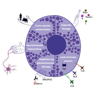
Dermatitis encompasses a spectrum of inflammatory skin disorders with aberrant immune
responses classified as type 1, type 2, and/or type 3. Major advances in the understanding
of the pathogenesis of atopic dermatitis (AD) have shed new light on how innate immune
responses critically regulate type 2 inflammation and itch. This article highlights
the diverse ways by which type 2 immune cells regulate diseases beyond AD. The discovery
of human Mas-related G protein–coupled receptor X2 on mast cells has revealed novel
T cell–independent and immunoglobulin E–independent mechanisms of allergic contact
dermatitis–associated and urticarial itch, respectively.
|
|
Scooped by
Gilbert C FAURE
October 8, 2020 4:10 AM
|
Interleukin 10 Interleukin-10 (IL-10) is an important anti-inflammatory cytokine (Fiorentino, Zlotnik, Mosmann, Howard, & O’Garra, 1991; Fiorentino, Zlotnik, Vieira, et al., 1991). From: Advances in Cancer Research, 2015 Related terms: View all Topics Role of IL-10 and the IL-10 Receptor in Immune Responses A. Howes, ... A. O'Garra, in Reference Module in Biomedical Sciences, 2014 Conclusions and Unanswered Questions on IL-10 IL-10 is an anti-inflammatory cytokine that maintains the balance of the immune response, allowing the clearance of infection while minimizing damage to the host. IL-10 can also dampen the harmful immune responses elicited in autoimmunity and allergy. The consequence of this activity, however, is that IL-10 can contribute to chronic infection. The importance of IL-10 in this balance is supported by a wealth of evidence gathered from studies in both the human and mouse systems. The immune-stimulatory roles of IL-10 are less well understood but may be a factor in influencing the role of IL-10 in antitumor and/or mucosal immune responses while maintaining the response that limits immunopathology. Further understanding of the sources of IL-10 in various contexts, how IL-10 production is regulated in different cell types, and precisely which cells IL-10 in different immunological settings will greatly enhance our ability to use IL-10 as a potential immune therapy in the future. Introduction to Mechanisms of Allergic Diseases Terufumi Kubo, ... Cezmi A. Akdis, in Middleton's Allergy Essentials, 2017 Interleukin-10 (IL-10) IL-10 plays a role in the control of allergy and asthma. IL-10 inhibits many effector cells and disease processes, and its levels are inversely correlated with disease incidence and severity. IL-10 is synthesized by a wide range of cell types, including B cells, monocytes, DCs, NK cells, and T cells. It inhibits proinflammatory cytokine production and Th1 and Th2 cell activation, which is likely attributable to the effects of IL-10 on APCs and its direct effects on T cell function (Table 1-3). IL-10 levels inversely correlate with the incidence and severity of asthmatic disease in the lung. In addition, the levels of IL-10 inversely correlate with skin-prick test reactivity to allergens. Beekeepers, who undergo multiple bee stings and are naturally tolerant to bee venom allergen have a high IL-10 response. IL-10 and IL-10-producing Treg and Breg cells play essential roles in immune tolerance to allergens. In addition, the roles of Treg and Breg cells and IL-10 have been shown in many autoimmune, organ transplantation, tumor tolerance conditions.6 Interleukin-10 YaoZhong Ding, ... Jonathan Bromberg, in Encyclopedia of Hormones, 2003 V.A Inflammation IL-10 has protective effects in experimental endotoxemia and rescues mice from LPS-induced toxic shock, which is correlated with reduced levels of serum TNFα. IL-10 inhibits the production of TNFα and macrophage inflammatory protein-2 (MIP-2); regulates hemodynamic parameters, leukocyte–endothelial cell interactions, and microvascular permeability; and reduces mortality in experimental endotoxemia. Mice treated with anti-IL-10 from birth or IL-10-deficient mice are more susceptible to endotoxin-induced shock than are normal mice. Human volunteers receiving IL-10 after endotoxin challenge suffer fewer systemic symptoms and less cytokine production. Cytokines in GVHD and GVL Kate A. Markey, ... Geoffrey R. Hill, in Immune Biology of Allogeneic Hematopoietic Stem Cell Transplantation (Second Edition), 2019 Clinical Evidence for Interleukin-10 IL-10 polymorphisms were among the first to be correlated with disease outcome in GVHD [187]. The seminal paper by Lin and colleagues demonstrated a clear and dramatic association of recipient IL-10 genotype with GVHD outcome. Donor IL-10 genotype has also been associated with significantly lower risk of grades III–IV aGVHD [188]. The phenotypic correlation of these polymorphisms does remain uncertain however, i.e., it is not clear whether they lead to gain or loss of IL-10 function. It has been hypothesized that the protection from aGVHD is due to enhanced IL-10 production from APC [184]. INTERLEUKINS | IL-10 T.J. Standiford, J.C. Deng, in Encyclopedia of Respiratory Medicine, 2006 Regulation of Production and Activity IL-10 is produced by a variety of cell types, including CD8+ and CD4+ T cells (e.g., T-helper-2 (Th2) cells, regulatory T cells), γδ-T cells, NK cells, NK T cells, B cells, dendritic cells, eosinophils, mast cells, and monocytic cells (e.g., microglia, monocytes, macrophages). Macrophages are the major source of IL-10, although regulatory T cells are now recognized as an important subpopulation of T cells that release IL-10. Nonleukocyte cell populations, including epithelial cells, keratinocytes, and melanoma cells, also express IL-10. In the lung, human alveolar macrophages (AMs), bronchial epithelial cells, and alveolar epithelial cells have been shown to express IL-10 mRNA and protein. In contrast, murine resident AMs secrete minimal amounts of IL-10, although these cells do express IL-10 receptors. Macrophages produce IL-10 in response to inflammatory or infectious stimuli, including lipopolysaccharide, TNF-α, and catecholamines. High levels of IL-10 are also produced under conditions that induce anergy and tolerance, such as repeated antigen stimulation. In these contexts, IL-10 likely provides negative feedback to dampen the magnitude of immune responses. Interestingly, several viruses, such as EBV, produce IL-10 or IL-10-like molecules (vIL-10). The genomes of other viruses, including members of the herpesvirus family, poxvirus, and primate cytomegaloviruses (CMV), also contain homologs of the IL-10 gene. Many vIL-10 proteins share extensive sequence homology with human IL-10. Viral IL-10 appears to engage the same IL-10 receptor in the host and is capable of producing some of the same biological effects as host-derived IL-10. Given the anti-inflammatory properties of IL-10, vIL-10 likely confers a survival advantage for these viruses by suppressing antiviral host immune responses. Interleukin 10 Receptor Signaling Dror S. Shouval, ... Scott B. Snapper, in Advances in Immunology, 2014 Abstract Interleukin 10 (IL10) is a key anti-inflammatory cytokine that can inhibit proinflammatory responses of both innate and adaptive immune cells. An association between IL10 and intestinal mucosal homeostasis became clear with the discovery that IL10 and IL10 receptor (IL10R)-deficient mice develop spontaneous intestinal inflammation. Similarly, patients with deleterious mutations in IL10, IL10RA, or IL10RB present with severe enterocolitis within the first months of life. Here, we review recent findings on how IL10- and IL10R-dependent signaling modulates innate and adaptive immune responses in the murine gastrointestinal tract, with implications of their role in the prevention of inflammatory bowel disease (IBD). In addition, we discuss the impact of IL10 and IL10R signaling defects in humans and their relationship to very early-onset IBD (VEO-IBD). Spinal Cord Injury Samuel David, ... V. Wee Yong, in Handbook of Clinical Neurology, 2012 Interleukin 10 Interleukin 10 (IL-10) is a potent anti-inflammatory cytokine which reduces inflammation in several disease models. Several reports indicate that IL-10 promotes tissue protection and functional recovery after SCI. Mice lacking IL-10 showed worse functional outcome and greater inflammatory response, apoptosis, tissue loss, and edema after spinal cord compression (Genovese et al., 2009) or excitotoxic injury (Abraham et al., 2004). Furthermore, the increased tissue damage observed in IL-10 null mice, could be reversed with IL-10 administration (Abraham et al., 2004). In addition, a single injection of systemically administered IL-10 improved locomotor recovery and reduced TNF-α expression after spinal cord contusion injury in rats (Bethea et al., 1999). However, in another study, the same group failed to observe any protective effects of IL-10 after a similar type of lesion in different strain of rat (Takami et al., 2002). More animal studies are needed to test various doses, mode of delivery and timing of treatment to establish the role of IL-10 in SCI. Interleukin 10 and its Receptor Vijay P. Khatri, Michael A. Caligiuri, in Encyclopedia of Immunology (Second Edition), 1998 Cellular source of IL-10 IL-10 is produced by several types of cells, including activated T lymphocytes, monocytes, B cells as well as nonhematopoietic sources such as keratinocytes, colon carcinoma and melanoma cells. In humans though TH2 T cell clones are the main source of IL-10, many TH1 clones will also secrete IL-10 following antigen-specific stimulation. In contrast, mIL-10 is produced by the TH2 subset of CD4+ T cells but not by TH1 or CD8+ T cells. In humans both CD4+ and CD8+ T cells secrete IL-10 following stimulation with anti-CD3, although significantly higher levels are produced by CD4+ T cells. Among the CD4+ T cells, CD45RO+ memory cells produce 10-fold higher level of IL-10 than naive T-cells (CD45RA+). Murine Ly-1 B cells as well as Epstein–Barr virus transformed human B cells produce IL-10. Human monocytes are also a major source of IL-10 in response to activation with lipopolysaccharide. Interestingly, kinetics studies reveal that IL-10 is synthesized later than other immunoregulatory cytokines by activated T cells or monocytes. This suggests that IL-10 may play a regulatory role for the later phases of the immune response. The Immune Basis of Allergic Lung Disease Stefanie C.M. Burleson, ... Michael R. Van Scott, in Comparative Biology of the Normal Lung (Second Edition), 2015 IL-10 IL-10, originally described as “cytokine synthesis inhibitor factor,” is produced by TH0, TH1, TH2, Treg, cytotoxic T cells, mast cells, and activated monocytes. IL-10 receptors are located on lymphoid, myeloid, and NK cells. IL-10 decreases TH2 signaling by downregulating MHC class II, B7.1/B7.2 and CD23 expression (Bjorgo et al., 2011; Bopp et al., 2009; Baumer et al., 2007; Heijink and Van Oosterhout, 2006). IL-10 suppresses monocyte and macrophage activity, including release of ROI, and downregulates secretion of pro-inflammatory cytokines such as IL-1β, TNF-α, IL-6, IL-8, and MIP-1α. In TH1 cells, IFN-γ and IL-2 synthesis are inhibited, as is synthesis of IL-4 and IL-5 by TH2 cells. In contrast, IL-10 can promote growth of mast cells, T cells, and B cells. However, even these responses can be bimodal, with inhibition observed at high concentrations of IL-10. IL-10 mRNA and protein are reduced in alveolar macrophages from patients with asthma, although the number of IL-10 expressing T cells and macrophages may be increased. In Brown Norway rats, IL-10 is reported to attenuate late-phase responses and eosinophilia postchallenge. In mice, treatment with IL-10 at the time of sensitization decreases eosinophilia (Chung and Barnes, 1999). Studies involving antibodies against IL-10 and IL-10R, as well as IL-10 knockout, have revealed attenuation of AHR and eosinophilia. This apparent inconsistency in the typical TH1 functioning of IL-10 might be explained by the finding that IL-10 can inhibit IL-13Rα2, a protein responsible for binding and inhibiting IL-13, thereby potentially increasing IL-13 effects. It has been noted that mice lacking both IL-10 and IL-13Rα2 exhibit a more severe manifestation of pulmonary allergic disease than mice deficient in IL-10 or IL-13Rα2 alone (Barnes, 2008). Sepsis Hector R. Wong, ... Cláudio Flauzino de Oliveira, in Pediatric Critical Care (Fourth Edition), 2011 Interleukin-10 Interleukin-10 is the best studied and most well known antiinflammatory cytokine.40,41 As an antiinflammatory cytokine, IL-10 serves to antagonize the proinflammatory effects of other cytokines and can thereby keep inflammation “in check.” IL-10 inhibits expression of cytokines such as TNF-α, IL-1β, and IL-8, and can inhibit expression of adhesion molecules. In addition, IL-10 can “deactivate” monocytes by downregulating the expression of MHC surface molecules. Thus IL-10 has a number of interesting properties that could potentially be leveraged therapeutically to limit excessive inflammation during sepsis. This theoretical consideration must be tempered by the ability of IL-10 to deactivate monocytes and thereby potentially impair the ability to adequately clear infection (i.e., the immune suppression paradigm depicted in Figure 103-1). Indeed, it has been reported that in children with multiple organ dysfunction syndrome, higher plasma IL-10 levels correlate with higher mortality, and higher monocyte mRNA levels of IL-10 correlate with increased length of stay in the ICU.42 Similar observations have been reported in adult patients with septic shock.43
|
|
Scooped by
Gilbert C FAURE
June 4, 2020 2:10 PM
|
Abstract The emergence and evolution of the complement system and mast cells (MCs) can be traced back to sea urchins and the ascidian Styela plicata, respectively. Acting as a cascade of enzymatic ...
|
|
Scooped by
Gilbert C FAURE
May 9, 2020 11:50 AM
|
T-cell immunoglobulin and mucin domain containing 4 (Tim-4) is a phosphatidylserine receptor and is selectively expressed on antigen presenting cells. Recently, Tim-4 was reported to be expressed on iNKT cells, B1 cells, and tumor cells, suggesting it has multiple biological functions. In this review, we mainly summarize the expression and regulation of Tim-4 in immune cells including T cells, macrophages, dendritic cells, NKT cells, B cells, and mast cells. The expression of Tim-4 in these cells implies that Tim-4 might participate in immune related diseases. Emerging evidence emphasizes a substantial role for Tim-4 in maintaining homeostasis by regulating various immune responses, including viral infection, allergy, autoimmunity, and tumor immunity. Here, we collectively evaluated the role of Tim-4 in health and diseases. This summary will be extremely useful to fully understand the function of Tim-4 in the pathogenesis of immune related diseases, which would provide novel clues for the diagnosis and treatment of diseases.
|
|
Scooped by
Gilbert C FAURE
February 6, 2020 2:24 AM
|
Il1rl1 (also known as ST2) is a member of the IL-1 superfamily, and its only known ligand is IL-33. ST2 exists in two forms as splice variants: a soluble form (sST2), which acts as a decoy receptor, sequesters free IL-33, and does not signal, and a membrane-bound form (ST2), which activates the MyD88/NF-κB signaling pathway to enhance mast cell, Th2, regulatory T cell (Treg), and innate lymphoid cell type 2 functions. sST2 levels are increased in patients with active inflammatory bowel disease, acute cardiac and small bowel transplant allograft rejection, colon and gastric cancers, gut mucosal damage during viral infection, pulmonary disease, heart disease, and graft-versus-host disease. Recently, sST2 has been shown to be secreted by intestinal pro-inflammatory T cells during gut inflammation; on the contrary, protective ST2-expressing Tregs are decreased, implicating that ST2/IL-33 signaling may play an important role in intestinal disease. This review will focus on what is known on its signaling during various inflammatory disease states and highlight potential avenues to intervene in ST2/IL-33 signaling as treatment options.
|
|
Scooped by
Gilbert C FAURE
August 9, 2019 6:44 AM
|
KEY POINTS IL-33 is expressed during early B cell development in both mice and humans. Deficiency of IL-33 promotes B cell fitness via a cell-endogenous mechanism. IL-33 expression is modulated in B cell malignancies. Abstract IL-33 is an IL-1 family member protein that is a potent driver of inflammatory responses in both allergic and nonallergic disease. This proinflammatory effect is mediated primarily by extracellular release of IL-33 from stromal cells and binding of the C-terminal domain of IL-33 to its receptor ST2 on targets such as CD4+ Th2 cells, ILC2, and mast cells. Notably, IL-33 has a distinct N-terminal domain that mediates nuclear localization and chromatin binding. However, a defined in vivo cell-intrinsic role for IL-33 has not been established. We identified IL-33 expression in the nucleus of progenitor B (pro-B) and large precursor B cells in the bone marrow, an expression pattern unique to B cells among developing lymphocytes. The IL-33 receptor ST2 was not expressed within the developing B cell lineage at either the transcript or protein level. RNA sequencing analysis of wild-type and IL-33–deficient pro-B and large precursor B cells revealed a unique, IL-33–dependent transcriptional profile wherein IL-33 deficiency led to an increase in E2F targets, cell cycle genes, and DNA replication and a decrease in the p53 pathway. Using mixed bone marrow chimeric mice, we demonstrated that IL-33 deficiency resulted in an increased frequency of developing B cells via a cell-intrinsic mechanism starting at the pro-B cell stage paralleling IL-33 expression. Finally, IL-33 was detectable during early B cell development in humans and IL33 mRNA expression was decreased in B cell chronic lymphocytic leukemia samples compared with healthy controls. Collectively, these data establish a cell-intrinsic, ST2-independent role for IL-33 in early B cell development. Footnotes This work was supported by National Institutes of Health Grants R01AI145265, R01AI124456, R01AI111820, R21AI145397 and U19AI095227 (all to R.S.P.), F30 AI114262 (to M.T.S.), R01CA226432 (to C.M.E.), and R01DK084246 (to P.L.K.); U.S. Department of Veterans Affairs Grant I01 BX004299 (to R.S.P.); Vanderbilt Medical Scientist Training Program Grant T32 GM007347; National Center for Research Resources Grant 1S10RR029557-01, National Center for Advancing Translational Sciences Grant UL1TR000445; and U.S. Department of Veterans Affairs Grant I01 BX 002882 (to P.L.K.). The RNA sequencing data presented in this article have been submitted to the Gene Expression Omnibus database (https://www.ncbi.nlm.nih.gov/geo/query/acc.cgi?acc=GSE132123) under accession number GSE132123. The online version of this article contains supplemental material. Received April 10, 2019. Accepted July 3, 2019. Copyright © 2019 by The American Association of Immunologists, Inc.

|
Suggested by
Société Francaise d'Immunologie
June 16, 2019 5:45 AM
|
Summary G‐protein–coupled receptor (GPCR) signaling is essential for the spatiotemporal control of leukocyte dynamics during immune responses. For efficient navigation through mammalian tissues, most leukocyte types express more than one GPCR on their surface and sense a wide range of chemokines and chemoattractants, leading to basic forms of leukocyte movement (chemokinesis, haptokinesis, chemotaxis, haptotaxis, and chemorepulsion). How leukocytes integrate multiple GPCR signals and make directional decisions in lymphoid and inflamed tissues is still subject of intense research. Many of our concepts on GPCR‐controlled leukocyte navigation in the presence of multiple GPCR signals derive from in vitro chemotaxis studies and lower vertebrates. In this review, we refer to these concepts and critically contemplate their relevance for the directional movement of several leukocyte subsets (neutrophils, T cells, and dendritic cells) in the complexity of mouse tissues. We discuss how leukocyte navigation can be regulated at the level of only a single GPCR (surface expression, competitive antagonism, oligomerization, homologous desensitization, and receptor internalization) or multiple GPCRs (synergy, hierarchical and non‐hierarchical competition, sequential signaling, heterologous desensitization, and agonist scavenging). In particular, we will highlight recent advances in understanding GPCR‐controlled leukocyte navigation by intravital microscopy of immune cells in mice. 1 INTRODUCTION Innate and adaptive immune responses depend on the coordination and spatiotemporal control of leukocyte dynamics between and within lymphoid organs and peripheral tissues. A wide spectrum of chemically diverse molecules including small proteins, peptides, lipids, nucleotides, and extracellular matrix fragments can act as chemoattractants, regulating leukocyte motility, migration, chemotaxis, positioning, and cell‐cell interaction. Leukocytes perceive these signals mainly through G‐protein–coupled receptors (GPCRs) that, upon agonist binding, induce intracellular promigratory responses.1, 2 Based on the cell type–specific expression of cell surface GPCR type combinations and intracellular GPCR‐controlling protein networks, each leukocyte subset has evolved specific trafficking paths to fulfill their designated effector functions in the body.3-5 Over the last decades, the analysis of GPCR‐deficient mouse models has advanced our understanding of how specific GPCR signals determine immune cell positioning and compartmentalization in situ. Recent work now also started to shed light on the establishment of chemokine patterns and chemotactic fields that guide leukocytes in mammalian tissues.6, 7 In particular, intravital imaging of control and GPCR‐deficient leukocytes in tissues of live anesthetized mice has brought unprecedented insight into the dynamic migration patterns of immune cell subsets in situ.8, 9 Despite these achievements, it is still largely unclear how leukocytes make directional decisions in lymphoid and inflamed organs where they constantly encounter GPCR signals emanating from multiple sources. The detailed mechanisms of how leukocytes sense, integrate, and prioritize between multiple chemotactic fields in situ and how this influences their physiological trafficking are still largely unknown. Dissecting these molecular events experimentally in mammalian tissues is very challenging and demanding. In particular, we are missing important pieces of information (eg, the exact distribution and concentrations of known chemottractant, GPCR dynamics in leukocytes) and cannot control for unknown chemotactic fields. Due to these current limitations many of the emerging concepts for leukocyte navigation in complex chemoattractant milieus come from studies in controlled in vitro systems2 or lower model organisms.10, 11 However, these concepts from reductionist models may not always be easily translatable to the complex situation in mammalian lymphoid organs and inflamed peripheral tissues. In recent years, several systematic studies in mice have started to disentangle how multiple GPCR signals shape leukocyte migration patterns in the complexity of mammalian tissues. In this review, we will summarize common concepts and mechanisms of GPCR‐controlled leukocyte navigation coming from studies with mouse and human leukocytes. We outline the protein networks that control GPCR signaling and GPCR activity and discuss how these may control leukocyte navigation strategies. In particular, we highlight our current knowledge on leukocyte navigation in complex chemoattractant fields and summarize them for three leukocyte subsets (neutrophils, T cells, and dendritic cells [DC]) with different physiological trafficking paths. For each cell type, we will critically evaluate the relevance of current concepts for leukocyte navigation in mouse tissue. While it is very likely that some navigation strategies are only relevant for certain immune cell types and adapted to specific physiological tissue environments, leukocyte subsets may also share basic strategies for directional decision‐making in lymphoid and inflamed tissues of mammals. 1.1 Concepts and mechanisms of GPCR‐controlled leukocyte navigation Many chemokines and chemoattractants are classical inducers of leukocyte polarity, adhesion, and locomotion. They act through GPCRs on intracellular signaling pathways to establish biochemical asymmetry within the cell, preceding the formation of a polarized cell with an actin‐rich front and a myosin II–rich rear.12, 13 Key molecular pathways involved in establishing functional polarity downstream of GPCRs in leukocytes depend on phosphoinositide 3‐kinases (PI3K) and members of the Rho family of small GTPases.14 Rac isoforms, Cdc42 and RhoA represent major signal hubs that act through several effector molecules to regulate actin polymerization and actomyosin cytoskeleton. The balanced interplay of these components shapes the typical amoeboid migration mode of each leukocyte subset.15, 16 In homogeneous fields of soluble chemokines and chemoattractants, most leukocytes undergo self‐polarization, before increasing their motility, migration speed, and movement in a random and undirected fashion (chemokinesis) (Figure 1A).17 Chemokinesis is considered a migratory pattern that helps leukocytes scan large tissue volumes. We have learnt from intravital microscopy studies in mice that lymphocytes show such continuous undirected (random‐walk) migration in lymphoid organs to enable encounters and interactions with other leukocytes.9 Together with other determining factors (eg, confinement between stromal tissue elements), uniform chemokine signals critically control chemokinesis by increasing the speed of randomly moving lymphocytes in situ.18 Once leukocytes perceive external gradients of soluble chemokines and chemoattractants, they can perform directional movement along the concentration gradients of these diffusing attractants (chemotaxis) (Figure 1B).17 Some leukocyte subsets have also been reported to be repelled from high chemoattractant concentrations (chemorepulsion), leading to their movement against the gradient (fugetaxis).19 However, the underlying molecular mechanisms of this process are still under investigation (Figure 1C).20, 21 Apart from soluble molecular cues, leukocytes also perceive surface‐bound attractants as gradients or as homogeneous fields and undergo movement along these surfaces in a directed (haptotaxis) (Figure 1B) or undirected manner (haptokinesis) (Figure 1A), respectively.16, 22 The aspect of leukocyte navigation along surface‐bound attractants is particularly relevant for chemokines with binding capacities to extracellular glycosaminoglycans (GAGs), which decorate many stromal cell elements in tissues.22, 23 This form of imprinting non‐diffusible chemokine gradients into the tissue has been shown critical for leukocyte trafficking across the blood endothelium and entry into lymphatic vessels.6, 23 The terms “chemotaxis” and “haptotaxis” subsume a spectrum of navigation strategies, which leukocytes can use to follow attractant gradients.24 When perceiving an external attractant gradient, some leukocytes (eg, unstimulated neutrophils in culture dish) undergo a full morphological change from unpolarized shape to the establishment of a front‐back axis, which then leads to directional movement along the gradient. In contrast, other leukocytes already migrate with pre‐established polarity (eg, DCs and neutrophils in 3D), before they steer along the gradient by redirecting their migration path. Alternatively, prepolarized leukocytes may move along a gradient without redirecting their path and start to accelerate the closer they get to the attractant source (eg, neutrophils in response to dying cells). Once at the highest attractant source, leukocytes may also actively decelerate and slow down their migration.24 As evidenced from intravital microscopy of select leukocyte subsets, we know that this spectrum of chemotactic responses also occurs in mouse tissues. While we acknowledge that chemokines and chemoattractants are central for many aspects of these migratory responses, we are missing a detailed understanding about how these signals shape the navigation of leukocytes in situ. Is the cellular movement guided by one or more attractant signal(s)? How do leukocytes navigate in complex gradient fields and sense the right direction? How do leukocytes sense their final destination and know where to stop? Are there inherent differences between leukocyte subsets in their navigation strategies? As many other cell types, leukocytes possess several mechanisms that directly regulate the functionality of GPCRs.13, 25-28 These are mainly controlled by GPCR interactions with three protein families: heterotrimeric (α, β, γ) G‐proteins, G‐protein–coupled receptor kinases (GRKs), and β‐arrestins. Upon stimulation, a GPCR functions as a guanine nucleotide exchange factor that catalyzes the exchange of GTP for GDP on Gα subunits of the heterotrimeric G‐protein. In leukocytes, Gα subunits of the pertussis toxin‐sensitive Gi subfamily play critical roles for the signaling of many chemokine and chemoattractant receptors. The dissociation of Gβγ from Gα‐GTP allows them both to independently activate downstream signaling pathways. Leukocyte chemotactic responses are largely controlled by the released Gβγ, which activates phosphoinositide 3‐kinase (PI3K) and phosphoinositide‐specific phospholipase Cβ (PLC)/inositol‐trisphosphate (IP3)/diacylglycerol effector pathways.29, 30 The termination of GPCR signaling relies on the intrinsic GTPase activity of the Gα subunit that hydrolyzes GTP and reassembles as Gα‐GDP with Gβγ to form an inactive heterotrimer. This Gα GTPase reaction is accelerated by a family of proteins, called “regulators of G‐protein signalling” (RGS proteins) to facilitate the rapid turning off GPCR signaling. Hence, RGS proteins are considered key desensitizers of G‐protein‐signaling pathways.27 In addition, another negative feedback loop evolved to prevent the overstimulation of cells through excessive GPCR stimulation. Following agonist binding and G‐protein activation, GPCRs become rapidly phosphorylated on their cytoplasmic tails and intracellular C‐termini by specific enzymes, usually members of the GRK family.31, 32 The pattern of receptor phosphorylation creates a barcode with high‐affinity binding sites for the recruitment of β‐arrestin proteins.33 As a consequence, the increased binding of β‐arrestin to the receptor sterically hinders the GPCR interaction with G‐proteins. This uncoupling of G‐proteins from the receptor, referred to as “receptor desensitization”, prevents further GPCR activation by repeated agonist stimulation over seconds to minutes.34 β‐arrestins can also control more long‐lasting desensitization of GPCRs over minutes to hours. As they can interact with clathrin‐coated pits, their binding to GPCRs also targets receptors for internalization. Besides, GPCR internalization can also follow other pathways (arrestin independent, clathrin independent) or not occur at all.35 Depending on the intracellular trafficking route, the internalized receptors may be subject to degradation, leading to the downregulation of cell surface GPCRs. Alternatively, targeting internalized GPCR to recycling endosomes and back to the plasma membrane can quickly replenish GPCRs on the cell surface.35 In addition to their important roles in receptor inactivation and internalization, GPCR‐recruited β‐arrestins also act as signaling mediators by directly coupling to numerous signaling molecules.36 At many GPCRs, β‐arrestin‐mediated signaling and classical G‐protein signaling can have distinct biochemical and physiological actions from one another. A relatively new paradigm on GPCR signaling considers the allosteric interaction among the ligand, the GPCR, and the transducers (β‐arrestins and GRKs) and how their contribution influences the bias for G‐protein– or β‐arrestin–mediated signaling.28 Resulting from this paradigm, the concept of biased agonism, ie, that different ligands at the same GPCR are able to activate some signaling pathways while blocking others, is of particular interest for many chemokine receptors with prominent roles in leukocyte navigation.37 In the following, we will point out the most commonly and recently discussed mechanisms, which would allow the control of leukocyte navigation. Given the before‐mentioned regulation mechanisms for GPCR functionality, a “One‐GPCR system” appears already sufficient to control chemokinesis and chemotaxis of leukocytes (Figure 2A‐E). To increase the sensitivity for attractant gradients, leukocytes can upregulate the cell surface expression of GPCRs. Some leukocytes store GPCRs in intracellular vesicles, which can fuse with the plasma membrane under conditions of leukocyte activation.38 While the release of GPCR from vesicular stores occurs within minutes to hours, most leukocytes often respond to external stimuli by changing their cell surface GPCR profile over hours to days. These changes are primarily based on the induction of gene‐transcription networks, which also include GPCR proteins, when leukocytes transition between cell states or undergo differentiation (Figure 2A). Multimerization of GPCRs, including homomerization of the same GPCR type, provides another level of GPCR regulation.25 A recent study highlighted the functional importance of GPCR oligomerization in promoting leukocyte chemotaxis (Figure 2B). By lowering cellular cholesterol, inflammatory stimuli enabled GPCR oligomers that acted as scaffolds for distinct promigratory signaling pathways to foster efficient leukocyte migration.39 However, GPCR multimerization has also been reported to dampen GPCR responsiveness.25 Several other mechanisms can attenuate the sensitivity of leukocytes for chemokines and chemoattractants. One involves the presence of a receptor antagonist that binds to the same GPCR as the gradient‐forming attractant agonist, but exerts blocking function for promigratory signaling (Figure 2C). More cell‐intrinsic mechanisms include the downregulation of GPCR expression by lowering GPCR mRNA stability40 or enhancing GPCR degradation.41 As discussed above, agonist‐induced receptor desensitization also attenuates the signaling responsiveness of GPCRs. Upon homologous desensitization, when GRKs phosphorylate agonist‐specific GPCRs, the further recruitment of β‐arrestins can lead to G‐protein uncoupling and internalization of GPCRs (Figure 2D). This process explains why many leukocyte chemotaxis responses toward single attractants are dose dependent and follow a bell‐shaped curve, with higher concentrations resulting in stasis.42 Homologous desensitization may be particularly relevant when leukocytes sense high attractant concentration in situ, eg, at the center of an attractant source.43 Under such conditions, the majority of cell surface GPCRs is likely to undergo desensitization, internalization, and degradation, which may then result in cellular unresponsiveness to continuous or repeated stimulation through the same attractant. As a strategy to remain responsive to subsequent stimulation in gradients, some leukocytes internalize GPCRs, deploy its ligand cargo into lysosomes, and quickly recycle unloaded GPCRs back to the plasma membrane.44 Thus, by eliminating chemotactic factors from the extracellular environments, classic GPCRs can acquire additional scavenging function. This “consumption” of external attractant may also impact the chemotactic milieu by reducing the strength of attractant cues.44 Instead of only binding one agonist, many “One‐GPCR systems” can bind and respond to two or more chemoattractants and activate downstream signaling (Figure 2E). Sensing these distinct attractants through the same GPCR in a spatial or temporal sequence can provide another navigation strategy. In our body, leukocytes are rarely exposed to only one chemotactic factor. For efficient trafficking between organs and coordinated movement within tissues, most leukocytes express more than one GPCR on their surface and sense a wide range of chemokines and chemoattractants. Directional decision‐making in lymphoid and inflamed organs requires leukocytes to constantly sense, compare, and integrate numerous GPCR signals emanating from multiple sources. To efficiently navigate in a complex milieu of attractant gradients, leukocytes need to respond to combinational signals and prioritize between competing signals. While this aspect of GPCR signal integration has already been of interest decades ago, it has regained momentum with the recent developments in fabricating microfluidic‐based chemotaxis devices. We will now briefly summarize common mechanisms and concepts for leukocyte navigation, which become already relevant in the presence of a second GPCR signaling system (“Two‐GPCR systems”) (Figure 3). As leukocytes often use combinations of more than two‐GPCR systems, these principles could be further expanded. Manifold scenarios of attractant interpretation are conceivable, when leukocytes sense two chemoattractant signals through two different GPCR types (Figure 3). The outcome for leukocyte guidance depends on several determining parameters: (a) the spatial distribution of each signal (uniform, graded), (b) the spatial orientation of both signals toward each other (same or opposing directions), and (c) the ratio of signaling input that leukocytes receive from both signals (equal, unequal). The latter point includes both cell‐intrinsic features and cell‐extrinsic conditions. Unequal signaling input can result from different cell surface expression of the two‐GPCR types or different strength of the downstream signaling pathway. In both cases, one chemoattractant signal dominates over the other. This can also occur in leukocytes with equal signaling input from both GPCR systems under conditions of unequal concentrations of both signals in the extracellular space. Many of these scenarios have already been addressed with select leukocyte subsets and their cell‐specific GPCR combinations in controlled in vitro microenvironments, and examples will be discussed in the next paragraphs about specific leukocyte subsets (neutrophils, T cell, and DCs). As discussed before, homogenous chemoattractant fields can induce leukocyte polarity and chemokinesis. A second‐graded signal “on top” can then redirect prepolarized leukocytes and guide them toward other locations. However, if the uniform attractant field is very potent, it will retain leukocytes and attenuate chemotaxis toward the second‐graded signal (Figure 3A).45-48 Under conditions when both attractant signals are oriented in the same direction and overlap, the two‐GPCR signals have usually additive effects for leukocyte chemotaxis (Figure 3B). In the case of promigratory signals, chemotactic factors act often synergistically to improve chemotaxis along the same direction.49 Physiologically, this might be of particular relevance for tissue sites where overlapping attractant gradients originate from the same local area. A special variant of GPCR signal synergism has initially been observed in migrating human and mouse neutrophil populations in vitro. During their chemotaxis in response to shallow N‐formyl‐peptides (fMLF) gradients, neutrophils released the lipid leukotriene B4 (LTB4) that acted on top of the early chemotactic factor as a signal relay molecule to improve chemotaxis of the whole neutrophil population.50 Under conditions when leukocytes encounter two spatially opposing gradients, the two‐GPCR signals compete for guiding leukocytes into one or the other direction (Figures 3C and 4). Leukocytes exposed to two opposing attractants with equal signaling strength will likely accumulate and find migratory equilibrium in the middle of both gradients (Figure 3C).47, 48, 51, 52 However, an unequal signaling input from both signals favors the movement of leukocytes into one direction (Figure 4A).47 It is known for decades that specific GPCR signals can dominate over other GPCR signals, which enables leukocytes to prioritize some chemoattractant signals over others (Figure 4B).42, 45 This aspect of GPCR crosstalk provided a first conceptual framework for neutrophils traveling in a stepwise fashion from the blood into infected tissues.45 Initially, it was discussed that heterologous desensitization of GPCRs might be an underlying mechanism for prioritizing chemotactic cues. In contrast to homologous desensitization (see above), heterologous GPCR desensitization is agonist independent and results from the phosphorylation of one GPCR by a kinase activated through the signaling cascade of another activated GPCR (Figure 3D). Thus, one‐GPCR signal can act as desensitizing signal for multiple other GPCR types, causing leukocytes to become refractory to several chemotactic stimuli within minutes. This rapid cross‐phosphorylation has been linked to second‐messenger kinases, particularly protein kinase C, which phosphorylates the GPCR at sites different from GRKs.53 For neutrophil navigation in opposing gradients, it was later proposed that the dominance of certain GPCR signals is regulated by events downstream of the receptors. According to this concept, referred to as “signaling hierarchy”, special GPCRs induce intracellular signaling pathways that can override signaling pathways downstream of other GPCR signals42, 54 (see below for details). Leukocyte populations in tissues rarely consist of identical cell clones, but are rather characterized by many cells with slightly heterogeneous expression of GPCR combinations. Thus, single‐cell heterogeneity on the basis of combinatorial GPCR expression may influence the population response in two opposing attractants with equal signaling strength (Figure 4C). In many physiological settings, leukocytes are thought to navigate step‐by‐step through sequential chemotactic fields. Under conditions of two spatially segregated chemotactic signals in sequence (Figure 3E), leukocytes may first move along a first attractant gradient (Figure 1A), and then subsequently sense the gradient of a second, more distant attractant. During the transition from first to second attractant gradient leukocytes then encounter a competitive situation between two gradients (Figure 3C). When the second signal is dominant over the first one, it is plausible that leukocytes are recruited to and then retained at the distant site. Importantly, even without any dominance between the signals, leukocytes can migrate away from a first attractant and travel toward a second, more distant attractant.45 In principle, this also allows leukocytes to leave chemotactic fields of high, saturating concentrations. Moreover, leukocytes remain responsive for additional attractant changes, while moving back and forth in the equilibrium between both gradients. Thus, interpreting two distinct signals in sequence allows efficient navigation patterns that are not possible with only one attractant presented in the same spatial array. We want to point out that the sequential sensing of attractant gradients can also be temporally controlled (Figure 3E). Leukocytes may undergo sensing of a first attractant gradient, while a second attractant arises newly in close vicinity. In the course of an immune response in lymphoid or inflamed tissues, this may result from chemoattractant secretion of nearby activated tissue‐resident cells or recently recruited, blood‐derived leukocytes. As most immune responses are choreographed by a sequence of timely separated cellular events, it is likely that this aspect of sequential sensing has strong influence on leukocyte navigation in situ. In our contemplation on leukocyte navigation by “Two‐GPCR systems”, we have focused until now on GPCRs with signaling function. However, many leukocyte subsets additionally express members of the atypical chemokine receptor (ACKR) family on their surface. Also referred to as scavenging receptors, ACKRs are GPCRs with promiscuous binding affinity for multiple chemokines. As they do not couple to G‐proteins, they are unable to induce intracellular signaling pathways. Instead, ACKRs can actively internalize and target chemokines for lysosomal degradation.55 Functional co‐expression of conventional chemokine receptors and ACKRs in leukocytes has been implicated in the fine‐tuning of leukocyte chemotaxis, with ACKRs acting as rheostats in restricting excessive migration in response to moderate chemokine levels (Figure 3F).56-58 As we have just outlined, leukocytes have evolved several molecular mechanisms that allow an enormous spectrum of GPCR‐controlled navigation strategies. Many of the emerging concepts for leukocyte navigation in complex chemoattractant fields have been developed in controlled in vitro systems, some of which by studying only one select leukocyte subset. Rigorous testing and proving the relevance of these concepts for leukocyte migration in the complexity of mammalian organs and tissues is technically very challenging. However, systematic studies in mice have started to delineate GPCR‐controlled navigation strategies and their influence on physiological trafficking patterns through lymphoid organs and inflamed peripheral tissues. In the following, we will discuss our current knowledge on the role of GPCR signals for the migration patterns of three leukocyte subsets (neutrophils, T cells, and DCs). Each of these immune cell types has evolved their own trafficking paths to fit their effector functions. We will highlight recent studies that conceptually advanced our understanding of GPCR‐controlled leukocyte navigation in mammalian tissues. 2 NEUTROPHILS Neutrophils are pivotal effector cells of our innate immune response and the first line of defense for eliminating bacteria and fungi in tissues.59 Recent studies also implicate regulatory functions of neutrophils in tissue homeostasis and cancer.60 In mammals, they are produced in the bone marrow and are continuously released into the bloodstream to patrol the intravascular compartment under homeostasis. Neutrophils circulate in the blood for ca. 12 hours under the influence of circadian rhythms, before they are mostly cleared into the bone marrow, but also other extramedullary tissues.61 In contrast to the general believe that naive peripheral tissues are free of neutrophils, recent work suggests that low numbers of neutrophils are present in many tissues under steady state.62 Upon inflammation and infection, the release of neutrophils from bone marrow increases their concentration in the blood by several folds.63 Moreover, large numbers of circulating neutrophils then infiltrate inflamed tissues where they sense activated vascular endothelium. In agreement with their critical role as first responders, neutrophils have evolved robust strategies to infiltrate inflamed tissues extremely fast and orient themselves to detect potential sites of microbial invasion with great precision. In many organs, the exit from the vascular compartment involves a sequence of exquisitely coordinated processes: (a) intravascular arrest, migration, and crawling along the activated endothelial cells, and (b) breaching multiple layers of the vascular wall (venular endothelium, venular basement membrane, and pericytes).64 Once neutrophils have traversed this barrier, they are primed for the rapid detection of damaged tissue or microbes in the extravascular space. At sites of cell death in the interstitial space, neutrophils undergo sequential phases of highly coordinated chemotaxis and cell cluster formation, now commonly referred to as neutrophil swarming.65, 66 As true sentinel cells, they sense almost any form of tissue insult, rapidly localize it in any inflamed organ and efficiently move toward the core of the inflammation or infection site. Neutrophils have been shown to express on their cell surface more than 30 GPCR types, which can all bind chemotactic molecules. Accordingly, neutrophils are responsive to a broad array of chemically diverse chemoattractants, including chemokines, lipids, complement factors, formylated peptides, nucleotides, alarmins, and proteolytic (ECM) fragments.67-69 Many of these chemotactic factors are usually present at sites of in vivo inflammation. How the crosstalk between these GPCR signals guides neutrophil recruitment in inflamed tissues has been of huge interest since decades. Early in vitro work with human polymorphonuclear neutrophils (PMN) characterized the strength of chemically distinct chemoattractant classes to desensitize calcium‐ion mobilization, as a read‐out of GPCR downstream signaling, to one another (Figures 2D and 3D).53, 70 Peptide chemoattractants, including N‐formylmethionyl‐leucyl‐phenylalanine (fMLF), the complement factor C5a, and the chemokine CXCL8 (IL‐8), cross‐desensitized one another to different extents (fMLF > C5a > IL‐8). Moreover, peptide chemoattractants, in particular fMLF, cross‐desensitized lipid chemoattractants, including platelet‐activating factor (PAF) and LTB4. In contrast, lipid and purinergic agonists failed to desensitize to the peptide chemoattractants.70 These results were extended to neutrophil chemotaxis in the presence of multiple chemoattractants, showing a hierarchy of resistance to competing signals, with fMLF and C5a inhibiting migration to other chemoattractants (Figure 3D).71, 72 Elegant under‐agarose chemotaxis experiments proposed the concept that neutrophils can navigate through complex chemoattractant fields by migrating in a step‐by‐step manner in response to one agonist source after another. Thus, their ultimate positions are determined combinatorially by the array of GPCRs they express and by the sequence of chemoattractant gradients they encounter (Figure 3E).45 In this concept, pathogen‐derived “end‐target” chemoattractants (N‐formyl peptides, C5a) will dominate over host tissue‐derived “intermediate‐target” chemotactic factors (CXCL8, LTB4) in their ability to guide neutrophil migration (Figure 3A, C, E).45 This dominance of end‐target attractants has been suggested to depend on the activation of the p38 MAPK pathway, which can override the phosphoinositide 3‐kinase (PI3K) pathway activated by intermediate‐target attractants.42, 54 Importantly, neutrophil navigation is also possible in a spatially sequential array of intermediate‐target chemoattractants (Figure 3E).45, 47, 51, 73 Another aspect of crosstalk between end‐target and intermediate‐target chemoattractants has not been considered in these early studies, but is also functionally relevant for neutrophil navigation. Already small amounts of fMLF or C5a can stimulate neutrophils to secrete intermediate‐target attractants, in particular LTB4.50, 74 This paracrine signaling mechanism becomes very relevant in chemotaxing neutrophil populations. Neutrophil‐released LTB4 functions as a signal relay molecule and amplifies in a feed‐forward manner the chemotactic response to subsaturating concentrations of end‐target attractants (Figure 3B).50, 75 Recent developments in microfabricated devices and microfluidic‐based chemotaxis systems now allow a more detailed analysis of neutrophil migration responses in complex chemoattractant milieus, providing deeper insight into neutrophil navigation in opposing gradients (Figure 3C),47, 73, 76-78 synergistic gradients (Figure 3B),49 and geometrically complex environments.49, 77 Moreover, even the analysis of GPCR signaling underlying neutrophil swarming dynamics is now possible due to novel micropatterning techniques.75 As human PMNs, mouse neutrophils express a range of functional GPCRs to sense N‐formyl peptides (through FPR1 and FPR2), C5a (mainly through C5ar1), LTB4 (mainly through LTB4R1/BLT1), and several chemokines including CXCL1, CXCL2, CXCL5 (through CXCR2), CXCL12 (through CXCR4), and several CC‐chemokines (through CCR1).67 Some of these GPCRs are present in secretory vesicles that can rapidly fuse with the plasma membrane upon neutrophil activation, allowing the cells to increase their responsiveness to chemotactic cues (Figure 2A).38 Other reported mechanisms to control GPCR functionality in neutrophils include competitive antagonism or partial agonism (Figure 2C),79 receptor multimerization (Figure 2B), differential agonist responses (Figure 2E),80 and agonist scavenging through simultaneous expression of ACKRs (Figure 3F).56 Homologous desensitization is a well‐known phenomenon for almost all neutrophil‐expressed GPCRs (Figure 2D). While GPCR transducers (G‐protein subunit types, GRKs, and arrestins) and RGS proteins have been linked to the function of some GPCRs in primary mouse neutrophils,32, 81-88 we are still missing a detailed understanding of their exact contributions to neutrophil navigation in complex chemoattractant fields. Clearly, neutrophils are equipped with many GPCR types and mechanisms to dynamically control GPCR activity, allowing them to use various navigation strategies in the presence of multiple chemotactic cues. Moreover, neutrophils have been shown to perform fugetaxis against the direction of a gradient of high concentration chemokine, a process that is mechanistically not well understood (Figure 1C).89 However, there are still many open questions of how these mechanisms contribute to the neutrophil migration patterns observed in situ. Intravital microscopy studies of GPCR‐deficient neutrophils provided novel insight into how GPCR signals choreograph the physiological trafficking paths of neutrophils. In the following, we will point out that GPCR signals are important throughout many steps in the lifecycle of a neutrophil. In particular, we will highlight studies that advanced our concepts of GPCR‐controlled neutrophil navigation in mouse tissues. 2.1 Bone marrow release: inhibiting chemokine‐mediated retention The majority of mature neutrophils in the body reside in the bone marrow, with an estimated ratio of 300:1 between neutrophils in the bone marrow and blood. Neutrophil release into the blood is controlled by the interplay of retention and mobilization signals, provided by two antagonistically operating chemokine systems (Figure 3A,C). During homeostasis, the chemokine CXCL12 is produced in the bone marrow and essential for the retention of mature neutrophils that express the corresponding chemokine receptor CXCR4.90 CXCL12 is expressed at highest levels by “CXCL12‐abundant reticular cells” (CAR cells), but also by other stromal cells of the bone marrow, including osteoblasts.63, 91 The expression levels of CXCL12 in stromal cells follow a circadian rhythm, controlled by the sympathetic nervous system.92 When neutrophils were freshly isolated from the bone marrow, they expressed only low levels of cell surface CXCR4, but contained high intracellular levels of CXCR4.90, 93 After placing these cells in medium lacking CXCL12, they upregulated cell surface CXCR4 within hours.90, 93 These findings suggested receptor desensitization and internalization in response to constitutively high local concentrations of CXCL12 in the bone marrow microenvironment (Figure 2D). Moreover, these data underlined that neutrophils receive constant CXCL12 signals, which position them in the bone marrow, probably by activating integrin‐mediated adhesion to stromal cells.94 While high concentrations of CXCL12 retain neutrophils in the bone marrow via CXCR4 signaling, low concentrations of CXCR2 ligands expressed on endothelial cells and produced by megakaryocytes facilitate neutrophil egress via CXCR2.63 Thus, CXCR2 signals recruit neutrophils out of the retentive CXCL12 field and direct them to the endothelial sinus walls for entry into the blood circulation (Figure 3A). This intricately balanced interplay between retention and mobilization signals can be shifted in both directions. G‐CSF treatment or acute inflammation, which increases blood serum levels of G‐CSF and CXCR2 ligands, shifts the balance toward neutrophil mobilization from the bone marrow.95-98 Several modes of action have been proposed for G‐CSF, either leading to the lowering of CXCL12 levels in the bone marrow or triggering the release of CXCR2 ligands from endothelial cells or megakaryocytes (summarized in63). The same shift toward neutrophil egress can be achieved by conditional gene depletions of CXCR4 or CXCL12 in mice.99, 100 The administration of the CXCR4 antagonist AMD3100 also results in a threefold increase in the number of circulating neutrophils and a corresponding decrease in bone marrow numbers.93, 101 Originally, this drug effect was interpreted as direct antagonism of CXCR4 expressed by neutrophils, thereby disrupting the CXCL12‐CXCR4 axis in the bone marrow (Figure 2C).90 Surprisingly, subsequent studies found that AMD3100 also acted on CXCR4 expressed by bone marrow endothelial cells, and inhibited the translocation of CXCL12 across bone marrow endothelial cells, suggesting a rapid rise of CXCL12 in the blood and reversal of the CXCL12 gradient (Figure 3B).102, 103 These data were questioned by intravital imaging studies that could not observe clear neutrophil egress from the bone marrow upon AMD3100 treatment. Instead, the increase in circulating neutrophils through AMD3100 was explained by the release of marginated neutrophils from the lung microvasculature.104 Moreover, AMD3100 had a blocking effect on the trafficking of aged blood neutrophils, which upregulate CXCR4 expression over time, enabling them to home back to the bone marrow for clearance.104 The exact mode of action of AMD3100 is still controversially discussed.90 Insight into the physiological consequences of shifting the chemokine balance in the opposite direction, toward retention in the bone marrow, comes from an extremely rare congenital human immunodeficiency disease. Warts, hypogammaglobulinemia, infections, and myelokathexis (WHIM) syndrome is an autosomal dominant human disorder caused by gain‐of‐function mutations in the C‐tail of CXCR4.105, 106 WHIM syndrome involves myelokathexis, a condition characterized by the retention of mature neutrophils in the bone marrow and peripheral neutropenia. WHIM‐associated truncations or mutations in the carboxy‐terminal tail of CXCR4 result in the loss of multiple serine residues, which are target sites for GRK‐mediated receptor phosphorylation. Truncated forms of CXCR4 are impaired in agonist‐induced homologous desensitization and internalization, and cause enhanced signaling upon CXCL12 stimulation.106 This leads to stronger anchorage of mature neutrophils in the bone marrow, counteracting CXCR2‐mediated chemotaxis toward vessels and neutrophil egress. However, administration of G‐CSF or ADM3100 to WHIM patients can overcome this imbalance and cause an increase in circulating neutrophil numbers.106 2.2 Getting to the site of inflammation: navigation along multiple chemokine fields Blood‐circulating neutrophils detect sites of tissue injury and infection by interacting with the luminal surface of activated venular endothelium. In response to tissue‐released inflammatory cytokines, endothelial cells upregulate P‐selectin, E‐selectin, and integrin ligands, such as ICAM‐1 and VCAM‐1, to promote interactions between neutrophils and the vessel wall. This induces neutrophils to undergo a multistep adhesion cascade along the endothelial lining before emigrating from the vessel into the inflamed tissue interstitium.107 Chemokines are present on the luminal surface of endothelial cells and cause arrest of rolling neutrophils by triggering the activation of integrins. Many studies showed an involvement of CXCR2 ligands, in particular CXCL1, but also CXCL2 and CXCL5, in this process.108, 109 These chemokines are primarily expressed and released by endothelial cells,110, 111 but also tissue‐resident cells, including pericytes, perivascular macrophages, and mast cells, can act as important chemokine sources.112-115 Due to their high‐affinity binding sites for heparin sulfate (HS) GAG most chemokines become immobilized by HS GAG on the luminal side of the endothelium,116 and defects in HS GAG synthesis have been shown to decrease immobilization of chemokines and neutrophil transmigration into inflamed tissues.117, 118 While the involvement of chemokines for neutrophil transmigration is widely established, there has been ongoing debate whether and how leukocytes sense stable chemokine gradients across the vessel wall on their way into the tissue (Figure 1B). There are ample references to support the common believe that chemotactic gradients between the apical and basolateral part of postcapillary venules promote diapedesis, ie, the outward passage of neutrophils through the vessel wall. Immunofluorescence stainings of HS GAG moieties revealed steep gradient of HS GAG scaffolds between the luminal and abluminal aspect of skin postcapillary venules.119 Under inflammatory conditions, HS GAG distribution became even more polarized by massive depositions of HS and laminin in the basolateral basement membrane of skin vessels.119 Given the GAG‐binding properties of many chemokines, it was proposed that these sharp HS GAG scaffold gradients might favor chemokine gradients across the venular wall. In agreement with this idea, excessive deposition of the GAG‐binding chemokine CXCL1 on the apical part of inflamed blood vessels resulted in decreased neutrophil diapedesis.120 Other supporting data come from experiments in which an extravascular source of CXCR2 ligands was placed in form of a gel on one side of a blood vessel.121 Under these conditions, neutrophil crawling was directed to emigrate preferentially from that side of the vessel. HS GAG on the surface of the endothelium was required to present CXCL2, forming a chemotactic gradient that was followed by neutrophils. This sequestration of endothelial chemokine occurred exclusively in venules, but not other blood vessel types.121 HS GAG, together with the atypical receptor ACKR1, was also discussed to establish gradients by transcytosis of chemokines.118, 122, 123 Transcytosis involves the internalization and transport of tissue‐derived inflammatory chemokines from the basal surface onto the luminal surface of endothelial cells. ACKR1 is specifically expressed by postcapillary venules and was shown to support promigratory chemokine patterns across the vessel wall for optimal leukocyte migration across the vessel wall.123, 124 Unlike other ACKR, ACKR1 does not act as classical scavenger receptor, and was initially discussed to promote the transcytosis of tissue‐derived inflammatory chemokines from perivascular pools to the apical surface of venules.122, 123 Novel insight into the functional role of ACKR1 and the directional cues that guide neutrophils during diapedesis come from recent elegant experiments of Nourshargh and colleagues.125 By studying TNF‐induced neutrophil emigration in the inflamed mouse cremaster muscle, it was shown that neutrophils encounter the two CXCR2 ligands CXCL1 and CXCL2 in a sequential manner during their passage through the vessel wall. In a three‐step process, neutrophils first sense CXCL1 produced by TNF‐stimulated endothelial cells on the luminal surface of venules, promoting crawling along the endothelium. Next, neutrophils encounter CXCL2 that is retained at endothelial cell junctions by ACKR1. Notably, CXCL2 is secreted by neutrophils themselves and then presented by ACKR1, which is enriched in endothelial junctions. This self‐guided migration response of neutrophils was critical for breaching of endothelial junctions and unidirectional transendothelial migration. Lastly, CXCL1 expressed by pericytes governed neutrophil crawling in the subendothelial space.125 While earlier work indicated relatively restricted expression of CXCL1 to vascular and tissue‐resident cells, and CXCL2 with activated neutrophils,126 these novel findings highlight unexpected non‐redundant roles for CXCL1 and CXCL2 to guide neutrophils through venular walls as governed by their distinct cellular sources.125 Having identified this critical spatiotemporal sequence of chemokine‐guided steps across the vessel wall (Figure 3E), it will be of future interest to understand how other tissue‐derived attractant signals may act on top of this and support further guidance into the underlying inflamed interstitium. As an example, neutrophils entering an inflamed peritoneum were suggested to encounter two sequential gradients in a step‐by‐step manner (Figure 3E).114 In a lipopolysaccharide‐induced peritonitis model, initial recruitment of neutrophils out of the blood vessel into the peritoneal tissue was regulated by rapid secretion of CXCL1 and CXCL2 from perivascular mast cells. Further guidance of extravasated neutrophils through the interstitium and into the peritoneal cavity depended on CXCL1 and CXCL2 produced by tissue‐resident interstitial macrophages.114 2.3 Getting to the site of inflammation: interpreting temporal cascades of chemically distinct chemoattractants Most of our discussions until now focused on the role of chemokines for neutrophil guidance in the tissue, but also putative non‐GAG‐binding, soluble chemoattractants including C5a, LTB4, and formylated peptides are sensed by neutrophils and relevant for special types of inflammatory reactions. A mouse model of autoimmune rheumatoid arthritis, the K/BxN serum transfer model, has provided unprecedented insight into how four different chemoattractant receptors, C5aR1, LTB4R1, CCR1, and CXCR2, collaborate to recruit neutrophils into the inflamed tissue.127 In this model, the inflammatory response is driven by immunoglobulin G autoantibodies to the ubiquitous glycolytic enzyme glucose‐6‐phosphate isomerase (GPI) leading to the formation of immune complexes that drive immune cell activation. Neutrophil infiltration into the joint is critical for the clinical signs of arthritis in this model. Several consecutive studies have elucidated a temporal cascade in which neutrophils encounter complement, lipid, and chemokine signals to enter the joint and contribute to disease progression (Figure 3E).74, 128-131 While it has long been known that immune complexes lead to the activation of the alternative complement system in arthritis,132, 133 it has only recently been found that C5aR signaling in circulating neutrophils is the key initiator for a cascade of events that ignite inflammation.131 The deposition of immune complexes in the joint space induces the generation of C5a, which unexpectedly binds to HS GAG on the synovial endothelium. Neutrophils sense C5a on the joint endothelium, induce integrin‐dependent arrest, and facilitate crawling along the vasculature. Subsequently, C5a signals stimulate neutrophils to secrete LTB4, which induces the entry of the first neutrophils into the joint and initiates the onset of disease.74, 128, 129, 131 After disease onset, recruited neutrophils in the joint secrete IL‐1β, which in turn stimulates fibroblast‐like synoviocytes to produce CCR1 ligands at first and later CXCR2 ligands.130 CCR1 ligands are required to recruit the next wave of neutrophils into the joint, and this recruitment of neutrophils is broadly amplified in the last step of the cascade when neutrophils themselves release CXCL2 to potently attract large numbers of neutrophils into the joint.67, 130 Thus, neutrophil entry into the inflamed joint is a prime example where neutrophil recruitment is choreographed by non‐redundant roles of distinct chemoattractants that collaborate sequentially in a temporal cascade (Figure 3E). Moreover, this process highlights the rapid production of complement and LTB4 to initiate a recruitment cascade at the local site of inflammation. By contrast, chemokines act later in this cascade to amplify and prolong the inflammatory response.67 2.4 At the site of inflammation: synergism through lipid‐mediated amplification Another example where complement‐induced LTB4 secretion rapidly shapes neutrophil migration patterns has recently been described.134 In a simplified fungemia mouse model, the fungi Candida albicans sequestered in the pulmonary vaculature and actively recruited neutrophils. Neutrophils showed swarm‐like migration patterns in response to sequestered yeast, similar to previously described swarming responses in other tissues.66 A small number of lung neutrophils responded within minutes and performed intravascular chemotaxis, before larger numbers of neutrophils were recruited and formed cell clusters. The immediate capture of live C. albicans by neutrophils depended on complement‐mediated chemotaxis.134 In addition, complement activation stimulated neutrophils to secrete LTB4, which amplified the directional recruitment and clustering of neutrophils in a synergistic manner (Figure 3B). This was in agreement with a previous study highlighting the importance of LTB4‐mediated signal amplification during neutrophil swarming in interstitial tissue spaces at sites of sterile skin injury and in bacteria‐infected lymph nodes.135 Neutrophil swarming is an essential process of the neutrophil tissue response and has been observed in diverse tissues under conditions of sterile inflammation and infection with various pathogens, including bacteria, fungi, and parasites.65, 66 The swarming response comprises sequential phases of highly coordinated chemotaxis followed by neutrophil accumulation and the formation of substantial neutrophil clusters, which has led to the multistep attraction model of neutrophil swarming.66 While the underlying signals and molecular players for some of these steps are still unclear, we know that neutrophil‐derived LTB4 acted on top of early released, yet unknown, chemoattractants and mediates intercellular signal relay among neutrophils to amplify interstitial recruitment in a feed‐forward manner (Figure 3B). This model was in accordance with earlier in vitro studies identifying LTB4 acted as a signal relay molecule (see above).50 As a consequence, LTB4‐LTB4R1 signaling enhances the radius of neutrophil recruitment from distant tissue sites and maintains the amplified chemotactic response of the neutrophil population. Together with signaling through other GPCRs, such as CXCR2 and FPR2, LTB4R1 signaling also optimizes neutrophil clustering.135 Importantly, LTB4 and CXCR2 signals are also relevant for the swarming response of human neutrophils.75 The formation of focalized dense neutrophil aggregates is considered beneficial for isolating sites of tissue wounding from viable surrounding tissue, containing microbial invaders and concentrating the microbicidal activity of a whole neutrophil population in one swarm center.66 However, neutrophil aggregation can also lead to tissue damage and removal of other cells.135, 136 In the context of the lung microvasculature, intravascular neutrophil swarms that formed in response to C. albicans infection lead to vessel occlusion and pulmonary hemorrhages, which were reverted in the absence of LTB4R1.134 How neutrophil‐derived LTB4 shapes and maintains a promigratory chemotactic field in vivo remains unclear. LTB4 is considered to have a short half‐life and can be rapidly turned into LTB4 metabolites, which act as natural inhibitors of LTB4‐mediated responses. However, it has also been demonstrated that neutrophils release LTB4 in form of exosomes, which may support more prolonged effects of LTB4 gradients.137 Interesting insights into this issue come from studies with diabetic mice receiving skin infections with methicillin‐resistant Staphylococcus aureus (MRSA), which leads to large neutrophil clusters in form of abscesses.138 Diabetic mice produce higher levels of LTB4 in the infected skin compared to non‐diabetic mice. Surprisingly, neutrophils in the skin of these mice failed to assemble into an organized abscess, as excessive LTB4 levels likely disrupted chemoattractant gradients in the skin and impaired directed neutrophil chemotaxis.138 These findings exemplify that the appearance or absence of neutrophil swarms depends on the specific mixture of chemoattractants in a specific inflammatory environment. Although not yet studied in detail, we know that several factors, including tissue damage size, presence of pathogens, induction of cell death, and neutrophil numbers, are important for shaping the exact neutrophil‐swarm phenotype in an inflamed tissue. All these factors can potentially initiate the release of chemotactic fields that may strengthen or attenuate LTB4‐mediated signal amplification in situ.66 2.5 Getting to the site of inflammation: following hierarchies True prioritization of chemoattractant fields has been observed for neutrophils in a model of thermal‐induced liver injury.139 In this model, large numbers of neutrophils are recruited over hours from the liver sinusoids to a focal site of sterile hepatic necrosis. The necrotic area releases several molecules that shape the adjacent hepatic environment and ultimately form two distinct chemotactic fields at different distances from the necrotic focus. During their search for damaged tissue neutrophils sequentially navigate through these two chemotactic fields. First, neutrophils follow an intravascular gradient of GAG‐bound immobilized CXCL2 that supports integrin‐dependent crawling toward the necrotic focus. This CXCL2 haptotactic gradient abruptly halts, approximately 150 μm proximal to the border of the necrotic tissue. Second, neutrophils at this border continue to migrate on top of platelets along non‐perfused vasculature, before entering the necrotic area.139, 140 When neutrophils were lacking the formyl‐peptide receptor FPR1, they migrated along the healthy sinusoids to the highest concentration at the end of the haptotactic CXCL12 gradient and never entered into the platelet‐rich and following necrotic zone. Given the important role of FPR1 signaling during this process, mitochondrial‐derived formyl peptides released from dying hepatocytes were suggested to attract neutrophils into the necrotic area. Of interest, FPR1 signaling has also been shown to regulate neutrophil recruitment into necrotic areas of hepatic ischemia‐reperfusion injury.141 In contrast to formyl peptides released from bacteria, endogenous formyl peptides are produced by mitochondria and become released upon cellular damage.142 Similar to bacterial formyl peptides, also mitochondrial formyl peptides can promote neutrophil chemotaxis in an FPR‐dependent manner.139, 142 Thus, neutrophil navigation in the liver injury model follows a spatial cascade of chemoattractants (Figure 3E) and appears to depend on the prioritization of an end‐target attractant (eg, fMLF) over an intermediate‐target attractant (eg, CXCL2) (Figure 3A). Recent elegant work by Kubes and colleagues revealed that neutrophils play a critical role in fully repairing the hepatic injury. In the course of this process, neutrophils at the border of the injury left the injury site and reentered healthy liver sinusoids.143 However, it remains to be shown if GPCR‐controlled mechanisms are involved in this form of reverse migration into the vasculature. 3 T CELLS T cells and B cells represent the cellular hallmark of the adaptive immune system. These lymphocytes are generally long‐lived cells and are driven by GPCR‐mediated guidance throughout their life cycle. Being released from the bone marrow as a common lymphoid progenitor, T‐cell precursor settle in the thymus where they tread a highly regulated developmental path, guided by GPCRs. Once T cells have been fully matured and selected, they exit the thymus and commence their recirculation pattern between secondary lymphoid organs (SLO). As the T‐cell receptor (TCR) repertoire needs to be highly diverse in order to raise the chance to efficiently detect foreign antigens against all kind of pathogens, the naive T‐cell precursor frequency for a specific antigenic determinant is very low (≈1/105‐1/106). Consequently, during a newly arising infection, antigen‐presenting DC need to interact with millions of T cells in order to activate the few rare naive T cells that optimally react to the foreign antigenic determinant. To facilitate this process T cells need to efficiently migrate between and navigate within SLOs to optimize such DC encounters. Once T cells are activated and primed by a DC carrying and displaying the proper antigen, they differentiate and change their GPCR expression pattern in order to leave the SLO and enter infected tissues via the blood stream. Here, GPCRs pilot them toward infected cells where they execute their specific effector function in order to eliminate or at least control the microbial invasion. In vitro experiments to address how lymphocytes respond to chemokines were primarily based on transwell, under agarose assays or 3D ECM‐like gels, showing that chemokinesis, chemotaxis, and homologous and heterologous desensitization apply to GPCR‐mediated navigation of T cells.144-149 However, these approaches have limitations when trying to address how T cells respond to more complex chemokine fields involving multiple GPCRs. With the development of microfluidic devices150 and more recently, microfluidic mazes, T‐cell exploratory patterns and chemotaxis in response to chemokines could be analyzed in more detail, while providing the investigator with a higher level of control and precision.151 Overall, however, there are only a few studies that have addressed complex GPCR interactions in T cells in vitro. In the following sections we will focus on what we have learned from in vivo experiments regarding the trafficking of T cells. 3.1 Searching for the antigen: exploration and compartmentalization In vivo, chemo‐ and haptokinesis are particularly prevalent in lymphoid organs and underlie the continuous undirected or random‐walk migratory activity of lymphocytes (Figure 1A).9 Functionally, this migratory behavior is required to enable encounters and consecutive interactions between leukocytes. In other words it reflects the search strategy of lymphocytes to detect rare interaction partners in a large and densely populated three‐dimensional space. Given the low precursor frequency of ly
|
|
Suggested by
Société Francaise d'Immunologie
December 11, 2018 3:30 AM
|
Summary The role of the host extracellular matrix (ECM) in infection tends to be neglected. However, the complex interactions between invading pathogens, host tissues and immune cells occur in the context of the ECM. On the pathogen side, a variety of surface and secreted molecules, including microbial surface components recognizing adhesive matrix molecules and tissue‐degrading enzymes, are employed that interact with different ECM proteins to effectively establish an infection at specific sites. Microbial pathogens can also hijack or misuse host proteolytic systems to modify the ECM, evade immune responses or process biologically active molecules such as cell surface receptors and cytokines that direct cell behaviour and immune defence. On the host side, the ECM composition and three‐dimensional ultrastructure undergo significant modifications, which have a profound impact on the specific signals that the ECM conveys to immune cells at the forefront of infection. Unexpectedly, activated immune cells participate in the remodelling of the local ECM by synthesizing ECM glycoproteins, proteoglycans and collagen molecules. The close interplay between the ECM and the innate immune response to microbial pathogens ultimately affects the outcome of infection. This review explores and discusses recent data that implicate an active role for the ECM in the immune response to infection, encompassing antimicrobial activities, microbial recognition, macrophage activation, phagocytosis, leucocyte population balance, and transcriptional and post‐transcriptional regulation of inflammatory networks, and may foster novel antimicrobial approaches. Introduction To successfully establish an infectious disease, most pathogenic bacteria adhere to the host, invade underlying tissues, and multiply and grow while evading or overcoming host immune defences.1 The underlying host–pathogen interactions are remarkably specific and involve a variety of substrates within the host. The interactions of pathogens with eukaryotic cells have been intensively studied in microbiology, immunology, cell biology, genetics and ecology research. Less attention has been given to the host extracellular matrix (ECM). However, the individual components of the ECM are substrates that pathogens can directly bind to or degrade, facilitating adhesion and penetration into the host; evidence for this started to accumulate 40 years ago.2, 3 Furthermore, the ECM is a fundamental component of the host cellular microenvironment where most of the events leading to infection, disease and tissue repair take place and is also a reservoir of diverse and tissue‐specific signals that feed into immunological pathways.4, 5 Recently, evidence has emerged that the ECM conveys specific signals to cells, thereby modulating essential immune functions, immune cell migration into and within infected tissues, and immune cell activation, proliferation and differentiation. Moreover, the ECM composition undergoes significant alterations during infection and, notably, specific immune cell types appear to contribute to that. Hence, regulatory circuits closely link the ECM and the immune system. Important questions to emerge from these studies include (i) does the ECM play an active role in infection rather than simply providing a scaffold for bacterial adhesion or being a barrier to breach; (ii) which ECM–pathogen interactions significantly impact the ability of pathogens to colonize and invade host tissues, and/or bypass host defences, and influence how host cells respond to pathogens; and (iii) can we interfere with these interactions to develop new antimicrobial strategies or improve existing ones. Here, we briefly review the ECM and its interactions with microbial pathogens, and discuss the evidence for a direct implication of the ECM in the immune response to infection. The ECM: an immunological perspective Most infections and the resulting immune responses are tissue‐specific. Each tissue has distinct ECM signatures that arise from complex and dynamic combinations of up to ~ 300 different proteins in varying concentrations and geometries within the three‐dimensional extracellular space. This ever increasing number of proteins that contribute to matrices (the ‘matrisome’) include approximately 43 collagen subunits, 36 proteoglycans and 200 glycoproteins.6 ECM molecular multiplicity and complexity are amplified by post‐translational modifications, which can unveil cryptic epitopes or generate new ones capable of triggering immune reactions, and alternative splicing, which generates several isoforms with distinct functions.7, 8 For instance, laminin α4 and α5 isoforms in basement membranes contribute to immune cell subtype selectivity during leucocyte recruitment to sites of inflammation.9, 10 Interstitial ECMs, loose fibril‐like matrices that fill the tissue stroma, and basement membranes, laminar sheets that anchor cell layers to underlying tissues, are basic forms of ECM. Additionally, specialized reticular fibre networks combining features of these two ECM structures are found in secondary lymphoid organs.11, 12 The ECM is a highly dynamic yet strictly regulated tissue component. Its composition and normal function are determined and maintained by a fine balance between ECM synthesis, orchestrated by cytokines such as transforming growth factor‐β (TGF‐β), and turnover, accomplished by matrix metalloproteinases (MMPs), a disintegrin and metalloproteinases (ADAMs) and a disintegrin and metalloproteinases with thrombospondin motifs (ADAMTSs), whose activity is controlled by tissue inhibitors of metalloproteinases (TIMPs).13 During injury and infection, these enzymes are produced by activated immune cells such as monocytes/macrophages and promote immune cell migration into sites of infection and tissue damage, and affect their ability to mount inflammatory responses.14 The central dogma of matrix biology describes how the ECM provides structural support for cells and contributes to the unique structure of a tissue. However, this is only one function of the ECM. Host immune responses are carried out in the context of the ECM, so when immune cells contact the ECM, they receive vital instructions for their survival, proliferation, differentiation and activation, in addition to support for adhesion and guidance for migration. Several mechanisms are responsible for the communication between the ECM and cells. One such mechanism is signalling through cell surface adhesion molecules and receptors, including integrins and discoidin‐domain receptors (reviewed in refs 15, 16). Others involve binding, storage, activation and release of secreted molecules with potent immunomodulatory activity, including cytokines, chemokines and growth factors.17, 18 For instance, TGF‐β has been shown in vitro and in vivo to affect activation, proliferation and differentiation of most immune cell types, modulating nearly all stages of the immune response.19 Moreover, bioactive ECM fragments generated by tissue destruction (e.g. hyaluronan and heparan sulphate oligosaccharides) and ECM molecules whose expression is specifically induced upon tissue injury (e.g. fibronectin isoforms containing extra domain A, versican and biglycan) promote inflammation by inducing pro‐inflammatory gene expression or exhibiting chemoattractant properties (reviewed in refs 20, 21). These molecules form a class of endogenous damage‐associated molecular patterns,22 which, by activating pattern recognition receptors (PRRs) such as toll‐like receptors (TLRs) in immune cells (e.g. macrophages and dendritic cells) and non‐immune cells (e.g. fibroblasts and epithelial cells) alert the immune system to tissue damage and infection, which initiates not only pathogen clearance, but also tissue repair.23 Hence, the ECM and the immune system are intertwined: signals from the ECM help to coordinate immune responses and, in turn, immune cells promote ECM repair and regeneration through the release of cytokines such as tumour necrosis factor (TNF), interferon‐γ (IFN‐γ) and TGF‐β, which regulate the expression of many ECM molecules. ECM–pathogen interactions A tale of adhesion and colonization of the host Tissue adherence represents the first essential mechanism that bacteria adopt to colonize the host. Failure to do so results in the organism being removed by physiological cleansing systems at sites of entry. The surface of host cells and the ECM are negatively charged and microbes employ a number of physico‐chemical forces to overcome these repulsive forces and establish interactions between a microbial ligand or ‘adhesin’ and a complementary molecule or ‘receptor’ on the host tissue. These interactions are highly specific, contributing to tissue tropism, species specificity and genetic specificity within a species. Bacterial adhesins are components of capsules, cell walls, pili or fimbriae, and those that bind to ECM components are called ‘microbial surface components recognizing adhesive matrix molecules’. Host receptors are usually glycoproteins found on the cell membrane and ECM components.24 The binding of microbial pathogens to specific ECM proteins has been extensively reviewed elsewhere1, 3, 25, 26 and will be briefly discussed here. The first report of a host ECM–pathogen interaction was published in 1978 by Kuusela et al.2 who studied the binding of Staphylococcus aureus to fibronectin and, a few years later, revealed two separate binding sites on fibronectin, in the N‐terminus and C‐terminus, respectively.27 Other groups further characterized the interaction of this ubiquitous and promiscuous ECM glycoprotein with Staphylococcus aureus and Streptococcus pyogenes, which has a remarkably large number of fibronectin binding adhesins.28-33 Several other ECM proteins such as laminin, collagen, heparan and chondroitin sulphate, vitronectin, thrombospondin, elastin, bone sialoprotein and tenascin‐C, have since been implicated in specific interactions with pathogenic bacteria26, 34 (Table 1). ECM molecule Pathogen Adhesin Effect of interaction Disease References Fibronectin Enterohaemorrhagic Escherichia coli O157:H7 Lpf fimbriae (LpfA1 major subunit) Colonization of GI tract Acute diarrhoea; bloody diarrhoea; haemolytic uraemic syndrome 130 Streptococcus pyogenes (group A streptococci, GAS) Protein F1 (functional upstream domain) Fibronectin links F1 to integrin receptors, helping bacterial uptake Tonsillopharyngitis; necrotizing fasciitis; myositis; streptococcal toxic shock syndrome 131-133 Protein F2 (C‐terminal domains) Host cell adhesion and internalization 33 SfbII (C‐terminal domain) Host cell adhesion and internalization 134 Fba Host cell adhesion and internalization 31* Staphylococcus aureus MntC Mucosal colonization Nosocomial infections, septicaemia, osteomyelitis, endocarditis, etc. 135 Salmonella typhimurium MisL (N‐terminal non‐conserved region) Intestinal colonization Gastroenteritis 136* ShdA 137 Plasma fibronectin Borrelia burgdorferi BBK32 Binding to fibronectin 13FnIII repeat module and intestinal colonization Lyme disease 35 Fibronectin N‐terminal proteolytic fragments (30 and 70 kDa) Streptococcus pyogenes Protein F2(C‐terminal domains) Fibronectin polymerization and colonization of vascular surfaces Tonsillopharyngitis; necrotizing fasciitis; myositis; streptococcal toxic shock syndrome 33 130 Laminin EnterohaemorrhagicEscherichia coli O157:H7 Lpf fimbriae (LpfA1 major subunit) Eukaryotic cell adhesion and internalization Acute diarrhoea; bloody diarrhoea; haemolytic uraemic syndrome 130 EnterohaemorrhagicEscherichia coli O157:H7 Lpf fimbriae (LpfA1 major subunit) Colonization of GI tract Acute diarrhoea; bloody diarrhoea; haemolytic uraemic syndrome 135 Staphylococcus aureus MntC Colonization of GI tract Nosocomial infections, septicaemia, osteomyelitis, endocarditis, etc. 138 Streptococcus gallolyticus (gallolyticus endocarditis isolates) FimB, gtf and pilB Mucosal colonization Infective endocarditis 139 Collagen I Borrelia burgdorferi ErpX Adhesion and invasion of endothelial cells Lyme disease 138 Streptococcus gallolyticus (gallolyticus endocarditis isolates) FimB, gtf and pilB Long‐term host tissue colonization Infective endocarditis 137 Salmonella typhimurium ShdA Adhesion and invasion of endothelial cells Gastroenteritis 140* Collagen II Enterococcus faecium Pilus subunits EmpA and EmpB Intestinal colonization and persistence UTIs, bacteraemia, and infective endocarditis 138 Collagen IV Streptococcus gallolyticus (gallolyticus endocarditis isolates) FimB, gtf and pilB Adherence to host tissue and biofilm formation Infective endocarditis 130 EnterohaemorrhagicEscherichia coli O157:H7 Lpf fimbriae (LpfA1 major subunit) Adhesion and invasion of endothelial cells Acute diarrhoea; bloody diarrhoea; haemolytic uraemic syndrome 135 Staphylococcus aureus MntC 138 Streptococcus gallolyticus (gallolyticus endocarditis isolates) FimB, gtf and pilB Colonization of GI tract Nosocomial infections, septicaemia, osteomyelitis, endocarditis, etc. 136* Collagen V Salmonella typhimurium MisL (N‐terminal non‐conserved region) Mucosal colonization Infective endocarditis 141 Collagen VI Enterococcus faecium EcbA Adhesion and invasion of endothelial cells Gastroenteritis 142* Tenascin‐C Legionella pneumophila Mip Intestinal colonization UTIs, bacteraemia, and infective endocarditis 138 Vitronectin Streptococcus gallolyticus (gallolyticus endocarditis isolates) FimB, gtf and pilB Host tissue adhesion and biofilm formation Legionellosis 138 Streptococcus gallolyticus (gallolyticus endocarditis isolates) FimB, gtf and pilB Adhesion to lung tissue and bacterial dissemination Infective endocarditis 143* Yersinia enterocolitica YadA Adhesion and invasion of endothelial cells Infective endocarditis 46 Thrombospondin Trypanosoma cruzi TcCRT Adhesion and invasion of endothelial cells Enteric and systemic diseases 144* Decorin Borrelia burgdorferi DbpA and DbpB Adhesion to host cells and tissue; improved bacterial survival Chagas disease 141 Nidogen 1 and 2 Enterococcus faecium SgrA Enhancement of cellular infection Lyme disease 145, 146 Soluble and immobilized fibrinogen(α‐ and β‐chains) Staphylococcus aureus ClfA, ClfB Specific localization to decorin‐rich niches in the tunica adventitia and myocardial connective tissue; persistence of infection UTIs, bacteraemia, and infective endocarditis Host tissue adhesion and biofilm formation Nosocomial infections, septicaemia, osteomyelitis, endocarditis, etc. Colonization of biomaterial implants; bacterial spread GI, gastrointestinal; UTI, urinary tract infection; LpfAI, long polar fimbrae subunit 1; SfbII, fibronectin surface binding protein II; Fba, fibronectin binding protein; MntC, manganese transport protein C; MisL, autotransport protein MisL; FimB, type I fimbriae regulatory protein FimB; gtf, glucosyltransferase; pilB, type 4 fimbrial assembly protein; EcbA, E. faecium collagen binding protein A; MIP, macrophage infectivity potentiator; YadA, adhesin YadA; TcCRT, Trypanosoma cruzi calreticulin; DdpA/ DdpB, decorin binding protein A and B; SgrA, serine‐glutamate repeat containing protein A; ClfA/ClfB, clumping factor A and B. Early studies were mostly limited by the use of binding assays, involving bacterial cells or recombinant adhesins and ECM molecules, and inhibition experiments to demonstrate these interactions. Cutting‐edge technologies, including live cell imaging and particle‐tracking methods, have now started to reveal the implications of these interactions in pathogenesis. For example, Niddam et al. showed that the Lyme disease spirochaete Borrelia burgdorferi exploits fibronectin to interact with vascular surfaces under physiological shear stress. Specifically, it recruits and induces polymerization of soluble plasma fibronectin that strengthens and stabilizes bacterial interactions with endothelia by a catch–bond mechanism.35 Breaking down barriers to invade the host Having colonized the host, most pathogens need to invade tissues to cause disease. This requires the breakdown of primary and/or secondary defences of the host, involving the crossing of basement membranes and interstitial matrices. Pathogens have developed distinct ways to modify the ECM.1, 36 They can directly degrade ECM components using ‘invasins’ or bacterial tissue‐degrading enzymes such as hyaluronidases and collagenases, causing local tissue damage. Specific examples of these interactions are reported in Table 2 and reviewed elsewhere.26, 36 Degradation of the ECM not only facilitates the spread of pathogens, but can also favour tissue necrosis, bacterial toxin diffusion and host cell adhesion, migration and survival. Pathogens can also indirectly modify the ECM by altering the synthesis and turnover of ECM components by host cells in response to their presence (discussed later in this review). Moreover, pathogens can hijack or misuse host proteolytic systems. For instance, Staphylococcus aureus,37 Haemophilus influenzae38 and Pseudomonas aeruginosa,39 among other common pathogenic bacteria, manipulate the plasminogen–plasmin system thereby degrading laminin and fibronectin and activating MMP zymogens that not only degrade all types of ECM proteins, but also process many biologically active molecules (e.g. cell surface receptors and cytokines) that direct cell behaviour and host defence. ECM molecule Pathogen Microbial enzyme Effect of ECM cleavage Disease References Laminin Psedomonas aeruginosa Elastase; alkaline protease Tissue invasion and necrosis Necrotizing pneumonia, septic shock, UTI, skin and soft‐tissue infections 147 # Clostridium difficile Cwp84 Tissue integrity loss; facilitation of toxin diffusion Pseudomembranous colitis and nosocomial diarrhoea 148 Collagen I Porphyromonas gingivalis Gingipains in P. gingivalis supernatant Tissue degradation Periodontal disease 149 Vibrio parahaemolyticus Metalloprotease VppC Tissue damage Acute gastroenteritis 150 Collagen I, II, III, IV, V and VI Clostridium histolyticum Class I and II collagenases (ColG, ColH) Necrotic tissue degradation; promote keratinocyte migration Gas gangrene, infective endocarditis 151, 152 Collagen IV Streptococcus gordonii Serine protease Basement membrane breakdown Infective endocarditis 153 Fibronectin Clostridium difficile Cwp84 Tissue integrity loss; facilitation of toxin diffusion Pseudomembranous colitis and nosocomial diarrhoea 148 Porphyromonas gingivalis Gingipains in P. gingivalis supernatant; HRgpA and RgpB gingipains Cleavage and inactivation of cell‐binding region of fibronectin; gingival fibroblast detachment and death; tissue destruction Periodontal disease 149, 154 # Vitronectin Clostridium difficile Cwp84 Tissue integrity loss; facilitation of toxin diffusion Pseudomembranous colitis and nosocomial diarrhoea 148 Tenascin‐C (large isoforms) Porphyromonas gingivalis HRgpA, RgpB and Kgp gingipains Enhanced anti‐adhesive activity of tenascin‐C; gingival fibroblasts detachment, apoptosis and tissue destruction Periodontal disease 154 # Cwp84, putative cell surface‐associated cysteine protease; HRgpA and RgpB, arginine‐gingipains; Kgp, lysine‐gingipains; UTI, urinary tract infection. Strategies to evade the host immune response? Although numerous pathogens undoubtedly exploit ECM components and ECM‐associated molecules to adhere to and degrade tissues for efficient host colonization and invasion, it is not always clear whether pathogens interact with these ECM molecules also to evade immune responses. For instance, Borrelia burgdorferi can avoid antibody‐mediated clearance and this may be partly explained by its specific interaction with decorin.36 The Haemophilus influenzae surface protein E binds to plasminogen, which is converted to plasmin, and uses plasmin for complement evasion and innate immune escape.37 Several other pathogenic bacteria take advantage of the host plasminogen system to facilitate their own spread and invasion through tissues.40, 41 Notably, Helicobacter pylori benefits from the complement regulatory property and the plasminogen‐binding ability of vitronectin to protect itself from innate immune responses.42 Furthermore, Group B streptococci and other Gram‐positive bacteria secrete hyaluronidases, whose activity allows immune evasion besides tissue invasion. Specifically, their hyaluronidases process pro‐inflammatory hyaluronan fragments into disaccharides, which block TLR2/4 signalling triggered by host‐derived hyaluronan fragments and pathogenic ligands, including lipopolysaccharide (LPS), thereby evading immune detection.43 Altered ECM dynamics in infection and its implications The ECM undergoes significant alterations upon infection that promote or inhibit the establishment of infection and the host response to it. Below is an overview of the changes in ECM synthesis, degradation and post‐translational modification during infection and a discussion of their implications in pathogenesis. ECM synthesis and deposition Signal transduction pathways activated upon pathogen entry and recognition, and by mediators of inflammation and tissue repair during infection all contribute to ECM synthesis and deposition. This involves structural ECM proteins (e.g. collagens, laminins and proteoglycans) as well as non‐structural ECM components termed ‘matricellular proteins’, which are normally absent or scarcely expressed in healthy tissues (e.g. osteopontin, thrombospondins, galectins, tenascins). For instance, rhinovirus activates TLR3 and TLR7/8 signalling in airway smooth muscle cells, which leads to increased deposition of fibronectin, perlecan and collagen IV, contributing to airway remodelling and facilitating the migration of airway smooth muscle cells to the infection site.44 Interleukin‐33 (IL‐33) signalling induced by Staphylococcus aureus enhances fibronectin and collagen IIIa expression and deposition, accelerating wound repair.45 Systems biology approaches elucidating the ECM interactome network regulated by Trypanosoma cruzi and its gp83 ligand, which mediates trypanosome attachment and entry, have shown that activation of gp83 receptors in the cell via extracellular signal‐regulated kinase 1/2 results in up‐regulation of laminin γ1 and thrombospondin expression to facilitate trypomastigote recruitment, enhancing cellular infection.46 In mice, Citrobacter rodentium infection induces osteopontin and fibronectin expression through integrin‐linked kinase activation, facilitating bacterial colonization of the intestine.47 High expression levels of osteopontin have also been shown to be induced in murine acute and chronic coxsackievirus B3‐myocarditis together with those of MMP‐3, TIMP1, urokinase‐type plasminogen activator and TGF‐β1 and, in turn, procollagen‐1α mRNA expression and fibrosis. Accordingly, osteopontin‐null mice are protected from viral myocarditis, and inhibition of osteopontin transcription by vitamin D decreases cardiac fibrosis in wild‐type animals.48 Viral myocarditis also results in up‐regulation of tenascin‐C before immune cell infiltration occurs and until scar tissue is formed.49 Elevated tenascin‐C levels have also been reported in patients with sepsis, parapneumonic infection, tuberculosis and Staphylococcus aureus infection.50-54 Furthermore, the lungs of mice infected with Gram‐negative bacteria show accumulation of versican, a chondroitin sulphate proteoglycan with multiple cytokine, chemokine, adhesion molecule and growth factor‐binding domains, that is implicated in the innate immune response.55 Similarly, when primary human lung fibroblasts are treated with the viral mimetic Poly I:C, they deposit a higher‐order structured ECM, rich in versican and hyaluronan, to which T cells avidly adhere and cease migration, an effect reversed by hyaluronidase treatment or versican antibody during matrix formation.56 Importantly, besides expression levels, infection can also alter the spatial distribution of ECM components as it emerges from influenza‐infected lungs, which show distinct regions that are enriched with either fibronectin or collagen.57 Hence, specific infectious diseases seem to generate distinct compositional changes of the ECM, inevitably influencing its biophysical structure and presentation of bioactive compounds that impact bacterial colonization and invasion, immune cell response and tissue repair. ECM degradation The catabolic machinery that breaks down and remodels the ECM is also altered upon infection, affecting the supportive, barrier and biological functions of the ECM. Tissue degrading enzymes such as MMPs play a crucial role in regulating immune cell recruitment: they cleave the basement membrane ECM; expose cryptic pro‐migratory sites of ECM components (e.g. γ2 chain of laminin 5); target non‐ECM proteins such as adhesion molecules (e.g. E‐cadherin); activate, deactivate or regulate the bioavailability of chemokines (e.g. monocyte cheomattractant protein‐1 and IL‐8) and cytokines (e.g. IL‐1β and TNF‐α); and shed cell surface receptors associated with cell migration (e.g. CD44), modulating inflammation (reviewed in refs 58-60). With the exception of neutrophils, tissue‐degrading enzymes are not stored, but require de novo synthesis that is strictly regulated and can be induced by pro‐inflammatory cytokines (e.g. TNF‐α and IL‐1) and also by bacterial products (e.g. LPS and chlamydial heat‐shock proteins).61 Several pathogens, including Mycobacterium bovis, Mycobacterium tuberculosis, Streptococcus pyogenes and Helicobacter pylori, induce expression and activity of a number of MMPs, including MMP‐1, MMP‐2, MMP‐7, MMP‐9 and MMP‐13.62-64 In particular, the extensive production of MMP‐2 and MMP‐9 during mycobacterial infection is regulated by macrophage‐derived and T‐cell‐derived cytokines and causes ECM breakdown. This may be necessary for cell recruitment and granuloma formation, both protective immune responses to mycobacteria. However, dysregulation of MMP production at late stages of the infection could contribute to tissue damage and, by compromising tissue integrity, may facilitate bacterial dissemination and persistence of infection.62 Systemic Escherichia coli infection, acute Lyme neuroborreliosis and pneumococcal meningitis can all lead to secretion of high amounts of MMP‐9. In meningitis, MMP‐9 has been suggested to contribute to blood–brain barrier destruction and neuronal injury.65-67 Similarly, gastrin‐dependent induction of MMP‐7 upon Helicobacter pylori infection has been implicated in the development of gastric cancer through the release of heparin‐binding epidermal growth factor.64 Chlamydia trachomatis, which causes trachoma and blindness, up‐regulates MMP‐7, MMP‐9, MMP‐12 and TIMP‐1 expression, while it down‐regulates MMP‐10 and SPARC (secreted protein, acidic, cysteine‐rich)‐like 1, a matricellular protein that regulates decorin production and collagen assembly.68 In this study the expression pattern of these ECM‐modifying enzymes was correlated with the clinical scarring grade and inflammation. Interestingly, a dual function has been shown for MMP‐12 in viral myocarditis caused by coxsackievirus type B3. While intracellular MMP‐12 causes IFN‐α secretion and host protection, extracellular MMP‐12 cleaves the IFN‐α receptor 2 binding site of IFN‐α, preventing an uncontrolled immune response.69 In the same model of viral myocarditis, MMP‐9 exerts a protective role by inactivating IFN‐β/γ. Indeed, MMP‐9‐null mice display higher viral load, infiltration of CD3+ cells and tissue damage.70 However, murine MMP‐9 can also enhance susceptibility to infection and increase morbidity and mortality. This is the case in Francisella tularensis pulmonary infection where MMP‐9 generates pro‐inflammatory ECM‐derived peptides (i.e. Pro‐Gly‐Pro peptide from collagen I), enhancing neutrophil infiltration to lungs.71 In addition to inducing MMP expression, pathogens can also activate pro‐MMPs by secreting their own activating enzymes, as in the case of Pseudomonas aeruginosa, which activates pro‐MMP‐2 using LasB, a thermolysin‐like metalloprotease.39 The role of dysregulated tissue‐degrading enzymes, individually or in combination, is not well understood in every infectious disease. However, it is clear that the MMP/TIMP system changes the ECM composition and biophysics and its presentation of bioactive molecules, generating environmental cues that are detected and processed by immune cells into signalling events that direct their behaviour and response to infection. ECM post‐translational modification Post‐translational modifications (PTMs) add complexity to the ECM. Some are generated by proteolytic cleavage, while others are generated by citrullination of arginine, glycosylation, cross‐linking, hydroxylation of prolines, nitrosylation of tyrosines and aspartate isomerization (reviewed in ref. 7). Given the ability of pathogens to hijack host enzymes or secrete enzymes targeting host molecules, it is tempting to ask whether PTMs in the ECM are altered in infection. Indeed, the Porphyromonas gingivalis enzyme peptidylarginine deiminase can convert arginine residues to citrulline in mammalian ECM proteins, including fibrinogen, collagen II, fibronectin and tenascin‐C.72-76 Although arginine is positively charged at neutral pH, citrulline is uncharged, increasing protein hydrophobicity and, hence, altering protein three‐dimensional structure and function. Notably, Porphyromonas gingivalis periodontal infection has been linked to rheumatoid arthritis, an autoimmune disease of the joints characterized by high levels of citrullinated proteins and anti‐citrullinated ECM protein antibodies.77 Trypanosoma cruzi, the aetiological agent of Chagas disease, which features extensive inflammation and fibrosis of the heart, has been reported to increase the expression of lysyl oxidase (LOX).78 This enzyme carries out cross‐linking of collagen fibres thereby altering matrix stiffness, which has been linked to cancer metastasis.79 By irreversibly altering collagen structure and function, LOX has been proposed to cause dysfunction of cardiomyocytes and, in turn, of the heart in Chagas disease. Similarly, dengue virus infection has been shown to suppress the expression of cartilage‐associated protein (CRTAP), alongside the protein associated to tight junctions (PATJ). CRTAP associates with the proteoglycan leprecan, which has collagen prolyl 3‐hydroxylase activity, and cyclophilin B in the endoplasmic reticulum. This trimeric complex is required for proline 3‐hydroxylation of collagen and, hence, collagen assembly. As the levels of CRTAP mRNA negatively correlate with viral replication, the authors of the study have speculated that CRTAP, together with PATJ, restrict dengue infection by influencing cell–cell adhesion.80 While evidence supporting a link between specific PTMs of ECM proteins and diseases such as arthritis and cancer is accumulating, the implications of altered PTMs of ECM components during infection are only beginning to emerge. For example, collagen fibre cross‐linking mediated by LOX can significantly alter tissue structure and ECM mechanics. Mammoto et al. recently showed that LOX‐dependent changes in ECM mechanics control vascular permeability and pulmonary oedema. In vivo, mouse lungs treated with LPS, which contributes to pulmonary oedema and acute respiratory distress syndrome in patients with sepsis,81 become much stiffer than untreated lungs and exhibit enhanced vascular permeability. Increased LOX expression and LOX and LOX1 protein isoform activity control alveolar architecture and vascular permeability, which are restored by LOX activity inhibition.82 Macrophages: not just destroyers of the ECM In bacterial and viral infection, activated macrophages [e.g. M(IFN‐γ), M(LPS)] have long been implicated in ECM destabilization and destruction through the secretion of tissue‐degrading enzymes, including MMP‐9. Similarly, parasitic infections involve activated macrophages [i.e. M(IL‐4)], which release proteases such as MMP‐1 and MMP‐12.83 However, macrophages can also produce a number of ECM proteins. Increasing evidence of this and its implications are discussed below. Fibronectin is the first ECM glycoprotein reported to be produced by human macrophages and IFN‐γ‐stimulated mouse peritoneal macrophages.84, 85 Moreover, bacterial components induce tenascin‐C expression in human monocyte‐derived and mouse bone‐marrow‐derived macrophages.53, 86 Lipopolysaccharide from Porphyromonas gingivalis induces thrombospondin‐1 production in THP‐1 cells,87 while LPS from Escherichia coli and IFN‐γ up‐regulate galectin‐1 and galectin‐3, including five galectin‐3 truncated forms, in primary human macrophages.88 In mice with viral myocarditis, macrophages infiltrating the heart are the main producers of osteopontin.48 Notably, in murine macrophages, LPS causes the formation of chromosomal loops in the osteopontin promoter by bridging nuclear factor‐κB (NF‐κB) and activator protein‐1 together, leading to osteopontin transcription,89 which is negatively regulated by GSK3b.90 Macrophage expression of proteoglycans has also been demonstrated. Treatment of bone marrow‐derived and alveolar macrophages with Escherichia coli LPS results in rapid induction of versican and hyaluronan synthase 1, and simultaneous inhibition of the major hyaluronan‐degrading enzymes (hyaluronidases 1/2).91 Serglycin, decorin and biglycan are also secreted by LPS‐activated mouse peritoneal macrophages.92-94 By using inhibition, chromatin immunoprecipitation and NF‐κB reporter gene assays, some of these studies have demonstrated that pathogenic activation of the NF‐κB signalling pathway downstream of TLR4 leads to ECM molecule transcription.86, 89, 93-95 Importantly, in the case of tenascin‐C, biglycan and decorin, which can activate TLR4, this can promote autocrine loops of inflammation.86, 93, 94 Hence, the infected cellular microenvironment influences TLR function and, in turn, TLR activation affects the microenvironment. Intriguingly, there is mounting evidence that macrophages can synthesize collagen molecules. In Drosophila, phagocytes (haemocytes) produce functional collagen IV, which controls key signalling events in the germline stem cell niche.96 The first reports of collagen synthesis by mammalian macrophages were published in the 1990s. The first showed collagen I synthesis in mouse peritoneal macrophages and the second demonstrated collagen VIII synthesis and secretion in human macrophages, which was decreased by IFN‐γ and LPS treatment.97, 98 Collagen VIII is a short‐chain, non‐fibrillar collagen that forms unique hexagonal lattice structures and possesses both structural and signalling properties. Later, human macrophages were shown to secrete collagen VI, which forms beaded filaments with a multidomain structure that interact with ECMs and cell surface receptors to anchor interstitial structures and cells within tissues. Expression of collagen VI was decreased by IFN‐γ and LPS, but increased upon IL‐4 treatment.99 Recently, the expression of all 28 collagen‐encoding mRNAs was quantified in steady‐state and LPS‐activated primary human macrophages and compared with that of human dermal fibroblasts, an abundant matrix source.95 Steady‐state macrophages expressed basal levels (lower than those in dermal fibroblasts) of collagen mRNAs with the exception of collagen III, X, XI, XVI, XX and XXVI. However, LPS specifically increased the expression of fibril‐associated collagens with interrupted triple helices (FACITs; collagen VII, XII, XV, XIX and XXI), the collagenase‐resistant collagen V, the collagen‐containing von Willebrand factor collagen XXVIII and collagen I, IV, XVIII, XXV and XXVII. Collagen II, VIII, IX, XIII, XIV, XXIII and XXIV expression was not increased. LPS also down‐regulated the expression of collagen VI, confirming previous studies,99 and collagen XIII and XVII.95 By secreting collagens, depending upon their mode of activation, macrophages may contribute to the ECM, and therefore to tissue stabilization and repair, and to cell–cell and cell–matrix interactions (e.g. in vitro, monocytes adhere strongly to collagen VI99). Furthermore, macrophages may bind to their secreted collagen molecules as they express several receptors known to interact with collagen (e.g. integrins and proteoglycans). However, collagen secretion by macrophages and its potential role in macrophage adhesion and the immune response in vivo remain to be clarified. The ECM: an integral part of the innate immune response to infection? Antimicrobial activity of the ECM The innate immune system employs endogenous peptides like α‐defensin and LL‐37, which bind to heparin and dermatan sulphate glycosaminoglycans and have antimicrobial properties. During the inflammatory response to infection, tissue‐degrading enzymes generate bioactive ECM fragments. A number of studies showed that heparin‐binding peptides derived from laminin isoforms, vitronectin, thrombospondin and fibronectin, exert antimicrobial activities against Gram‐positive and Gram‐negative bacteria, and the fungus Candida albicans.100-102 Certain ECM proteins are also found in biological fluids. One such protein is tenascin‐C, which has been found in human breast milk (2·2–671 μg/ml) where it acts as an innate broad‐spectrum human immunodeficiency virus 1 (HIV‐1) ‐neutralizing protein.103 Tenascin‐C directly captures HIV‐1 virions by binding to the HIV‐1 envelope gp120 protein at a CD4‐inducible epitope that overlaps the chemokine co‐receptor binding site. Accordingly, tenascin‐C depletion abolishes the HIV‐1‐neutralizing activity of milk.103 Direct antimicrobial activity has also been reported for the ECM‐associated protein MMP‐12, which is abundantly expressed in mature tissue macrophages and mobilized to macrophage phagolysosomes after the ingestion of bacteria. Inside phagolysosomes, MMP‐12 adheres to bacterial cell walls and disrupts cellular membranes, killing the bacteria. Notably, the C‐terminal domain of MMP‐12, but not its catalytic domain, contains a four‐amino‐acid sequence on an exposed β loop of the protein that is unique in nature and confers antimicrobial activity.104 Together, these data may help the search for safe, endogenous antimicrobial molecules from complex biological mixtures. Furthermore, binding epitopes could serve as templates for de novo synthesis of novel antimicrobial molecules. ECM‐mediated recognition of microbial pathogens, macrophage activation and phagocytosis The innate immune system employs highly conserved receptors, namely PRRs, to recognize conserved motifs in microbial pathogens, called pathogen‐associated molecular patterns. Mindin, a member of the mindin‐F‐spondin family of secreted ECM proteins, has emerged as a unique pattern recognition molecule in the ECM for microbial pathogens and has been proposed to function as an integral part of the innate immune response.105 He et al., showed that genetic ablation of mindin confers resistance to LPS‐induced shock and systemic Salmonella typhimurium and Streptococcus pneumoniae infections in vivo. Moreover, mindin‐null mice feature impaired bacterial clearance in lungs infected with Gram‐positive group B streptococcus or Haemophilus influenzae. In vitro, macrophages and mast cells lacking mindin display impaired TNF‐α and IL‐6 production and defective phagocytosis. By using recombinant mindin, the authors showed that this ECM protein recognizes carbohydrate moieties of Gram‐positive and Gram‐negative bacterial components and, by binding to them, it agglutinates bacteria. As glucose inhibited not only mindin binding to pathogens but also macrophage activation by LPS, the authors concluded that mindin‐mediated carbohydrate recognition of microbial pathogens is a secondary stimulation necessary for the activation of macrophages and mast cells105 (Fig. 1). Notably, mindin is not a universal, but is a specific pattern recognition molecule as it only recognizes and opsonizes certain bacteria. Following this, the innate immune function of mindin has been extended to include promoting influenza virus clearance from the nasal cavity by allowing efficient macrophage activation.106 Furthermore, the proteoglycan lumican, whose core protein contains tandem repeats of leucine‐rich motifs similarly to PRRs, interacts with CD14 on the surface of macrophages and neutrophils, promoting CD14‐TLR4‐mediated responses to LPS (Fig. 1). Hence, lumican‐null mice are hyporesponsive to LPS‐induced septic shock.107 In a Pseudomonas aeruginosa model of lung infection, mortality of lumican‐deficient mice is increased as animals fail to clear bacteria from tissues. This study showed that CD14‐mediated phagocytosis of Escherichia coli and Pseudomonas aeruginosa bacteria by macrophages is impaired in the absence of lumican and identified Tyr‐20 as a vital residue for CD14 binding and phagocytosis.108 Finally, infection of the cornea with Pseudomonas aeruginosa readily increases lumican expression before inflammatory cell infiltration, and lumican‐null mice display poor resolution of bacterial keratitis and sustained production of pro‐inflammatory cytokines.109 Another ECM protein that senses a number of microbial pathogens is galectin‐3, which binds to carbohydrate structures on glycoproteins and glycolipids (e.g. N‐acetyl‐d‐lactosamine and LPS) from (myco)bacteria, protozoan parasites and yeast.110 Although the in vivo function of galectin‐3 during infection has not been fully investigated, upon microorganism recognition, it contributes to macrophage‐mediated phagocytosis, at least in vitro111 (Fig. 1). Whether these ECM components exert the same essential immune functions in humans represents an outstanding, important question. Leucocyte population balance in infection: emerging roles for ECM components Severe infection demands large numbers of leucocytes that are compensated by the emergency myelopoiesis response, which is initiated by activated PRRs and cytokines, including IL‐6, granulocyte–macrophage colony‐stimulating factor and granulocyte colony‐stimulating factor. This protects the host from systemic infection by quickly generating the required leucocyte population. Kanayama et al.112 have recently found that osteopontin skewed the balance of myeloid and lymphoid cell populations during systemic infection with Candida albicans. Specifically, the authors showed that osteopontin limited the supply of neutrophils and Ly6C+ monocytes–macrophages by enhancing the apoptosis of common myeloid and granulocyte–macrophage progenitors through a down‐regulation of the expression of the apoptosis inhibitor survivin. This resulted in greater fungal load in kidneys and significantly higher mortality of wild‐type mice with systemic fungal infection compared with osteopontin‐deficient littermates. The detrimental effect of osteopontin was observed early, 24 hr after infection.112 However, it is unclear if this effect is maintained, exacerbated or reversed at later stages of infection. Dendritic cells (DCs) are the main antigen‐presenting cells and, upon capturing microbial pathogens, mature and migrate to lymphoid tissues where they activate naive T cells. Distinct classes of microbes elicit lineage‐specific responses from the effector T‐cell repertoire. Type 1 helper T (Th1) cells are involved in infection by intracellular bacteria and viruses, Th2 cells in parasitic infection, and Th17 cells in infection by extracellular pathogens and facultative and obligate intracellular bacteria and fungi. Regulatory T cells hold the inflammatory response in check. Specific ECM proteins have been shown to contribute to the T‐cell polarizing function of DCs without affecting DC development. For instance, generation of Th17 cells by Escherichia coli LPS or Mycobacterium tuberculosis stimulated bone marrow‐derived DCs from tenascin‐C‐null mice is significantly impaired.113 Furthermore, the expression of galectin‐3 in DCs controls the magnitude of T‐cell priming in vitro and in vivo during helminthic infection with Schistosoma mansoni. Galectin‐3‐deficient mice have significantly fewer T cells in their spleen and higher cellular and humoral Th1 responses.114 Although this study shows that galectin‐3 expression by DCs modulates the proliferation and cytokine release by T cells, it does not explain the mechanism responsible for the biased Th1 response. Efficient T‐cell priming by DCs has also been shown to depend on mindin.115 When DCs from mindin‐null mice are activated with bacterial components, including LPS from Salmonella typhosa or Escherichia coli and lipoteichoic acid from Staphylococcus aureus, CD4+ T‐cell priming is 60–70% lower than that of wild‐type mice. Investigation of this demonstrated that DCs interact with mindin via integrins α4β1 and α5β1, leading to up‐regulated expression of the Rho GTPases Rac1/2, which are known to regulate DC priming of T cells.115 As DCs link innate to adaptive immunity and unbalanced effector T‐cell populations lead to pathological inflammation, understanding how the ECM can regulate T‐cell responses is crucial. Transcriptional and post‐transcriptional roles for ECM molecules in host defence signalling pathways Pathogen recognition via PRRs initiates inflammatory signalling pathways that are tightly regulated to allow microbial clearance with minimal damage to the host. Recent research implicates a role for ECM and ECM‐associated proteins in regulating inflammatory networks at the transcriptional and post‐transcriptional levels during the immune response to infection. An elegant study by Marchant et al.69 found a transcriptional role for MMP‐12 (macrophage elastase) in immunity against viral infection. During coxsackievirus type B3 and respiratory syncytial virus infections, MMP‐12‐null mice display increased viral load, mortality and lower levels of IFN‐α, which is essential for viral immunity. Mechanistically, secreted MMP‐12 is taken up by virus‐infected cells and traffics to the nucleus, where it binds to the NFKBIA promoter, driving its transcription, which is essential for optimal IFN‐α secretion and host protection. Additionally, MMP‐12 regulates specific substrates by two distinct mechanisms: (i) through DNA binding of gene exons (e.g. exons encoding PSME3, the immunoproteasome cap protein, and SPARC‐like protein 1, which decreases their mRNA and protein levels in MMP‐12‐null mice); and (ii) extracellularly, through substrate protein cleavage (e.g. IFN‐α receptor 2 binding site). Hence, MMP‐12 clears systemic IFN‐α and, accordingly, selective inhibition of extracellular MMP‐12 in infected wild‐type mice elevates systemic IFN‐α levels and reduces viral replication.69 At the post‐transcriptional level, a role for decorin and tenascin‐C has been found in regulating microRNAs in LPS‐induced sepsis.53, 93, 116 Merline et al. detected increased decorin levels in patients with sepsis and mice with LPS‐induced sepsis, and elevated IL‐10 amounts in decorin‐null mice. They demonstrated that, in the presence of LPS, decorin reduces the levels of the anti‐inflammatory cytokine IL‐10 through two mechanisms. First, it activates extracellular signal‐regulated kinase and p38 mitogen‐activated protein kinases downstream of TLR2 and TLR4, thereby inducing the expression of the pro‐inflammatory modulator programmed cell death 4 (PDCD4), which translationally represses IL‐10. Second, decorin inhibits TGF‐β1 signalling, leading to lower levels of the microRNA miR‐21, which represses PDCD4 expression and so decreases IL‐10 levels.93 Soon after, tenascin‐C was shown to orchestrate the secretion of specific cytokine subsets in response to LPS. Specifically, tenascin‐C regulates the biosynthesis of the LPS‐responsive miRNA miR‐155, allowing optimal TNF‐α production in macrophages and effective immune response to LPS in vivo.53 Hence, specific ECM and ECM‐associated proteins possess previously unknown transcriptional and post‐transcriptional regulatory activities, which fine‐tune the innate immune response to infection. The ECM in infectious granulomas Tuberculosis, syphilis, toxoplasmosis, infectious mononucleosis and measles are a few examples of infectious diseases characterized by the formation of granulomas. Traditionally considered host‐protective structures, infectious granulomas are compact, organized immune cell clusters that are generated in response to specific pathogens. Granulomas contain large numbers of mature macrophages, which can fuse into multinucleated giant cells or differentiate into foam cells. Neutrophils, dendritic cells, B, T and natural killer cells, and fibroblasts are also found in granulomas, which are surrounded by epithelial cells. Granulomas also contain ECM proteins, whose expression and function in infectious granulomatous disease pathogenesis has only recently been investigated. In humans, osteopontin has been detected in granulomas of diverse aetiology;117 in those caused by Paracoccidioides brasiliensis, it localizes in ECM, macrophages and multinucleated giant cells at the centre of lesions in the early phase of infection.118 In tuberculosis granulomas, osteopontin is markedly expressed within lymphocytes, macrophages, epithelioid cells and multinucleated giant cells, but not in the central necrotic core.54 These granulomas also stain strongly for tenascin‐C in the surrounding fibrotic rings and weakly inside, in the ECM. A diffuse ECM‐associated staining of galectin‐9 is detected in the granuloma and in epithelioid and multinucleated giant cells. Notably, osteopontin, tenascin‐C and galectin‐9 are absent in non‐infectious Crohn disease granulomas.54 Analysis of human lung biopsies from patients with atypical mycobacteriosis and tuberculosis revealed expression of tenascin‐C and precursor proteins of collagens I and III around granulomas. Precursor proteins of collagen I were also found within granulomas that co‐localized with myofibroblasts.119 In mice infected with Paracoccidioides brasiliensis, Gonzalez et al. detected increased expression of laminin, fibronectin, fibrinogen, collagen I, collagen III, elastic fibres and proteoglycans during granuloma formation.120 Analysis of their arrangement showed that they were mostly surrounding the granuloma, but sporadically inside it. Initially arranged in a disorganized manner, ECM fibres later acquired a compact, concentric arrangement, which originated from an anchorage point that may contribute to tissue integrity and enhance distribution of growth factors and cytokines.120 Notably, during chronic mycobacterial infection, the fibrinolytic system has been shown to limit progressive fibrosis with plasminogen regulating the turnover of ECM proteins within the granuloma.121 Recent studies on ECM turnover in tuberculosis granuloma have helped to rewrite tuberculosis immunopathology. Traditionally considered a host‐protective structure that ‘quarantines’ the infecting mycobacteria, tuberculosis granuloma has been implicated in the expansion and dissemination of infection. Specifically, caseous necrosis leading to ECM destruction and bacterial dissemination was thought to be the cornerstone of tuberculosis pathogenesis. Conversely, collagen destruction has now been proposed to initiate caseous necrosis. Elkington et al.122 have first shown that MMP‐1‐expressing mice develop collagen destruction within granulomas upon infection with Mycobacterium tuberculosis in the absence of caseous necrosis. They then demonstrated that proteolytic collagen destruction of the lung ECM is the initial pathological event that reduces the survival of Mycobacterium tuberculosis‐infected cells, resulting in caseous necrosis and cavitation, and so diverting the immune response in favour of the pathogen. Conversely, intact collagen fibrils increase survival of infected cells.123, 124 In line with this, Parasa et al. reported up‐regulation of MMP‐1, MMP‐3, MMP‐9 and MMP‐12 in a human lung‐tissue model and in biopsies from patients with non‐cavitary tuberculosis. Global MMP inhibition via marimastat reduced granuloma formation and bacterial load.125 Future work should take into account the hypoxic conditions in infectious granulomas and include unbiased global analyses of ECM and ECM‐associated molecules in granulomas at various disease stages and gain‐ and loss‐of‐function experiments in three‐dimensional systems and/or genetically modified animals to understand the role of the ECM in granuloma formation and function. Concluding remarks and future perspectives The ECM and the innate immune response to infection are inextricably linked. Largely ignored or overlooked, the diverse yet specific functions of the ECM in infection influence the establishment and dissemination of microbial pathogens in host tissues as well as the outcome of the immune response to the infection (Fig. 2). In addition to the studies discussed here, ECM immunological research, which is expanding into the areas of prion disease,126 virus transmission,127 exosomes in viral pathogenesis128 and vaccine development,129 shows the breadth and complexity of ECM activity and regulation in infection. The ECM undergoes significant changes upon microbial invasion, magnifying the complexity of the cellular microenvironment at sites of infection. This presents challenges in setting up appropriate model systems and identifying the ECM and immunological pathways that are directly responsible for the outcome of individual infectious diseases. Unbiased omics, systems biology and genome editing approaches are promising resources for defining these pathways, designing mechanistic studies, and, in the longer‐term, elucidating them in the context of the microbiota. As antimicrobial resistance is of global concern and tackling it is becoming increasingly challenging, investigating the now evident role of the ECM in infection may reveal novel therapeutic strategies or improve existing ones. It may also inform biomaterials and tissue engineering in their effort to prolong the lifespan and integrity of medical devices that are compromised by infection. Acknowledgements The authors are supported by an Anne McLaren Fellowship (University of Nottingham; awarded to AMP) and a BBSRC‐DTP studentship (HT). We thank E.P. Ling for assistance with the literature search and N. Zordan for help with the figures. Disclosures None to declare. References Notes : GI, gastrointestinal; UTI, urinary tract infection; LpfAI, long polar fimbrae subunit 1; SfbII, fibronectin surface binding protein II; Fba, fibronectin binding protein; MntC, manganese transport protein C; MisL, autotransport protein MisL; FimB, type I fimbriae regulatory protein FimB; gtf, glucosyltransferase; pilB, type 4 fimbrial assembly protein; EcbA, E. faecium collagen binding protein A; MIP, macrophage infectivity potentiator; YadA, adhesin YadA; TcCRT, Trypanosoma cruzi calreticulin; DdpA/ DdpB, decorin binding protein A and B; SgrA, serine‐glutamate repeat containing protein A; ClfA/ClfB, clumping factor A and B. Cwp84, putative cell surface‐associated cysteine protease; HRgpA and RgpB, arginine‐gingipains; Kgp, lysine‐gingipains; UTI, urinary tract infection. Citing Literature Number of times cited: 1 Abha Sahni, Hema Narra, Jignesh Patel and Sanjeev Sahni, MicroRNA-Regulated Rickettsial Invasion into Host Endothelium via Fibroblast Growth Factor 2 and Its Receptor FGFR1, Cells, 10.3390/cells7120240, 7, 12, (240), (2018). Crossref
|
|
|
Scooped by
Gilbert C FAURE
January 4, 5:23 AM
|
Every leukocyte class shall have its day in atherosclerosis. With an international team we summarize the current evidence supporting mechanisms by which mast cells can participate in this disease. Inflammation has many faces, and reaginic antibody (IgEs) can link adaptive to innate immunity unleashing a multitude of mast cell mediators that can modulate arterial and microvascular biology. #atherosclerosis #angiogenesis #inflammation #allergy #cardiology #cardiovascularmedicine #heartattack #myocardialinfarction #immunology #microvasculature
|
|
Scooped by
Gilbert C FAURE
January 5, 2025 4:59 AM
|
𝗧𝗬𝗣𝗘𝗦 𝗢𝗙 𝗜𝗠𝗠𝗨𝗡𝗘 𝗖𝗘𝗟𝗟𝗦 𝗔𝗡𝗗 𝗧𝗛𝗘𝗜𝗥 𝗙𝗨𝗡𝗖𝗧𝗜𝗢𝗡𝗦
1. Mast Cell
🔵 Dilates blood vessels.
🔵 Induces inflammation via… | 14 comments on LinkedIn
|
|
Scooped by
Gilbert C FAURE
October 9, 2023 4:03 AM
|
Abstract. Mast cells (MCs) are multifunctional immune cells that express a diverse repertoire of surface receptors and pre-stored bioactive mediators. They are
|
|
Scooped by
Gilbert C FAURE
July 19, 2023 2:45 AM
|
Combining whole-tissue imaging and single-cell RNA sequencing datasets, Tauber et al. present a pan-organ analysis of mast cells in mice and humans at steady st
|
|
Scooped by
Gilbert C FAURE
August 2, 2021 4:00 AM
|
Th2 Cell Th2 cells stimulate B cell and eosinophil proliferation and reduce IFN-γ production by Th1 cells, thereby promoting humoral and allergic responses. From: Neurobiology of Disease, 2007 Related terms: View all Topics Effector CD4+ T Cells in the Intestines Craig L. Maynard, Casey T. Weaver, in Mucosal Immunology (Fourth Edition), 2015 Th2 Cells Th2 cells augment the eradication of parasitic helminthes that induce expression of IL-4 by innate immune cells, such as basophils and tissue-resident mast cells. IL-4 signaling to antigen-activated, previously naïve CD4 T cells results in activation of STAT6 and subsequent induction of the transcription factor GATA-3 (Bonecchi et al., 1998). Via secretion of IL-4, IL-5, and IL-13, Th2 cells orchestrate B cell class switching to IgE (Bonecchi et al., 1998), thereby priming basophils and mast cells for granule release, recruit eosinophils, and enhance mucus production, respectively. Human Th2 cells can be distinguished by surface expression of CCR4 and CRTH2 (Bonecchi et al., 1998; Abe et al., 1999; Nagata et al., 1999). Host Defenses in Skin Hui Xu, ... Craig A. Elmets, in Clinical Immunology (Fifth Edition), 2019 Th2 responses. Th2 cells are involved in type 2 immune responses, which are important for eradication of extracellular parasites and bacterial infection. They produce IL-4, IL-5, IL-10, and IL-13, which are important for the induction and development of humoral immune responses. IL-4 and IL-13 activate B-cell proliferation, Ig class-switching, and antibody production. Th2 cell-mediated inflammation is characterized by the presence of eosinophils and basophils, as well as extensive mast cell degranulation—a process dependent on cross-linking surface-bound IgE.24 IL-5 is a potent hematopoietic cytokine, which stimulates bone marrow production of eosinophils as well as activation and chemotaxis of eosinophils and basophils to affected tissue. In mice, Th2-cell deficiency profoundly increases susceptibility to Leishmania infection in skin. In humans, Th2 cells appear to play a critical role in the pathogenesis of atopic dermatitis (Chapter 44). A recent clinical trial with dupilumab, a fully human mAb that targets the IL-4 receptor-αα and blocks IL-4 and IL-13 signaling, improved atopic symptoms . Role of CD4+ T Cells in the Pathophysiology of Multiple Sclerosis Fumitaka Sato, ... Ikuo Tsunoda, in Multiple Sclerosis, 2016 Role of Th2 cells Th2 cells may play a protective role in MS, as Th2 immune responses have been shown to increase during remission in RRMS (Araki et al., 2003; Clerici et al., 2001). Decreased disease progression and exacerbation of MS during pregnancy have been associated with Th2-biased immune responses (Al-Shammri et al., 2004), although the exact mechanism remains unclear. Suppression of MS disease activities by immunomodulatory drugs, such as glatiramer acetate, has also been associated with enhanced Th2 immune responses (Weber et al., 2007). Experimentally, Th2 cells have been shown to regulate EAE and TMEV-IDD. In EAE induced with mouse spinal cord homogenate, injection of anti-IL-4 neutralizing mAb during the induction phase rendered resistant BALB/c mice susceptible to EAE (Constantinescu et al., 2001). The adoptive transfer of PLP-specific Th2 cell clones at the time of sensitization or disease onset prevented EAE in mice sensitized with PLP (Kuchroo et al., 1995). While T cell immunoglobulin mucindomain containing (TIM)2 has been shown to be preferentially expressed on the surface of Th2 cells and to negatively regulate Th2 immune responses, blockade of TIM-2/TIM-2 ligand interaction by administration of soluble TIM-2 fusion protein delayed the onset and decreased the severity of PLP-induced EAE by enhancing Th2 immune responses (Chakravarti et al., 2005). In TMEV-IDD, Th2 immune responses have also been demonstrated to suppress inflammatory demyelination in the CNS. Hill et al. (1998) demonstrated that during the early chronic phase of TMEV infection, infected mice treated with IL-4 developed less severe inflammatory demyelination compared with controls. Thus, the findings in EAE and TMEV-IDD suggest that Th1 cells could contribute to the pathogenesis of MS, while Th2 cells may play a protective role (Table 3). Cell-Mediated Defense against Infection Tobias M. Hohl, in Mandell, Douglas, and Bennett's Principles and Practice of Infectious Diseases (Eighth Edition), 2015 Th2 Cells Th2 cells express a range of cytokines that influence B-cell differentiation and antibody production, eosinophil recruitment, and mucus production. The signature cytokines produced by Th2 cells are IL-4, IL-5, and IL-13, but Th2 cells can also produce IL-9, IL-10, IL-25, and amphiregulin.20 Th2 responses are generated when naïve T cells are exposed to IL-4 at the time of T-cell priming. In the setting of low antigen concentrations, IL-4 can be produced by responding T cells.21 After antigenic challenge, IL-4 can also be produced by mast cells and basophils in the vicinity of T-cell priming.22,23 IL-4 signals naïve T cells via the STAT6 pathway to express GATA3, the master regulator of Th2 differentiation,24 a process that can be enhanced by IL-4– and STAT6-independent GATA3 activation,25 all of which drives the expression of additional downstream activators. Although Th2 cells are best known for causing or contributing to allergic diseases such as atopic dermatitis, allergic rhinitis, and asthma, Th2 cells also contribute to defense against infections, particularly helminth infections of the gastrointestinal tract.26 In this setting, eosinophil recruitment, IgE production, and mucus hypersecretion can enhance parasite expulsion in an IL-4 and IL-13 signaling–dependent manner, a notion that is supported by murine studies of Nippostrongylus brasiliensis infection.27,28 The secretion of amphiregulin by Th2 cells can stimulate intestinal epithelial cell proliferation and expulsion of Trichuris muris, a nematode that infects mice.29 Besides Th2 cells, tissue-resident and Th2 cytokine-secreting innate lymphoid cells represent a significant source of IL-13 during the early stages of parasitic infection and promote expulsion.30-32 Aberrant Th2 responses to pathogens that require IFN-γ and Th1 responses for control can result in progressive infections and lethality. For example, Leishmania major infection of certain mouse strains induces Th2 responses that result in progressive in vivo replication and host death.33,34 In contrast, mouse strains that respond to L. major with Th1 responses clear and survive experimental infections. The mechanisms that determine whether an L. major–specific T-cell response will be predominately Th1 or Th2 are complex.35 In some mouse strains, Th2 responses occur because of T-cell responses to one dominant antigen called LACK (Leishmania analogue of the receptors of activated C kinase).36 In the absence of a T-cell response to this specific antigen, the responding CD4+ T cells differentiate into Th1 cells. In humans, the type of disease associated with Mycobacterium leprae infection is also tied to CD4+ T-cell differentiation. Th1 differentiation is associated with tuberculoid leprosy, a paucibacillary infection in which IFN-γ–producing T cells enhance microbial killing. The induction of type I interferon and IL-10 signaling in innate immune cells during leprosy can antagonize IFN-γ–dependent protection.37 Th2 differentiation is associated with high tissue densities of M. leprae and more robust, but ineffective, antibody responses.38,39 T Cells and Their Effector Functions Ruben C. Fragoso, ... Steven J. Burakoff, in Encyclopedia of Cancer (Second Edition), 2002 IV.B.2 Th2 T Cells Th2 cells promote IgE production and eosinophil function, which are the key players in the pathogenesis of allergic inflammation and immunity against parasitic infections. Cytokines such as IL-4 and IL-5 released by Th2 cells stimulate, respectively, B-cell switching to the production of IgE antibody and activation of eosinophils. The coordinate actions of these effector mechanisms result in heightened immunity against, for example, helminthic parasites, which can be coated with IgE and destroyed by the toxic granular contents of eosinophils. The balance between Th1 and Th2 cells may serve to determine the outcome of an infection. The Th1-mediated response is an effective deterrent for the protozoan parasite Leishmania major. In strains of mice with a genetic predisposition to mount predominately Th2 responses, infection by L. major results in a severe cutaneous and systemic disease that cannot be eliminated effectively. In contrast, if mice were vaccinized with leishmania antigens coadministered with IL-12 to induce a Th1 response, the mice are protected from subsequent challenges with L. major. In an analogous manner, responses to Mycobacterium leprae in humans can have two sharply different outcomes depending on the polarization of Th cells. In lepromatous leprosy, a Th2-dominated response can result in diffuse and destructive lesions due to an ineffective response against M. leprae antigens. In contrast, patients who develop a strong Th1-mediated immunity have a less destructive disease called tuberculoid leprosy. T-Cell Immunity Shannon A. Carty, ... Gary A. Koretzky, in Hematology (Seventh Edition), 2018 Th2 Cells Th2 cells are critical for the immune response against extracellular parasites, such as helminths, through production of IL-4, IL-5, and IL-13. At initial sites of parasitic infection, epithelial cells of the target organs, including the skin, lungs, and intestines, and resident cells of the innate immune system sense parasite-derived products and produce Th2-inducing cytokines, including thymic stromal lymphopoietin (TSLP), IL-4, IL-25, and IL-33. These cytokines then act on innate immune cells, including basophils and DCs, as well as directly on naive CD4+ cells to promote Th2 differentiation. Recent work has provided insight into how cytokine signaling, particularly IL-4 signaling, promotes Th2 differentiation. Through interaction with its receptor, IL-4 activates STAT6. STAT6 plays a vital role in Th2 differentiation, as evidenced by the profound reduction in development of this lineage in Stat6-deficient mice. STAT6 activation leads to its nuclear translocation and subsequent induction of the transcription factor GATA3, which, like T-bet for Th1 cells, is considered the master regulator of Th2 differentiation. GATA3 regulates Th2 cytokine production by binding and activating the “Th2 locus,” which includes the genes encoding IL-4, IL-5, and IL-13. When GATA3 function is abrogated, Th2 differentiation is virtually absent both in vitro and in vivo. In mature differentiated Th2 cells, GATA3 deficiency results in loss of IL-5 and IL-13 production. GATA3 is both necessary and sufficient for Th2 differentiation because forced expression either by retroviral constructs or transgenic expression promotes Th2 differentiation and represses Th1 differentiation. Repression of Th1 development occurs at least partially through GATA3-dependent inhibition of STAT4, thus interfering with Ifng gene transcription. TCR signal strength also is involved in determining if a naive T cell will differentiate into a Th1 or Th2 cell. Studies in mice using altered peptide ligands that have decreased affinity for particular TCRs and experiments using limiting doses of antigen have demonstrated that diminished TCR stimulation promotes Th2 cell differentiation. Differences in costimulation also affect Th2 pathway differentiation. Mice deficient in CD28 or its ligand have a more pronounced defect in Th2 responses, suggesting that these molecules may play a greater role in promoting Th2 differentiation than Th1 differentiation. IL-4 produced by mature Th2 cells acts in a positive feedback loop to promote further Th2 cell differentiation in naive T cells as they encounter antigen. Th2-derived IL-4 also mediates IgE class switching in B cells. Soluble IgE binds to and crosslinks its high-affinity receptor FcεRI on basophils and mast cells, promoting production of histamine and serotonin as well as several cytokines, including IL-4, IL-13, and TNF-α. IL-5 produced from Th2 cells recruits eosinophils, whereas Th2-derived IL-13 promotes both the expulsion of helminths during parasitic infection and also the induction of airway hypersensitivity. Th2 responses are critical for immunity against extracellular parasites, but excessive Th2 responses are associated with the pathologic conditions of allergy and airway hypersensitivity. The increase in asthma in the developed world has been linked to an imbalance of Th subsets with skewing toward “Th2-ness” in the population. Additional work is necessary to more firmly establish a molecular immunologic link to the epidemiology of these diseases. Chronic Inflammation and Atherosclerosis Jan Nilsson, ... Andreas Edsfeldt, in Early Vascular Aging (EVA), 2015 Interleukin-10 Th2 cells, Tregs, B-cells, monocytes, and macrophages are all potential sources of IL-10. The anti-inflammatory effects of IL-10 are mediated by inhibition of T-cell proliferation, macrophage apoptosis, antigen presentation, collagenase expression, and inflammatory cytokine production. In mice, IL-10 deficiency is associated with increased inflammatory cell invasion, a greater plaque burden, and an increased inflammatory cytokine response [40]. Human studies on circulating IL-10 revealed that high plasma levels of IL-10 are associated with an improved outcome and a lower risk for recurrent events in patients with acute coronary syndromes [41,42]. Group 2 Innate Lymphoid Cells in the Regulation of Immune Responses Ben Roediger, Wolfgang Weninger, in Advances in Immunology, 2015 7.8 IL-4/IL-4Rα Like Th2 cells, ILC2 cells express a functional IL-4 receptor (Doherty et al., 2012; Motomura et al., 2014), at least in the lung, and have been shown to produce IL-13 and IL-9 in response to IL-4 in vitro (Motomura et al., 2014). IL-4 was also shown to augment IL-2-driven proliferation of ILC2 cells in vitro (Motomura et al., 2014), which may relate to the STAT6 dependency of ILC2 cell proliferation in vivo (discussed further below). Animal Models of Immunity to Female Genital Tract Infections and Vaccine Development Charu Kaushic, ... Kenneth W. Beagley, in Mucosal Immunology (Fourth Edition), 2015 Th2 Cells CD4+ Th2 cells do not protect against chlamydial infection (Wang et al., 1999; Yang, 2001; Hawkins et al., 2002) and can exacerbate pathology (Chen et al., 2010; Wang et al., 1999; Perry et al., 1997) because of suppression of Th1 immunity. However, activation of Th2 cells is important for the production of IgG and IgA, both of which reduce infection in vivo. Th2 cells also may act as regulators of the Th1 response to limit tissue pathology after resolution of infection (Debattista et al., 2003). Indeed, it has been suggested that a human vaccine to prevent ascending infection and tissue inflammation should aim to elicit primarily a Th2 response to limit collateral damage (Vicetti Miguel and Cherpes, 2012). This approach would certainly be contrary to the current dogma driving vaccine research (see below).
|
|
Scooped by
Gilbert C FAURE
June 1, 2021 1:24 PM
|
Viewpoint Free access | 10.1172/JCI151092 Is complement the culprit behind COVID-19 vaccine-related adverse reactions? Dimitrios C. Mastellos,1 Panagiotis Skendros,2 and John D. Lambris3 Published May 4, 2021 - More info View PDF Rare thrombotic reactions to adenovirus-based COVID-19 vaccines Undeniably, a global and coordinated COVID-19 vaccination effort is a prerequisite for taming the spread of emerging SARS-CoV-2 variants and achieving population-wide immunity that can thwart future viral surges. Ongoing vaccination programs have relied on both mRNA and adenoviral-based formulations eliciting potent humoral responses and showing safety in the majority of the population vaccinated so far. However, rare vaccine-related adverse events were recently reported in individuals who received the adenoviral-encoded ChAdOx1 nCov-19 and Ad26.COV2.S vaccines (1–3). Most of these cases developed acute thrombotic complications (cerebral venous thrombosis or disseminated intravascular coagulation) and severe thrombocytopenia following the first dose of the vaccine, without any prior history of heparin exposure or known risk factors for thrombophilia. This rare syndrome, termed vaccine-induced immune thrombotic thrombocytopenia (VITT), clinically resembles the hallmarks of autoimmune heparin-induced thrombocytopenia (autoimmune HIT) and is linked to the consistent presence of circulating autoantibodies against platelet factor 4 (PF4), which can directly activate platelets in the absence of heparin (2). The prothrombotic action of anti-PF4 antibodies likely involves antibody-mediated platelet activation through IgG-FcγR interactions and similar FcR-mediated engagement of immune effector cells such as monocytes/macrophages and neutrophils (Figure 1). Since VITT is a newly described syndrome, its clinical management mostly relies on its similarities with HIT and non–heparin-dependent autoimmune thrombotic thrombocytopenias. While nonheparin anticoagulants and intravenous immunoglobulin (IVIg) have been proposed as first-line treatments for patients with VITT, these approaches are not fail-safe and entail drawbacks that may curtail their effectiveness (e.g., risk of promiscuous FcR activation, acute anaphylaxis, or even thromboembolic events in the case of intravenous immunoglobulin) (4). Several second-line therapies for HIT have also been suggested, including plasmapheresis, especially when nonheparin anticoagulation is contraindicated because of major bleeding events (5). Thus, more insight into the precise pathophysiology of VITT is needed to enable more personalized medicine approaches. Figure 1 A schematic diagram of the plausible mechanisms by which complement may contribute to the VITT-associated prothrombotic response. Vaccine-induced immune thrombotic thrombocytopenia (VITT) has been described in individuals presenting with high titers of platelet-activating anti–platelet factor 4 (PF4) autoantibodies. These antibodies recognize large multiantigenic complexes comprising PF4 and polyanionic structures similar to heparin, and resemble autoimmune heparin-induced thrombocytopenia (HIT). Complement activation can be triggered in multiple ways in patients presenting with VITT and anti-PF4 antibodies. First, the formation of complexes between PF4 and certain vaccine constituents (e.g., adenoviral capsid proteins or DNA) (23) may serve as a scaffold for C3 activation, similarly to what has been observed with PF4-heparin ultra-large complexes. In addition, complement activation can be triggered by the binding of C1q to the anti-PF4 immune complexes that are deposited on the endothelium, monocyte, or platelet surface via binding to polyanionic structures such as glycosaminoglycans (GAGs). Classical pathway activation leads to C3 cleavage, amplification of complement responses via the alternative pathway, downstream generation of proinflammatory C3a and C5a anaphylatoxins, and the formation of the C5b-9 complex. Complement activation fragments mediate a broad range of thromboinflammatory reactions by interacting with complement receptors on platelets, monocytes, and neutrophils. These interactions can induce or enhance FcR-mediated platelet activation, neutrophil-platelet aggregation, and release of TF-loaded neutrophil extracellular traps (NETs) from activated neutrophils. All these processes are fueled by complement activation and can collectively contribute to a prothrombotic environment that may lead to VITT. Moreover, C3-opsonized immune complexes can enhance FcγR-dependent effector responses on platelets further promoting thrombotic responses. Abbreviations: CP, classical pathway; AP, alternative pathway; TF, tissue factor. A proposed role for complement Complement is a host innate immune sentinel comprising over 50 proteins that can be swiftly activated through any of three initiating pathways (classical, lectin, or alternative) that sense microbial invaders or other inflammatory cues, such as immune complexes. In fact, complement activation has been implicated as a perpetrator of immune complex–driven thromboinflammation in autoimmune pathologies such as the antiphospholipid syndrome, a systemic disorder that increases risk for blood clots (6). Prompted by the striking findings in patients who developed VITT after a single dose of adenoviral-associated vaccine, we posit here that the observed thrombotic complications may partly reflect immune complex–triggered complement activation through the classical pathway. PF4-containing immune complexes can be recognized by C1q, a soluble pattern recognition molecule of the classical pathway that binds to the Fc portion of IgG molecules. Anti-PF4 complex–driven classical pathway activation can lead to C3 activation, alternative pathway–mediated amplification of complement responses, and downstream generation of potent proinflammatory mediators and effectors (C3a, C5a and membrane attack complex) that can potentiate thromboinflammation (7). In fact, the thrombogenic capacity of distinct complement activation fragments has been linked to a broad spectrum of immune-related mechanisms that underpin the immunothrombosis of severe COVID-19 (7, 8). Considering that certain allelic combinations of complement gene polymorphisms within the population confer differential susceptibility to deregulated complement activity, it is tempting to speculate that these vaccinated individuals may represent cases where complement deregulation occurs due to genetic alterations that result in ineffective host regulatory control (9). Deregulated complement responses can perpetuate a vicious cycle of inflammatory tissue damage that renders the vascular endothelium and platelet/monocyte compartment more thrombogenic. In this regard, circulating anti-PF4 immune complexes may well serve as a first hit that ignites complement activation, in a way similar to complement-mediated thrombotic microangiopathies (10). The fact that a fraction of these anti-PF4 autoantibodies may recognize antigenic complexes between PF4 and polyanionic structures (such as GAGs or heparan sulfate) raises the possibility that these immune complexes may skew the binding of endogenous complement regulators to host polyanionic surfaces, thereby altering the capacity of these surfaces to withstand autologous complement attack. C3 activation may play a pivotal pathogenic role in this vaccine-induced immunothrombotic phenotype. C3-opsonized anti-PF4 immune complexes can enhance the phagocytic and inflammatory properties of monocytes and neutrophils through the synergistic engagement of FcγR and complement receptors, thereby increasing the thrombogenic milieu in the vasculature (Figure 1). C3 fragment deposition on the endothelium and on platelets/neutrophils can potentiate thromboinflammatory responses by promoting CR3-dependent platelet-neutrophil interactions that favor platelet adhesion and clotting (11). Also, C3aR signaling on platelets can further drive thrombotic responses (12). Of note, it has been shown that PF4–heparin complexes, a hallmark of HIT pathology, can potently activate C3 in the fluid phase and C3 inhibitors such as Cp40 can abrogate C3 activation triggered by HIT-associated PF4–heparin complexes (13). In line with this, the Cp40-based drug candidate AMY-101 has been shown to effectively decrease both neutrophils and neutrophil extracellular trap (NET) formation, two major drivers of HIT-associated thrombosis, in patients with COVID-19 (7, 14–16). Potential treatment approaches In an analogy to HIT, clinical-stage complement C3 inhibitors may represent a novel therapeutic strategy for ameliorating the thrombotic complications elicited in VITT. On one hand, C3 inhibition may quench anti-PF4 antibody production by reducing antigen uptake or processing in the lymphoid tissue. On the other hand, it can broadly suppress multiple paths of immune complex–driven thromboinflammation in VITT (Figure 1). Moreover, C3 inhibition may offer broader therapeutic control of thromboembolic reactions mediated by pathogenic autoantibodies. In this regard, C3 activation, but not C5, was shown to be indispensable for antiphospholipid antibody-induced tissue factor (TF) activation and thrombosis in antiphospholipid syndrome (17). The recently reported resolution of vaccine-induced thrombotic microangiopathy in two patients following combined treatment with anticoagulants and the C5 inhibitor eculizumab further corroborates our hypothesis that therapeutic complement modulation may offer significant clinical benefits to patients with VITT (18). In this regard, C3 inhibitors may exert a broader therapeutic effect than anti-C5 agents by suppressing both terminal pathway activation and thrombogenic pathways operating upstream of C5 (14, 19). Because this newly described immune syndrome is still poorly defined, it would be of great benefit to patients with VITT if the algorithm for VITT clinical management considered diagnostic monitoring of complement biomarkers as a means of validating the use of specific complement inhibitors. As a cautionary note, we should stress that plasma levels of complement proteins reflect both turnover and synthesis. Therefore, the accurate monitoring of complement activity should rely on markers of ongoing activation combined with corroborative functional assays that gauge the impact of complement activation on the tissue or cellular level. mRNA vaccine reactions In addition to these cases of VITT in individuals receiving adenovirus vectored vaccines, the rollout of COVID-19 mRNA vaccines has brought to the spotlight rare adverse events involving a maladaptive immune reaction to certain vaccine constituents. Rare cases of severe allergic reactions (anaphylaxis) were reported following the first dose of the Pfizer–BioNTech or Moderna SARS-CoV-2 mRNA vaccines (20). Such allergic reactions are likely triggered by the PEG or lipid moieties of these mRNA vaccines. They involve complement and mast cell activation in an IgE-independent manner and closely resemble the complement mediated pseudo-allergy reaction (CARPA) that has been described in the case of liposomal carriers (21). Complement activation in this case may be initiated by IgG or IgM directed against the PEGylated lipids of these vaccine formulations. Concluding remarks Taken together, these paradigms of vaccine-related adverse reactions highlight the potential contribution of complement activation to vaccine-related pathology, raise the importance of incorporating complement diagnostic monitoring in the early clinical management of such patients, and endorse complement inhibitors as potential treatment options. In addition to the complement-activating capacity of anti-PF4 antibodies, we cannot rule out the possibility that complement activation in VITT may also be fueled by preformed (natural IgM) or induced IgG antibodies against adenoviral capsid antigens or direct interaction of pattern recognition molecules with capsid elements (22). The relevance of these plausible mechanisms to VITT pathology warrants further investigation. Footnotes Conflict of interest: JDL is the founder of Amyndas Pharmaceuticals, which is developing complement inhibitors for therapeutic purposes. He is the inventor on patents or patent applications that describe the use of complement inhibitors for therapeutic purposes, some of which are developed by Amyndas Pharmaceuticals: US patents 8946145/9371365 (Modified compstatin with peptide backbone and C-terminal modifications), 9630992 (Compstatin analogs with improved pharmacokinetic properties), 9579360 (Methods of treating or preventing periodontitis and diseases associated with periodontitis). JDL is also the inventor of the compstatin technology licensed to Apellis Pharmaceuticals, i.e., 4(1MeW)7W/POT-4/APL-1 and PEGylated derivatives such as APL-2/pegcetacoplan/empaveli and APL-9: US patents 6319897 (Peptides which inhibit complement activation), 7989589 (Compstatin analogs with improved activity), 7888323 (Potent compstatin analogs). Copyright: © 2021, American Society for Clinical Investigation. Reference information: J Clin Invest. 2021;131(11):e151092. https://doi.org/10.1172/JCI151092. References Muir K-L, et al. Thrombotic thrombocytopenia after Ad26.COV2.S vaccination. [published online April 14, 2021]. N Engl J Med. https://doi.org/10.1056/NEJMc2105869. View this article via: PubMed Google Scholar Scully M, et al. Pathologic antibodies to platelet factor 4 after ChAdOx1 nCoV-19 vaccination. [published online April 16, 2021]. N Engl J Med. https://doi.org/10.1056/NEJMoa2105385. View this article via: PubMed Google Scholar Schultz NH, et al. Thrombosis and thrombocytopenia after ChAdOx1 nCoV-19 vaccination. [published online April 9, 2021]. N Engl J Med. https://doi.org/10.1056/NEJMoa2104882. View this article via: PubMed Google Scholar Kapoor M, et al. Thromboembolic risk with IVIg: incidence and risk factors in patients with inflammatory neuropathy. Neurology. 2020;94(6):e635–e638. View this article via: PubMed CrossRef Google Scholar Marchetti M, et al. Heparin-induced thrombocytopenia: a review of new concepts in pathogenesis, diagnosis, and management. J Clin Med. 2021;10(4):683. View this article via: PubMed CrossRef Google Scholar Chaturvedi S, et al. Complement in the pathophysiology of the antiphospholipid syndrome. Front Immunol. 2019;10:449. View this article via: PubMed Google Scholar Skendros P, et al. Complement and tissue factor-enriched neutrophil extracellular traps are key drivers in COVID-19 immunothrombosis. J Clin Invest. 2020;130(11):6151–6157. View this article via: JCI PubMed CrossRef Google Scholar Risitano AM, et al. Complement as a target in COVID-19? Nat Rev Immunol. 2020;20(6):343–344. View this article via: PubMed CrossRef Google Scholar Heurich M, et al. Common polymorphisms in C3, factor B, and factor H collaborate to determine systemic complement activity and disease risk. Proc Natl Acad Sci U S A. 2011;108(21):8761–8766. View this article via: PubMed CrossRef Google Scholar Gavriilaki E, Brodsky RA. Complementopathies and precision medicine. J Clin Invest. 2020;130(5):2152–2163. View this article via: JCI PubMed CrossRef Google Scholar Hamad OA, et al. Contact activation of C3 enables tethering between activated platelets and polymorphonuclear leukocytes via CD11b/CD18. Thromb Haemost. 2015;114(6):1207–1217. View this article via: PubMed Google Scholar Sauter RJ, et al. Functional relevance of the anaphylatoxin receptor C3aR for platelet function and arterial thrombus formation marks an intersection point between innate immunity and thrombosis. Circulation. 2018;138(16):1720–1735. View this article via: PubMed CrossRef Google Scholar Khandelwal S, et al. Novel immunoassay for complement activation by PF4/heparin complexes. Thromb Haemost. 2018;118(8):1484–1487. View this article via: PubMed CrossRef Google Scholar Mastellos DC, et al. Complement C3 vs C5 inhibition in severe COVID-19: early clinical findings reveal differential biological efficacy. Clin Immunol. 2020;220:108598. View this article via: PubMed Google Scholar Gollomp K, et al. Neutrophil accumulation and NET release contribute to thrombosis in HIT. JCI Insight. 2018;3(18):99445. View this article via: JCI Insight PubMed Google Scholar Perdomo J, et al. Neutrophil activation and NETosis are the major drivers of thrombosis in heparin-induced thrombocytopenia. Nat Commun. 2019;10(1):1322. View this article via: PubMed CrossRef Google Scholar Müller-Calleja N, et al. Complement C5 but not C3 is expendable for tissue factor activation by cofactor-independent antiphospholipid antibodies. Blood Adv. 2018;2(9):979–986. View this article via: PubMed CrossRef Google Scholar Tiede A, et al. Prothrombotic immune thrombocytopenia after COVID-19 vaccine. [published online April 30, 2021]. Blood. https://doi.org/10.1182/blood.2021011958. View this article via: PubMed Google Scholar Hillmen P, et al. Pegcetacoplan versus eculizumab in paroxysmal nocturnal hemoglobinuria. N Engl J Med. 2021;384(11):1028–1037. View this article via: PubMed CrossRef Google Scholar Castells MC, Phillips EJ. Maintaining safety with SARS-CoV-2 vaccines. N Engl J Med. 2021;384(7):643–649. View this article via: PubMed CrossRef Google Scholar Klimek L, et al. Allergenic components of the mRNA-1273 vaccine for COVID-19: possible involvement of polyethylene glycol and IgG-mediated complement activation. [published online March 3, 2021]. Allergy. https://doi.org/10.1111/all.14794. View this article via: PubMed Google Scholar Chéneau C, Kremer EJ. Adenovirus-extracellular protein interactions and their impact on innate immune responses by human mononuclear phagocytes. Viruses. 2020;12(12):1351. View this article via: PubMed CrossRef Google Scholar Greinacher A, et al. Towards understanding ChAdOx1 nCov-19 vaccine-induced immune thrombotic thrombocytopenia (VITT) [preprint]. https://doi.org/10.21203/rs.3.rs-440461/v1 Posted on Research Square April 20, 2021. Version history Version 1 (May 4, 2021): In-Press Preview Version 2 (June 1, 2021): Electronic publication
|
|
Scooped by
Gilbert C FAURE
August 8, 2020 4:14 AM
|
Background— Mast cells are powerful producers of multiple cytokines and chemical mediators playing a pivotal role in the pathogenesis of various cardiovascular diseases. We examined the role of mas...
|
|
Scooped by
Gilbert C FAURE
June 4, 2020 2:56 AM
|
A key mystery about the COVID-19 pandemic is why the disease presents as mild or asymptomatic in the majority of the population, while presenting as severe or even life-threatening in approximately 15% of the population. An additional mystery is why COVID-19 generally presents more severely in adults than in children. A common underlying disease, Mast Cell Activation Syndrome (MCAS), may provide vital clues to solving these mysteries. MCAS is thought to be present in approximately 17% of the population, but most individuals who have MCAS remain undiagnosed. Our hypothesis is that many – perhaps even most – severe cases of COVID-19 can be attributed to pre-existing Mast Cell Activation Disease in those patients.
Severe cases of COVID-19 are characterized by multi-system inflammation, caused by the body’s overreaction to the virus. Many times, it is this overreaction, not the virus itself, which becomes life-threatening. Mast cells which catastrophically over-activate in response to the virus could very well explain this phenomenon.
Mast cells are tissue-resident immune system cells which act as the “sentries” of the immune system. They maintain numerous receptors on their surface, including histamine receptors as well as receptors to detect invading pathogens (PAMPS). When an invading pathogen (such as a virus) is detected, the mast cells release a variety of chemical mediators in a process called degranulation. These mediators both facilitate and coordinate the immune response, as well as provide signals to potentially every system in the body. In a normal individual, this degranulation process is well regulated and proportional to the detected threat. In an individual with MCAS, defective mast cells activate out of proportion with the need to defend the body from the perceived danger. This hyperreactivity can result in inappropriate continued activation even after the infection has passed. In our model, this underlying hyperreactivity is the “gun”, and the virus which causes COVID-19 merely “pulls the trigger”. In MCAS patients, some of the mast cell’s surface receptors (which typically function like an on/off switch) can become stuck in the “on” position. When this happens, MCAS patients can severely react to trace amounts of triggers which would be undetectable to normal individuals. When stuck in this “on” position, the mast cells will continue to release their proinflammatory mediators. This inappropriate activation in turn leads to the presentation of a multi-system inflammatory disease, affecting multiple, diverse systems in the body.
Mast cells are central to the body’s immune response – as long as mast cells are activating in response to a perceived viral threat, they will both signal for more white blood cells to migrate to the infection site, and also cause the nearby tissue to expand in order to allow sufficient room for this migration to occur. Furthermore, the histamine released by mast cells activates the receptors on neighboring mast cells, causing them to degranulate, which then activates their neighboring mast cells in turn. In this manner, a cascading activation effect can sweep through all nearby mast cells, causing them to continually degranulate and release their mediators into the body. These inflammatory “cytokine storms”, which are characteristic in severe COVID-19, can affect such disparate systems as the lungs, the skin, and the blood. As a result, some of the unusual symptoms of the disease, such as blood clotting, skin rashes, and lung failure can all be explained by an underlying case of MCAS which becomes triggered by COVID-19.
Further evidence for our hypothesis is seen in the correlation of the risk factors for severe COVID-19 with the most common symptoms of MCAS. High blood pressure is the primary comorbidity for COVID-19 patients who go on to develop Acute Respiratory Distress Syndrome. High blood pressure is also the second most common comorbidity seen in MCAS. Heartburn, the most common comorbidity seen in MCAS, has also been found to be highly prevalent in hospitalized COVID-19 patients. Severe COVID-19 is also correlated with a number of other inflammatory diseases, which are also comorbid with MCAS. The symptoms of the recently described Multisystem Inflammatory Syndrome in Children also fit an underlying diagnosis of MCAS extremely well.
MCAS is frequently a lifelong, progressive disease, which tends to “step up” in severity over the patient’s lifetime. In the event our hypothesis is correct, this feature also describes why it is far more common to see severe COVID-19 in adults than in children. In our model, adults are far more likely to have severe MCAS, so they are far more likely to have severe COVID-19.
Some of the treatments which are showing promise for severe COVID-19 – such as the drugs tocilizumab, famotidine, and vitamin D – are exactly the drugs one would expect to be effective if an underlying case of MCAS is present. The early evidence which shows famotidine (an H2 antihistamine sold under the brand name Pepcid) as a potentially effective treatment for severe COVID-19 is particularly striking. Two separate studies have found that famotidine use is associated with a 50% reduction in severe COVID-19 outcomes. Famotidine is a common over the counter medication which has no known anti-viral properties. However, it is commonly used to treat MCAS by suppressing overly active mast cells. If additional famotidine studies continue to substantiate the 50% reduction in severe COVID-19 outcomes, then it becomes imperative to determine the drug’s mechanism of action. In our model, famotidine is doing in COVID-19 patients what it is known to do in MCAS patients – block the histamine cascades, stabilize the patients mast cells and prevent the patient’s immune system from inappropriately overreacting.
If an underlying case of MCAS is indeed the “gun” and the virus that causes COVID-19 merely “pulls the trigger”, then awareness of this connection is vital to proper treatment. This awareness can then lead to the necessary actions for better outcomes and a lower mortality rate during the COVID-19 pandemic.
Mast Cell Activation Syndrome affects a large minority of the population, but it is a disease that most people have never heard of. If MCAS is indeed the key to the COVID-19 mystery, there may be no better time than this moment to raise the profile of this important disease.
(Full Paper Below)
|
|
Scooped by
Gilbert C FAURE
February 17, 2020 1:50 PM
|
Mast cell functions beyond allergic diseases remain enigmatic. To provide structural
information, Plum et al. performed comprehensive proteome analyses on primary human and mouse mast cells, revealing the cells' unique lineage within the immune system, putative roles in neuroimmune interactions,...
|
|
Scooped by
Gilbert C FAURE
September 13, 2019 3:47 AM
|
The critical role of angiogenesis in promoting tumor growth and metastasis is strongly established. However, tumors show considerable variation in angiogenic characteristics and in their sensitivity to antiangiogenic therapy. Tumor angiogenesis involves not only cancer cells but also various tumor-associated leukocytes (TALs) and stromal cells. TALs produce chemokines, cytokines, proteases, structural proteins, and microvescicles. Vascular endothelial growth factor (VEGF) and inflammatory chemokines are not only major proangiogenic factors but are also immune modulators, which increase angiogenesis and lead to immune suppression. In our review, we discuss the regulation of angiogenesis by innate immune cells in the tumor microenvironment, specific features, and roles of major players: macrophages, neutrophils, myeloid-derived suppressor and dendritic cells, mast cells, γδT cells, innate lymphoid cells, and natural killer cells. Anti-VEGF or anti-inflammatory drugs could balance an immunosuppressive microenvironment to an immune permissive one. Anti-VEGF as well as anti-inflammatory drugs could therefore represent partners for combinations with immune checkpoint inhibitors, enhancing the effects of immune therapy.
|
|
Scooped by
Gilbert C FAURE
July 20, 2019 6:44 AM
|
Bacteria use quorum-sensing signaling for cross-species communication. Pundir et al.
report that host mast cells detect Gram-positive-bacteria-derived quorum-sensing molecules
via the Mrgpr receptors. Mrgpr activation triggers antibacterial activity and immune
cell recruitment to efficiently clear bacteria, while animals deficient in Mrgpr are
hypersusceptible to bacterial infection.
|
|
Scooped by
Gilbert C FAURE
June 15, 2019 12:38 PM
|
Il1rl1 (also known as ST2) is a member of the IL-1 superfamily and its only known ligand is IL-33. ST2 exists in two forms as splice variants: a soluble form (sST2), which acts as a decoy receptor, sequesters free IL-33, and does not signal, and a membrane bound form (ST2), which activates the MyD88/NF-κB signaling pathway to enhance mast cell, Th2, regulatory T cell (Treg), and innate lymphoid cell type 2 (ILC2) functions. sST2 levels are increased in patients with active inflammatory bowel disease, acute cardiac and small bowel transplant allograft rejection, colon and gastric cancers, gut mucosal damage during viral infection, pulmonary disease, heart disease, and graft-versus-host disease (GVHD). Recently, sST2 has been shown to be secreted by intestinal pro-inflammatory T cells during gut inflammation; on the contrary, protective ST2 expressing Tregs are decreased, implicating that ST2/IL-33 signaling may play an important role in intestinal disease. This review will focus on what is known on its signaling during various inflammatory disease states and highlight potential avenues to intervene in ST2/IL-33 signaling as treatment options.
|