Your new post is loading...

|
Scooped by
Gilbert C FAURE
October 13, 2020 2:29 AM
|
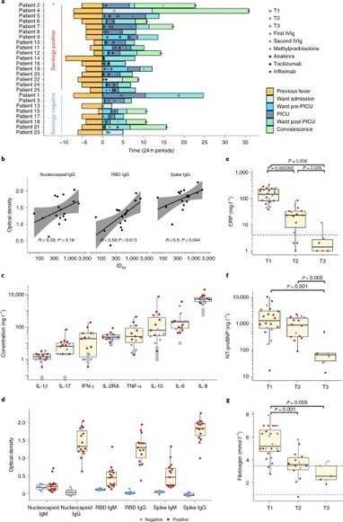
A wide spectrum of clinical manifestations has become a hallmark of the severe acute respiratory syndrome coronavirus 2 (SARS-CoV-2) COVID-19 pandemic, although the immunological underpinnings of diverse disease outcomes remain to be defined. We performed detailed characterization of B cell response …
|
|
Scooped by
Gilbert C FAURE
October 6, 2020 3:38 AM
|
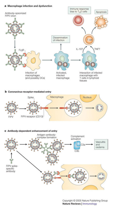
How the body responds to a coronavirus infection.
|
|
Scooped by
Gilbert C FAURE
September 29, 2020 3:17 AM
|
Severe acute respiratory syndrome coronavirus–2 (SARS-CoV-2) is the novel coronavirus initially detected in Wuhan, China, and is responsible for the w…
|
|
Scooped by
Gilbert C FAURE
September 14, 2020 6:03 AM
|
The COVID-19 pandemic is shining a spotlight on the field of immunology like never before. To appreciate the diverse ways in which immunologists have contributed, Nature Reviews Immunology invited the president of the International Union of Immunological Societies and the presidents of 15 other national immunology societies to discuss how they and their members responded following the emergence of severe acute respiratory syndrome coronavirus 2 (SARS-CoV-2). For this Viewpoint, Nature Reviews Immunology asked the presidents of 16 immunology societies from around the world to discuss how their society and its members responded to the COVID-19 pandemic. Their answers highlight the incredible contributions that immunologists around the globe have made following the emergence of severe acute respiratory syndrome coronavirus 2 (SARS-CoV-2).
|
|
Scooped by
Gilbert C FAURE
August 21, 2020 2:25 AM
|
Abstract. Beside the commonly described pulmonary expression of the coronavirus disease 2019 (COVID-19), major vascular events have been reported. The objectiv
|
|
Scooped by
Gilbert C FAURE
August 7, 2020 2:50 AM
|
Severe acute respiratory syndrome coronavirus 2 (SARS-CoV-2) has caused the rapidly unfolding coronavirus disease 2019 (COVID-19) pandemic1,2. Clinical manifestations of COVID-19 vary, ranging from asymptomatic infection to respiratory failure. The mechanisms determining such variable outcomes remain unresolved. Here, we investigated SARS-CoV-2 spike glycoprotein (S)-reactive CD4+ T cells in peripheral blood of patients with COVID-19 and SARS-CoV-2-unexposed healthy donors (HD). We detected SARS-CoV-2 S-reactive CD4+ T cells in 83% of patients with COVID-19 but also in 35% of HD. S-reactive CD4+ T cells in HD reacted primarily to C-terminal S epitopes, which show a higher homology to spike glycoproteins of human endemic coronaviruses, compared to N-terminal epitopes. S-reactive T cell lines generated from SARS-CoV-2-naive HD responded similarly to C-terminal S of human endemic coronaviruses 229E and OC43 and SARS-CoV-2, demonstrating the presence of S-cross-reactive T cells, probably generated during past encounters with endemic coronaviruses. The role of pre-existing SARS-CoV-2 cross-reactive T cells for clinical outcomes remains to be determined in larger cohorts. However, the presence of S-cross-reactive T cells in a sizable fraction of the general population may affect the dynamics of the current pandemic, and has important implications for the design and analysis of upcoming COVID-19 vaccine trials.
|
|
Scooped by
Gilbert C FAURE
July 28, 2020 4:30 AM
|
Abstract Cytokine storm syndrome (CSS) is a critical clinical condition induced by a cascade of cytokine activation, characterized by overwhelming systemic inflammation, hyperferritinaemia, haemody...
|
|
Scooped by
Gilbert C FAURE
July 25, 2020 4:40 AM
|
Understanding the clinical presentation of severe acute respiratory syndrome coronavirus 2 (SARS-CoV-2) infection and prognosis in children is a major issue. Children often present mild symptoms, and some severe forms require paediatric intensive care, with in some cases a fatal prognosis. Our aim was to identify the epidemiological characteristics, clinical presentation, and prognosis of children with coronavirus disease 2019 (Covid-19) hospitalized in Paris suburb hospitals. In this prospective, observational, multicentre study, we included children hospitalized in paediatric departments of Paris suburb hospitals from 23 March 2020 to 10 May 2020, during the national lockdown in France with confirmed SARS-CoV-2 infection (positive RNA test on a nasopharyngeal swab) or highly suspected infection (clinical, biological, and/or radiological data features suggestive for SARS-CoV-2 infection). A total of 192 children were included for confirmed (n = 157) or highly suspected (n = 35) SARS-CoV-2 infection. The median age was one year old (interquartile range 0.125–11) with a sex ratio 1.3:1. Fever was recorded in 147 (76.6%) children and considered poorly tolerated in 29 (15.1%). The symptoms ranged from rhinorrhoea (34.4%) and gastrointestinal (35.5%) to respiratory distress (25%). Only 10 (5.2%) children had anosmia and five (2.6%) had chest pain. An underlying condition was identified in almost 30% of the children in our study. Overall, 24 (12.5%) children were admitted to paediatric intensive care units, 12 required mechanical ventilation, and three died. For children in Paris suburbs, most cases of Covid-19 showed mild or moderate clinical expression. However, one-eighth of children were admitted to paediatric intensive care units and three died.
|
|
Scooped by
Gilbert C FAURE
July 5, 2020 5:36 AM
|
At the end of 2002, the first cases of severe acute respiratory syndrome (SARS) were reported, and in the following year, SARS resulted in considerable mortality and morbidity worldwide. SARS is caused by a novel species of coronavirus (SARS-CoV) and ...
|
|
Scooped by
Gilbert C FAURE
June 30, 2020 8:27 AM
|
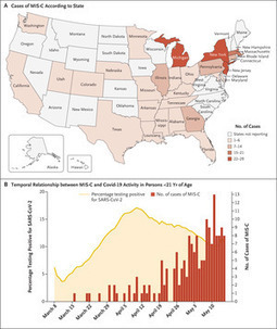
Abstract Background Understanding the epidemiology and clinical course of multisystem inflammatory syndrome in children (MIS-C) and its temporal association with coronavirus disease 201
|
|
Rescooped by
Gilbert C FAURE
from Virus World
June 25, 2020 5:32 AM
|
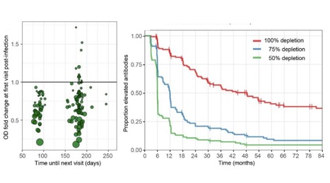
In the current COVID-19 pandemic a key unsolved question is the duration of acquired immunity in recovered individuals. The recent emergence of SARS-CoV-2 precludes a direct study on this virus, but the four seasonal human coronaviruses may reveal common characteristics applicable to all human coronaviruses. We monitored healthy subjects over a time span of 35 years (1985-2020), providing a total of 2473 follow up person-months, and determined a) the time to reinfection by the same seasonal coronavirus and b) the dynamics of coronavirus antibody depletion post-infection. An alarmingly short duration of protective immunity to coronaviruses was found. Reinfections occurred frequently at 12 months post-infection and there was for each virus a substantial reduction in antibody levels as soon as 6 months post-infection. Preprint Available (June 16, 2020): https://doi.org/10.1101/2020.05.11.20086439
Via Juan Lama
|
|
Scooped by
Gilbert C FAURE
May 29, 2020 8:53 AM
|
Coronavirus in nonhuman primates We urgently need vaccines and drug treatments for coronavirus disease 2019 (COVID-19). Even under these extreme circumstances, we must have animal models for rigorous testing of new strategies. Rockx et al. have undertaken a comparative study of three human coronaviruses in cynomolgus macaques: severe acute respiratory syndrome–coronavirus (SARS-CoV) (2002), Middle East respiratory syndrome (MERS)–CoV (2012), and SARS-CoV-2 (2019), which causes COVID-19 (see the Perspective by Lakdawala and Menachery). The most recent coronavirus has a distinct tropism for the nasal mucosa but is also found in the intestinal tract. Although none of the older macaques showed the severe symptoms that humans do, the lung pathology observed was similar. Like humans, the animals shed virus for prolonged periods from their upper respiratory tracts, and like influenza but unlike the 2002 SARS-CoV, this shedding peaked early in infection. It is this cryptic virus shedding that makes case detection difficult and can jeopardize the effectiveness of isolation. Science, this issue p. 1012; see also p. 942 Abstract The current pandemic coronavirus, severe acute respiratory syndrome–coronavirus 2 (SARS-CoV-2), was recently identified in patients with an acute respiratory syndrome, coronavirus disease 2019 (COVID-19). To compare its pathogenesis with that of previously emerging coronaviruses, we inoculated cynomolgus macaques with SARS-CoV-2 or Middle East respiratory syndrome (MERS)–CoV and compared the pathology and virology with historical reports of SARS-CoV infections. In SARS-CoV-2–infected macaques, virus was excreted from nose and throat in the absence of clinical signs and detected in type I and II pneumocytes in foci of diffuse alveolar damage and in ciliated epithelial cells of nasal, bronchial, and bronchiolar mucosae. In SARS-CoV infection, lung lesions were typically more severe, whereas they were milder in MERS-CoV infection, where virus was detected mainly in type II pneumocytes. These data show that SARS-CoV-2 causes COVID-19–like disease in macaques and provides a new model to test preventive and therapeutic strategies. After the first reports of an outbreak of an acute respiratory syndrome in China in December 2019, a novel coronavirus, severe acute respiratory syndrome–coronavirus 2 (SARS-CoV-2), was identified (1, 2). As of 14 March 2020, over 140,000 cases were reported worldwide with more than 5400 deaths, surpassing the combined number of cases and deaths of two previously emerging coronaviruses, SARS-CoV and Middle East respiratory syndrome (MERS)–CoV (3). The disease caused by this virus, coronavirus disease 2019 (COVID-19), is characterized by a range of symptoms, including fever, cough, dyspnoea, and myalgia in most cases (2). In severe cases, bilateral lung involvement with ground-glass opacity is the most common chest computed tomography (CT) finding (4). Similarly to the 2002–2003 outbreak of SARS, the severity of COVID-19 disease is associated with increased age and/or a comorbidity, although severe disease is not limited to these risk groups (5). However, despite the large number of cases and deaths, limited information is available on the pathogenesis of this virus infection. Two reports on the histological examination of the lungs of three patients showed bilateral diffuse alveolar damage (DAD), pulmonary edema, and hyaline membrane formation, indicative of acute respiratory distress syndrome (ARDS), as well as characteristic syncytial cells in the alveolar lumen (6, 7), similar to findings during the 2002–2003 outbreak of SARS-CoV (8). The pathogenesis of SARS-CoV infection was previously studied in a nonhuman primate model (cynomolgus macaques) where aged animals were more likely to develop disease (9–13). In the current study, SARS-CoV-2 infection was characterized in the same animal model and compared with infection with MERS-CoV and historical data on SARS-CoV (9, 10, 12). First, two groups of four cynomolgus macaques [both young adult (young), 4 to 5 years of age; and old adult (aged), 15 to 20 years of age] were inoculated by a combined intratracheal (IT) and intranasal (IN) route with a SARS-CoV-2 strain from a German traveler returning from China. No overt clinical signs were observed in any of the infected animals, except for a serous nasal discharge in one aged animal on day 14 post inoculation (p.i.). No significant weight loss was observed in any of the animals during the study. By day 14 p.i., all remaining animals seroconverted as revealed by the presence of SARS-CoV-2–specific antibodies against the virus S1 domain and nucleocapsid proteins in their sera (fig. S1). As a measure of virus shedding, nasal, throat, and rectal swabs were assayed for virus by reverse transcription–quantitative polymerase chain reaction (RT-qPCR) and virus culture. In nasal swabs, SARS-CoV-2 RNA peaked by day 2 p.i. in young animals and by day 4 p.i. in aged animals and was detected up to at least day 8 p.i. in two out of four animals and up to day 21 p.i. in one out of four animals (Fig. 1A). Overall, higher levels of SARS-CoV-2 RNA were detected in nasal swabs of aged animals compared with young animals. SARS-CoV-2 RNA in throat swabs peaked at day 1 p.i. in young and day 4 p.i. in aged animals and decreased more rapidly over time by comparison with the nasal swabs but could still be detected intermittently up to day 10 p.i. (Fig. 1B). Low levels of infectious virus were cultured from throat and nasal swabs up to day 2 and 4, respectively (table S1). In support of virus shedding by these animals, environmental sampling was performed to determine potential contamination of surfaces. Environmental sampling indicated the presence of low levels of SARS-CoV-2 RNA on surfaces through both direct contact (hands) and indirect contamination within the isolator (table S2). SARS-CoV-2 RNA was only detected in a rectal swab from one animal on day 14 p.i., and no viral RNA was detected in whole blood at any time point throughout the study. On autopsy of four macaques on day 4 p.i., two had foci of pulmonary consolidation in the lungs (Fig. 2A). One animal (aged: 17 years) showed consolidation in the right middle lobe, representing less than 5% of the lung tissue. A second animal (young: 5 years) had two foci in the left lower lobe, representing about 10% of the lung tissue (Fig. 2A). The consolidated lung tissue was well circumscribed, red-purple, level, and less buoyant than normal. The other organs in these two macaques, as well as the respiratory tract and other organs of the other two animals, were normal. Virus replication was assessed by RT-qPCR on day 4 p.i. in tissues from the respiratory, digestive, urinary, and cardiovascular tracts and from endocrine and central nervous systems, as well as from various lymphoid tissues. Virus replication was primarily restricted to the respiratory tract (nasal cavity, trachea, bronchi, and lung lobes) with highest levels of SARS-CoV-2 RNA in lungs (Fig. 1C). In three out of four animals, SARS-CoV-2 RNA was also detected in ileum and tracheo-bronchial lymph nodes (Fig. 1C). The main histological lesion in the consolidated pulmonary tissues of both the young and aged animals involved the alveoli and bronchioles and consisted of areas with acute or more advanced DAD (Fig. 2B). In these areas, the lumina of alveoli and bronchioles were variably filled with protein-rich edema fluid, fibrin, and cellular debris; alveolar macrophages; and fewer neutrophils and lymphocytes (Fig. 2, C to E). There was epithelial necrosis with extensive loss of epithelium from alveolar and bronchiolar walls. Hyaline membranes were present in a few damaged alveoli. In areas with more advanced lesions, the alveolar walls were moderately thickened and lined by cuboidal epithelial cells (type II pneumocyte hyperplasia), and the alveolar lumina were empty. Alveolar and bronchiolar walls were thickened by edema fluid, mononuclear cells, and neutrophils. There were aggregates of lymphocytes around small pulmonary vessels. Moderate numbers of lymphocytes and macrophages were present in the lamina propria and submucosa of the bronchial walls, and a few neutrophils were detected in the bronchial epithelium. Regeneration of epithelium was seen in some bronchioles, visible as an irregular layer of squamous to high cuboidal epithelial cells with hyperchromatic nuclei. There were occasional multinucleated giant cells (syncytia) free in the lumina of bronchioles and alveoli (Fig. 2F) and, based on positive pan-keratin staining and negative CD68 staining, these appeared to originate from epithelial cells (Fig. 2F, inset). SARS-CoV-2 antigen expression was detected in moderate numbers of type I pneumocytes and a few type II pneumocytes within foci of DAD (Fig. 2, G and H, and fig. S2). The pattern of staining was similar to that in lung tissue from SARS-CoV–infected macaques (positive control). SARS-CoV-2 antigen expression was not observed in any of the syncytia. In addition, SARS-CoV-2 antigen expression was detected in nonlesional tissues of all lung lobes in three out of four macaques (both young and one aged) in a few type I and II pneumocytes, bronchial ciliated epithelial cells, and bronchiolar ciliated epithelial cells. The other aged macaque, without virological or pathological evidence of SARS-CoV-2 infection in the lungs, did have SARS-CoV-2 antigen expression in ciliated epithelial cells of nasal septum (Fig. 2I), nasal concha, and palatum molle, in the absence of associated histopathological changes. No SARS-CoV antigen expression was detected in other sampled tissues, including brain and intestine. To assess the severity of infection with SARS-CoV-2 compared with MERS-CoV, we inoculated young cynomolgus macaques (3 to 5 years of age) with MERS-CoV via the IN and IT route. All animals remained free of clinical signs. At day 21 p.i., all remaining animals (n = 2) seroconverted as revealed by the presence of MERS-CoV–specific antibodies in their sera by ELISA (fig. S3). MERS-CoV RNA was detected in nasal (Fig. 3A) and throat swabs (Fig. 3B) on days 1 to 11 p.i., with peaks on days 1 and 2 p.i., respectively. Low levels [between 1 and 85 median tissue culture infectious dose (TCID50) equivalent/ml] of MERS-CoV RNA were detected in rectal swabs on days 2 and 3 p.i. At autopsy of four macaques at day 4 p.i., three animals had foci of pulmonary consolidation, characterized by slightly depressed areas in the lungs, representing less than 5% of the lung tissue (Table 1). Similar to SARS-CoV-2 infection in both young and aged animals, on day 4 p.i., MERS-CoV RNA was primarily detected in the respiratory tract of inoculated animals (Fig. 3C). Infectious virus titers were comparable to those of SARS-CoV-2 infection, but lower compared to SARS-CoV infection, of young macaques (Table 1). In addition, MERS-CoV RNA was detected in the spleen (Table 1). Table 1 Comparative pathogenesis of SARS-CoV-2, MERS-CoV, and SARS-CoV infections in cynomolgus macaques. Max, maximum; Ref., reference. View this table: View popup View inline Consistent with the presence of virus in the lower respiratory tract at day 4 p.i., histopathological changes characteristic for DAD were observed in the lungs of inoculated animals (Fig. 3D). The alveolar septa were thickened owing to infiltration of neutrophils and macrophages, and to moderate type II pneumocyte hyperplasia and hypertrophy. In the alveolar lumina, there were increased numbers of alveolar macrophages and some edema fluid containing fibrin and some neutrophils (Fig. 3D). Few syncytial cells were seen in the alveolar lumina. MERS-CoV antigen was not detected in tissues on day 4 p.i. in any part of the respiratory tract. We therefore sampled four young macaques at day 1 p.i. At this time, we observed multifocal expression of viral antigen, predominantly in type II pneumocytes and occasionally in type I pneumocytes, bronchiolar and bronchial epithelial cells, and some macrophages (Fig. 3D). In summary, we inoculated young and aged cynomolgus macaques with a low-passage clinical isolate of SARS-CoV-2, which resulted in productive infection in the absence of overt clinical signs. Recent studies in human cases have shown that presymptomatic and asymptomatic cases can also shed virus (14, 15). Increased age did not affect disease outcome, but there was prolonged viral shedding in the upper respiratory tract of aged animals. Prolonged shedding has been observed in both SARS-CoV-2 and SARS-CoV patients (16, 17). SARS-CoV-2 shedding in our asymptomatic model peaked early in the course of infection, similar to what is seen in symptomatic patients (16). Also, SARS-CoV-2 antigen was detected in ciliated epithelial cells of nasal mucosae at day 4 p.i., which was not seen for SARS-CoV (10) or MERS-CoV infections (this study) in this animal model. Viral tropism for the nasal mucosa fits with efficient respiratory transmission, as has been seen for influenza A virus (18). This early peak in virus shedding for SARS-CoV-2 is similar to that of influenza virus shedding (19) and may explain why case detection and isolation may not be as effective for SARS-CoV-2 as it was for the control of SARS-CoV (20). SARS-CoV-2 was primarily detected in tissues of the respiratory tract; however, SARS-CoV-2 RNA was also detectable in other tissues such as intestines, in line with a recent report (21). Similar results regarding viral shedding and tissue and cell tropism were recently also reported after SARS-CoV-2 inoculation in rhesus macaques. However, unlike in our model, SARS-CoV-2 infection in rhesus macaques does result in transient respiratory disease and weight loss (22, 23). Two out of four animals had foci of DAD on day 4 p.i. The colocalization of SARS-CoV-2 antigen expression and DAD provides strong evidence that SARS-CoV-2 infection caused this lesion. The histological character of the DAD, including alveolar and bronchiolar epithelial necrosis, alveolar edema, hyaline membrane formation, and accumulation of neutrophils, macrophages, and lymphocytes, corresponds with the limited pathological analyses of human COVID-19 cases (6, 7). In particular, the presence of syncytia in the lung lesions is characteristic of respiratory coronavirus infections. Whereas MERS-CoV primarily infects type II pneumocytes in cynomolgus macaques, both SARS-CoV and SARS-CoV-2 also infect type I pneumocytes. Injury to type I pneumocytes can result in pulmonary edema, and formation of hyaline membranes (24), which may explain why hyaline membrane formation is a hallmark of SARS and COVID-19 (7, 10) but not frequently reported for MERS (25, 26). These data show that cynomolgus macaques are permissive to SARS-CoV-2 infection, shed virus for a prolonged period of time, and display COVID-19–like disease. In this nonhuman primate model, SARS-CoV-2 replicates efficiently in respiratory epithelial cells throughout the respiratory tract, including nasal cavity, bronchi, bronchioles, and alveoli. Replication in the upper respiratory tract fits with efficient transmission between hosts, whereas replication in the lower respiratory tract fits with the development of lung disease. An in-depth comparison of infection with SARS-CoV, MERS-CoV, and SARS-CoV-2 in this model may identify key pathways in the pathogenesis of these emerging viruses. This study provides a new infection model that will be critical in the evaluation and licensure of preventive and therapeutic strategies against SARS-CoV-2 infection for use in humans, as well as for evaluating the efficacy of repurposing species-specific existing treatments, such as pegylated interferon (12). Supplementary Materials science.sciencemag.org/content/368/6494/1012/suppl/DC1 Materials and Methods Figs. S1 to S3 Tables S1 and S2 References (27–30) MDAR Reproducibility Checklist http://www.sciencemag.org/about/science-licenses-journal-article-reuse This is an article distributed under the terms of the Science Journals Default License. References and Notes ↵ J. F. Chan, S. Yuan, K.-H. Kok, K. K.-W. To, H. Chu, J. Yang, F. Xing, J. Liu, C. C.-Y. Yip, R. W.-S. Poon, H.-W. Tsoi, S. K.-F. Lo, K.-H. Chan, V. K.-M. Poon, W.-M. Chan, J. D. Ip, J.-P. Cai, V. C.-C. Cheng, H. Chen, C. K.-M. Hui, K.-Y. Yuen, A familial cluster of pneumonia associated with the 2019 novel coronavirus indicating person-to-person transmission: A study of a family cluster. Lancet 395, 514–523 (2020). doi:10.1016/S0140-6736(20)30154-9pmid:31986261OpenUrlCrossRefPubMed ↵ C. Huang, Y. Wang, X. Li, L. Ren, J. Zhao, Y. Hu, L. Zhang, G. Fan, J. Xu, X. Gu, Z. Cheng, T. Yu, J. Xia, Y. Wei, W. Wu, X. Xie, W. Yin, H. Li, M. Liu, Y. Xiao, H. Gao, L. Guo, J. Xie, G. Wang, R. Jiang, Z. Gao, Q. Jin, J. Wang, B. Cao, Clinical features of patients infected with 2019 novel coronavirus in Wuhan, China. Lancet 395, 497–506 (2020). doi:10.1016/S0140-6736(20)30183-5pmid:31986264OpenUrlCrossRefPubMed ↵ E. Mahase, Coronavirus covid-19 has killed more people than SARS and MERS combined, despite lower case fatality rate. BMJ 368, m641 (2020). doi:10.1136/bmj.m641pmid:32071063OpenUrlFREE Full Text ↵ H. Shi, X. Han, N. Jiang, Y. Cao, O. Alwalid, J. Gu, Y. Fan, C. Zheng, Radiological findings from 81 patients with COVID-19 pneumonia in Wuhan, China: A descriptive study. Lancet Infect. Dis. 20, 425–434 (2020). doi:10.1016/S1473-3099(20)30086-4pmid:32105637OpenUrlCrossRefPubMed ↵ N. Chen, M. Zhou, X. Dong, J. Qu, F. Gong, Y. Han, Y. Qiu, J. Wang, Y. Liu, Y. Wei, J. Xia, T. Yu, X. Zhang, L. Zhang, Epidemiological and clinical characteristics of 99 cases of 2019 novel coronavirus pneumonia in Wuhan, China: A descriptive study. Lancet 395, 507–513 (2020). doi:10.1016/S0140-6736(20)30211-7pmid:32007143OpenUrlCrossRefPubMed ↵ Z. Xu, L. Shi, Y. Wang, J. Zhang, L. Huang, C. Zhang, S. Liu, P. Zhao, H. Liu, L. Zhu, Y. Tai, C. Bai, T. Gao, J. Song, P. Xia, J. Dong, J. Zhao, F.-S. Wang, Pathological findings of COVID-19 associated with acute respiratory distress syndrome. Lancet Respir. Med. 8, 420–422 (2020). doi:10.1016/S2213-2600(20)30076-Xpmid:32085846OpenUrlCrossRefPubMed ↵ S. Tian, W. Hu, L. Niu, H. Liu, H. Xu, S.-Y. Xiao, Pulmonary pathology of early-phase 2019 novel coronavirus (COVID-19) pneumonia in two patients with lung cancer. J. Thorac. Oncol. S1556-0864(20)30132-5 (2020). doi:10.1016/j.jtho.2020.02.010pmid:32114094OpenUrlCrossRefPubMed ↵ J. M. Nicholls, L. L. M. Poon, K. C. Lee, W. F. Ng, S. T. Lai, C. Y. Leung, C. M. Chu, P. K. Hui, K. L. Mak, W. Lim, K. W. Yan, K. H. Chan, N. C. Tsang, Y. Guan, K. Y. Yuen, J. S. Peiris, Lung pathology of fatal severe acute respiratory syndrome. Lancet 361, 1773–1778 (2003). doi:10.1016/S0140-6736(03)13413-7pmid:12781536 ↵ R. A. Fouchier, T. Kuiken, M. Schutten, G. van Amerongen, G. J. J. van Doornum, B. G. van den Hoogen, M. Peiris, W. Lim, K. Stöhr, A. D. M. E. Osterhaus, Koch’s postulates fulfilled for SARS virus. Nature 423, 240 (2003). doi:10.1038/423240apmid:12748632 ↵ T. Kuiken, R. A. M. Fouchier, M. Schutten, G. F. Rimmelzwaan, G. van Amerongen, D. van Riel, J. D. Laman, T. de Jong, G. van Doornum, W. Lim, A. E. Ling, P. K. S. Chan, J. S. Tam, M. C. Zambon, R. Gopal, C. Drosten, S. van der Werf, N. Escriou, J.-C. Manuguerra, K. Stöhr, J. S. M. Peiris, A. D. M. E. Osterhaus, Newly discovered coronavirus as the primary cause of severe acute respiratory syndrome. Lancet 362, 263–270 (2003). doi:10.1016/S0140-6736(03)13967-0pmid:12892955 S. L. Smits, A. de Lang, J. M. A. van den Brand, L. M. Leijten, W. F. van IJcken, M. J. C. Eijkemans, G. van Amerongen, T. Kuiken, A. C. Andeweg, A. D. M. E. Osterhaus, B. L. Haagmans, Exacerbated innate host response to SARS-CoV in aged non-human primates. PLOS Pathog. 6, e1000756 (2010). doi:10.1371/journal.ppat.1000756pmid:20140198OpenUrlCrossRefPubMed ↵ B. L. Haagmans, T. Kuiken, B. E. Martina, R. A. M. Fouchier, G. F. Rimmelzwaan, G. van Amerongen, D. van Riel, T. de Jong, S. Itamura, K.-H. Chan, M. Tashiro, A. D. M. E. Osterhaus, Pegylated interferon-alpha protects type 1 pneumocytes against SARS coronavirus infection in macaques. Nat. Med. 10, 290–293 (2004). doi:10.1038/nm1001pmid:14981511 ↵ B. Rockx, F. Feldmann, D. Brining, D. Gardner, R. LaCasse, L. Kercher, D. Long, R. Rosenke, K. Virtaneva, D. E. Sturdevant, S. F. Porcella, J. Mattoon, M. Parnell, R. S. Baric, H. Feldmann, Comparative pathogenesis of three human and zoonotic SARS-CoV strains in cynomolgus macaques. PLOS ONE 6, e18558 (2011). doi:10.1371/journal.pone.0018558pmid:21533129OpenUrlCrossRefPubMed ↵ C. Li, F. Ji, L. Wang, L. Wang, J. Hao, M. Dai, Y. Liu, X. Pan, J. Fu, L. Li, G. Yang, J. Yang, X. Yan, B. Gu, Asymptomatic and Human-to-Human Transmission of SARS-CoV-2 in a 2-Family Cluster, Xuzhou, China. Emerg. Infect. Dis. 26 (2020). doi:10.3201/eid2607.200718pmid:32228809OpenUrlCrossRefPubMed ↵ A. Kimball, K. M. Hatfield, M. Arons, A. James, J. Taylor, K. Spicer, A. C. Bardossy, L. P. Oakley, S. Tanwar, Z. Chisty, J. M. Bell, M. Methner, J. Harney, J. R. Jacobs, C. M. Carlson, H. P. McLaughlin, N. Stone, S. Clark, C. Brostrom-Smith, L. C. Page, M. Kay, J. Lewis, D. Russell, B. Hiatt, J. Gant, J. S. Duchin, T. A. Clark, M. A. Honein, S. C. Reddy, J. A. Jernigan, A. Baer, L. M. Barnard, E. Benoliel, M. S. Fagalde, J. Ferro, H. G. Smith, E. Gonzales, N. Hatley, G. Hatt, M. Hope, M. Huntington-Frazier, V. Kawakami, J. L. Lenahan, M. D. Lukoff, E. B. Maier, S. McKeirnan, P. Montgomery, J. L. Morgan, L. A. Mummert, S. Pogosjans, F. X. Riedo, L. Schwarcz, D. Smith, S. Stearns, K. J. Sykes, H. Whitney, H. Ali, M. Banks, A. Balajee, E. J. Chow, B. Cooper, D. W. Currie, J. Dyal, J. Healy, M. Hughes, T. M. McMichael, L. Nolen, C. Olson, A. K. Rao, K. Schmit, N. G. Schwartz, F. Tobolowsky, R. Zacks, S. Zane; Public Health – Seattle & King County; CDC COVID-19 Investigation Team, Asymptomatic and Presymptomatic SARS-CoV-2 Infections in Residents of a Long-Term Care Skilled Nursing Facility - King County, Washington, March 2020. MMWR Morb. Mortal. Wkly. Rep. 69, 377–381 (2020). doi:10.15585/mmwr.mm6913e1pmid:32240128OpenUrlCrossRefPubMed ↵ L. Zou, F. Ruan, M. Huang, L. Liang, H. Huang, Z. Hong, J. Yu, M. Kang, Y. Song, J. Xia, Q. Guo, T. Song, J. He, H.-L. Yen, M. Peiris, J. Wu, SARS-CoV-2 Viral Load in Upper Respiratory Specimens of Infected Patients. N. Engl. J. Med. 382, 1177–1179 (2020). doi:10.1056/NEJMc2001737pmid:32074444OpenUrlCrossRefPubMed ↵ J. S. Peiris, C. M. Chu, V. C. C. Cheng, K. S. Chan, I. F. N. Hung, L. L. M. Poon, K. I. Law, B. S. F. Tang, T. Y. W. Hon, C. S. Chan, K. H. Chan, J. S. C. Ng, B. J. Zheng, W. L. Ng, R. W. M. Lai, Y. Guan, K. Y. Yuen; HKU/UCH SARS Study Group, Clinical progression and viral load in a community outbreak of coronavirus-associated SARS pneumonia: A prospective study. Lancet 361, 1767–1772 (2003). doi:10.1016/S0140-6736(03)13412-5pmid:12781535 ↵ M. Richard, J. M. A. van den Brand, T. M. Bestebroer, P. Lexmond, D. de Meulder, R. A. M. Fouchier, A. C. Lowen, S. Herfst, Influenza A viruses are transmitted via the air from the nasal respiratory epithelium of ferrets. Nat. Commun. 11, 766 (2020). doi:10.1038/s41467-020-14626-0pmid:32034144OpenUrlCrossRefPubMed ↵ C. C. Li, L. Wang, H.-L. Eng, H.-L. You, L.-S. Chang, K.-S. Tang, Y.-J. Lin, H.-C. Kuo, I.-K. Lee, J.-W. Liu, E.-Y. Huang, K. D. Yang, Correlation of pandemic (H1N1) 2009 viral load with disease severity and prolonged viral shedding in children. Emerg. Infect. Dis. 16, 1265–1272 (2010). doi:10.3201/eid1608.091918pmid:20678321 ↵ X. Pang, Z. Zhu, F. Xu, J. Guo, X. Gong, D. Liu, Z. Liu, D. P. Chin, D. R. Feikin, Evaluation of control measures implemented in the severe acute respiratory syndrome outbreak in Beijing, 2003. JAMA 290, 3215–3221 (2003). doi:10.1001/jama.290.24.3215pmid:14693874 ↵ F. Xiao, M. Tang, X. Zheng, Y. Liu, X. Li, H. Shan, Evidence for gastrointestinal infection of SARS-CoV-2. Gastroenterology S0016-5085(20)30282-1 (2020). doi:10.1053/j.gastro.2020.02.055pmid:32142773OpenUrlCrossRefPubMed ↵W. D. Linlin Bao et al., Reinfection could not occur in SARS-CoV-2 infected rhesus macaques. bioRxiv 2020.03.13.990226 (2020). https://doi.org/10.1101/2020.03.13.990226. ↵V. J. Munster et al., Respiratory disease and virus shedding in rhesus macaques inoculated with SARS-CoV-2. bioRxiv 2020.03.21.001628 (2020). https://doi.org/10.1101/2020.03.21.001628. ↵ L. B. Ware, M. A. Matthay, The acute respiratory distress syndrome. N. Engl. J. Med. 342, 1334–1349 (2000). doi:10.1056/NEJM200005043421806pmid:10793167 ↵ W. J. Shieh, C.-H. Hsiao, C. D. Paddock, J. Guarner, C. S. Goldsmith, K. Tatti, M. Packard, L. Mueller, M.-Z. Wu, P. Rollin, I.-J. Su, S. R. Zaki, Immunohistochemical, in situ hybridization, and ultrastructural localization of SARS-associated coronavirus in lung of a fatal case of severe acute respiratory syndrome in Taiwan. Hum. Pathol. 36, 303–309 (2005). doi:10.1016/j.humpath.2004.11.006pmid:15791576 ↵ D. L. Ng, F. Al Hosani, M. K. Keating, S. I. Gerber, T. L. Jones, M. G. Metcalfe, S. Tong, Y. Tao, N. N. Alami, L. M. Haynes, M. A. Mutei, L. Abdel-Wareth, T. M. Uyeki, D. L. Swerdlow, M. Barakat, S. R. Zaki, Clinicopathologic, Immunohistochemical, and Ultrastructural Findings of a Fatal Case of Middle East Respiratory Syndrome Coronavirus Infection in the United Arab Emirates, April 2014. Am. J. Pathol. 186, 652–658 (2016). doi:10.1016/j.ajpath.2015.10.024pmid:26857507OpenUrlCrossRefPubMed ↵ V. S. Raj, H. Mou, S. L. Smits, D. H. W. Dekkers, M. A. Müller, R. Dijkman, D. Muth, J. A. A. Demmers, A. Zaki, R. A. M. Fouchier, V. Thiel, C. Drosten, P. J. M. Rottier, A. D. M. E. Osterhaus, B. J. Bosch, B. L. Haagmans, Dipeptidyl peptidase 4 is a functional receptor for the emerging human coronavirus-EMC. Nature 495, 251–254 (2013). doi:10.1038/nature12005pmid:23486063 B. L. Haagmans, J. M. A. van den Brand, L. B. Provacia, V. S. Raj, K. J. Stittelaar, S. Getu, L. de Waal, T. M. Bestebroer, G. van Amerongen, G. M. G. M. Verjans, R. A. M. Fouchier, S. L. Smits, T. Kuiken, A. D. M. E. Osterhaus, Asymptomatic Middle East respiratory syndrome coronavirus infection in rabbits. J. Virol. 89, 6131–6135 (2015). doi:10.1128/JVI.00661-15pmid:25810539 V. M. Corman, O. Landt, M. Kaiser, R. Molenkamp, A. Meijer, D. K. W. Chu, T. Bleicker, S. Brünink, J. Schneider, M. L. Schmidt, D. G. J. C. Mulders, B. L. Haagmans, B. van der Veer, S. van den Brink, L. Wijsman, G. Goderski, J.-L. Romette, J. Ellis, M. Zambon, M. Peiris, H. Goossens, C. Reusken, M. P. G. Koopmans, C. Drosten, Detection of 2019 novel coronavirus (2019-nCoV) by real-time RT-PCR. Euro Surveill. 25, 25 (2020). doi:10.2807/1560-7917.ES.2020.25.3.2000045pmid:31992387OpenUrlCrossRefPubMed ↵ N. M. A. Okba, M. A. Müller, W. Li, C. Wang, C. H. GeurtsvanKessel, V. M. Corman, M. M. Lamers, R. S. Sikkema, E. de Bruin, F. D. Chandler, Y. Yazdanpanah, Q. Le Hingrat, D. Descamps, N. Houhou-Fidouh, C. B. E. M. Reusken, B.-J. Bosch, C. Drosten, M. P. G. Koopmans, B. L. Haagmans, Severe acute respiratory syndrome coronavirus 2-specific antibody responses in coronavirus disease 2019 patients. Emerg. Infect. Dis. 26, 10.3201/eid2607.200841 (2020). doi:10.3201/eid2607.200841pmid:32267220OpenUrlCrossRefPubMed Acknowledgments: We thank L. de Meulder, A. van der Linden, I. Chestakova, and F. van der Panne for technical assistance. We also thank Y. Kap, D. Akkermans, V. Vaes, M. Sommers, and F. Meijers for assistance with the animal studies. Funding: This research is (partly) financed by the NWO Stevin Prize awarded to M.K. by the Netherlands Organisation for Scientific Research (NWO), H2020 grant agreement 874735-VEO to M.K., NIH/NIAID contract HHSN272201400008C to R.F., and H2020 grant agreement 101003651–MANCO to B.H. Author contributions: Conceptualization, B.R., T.K., R.F., M.K., RdS, M.F. B.H.; investigation, B.R., T.K., S.H., T.B., M.L., D.d.M., G.v.A., J.v.d.B., N.O., D.S., P.v.R., L.L.; resources, B.H., C.D., E.V., B.V., J.L.; supervision, B.R. and B.H.; writing, original draft, B.R., T.K., and B.H.; writing–review and editing, all authors; funding acquisition: S.H., B.H., R.F., and M.K. Animal welfare: Research was conducted in compliance with the Dutch legislation for the protection of animals used for scientific purposes (2014, implementing EU Directive 2010/63) and other relevant regulations. The licensed establishment where this research was conducted (Erasmus MC) has an approved OLAW Assurance no. A5051-01. Research was conducted under a project license from the Dutch competent authority and the study protocol (no. 17-4312) was approved by the institutional Animal Welfare Body. Competing interests: T.B., R.F., and B.H. are listed as inventors on a patent application related to MERS-coronavirus diagnostics, therapy and vaccines. Other authors declare no competing interests. Data and materials availability: All data are available in the main text or the supplementary materials. This work is licensed under a Creative Commons Attribution 4.0 International (CC BY 4.0) license, which permits unrestricted use, distribution, and reproduction in any medium, provided the original work is properly cited. To view a copy of this license, visit https://creativecommons.org/licenses/by/4.0/. This license does not apply to figures/photos/artwork or other content included in the article that is credited to a third party; obtain authorization from the rights holder before using such material.
|
|
Scooped by
Gilbert C FAURE
May 15, 2020 2:34 PM
|
New findings suggest past infections may offer some protection against the novel coronavirus
|
|
|
Scooped by
Gilbert C FAURE
October 6, 2020 10:15 AM
|
Abstract The clinical progression of the severe acute respiratory syndrome coronavirus 2 (SARS‐CoV‐2) infection (COVID‐19) to critical illness is associated with an exaggerated immune response, lea...
|
|
Scooped by
Gilbert C FAURE
October 1, 2020 9:47 AM
|
Dr Ian Frayling says he is suffering with frequent, all-consuming tiredness, along with nausea, irritable bowel syndrome and lack of energy
|
|
Rescooped by
Gilbert C FAURE
from Virus World
September 23, 2020 4:40 AM
|
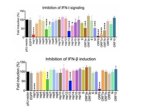
The coronavirus disease 2019 (COVID-19) is determined by SARS-CoV-2 replication and host immune response, but studies evaluating viral evasion of immune response are lacking. Here we employed unbiased screening to identify SARS-CoV-2 proteins that antagonize type-I interferon (IFN-I) response. Three proteins were found to antagonize IFN-I production via distinct mechanisms: nsp6 binds TBK1 to suppress IRF3 phosphorylation; nsp13 binds and blocks TBK1 phosphorylation; and ORF6 binds importin KPNA2 to inhibit IRF3 nuclear translocation. Two sets of viral proteins were identified to antagonize IFN-I signaling through blocking STAT1/STAT2 phosphorylation or nuclear translocation. Remarkably, SARS-CoV-2 nsp1 and nsp6 suppressed IFN-I signaling more efficiently than SARS-CoV and MERS-CoV. Thus, when treated with IFN-I, a SARS-CoV2 replicon replicated to a higher level than chimeric replicons containing nsp1 or nsp6 from SARS-CoV or MERS-CoV. Altogether, the study has provided insights on SARS-CoV-2 evasion of IFN-I response and its potential impact on viral transmission and pathogenesis. Published in Cell Reports (September 19, 2020):
Via Juan Lama

|
Suggested by
Société Francaise d'Immunologie
September 11, 2020 3:22 PM
|
SARS-CoV-2 is an enveloped, positive-sense, single-stranded RNA beta coronavirus belonging to the order of nidovirales. In the first three months since its ...
|
|
Scooped by
Gilbert C FAURE
August 18, 2020 9:37 AM
|
Recent reports highlight a new clinical syndrome in children related to severe acute respiratory syndrome coronavirus 2 (SARS-CoV-2)1—multisystem inflammatory syndrome in children (MIS-C)—which comprises multiorgan dysfunction and systemic inflammation2–13. We performed peripheral leukocyte phenotyping in 25 children with MIS-C, in the acute (n = 23; worst illness within 72 h of admission), resolution (n = 14; clinical improvement) and convalescent (n = 10; first outpatient visit) phases of the illness and used samples from seven age-matched healthy controls for comparisons. Among the MIS-C cohort, 17 (68%) children were SARS-CoV-2 seropositive, suggesting previous SARS-CoV-2 infections14,15, and these children had more severe disease. In the acute phase of MIS-C, we observed high levels of interleukin-1β (IL-1β), IL-6, IL-8, IL-10, IL-17, interferon-γ and differential T and B cell subset lymphopenia. High CD64 expression on neutrophils and monocytes, and high HLA-DR expression on γδ and CD4+CCR7+ T cells in the acute phase, suggested that these immune cell populations were activated. Antigen-presenting cells had low HLA-DR and CD86 expression, potentially indicative of impaired antigen presentation. These features normalized over the resolution and convalescence phases. Overall, MIS-C presents as an immunopathogenic illness1 and appears distinct from Kawasaki disease. Characterization of a cohort of children with multisystem inflammatory syndrome associated with SARS-CoV-2 infection provides insights into the immunopathogenic features of the disease.
|
|
Scooped by
Gilbert C FAURE
August 1, 2020 4:26 AM
|
The pathogenesis of coronavirus disease 2019 (COVID-19) may be envisaged as the dynamic
interaction between four vicious feedback loops chained or happening at once. These are the viral loop, the hyperinflammatory loop, the non-canonical renin-angiotensin system (RAS) axis loop, and the...
|
|
Scooped by
Gilbert C FAURE
July 27, 2020 8:22 AM
|
Recent studies have provided insights into the pathogenesis of coronavirus disease 2019 (COVID-19)1–4. Yet, longitudinal immunological correlates of disease outcome remain unclear. Here, we serially analysed immune responses in 113 COVID-19 patients with moderate (non-ICU) and severe (ICU) disease. Immune profiling revealed an overall increase in innate cell lineages with a concomitant reduction in T cell number. We identify an association between early, elevated cytokines and worse disease outcomes. Following an early increase in cytokines, COVID-19 patients with moderate disease displayed a progressive reduction in type-1 (antiviral) and type-3 (antifungal) responses. In contrast, patients with severe disease maintained these elevated responses throughout the course of disease. Moreover, severe disease was accompanied by an increase in multiple type 2 (anti-helminths) effectors including, IL-5, IL-13, IgE and eosinophils. Unsupervised clustering analysis identified 4 immune signatures, representing (A) growth factors, (B) type-2/3 cytokines, (C) mixed type-1/2/3 cytokines, and (D) chemokines that correlated with three distinct disease trajectories of patients. The immune profile of patients who recovered with moderate disease was enriched in tissue reparative growth factor signature (A), while the profile for those with worsened disease trajectory had elevated levels of all four signatures. Thus, we identified development of a maladapted immune response profile associated with severe COVID-19 outcome and early immune signatures that correlate with divergent disease trajectories.
|
|
Scooped by
Gilbert C FAURE
July 6, 2020 2:54 AM
|
Click here to edit the content Six months of coronavirus: the mysteries scientists are still racing to solve
|
|
Scooped by
Gilbert C FAURE
June 30, 2020 1:25 PM
|
It’s now clear that nearly everyone who recovers from coronavirus disease 2019 (COVID-19) produces antibodies that specifically target SARS-CoV-2, the novel coronavirus that causes the infection. Yet many critical questions remain. A major one is: just how well do those particular antibodies neutralize the virus to fight off the infection and help someone recover from…
|
|
Scooped by
Gilbert C FAURE
June 26, 2020 5:00 AM
|
From serology to T-cells, there’s still a lot we don’t know about how immunity to coronavirus works, says immunology lecturer Zania Stamataki
|
|
Scooped by
Gilbert C FAURE
May 31, 2020 2:44 PM
|
We developed and validated 2 species-independent protein-based assays to detect Middle East respiratory syndrome coronavirus functional antibodies that can block virus receptor-binding or sialic acid-attachment.
|
|
Scooped by
Gilbert C FAURE
May 18, 2020 10:27 AM
|
There’s still a lot to learn about SARS-CoV-2, the novel coronavirus that causes COVID-19. But it has been remarkable and gratifying to watch researchers from around the world pull together and share their time, expertise, and hard-earned data in the urgent quest to control this devastating virus.
|