Your new post is loading...

|
Scooped by
I2BC Paris-Saclay
February 19, 2024 10:25 AM
|
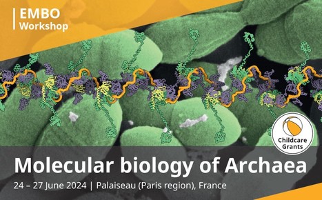
EMBO conference "Molecular Biology of Archaea" 23-27th of june 2024 in Palaiseau (near Paris)!
The registration in now open for the next EMBO conference "Molecular Biology of Archaea" that will take place near Paris (Palaiseau) on 23-27th of june 2024! Archaea are fascinating organisms with a bacteria-like morphology, but information-processing systems that are homologous to eukaryotic counterparts, for instance replication, transcription and translation. Recently, novel groups of archaea (e.g. Asgard archaea) have been discovered that represent the closest living relatives to eukaryotes and shed new light on their evolution. Archaeal research has led to seminal change-of-concept mechanistic discoveries in the area of molecular biology, and many studies are currently aiming to unravel the global environmental impact of archaea. Their viruses have a remarkable diversity of morphotypes, exceeding that of bacterial or eukaryotic viruses.The EMBO Workshop on Molecular biology of Archaea will cover cutting-edge research in various fields with a focus on archaeal molecular biology, evolution, and ecology and their interconnections. The proposed talks will present data at different scales ranging from single molecule and structural studies to archaeal physiology, metabolism and ecological impact. This is of high interest as molecular studies of archaea are a very active field, reflecting the wealth of metagenomics information revealing novel biology that can now be addressed by the latest experimental and computational techniques. all information: https://meetings.embo.org/event/24-archaea contact: Tamara BASTA-LE BERRE <tamara.basta@i2bc.paris-saclay.fr>
|
|
Scooped by
I2BC Paris-Saclay
February 19, 2024 10:14 AM
|
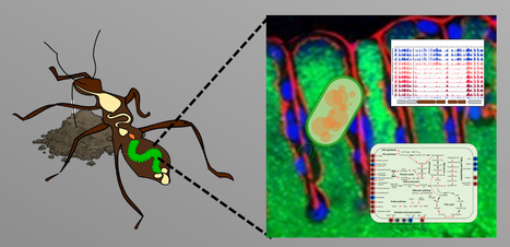
Transposon sequencing reveals genes enabling insect gut colonization by the symbiont Caballeronia insecticola
High-througput genetic screens with Tn-seq in an insect gut bacterium reveals gut-derived nutrients consumed by the symbiont. Caballeronia insecticola is a bacterium belonging to the Burkholderia sensu lato, able to colonize multiple environments like soils and the gut of the bean bug Riptortus pedestris. We constructed a saturated Himar1 mariner transposon library and revealed by transposon-sequencing (Tn-seq) that 498 protein-coding genes constitute the essential genome of C. insecticola for growth in free-living conditions. By comparing essential gene sets of C. insecticola and seven related Burkholderia s.l. strains, only 120 common genes were identified indicating that a large part of the essential genome is strain-specific. In order to reproduce specific nutritional conditions that are present in the gut of R. pedestris, we grew the mutant library in minimal media supplemented with candidate gut nutrients and identified several condition-dependent fitness-defect genes by Tn-seq. To validate the robustness of the approach, insertion mutants in six fitness genes were constructed and their growth-deficiency in media supplemented with the corresponding nutrient was confirmed. The mutants were further tested for their efficiency in R. pedestris gut colonization, confirming that gluconeogenic carbon sources, taurine and inositol, are nutrients consumed by the symbiont in the gut. Thus, our study provides insights about specific contributions provided by the insect host to the bacterial symbiont. More information: https://doi.org/10.1093/ismeco/ycad001 Contact: Peter MERGAERT <peter.mergaert@i2bc.paris-saclay.fr>
|
|
Scooped by
I2BC Paris-Saclay
February 19, 2024 10:10 AM
|
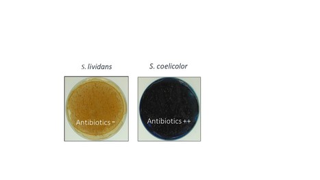
The stringent response is strongly activated in the high antibiotic producer, Streptomyces coelicolor
The stringent response controls positively antibiotic biosynthesis. Antibiotics are thus part of the stringent response. In most bacteria, the stringent response (SR) was initially characterized as a response to nitrogen (N) limitation resulting into the depletion of aminoacylated tRNAs leading to the stalling of ribosomes on mRNA. The recruitment of the ppGpp synthetase, RelA, at the stalled ribosomes activates (p)ppGpp synthesis from GTP. ppGpp that is the mediator of the SR controls negatively, at the transcriptional and translational levels, the expression of most ribosomal proteins leading to the down regulation of the translational process and thus of growth.
The model strains Streptomyces coelicolor (SC) and Streptomyces lividans (SL), strong and weak producers of the same antibiotics, respectively, were grown in condition of phosphate (Pi) limitation or proficiency and the abundance of proteins of their translational apparatus was compared. This study revealed that the expression of RelA was induced in Pi limitation suggesting that, besides N limitation, Pi limitation also contributes to the triggering of the SR. Interestingly, most proteins of the translational apparatus had a similar or slightly higher abundance in SL than in SC, in Pi limitation whereas most of these proteins were far more abundant in SL than in SC, in Pi proficiency. This indicated an alleviation of the SR in Pi proficiency in SL, but not in SC. This suggested an alteration of Pi up-take and/or Pi-mediated regulation in SC whose molecular basis remain to be elucidated.
Interestingly, the production of specialized metabolites in SC (CDA, RED and ACT) is usually concomitant of phases of growth slow down and it is known that ppGpp controls positively the expression of their biosynthetic pathways. Their production could thus be considered as part of the SR. Indeed, these metabolites were proposed to regulate negatively, through different processes, the energetic metabolism and thus the generation of ATP, in SC, a process that might contribute to the slower growth rate of SC compared to SL. More information: https://www.sciencedirect.com/science/article/pii/S0923250823001547?via%3Dihub Contact: Marie-Joëlle VIROLLE <marie-joelle.virolle@i2bc.paris-saclay.fr>
|
|
Scooped by
I2BC Paris-Saclay
January 30, 2024 10:07 AM
|
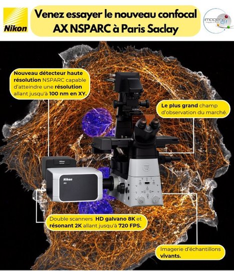
Nikon presents the AX-NSPARC at Imagerie-Gif - From January 30 to February 9 -
Nikon is presenting its latest confocal microscope to the Paris-Saclay community, equipped with a new detector for very high-resolution images (XY : 100 nm). From January 30 to February 9, 8 days of demonstrations on the Gif Imaging platform will enable you to test the microscope with your own samples . Don't hesitate to come and take advantage of this opportunity. More information: here
|
|
Scooped by
I2BC Paris-Saclay
January 30, 2024 9:21 AM
|
Antigen self-anchoring onto bacteriophage T5 capsid-like particles for vaccine design
Self-anchoring of large antigens onto Capsid-Like Particles derived from bacteriophage T5 paves the way for the development of a new vaccination platform that is highly immunogenic without the need for extrinsic adjuvant. The constant need for immunization to prevent life-threatening diseases worldwide is urging the search for new vaccines. The promises of vaccines based on virus-like particles stimulate demand for universal non-infectious virus-like platforms that can be efficiently grafted with large antigens. In this study, we harnessed the modularity and extreme affinity of the decoration protein pb10 for the capsid of bacteriophage T5 to design a novel Ag delivery platform. DNA-free T5 capsid-like particles (T5-CLP) were decorated with chimeric proteins formed of pb10 fused to the model antigen ovalbumin (Ova). SPR experiments demonstrated that these proteins retained picomolar affinity for T5-CLP, while cryo-EM studies attested to the full occupancy of the 120 capsid binding sites. Mice immunisation with CLP-bound pb10-Ova chimeras elicited strong long-lasting anti-Ova humoral responses involving a large panel of isotypes, as well as CD8+ T cell responses, without any extrinsic adjuvant. Therefore, T5-CLP constitutes a unique DNA-free bacteriophage capsid able to display a regular array of large antigens through highly efficient chemical-free anchoring. Its ability to elicit robust immune responses paves the way for further development of this novel vaccination platform. More information: https://www.nature.com/articles/s41541-023-00798-5 Contact: Pascale BOULANGER <pascale.boulanger-biard@i2bc.paris-saclay.fr>
On les trouve chez les animaux comme chez les plantes, les bactéries ou les virus. Elles permettent le battement régulier de votre cœur, la digestion de votre dernier repas, maintiennent votre corps à une température constante, participent à la lutte contre les infections qui vous menacent et bien plus encore... Les protéines sont partout où la vie existe et la science les étudie depuis des décennies, mais elles sont si nombreuses et si diverses que leur monde reste encore à explorer. Nous vous invitons à découvrir ces recherches passionnantes en compagnie de Marie-Hélène Le Du, chercheuse CEA à l'Institut de Biologie Intégrative de la Cellule – I2BC (DRF/Joliot/I2BC). RDV le mardi 30 janvier 2024 à 20h. Entrée libre sous réserve de places disponibles (le nombre de places est limité à 280). Vous pourrez aussi suivre cette conférence en direct sur la chaîne Youtube du CEA.
Via Life Sciences UPSaclay
|
|
Scooped by
I2BC Paris-Saclay
December 27, 2023 8:28 AM
|
Visualizing the DNA repair process by a photolyase at atomic resolution
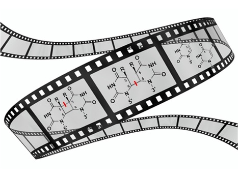
Molecular movies? No problem for time-resolved serial femtosecond X-ray crystallography! We have obtained 18 snapshots visualizing the whole DNA repair by a photolyase: from the initial electron transfer to the active site recovery and DNA reannealing. Photolyases, a ubiquitous class of flavoproteins, use blue light to repair DNA photolesions. In this work, we determined the structural mechanism of the photolyase-catalyzed repair of a cyclobutane pyrimidine dimer (CPD) lesion using time-resolved serial femtosecond crystallography (TR-SFX). We obtained 18 snapshots that show time-dependent changes in four reaction loci. We used these results to create a movie that depicts the repair of CPD lesions in the picosecond-to-nanosecond range, followed by the recovery of the enzymatic moieties involved in catalysis, completing the formation of the fully reduced enzyme-product complex at 500 nanoseconds. Finally, back-flip intermediates of the thymine bases to reanneal the DNA were captured at 25 to 200 microseconds. Our data cover the complete molecular mechanism of a photolyase and, importantly, its chemistry and enzymatic catalysis at work across a wide timescale and at atomic resolution. More information: https://www.science.org/doi/10.1126/science.add7795 Contact: Pavel MULLER <pavel.muller@i2bc.paris-saclay.fr>
|
|
Scooped by
I2BC Paris-Saclay
December 27, 2023 7:57 AM
|
First anti-CRISPR protein that inhibits the CRISPR-Cas defense system in Clostridioides difficile
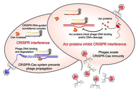
CRISPR-Cas adaptive immunity defends prokaryotes against their viruses named phages. In response, phages have evolved counter defense strategies to fight back. This study describes the identification of a phage protein to evade CRISPR-Cas immunity in a human pathogen Clostridioides difficile. Clostridioides difficile is the widespread anaerobic spore-forming bacterium that is a major cause of potentially lethal nosocomial infections associated with antibiotic therapy worldwide. Due to the increase in severe forms associated with a strong inflammatory response and higher recurrence rates, a current imperative is to develop synergistic and alternative treatments of C. difficile infections. In particular, phage therapy is regarded as a potential substitute for existing antimicrobial treatments. However, it faces challenges because C. difficile have highly active CRISPR-Cas immunity, which may be a specific adaptation to phage-rich and highly crowded gut environment. To overcome this defense, C. difficile phages must employ anti-CRISPR mechanisms. Here, we present the first anti-CRISPR protein that inhibits the CRISPR-Cas defense system in this pathogen. Our work offers insights into the interactions between C. difficile and its phages, paving the way for future CRISPR-based applications and development of effective phage therapy strategies combined with the engineering of virulent C. difficile infecting phages. More information: doi: 10.1128/msphere.00401-23 Contact: Olga SOUTOURINA <olga.soutourina@i2bc.paris-saclay.fr>
|
|
Scooped by
I2BC Paris-Saclay
December 27, 2023 7:52 AM
|
3D models of fungal chromosomes to enhance visual integration of omics data
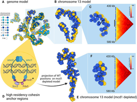
It has become easier to build 3D models of the entire genome based on Hi-C data, and thus benefit from the methodology and visualization tools developed for structural biology. In this paper, we show how seeing the spatial organization of chromosomes can bring new perspectives to omics data integration. The functions of eukaryotic chromosomes and their spatial architecture in the nucleus are reciprocally dependent. Hi-C experiments are routinely used to study chromosome 3D organization by probing chromatin interactions. Standard representation of the data has relied on contact maps that show the frequency of interactions between parts of the genome. In parallel, it has become easier to build 3D models of the entire genome based on the same Hi-C data, and thus benefit from the methodology and visualization tools developed for structural biology. 3D modeling of entire genomes leverages the understanding of their spatial organization. However, this opportunity for original and insightful modeling is underexploited. In this paper, we show how seeing the spatial organization of chromosomes can bring new perspectives to omics data integration. We assembled state-of-the-art tools into a workflow that goes from Hi-C raw data to fully annotated 3D models and we re-analysed public omics datasets available for three fungal species. Besides the well-described properties of the spatial organization of their chromosomes (Rabl conformation, hypercoiling and chromosome territories), our results highlighted (i) in Saccharomyces cerevisiae, the backbones of the cohesin anchor regions, which were aligned all along the chromosomes, (ii) in Schizosaccharomyces pombe, the oscillations of the coiling of chromosome arms throughout the cell cycle and (iii) in Neurospora crassa, the massive relocalization of histone marks in mutants of heterochromatin regulators. 3D modeling of the chromosomes brings new opportunities for visual integration of omics data. This holistic perspective supports intuition and lays the foundation for building new concepts. More information: https://academic.oup.com/nargab/article/5/4/lqad104/7458894 Contact: Gaëlle LELANDAIS <gaelle.lelandais@i2bc.paris-saclay.fr>
|
|
Scooped by
I2BC Paris-Saclay
December 1, 2023 9:15 AM
|
New: NGS small RNA library preparation training
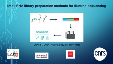
The I2BC NGS facility organizes a new training on the preparation of small RNA libraries with technical tips and tricks to conduct successful small RNA NGS experiments. MicroRNAs (miRNAs) and other types of small RNA (sRNA) molecules play important roles in the cell and their dysregulation has been linked to various diseases. Indeed, circulating miRNAs are found in various human body fluids and are of interest as potential new non-invasive biomarkers. Next-generation sequencing (NGS) allows a very sensitive and quantitative detection of sRNA expression profiles. However, NGS library preparation from sRNAs is technically relatively challenging as compared to NGS protocols for other applications. Classical sRNA library preparation methods often introduce serious biases leading to sequencing data that do not represent the original sRNA expression profiles. The new training organized by the I2BC NGS facility focusses on technical aspects of NGS library preparation from different types of sRNAs. These include natural sRNAs such as miRNAs, and artificial sRNAs such as ribosome-protected fragments (Ribo-seq) or co-immunoprecipitated sRNA fragments (e.g. CLIP-seq/RIP-seq). Various sRNA library preparation protocols exist and during the training advantages and pitfalls of these methods will be discussed. Specific adaptations required for different types of sRNA will be highlighted. A practical course will teach the participants how to prepare sRNA NGS libraries themselves and how to perform quality control. Together, this training will enable participants to prepare sRNA libraries themselves autonomously, to validate the quality of their libraries and to perform successful NGS experiments by being cautious of technical pitfalls. More information: https://cnrsformation.cnrs.fr/preparer-des-banques-ngs-a-partir-des-petits-arn-pour-la-technologie-illumina
|
|
Scooped by
I2BC Paris-Saclay
December 1, 2023 8:46 AM
|
The large ring-shaped BRCA2-HSF2BP complex is disassembled by BRME1, thus promoting Homologous Recombination: a cryo-EM study.
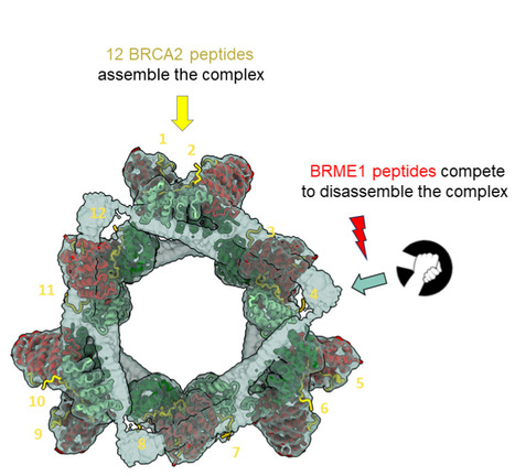
A short disordered and conserved motif of the tumor suppressor BRCA2 triggers the assembly of a large ring-shaped 24-mer of the meiotic HSF2BP protein, whereas the disordered C-terminal motif of the meiotic BRME1 dissociates this ring complex. In meiotic homologous recombination (HR), BRCA2 facilitates loading of the recombinases RAD51 and DMC1 at the sites of double-strand breaks (DSB). The HSF2BP-BRME1 complex interacts with BRCA2. Its absence causes a severe reduction in recombinase loading at meiotic DSB. We previously showed that, in somatic cancer cells ectopically producing HSF2BP, DNA damage can trigger HSF2BP-dependent degradation of BRCA2, which prevents HR. Here we report that, upon binding to BRCA2, HSF2BP forms octameric rings that are able to assemble into a large ring-shaped 24-mer. Addition of BRME1 leads to dissociation of both of these ring structures, and cancels the disruptive effect of HSF2BP on cancer cell resistance to DNA damage. It also prevents BRCA2 degradation during inter-strand DNA crosslink repair in Xenopus egg extracts. We propose that the control of HSF2BP-BRCA2 oligomerization by BRME1 ensures timely assembly of the ring complex that concentrates BRCA2 and controls its turnover, thus promoting HR. More information: https://www.science.org/doi/10.1126/sciadv.adi7352 Contact: Sophie ZINN JUSTIN <sophie.zinn@i2bc.paris-saclay.fr>
Portrait Jeune Chercheuse - Ambre Sala, chercheuse en biologie cellulaire
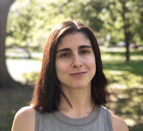
Chargée de recherche au CNRS, Ambre Sala a rejoint le département de biologie cellulaire de l’I2BC en février 2023, ou elle dirige l’équipe « Homéostasie des protéines dans le développement et le vieillissement ». Elle s’intéresse aux mécanismes qui régulent l'homéostasie des protéines (protéostasie), qui sont essentiels pour maintenir un protéome correctement replié et fonctionnel dans toutes les cellules. Ses recherches dans ce domaine se concentrent sur les chaperons moléculaires, qui assistent le repliement et le trafic des protéines et empêchent leur agrégation, ainsi que les voies de réponse au stress qui régulent le niveau d’expression des chaperons pour rétablir la protéostasie lors de de l’accumulation de protéines mal repliées. Pendant son doctorat dans le laboratoire de Pierre Genevaux à l'Université de Toulouse, elle a étudié l'évolution du chaperon bactérien SecB depuis son rôle canonique dans l'exportation des protéines via le translocon Sec vers le contrôle spécifique de systèmes toxine-antitoxine (TA) qui régulent la croissance bactérienne. Ces travaux ont apporté des connaissances fondamentales sur la remarquable plasticité du système chaperon SecB, et le processus d’évolution d’un chaperon générique vers le contrôle spécifique d'un système TA pour coupler la protéostasie à la croissance bactérienne. Pour ses études postdoctorales, Ambre a rejoint le laboratoire de Richard Morimoto à Northwestern University pour se concentrer sur la protéostasie et la réponse au stress dans le contexte du vieillissement. En utilisant le modèle Caenorhabditis elegans, elle a développé des approches de génétique, d'imagerie et de transcriptomique pour identifier et caractériser une nouvelle voie transcellulaire qui relie l'intégrité de l'embryon dans l'utérus à la protéostasie maternelle et à la résilience au stress. Ces travaux ont éclairé des stratégies systémiques qui coordonnent la reproduction, la protéostasie et le métabolisme pour déterminer la robustesse de l'organisme pendant le vieillissement. Ambre a rejoint l’I2BC après avoir obtenu une position de chercheur CNRS ainsi qu’un financement ATIP-Avenir en 2022 pour étudier les mécanismes qui protègent le protéome des cellules de la lignée germinale pour assurer la reproduction. L’équipe développe des approches génétiques, biochimiques et d'imagerie qui exploitent les avantages du modèle C. elegans, pour identifier les rôles cellulaires et la composition des réseaux de chaperons qui soutiennent la fonction des cellules germinales et leur résilience face au stress protéotoxique. « You can‘t always get what you want, but if you try sometimes, you just might find, you get what you need. » - Linda Randall, quoting the Rolling stones to describe chaperones overlapping functions. Contact : ambre.sala@i2bc.paris-saclay.fr
Via Life Sciences UPSaclay
|
|
Scooped by
I2BC Paris-Saclay
October 23, 2023 5:12 AM
|
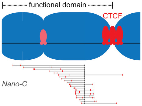
Clustered binding of the CTCF protein creates functional domains in the human and mouse genome with improved structure.
Clusters of binding sites for the CTCF protein create boundaries between functional domains in the mouse and human genomes. Scientists at the I2BC have determined the characteristics of these clusters and how individual sites contribute to domain separation. Topologically Associating Domains (TADs) regionalize the human and mouse genome into domains that are essential for processes like gene regulation and DNA repair. Most boundaries between TADs bind the CTCF insulator protein, which is responsible for the blocking of interactions between neighboring TADs. CTCF is thereby directly responsible for improved functional precision and reduced regulatory interference between domains in the genome. Although a clustering of CTCF binding sites had previously been observed at TAD boundaries, its structural impact remained to be determined.
Scientists in the Chromatin Dynamics group at the I2BC, in collaboration with groups at the Ecole Normale Supérieure (Paris) and the University of Pennsylvania (USA), found that three features of CTCF binding are enriched at TAD boundaries, including the presence of closely-spaced copies of its DNA binding motifs, a reduced distance between clusters of motifs and an enrichment of clusters with improved binding affinity. Using Nano-C technology, a new approach to measure the blocking of DNA interactions, they subsequently determined the blocking capacity of individual sites of CTCF binding. These studies reveal that these sites individually contribute to the blocking of interactions between domains, but in an incomplete manner. Grouping of multiple sites of CTCF binding in the genome thus creates a stepwise insulation between neighboring TADs, thereby improving the focus of genomic functions. More information: https://doi.org/10.1038/s41467-023-41265-y Contact: Daan NOORDERMEER <daan.noordermeer@i2bc.paris-saclay.fr>
|
|
|
Scooped by
I2BC Paris-Saclay
February 19, 2024 10:19 AM
|
Prize "Relève de l'étoile" for the excellence of the research work carried out by Alexia Royer
Alexia Royer received the prize "Relève de l'étoile" for her article “Clostridioides difficile S-Layer Protein A (SlpA) Serves as a General Phage Receptor” published in Microbiology Spectrum in February 2023. Antibiotics have been used for more than 80 years to treat bacterial infections, but bacteria have developed resistance to these treatments, making them difficult to eradicate. One of the side effects of antibiotics is that they tend to kill a broad spectrum of bacteria, including “good bacteria” important for the proper functioning of the human body. The resulting bacterial imbalance, called dysbiosis, will open the door to opportunistic bacteria such as Clostridioides difficile. This bacterium is the main cause of severe diarrhea in industrialized countries. As the infection is now treated with antibiotics, which are themselves responsible for the infection, many relapses are reported. In the laboratories of Professor Louis-Charles Fortier and Professor Olga Soutourina, we are interested in phage therapy, which is a treatment based on the use of bacteria-killing viruses called bacteriophages. Phages have the advantage of being specific and targeting a species or a few bacterial strains unlike antibiotics. Thus, they could be used as an alternative or complementary therapy to antibiotics to treat patients in therapeutic failure. The key step to set up a phage therapy is to understand the mechanism of recognition of the bacteria by phages. For a phage to attack and kill a bacterium, it must first attach to a receptor on its surface. In the article, we identified the SlpA protein as the receptor on the surface of the bacteria that is targeted by phages. We have shown that the infection specificity of phages is linked to the presence of different forms of SlpA, called isoforms, each strain expressing one isoform. We also identified a domain of SlpA, named D2, playing a role in receptor recognition by certain phages. These data are essential because they improve our understanding of phage/bacteria interactions and make phage therapy possible one day against C. difficile infections. https://frq.gouv.qc.ca/histoire-et-rapport/releve-etoile-jacques-genest-decembre-2023/ Alexia ROYER <alexia.royer@i2bc.paris-saclay.fr>
|
|
Scooped by
I2BC Paris-Saclay
February 19, 2024 10:12 AM
|
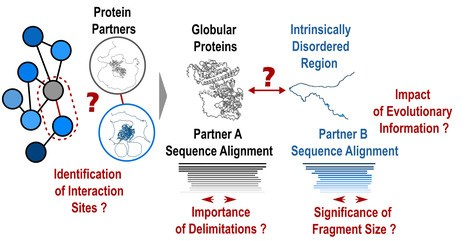
Protein-protein interactions: how to push forward the limits of the revolutionary AlphaFold2 programme?
AlphaFold2 is revolutionising protein structure prediction and structural biology practices. However, it may prove less effective for certain protein assemblies, particularly when they depend on intrinsically disordered regions. In an article published in Nature Communications, researchers from the I2BC show that applying a fragmentation strategy to the protein partners of such assemblies very significantly improves AlphaFold2’s prediction capacity. Mapping protein-protein interaction networks is essential for understanding the dynamics of cellular functions and their cross-regulation. Precise knowledge of interaction sites makes it possible to specifically perturb the proteins in these networks and understand the synergies and competitions that ensure cell function. Unfortunately, a great amount of structural information is still lacking to provide a detailed understanding of the organisation of interaction networks. The AlphaFold2 artificial intelligence programme has demonstrated a remarkable ability to predict the structures of protein assemblies that have co-evolved over long time scales. Its performance remained poorly characterised for assemblies involving intrinsically disordered regions, which often mediate transient and dynamic interactions during evolution. In a study published in the journal Nature Communications, researchers from the AMIG team at the Institut de Biologie Intégrative de la Cellule – I2BC (CNRS/CEA/UPSaclay, Gif-sur-Yvette) have shown that AlphaFold2 performs poorly if large disordered regions are used directly for prediction (40% success rate). A protein fragmentation strategy was found to be particularly well adapted to predicting the interfaces between folded domains and small protein motifs that fold on contact with the partner. Applied on a large scale using the Jean Zay HPC infrastructure on more than 900 complexes, this strategy achieved a success rate of almost 90%, a very encouraging result for the systematic screening of protein interaction networks. Nevertheless, the study calls for vigilance with regard to the risks of detecting false positives, which will be at the heart of future developments in artificial intelligence strategies such as AlphaFold2. More information: https://www.nature.com/articles/s41467-023-44288-7 Contact: Jessica ANDREANI and Raphaël GUEROIS <jessica.andreani@i2bc.paris-saclay.fr> <raphael.guerois@i2bc.paris-saclay.fr>
|
|
Scooped by
I2BC Paris-Saclay
February 19, 2024 10:08 AM
|
Composition of Poxvirus Core Revealed
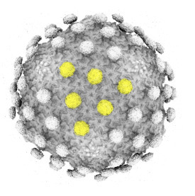
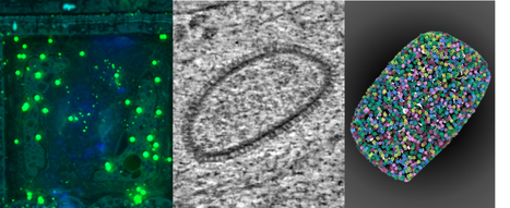
A collaboration between groups at I2BC, CSSB, MPI, and PEI, reveals the structure and the flexibility of A10 trimers that compose the palisade layer of the Vaccinia virus core encasing the viral genome. Vaccinia virus is the model for the family of poxviruses, however, the structure of the viral particle used to propagate infection is poorly understood. The group of Emmanuelle Quemin at I2BC together with collaborators at CSSB in Hamburg, MPI for Biophysics in Frankfurt, and PEI in Langen, have revealed the composition and architecture of Vaccinia virus core that encases the viral genome. Their findings have been published in Nature Structural & Molecular Biology.
By combining cryo-electron tomography with subtomogram averaging and AlphaFold2, the authors were able to identify components of the core of Vaccinia virus. During entry, there is the fusion of the viral and cellular membranes that lead to the subsequent release of the viral core inside the host cell. As these are rare and fast events difficult to tackle by cellular cryo electron tomography, the researchers studied the core both in situ and in vitro, using virusal particles treated with detergents to access so-called “naked” cores containing the viral genome still. In parallel to the large dataset obtained in vitro, entering cores found inside cells were also analysed under native conditions at CSSB cryo-EM facility directed by Kay Grünewald in Hamburg, Germany,
The four groups involved focused more specifically on the outer layer of the core called the palisade that displayed a dense and organized surface of tubular protrusions that we refer to as stakes. Their study determined that these protrusions are trimers of the viral protein A10, previously known as one of the major core proteins. Here, the stakes appeared as more randomly organized than reported in other recent published work and have an inherent flexibility.
While some poxviruses can spread in human populations as recently exemplified with the Mpox virus multi-country outbreak, improving our understanding of Vaccinia virus and its core sub-structure is key to shed light on conserved mechanisms of poxvirus infection and pathogenicity. More information: https://www.nature.com/articles/s41594-024-01218-5 Contact: Emmanuelle QUEMIN <emmanuelle.quemin@i2bc.paris-saclay.fr>
|
|
Scooped by
I2BC Paris-Saclay
January 30, 2024 9:23 AM
|
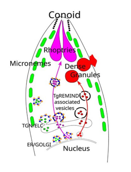
Toxoplasma membrane inositol phospholipid binding protein TgREMIND is essential for secretory organelle function and host infection
Apicomplexan parasites possess specialized secretory organelles called rhoptries, micronemes, and dense granules that play a vital role in host infection. In this study, we demonstrate that TgREMIND, a protein found in Toxoplasma gondii, is necessary for the biogenesis of rhoptries and dense granules. TgREMIND contains a Fes-CIP4 homology-Bin/Amphiphysin/Rvs (F-BAR) domain, which binds to membrane phospholipids, as well as a novel uncharacterized domain that we have named REMIND (regulator of membrane-interacting domain). Both the F-BAR domain and the REMIND are crucial for TgREMIND functions. When TgREMIND is depleted, there is a significant decrease in the abundance of dense granules and abnormal transparency of rhoptries, leading to a reduction in protein secretion from these organelles. The absence of TgREMIND inhibits host invasion and parasite dissemination, demonstrating that TgREMIND is essential for the proper function of critical secretory organelles required for successful infection by Toxoplasma. More information: doi: 10.1016/j.celrep.2023.113601 Contact: Stanislas TOMAVO <stanislas.tomavo@i2bc.paris-saclay.fr>
|
|
Scooped by
I2BC Paris-Saclay
January 30, 2024 9:19 AM
|
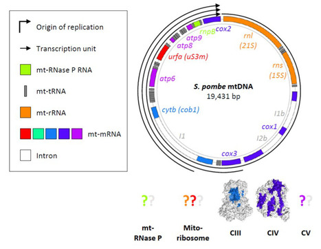
Schizosaccharomyces pombe as a fundamental model for research on mitochondrial gene expression: Progress, achievements and outlooks
This is a comprehensive and critical review on mitochondrial gene expression in fission yeast, presenting up-to-date knowledge, and emphasising numerous contributions of the unicellular model to both fundamental and biomedical research. Schizosaccharomyces pombe (fission yeast) is an attractive model for mitochondrial research. The organism resembles human cells in terms of mitochondrial inheritance, mitochondrial transport, sugar metabolism, mitogenome structure, and dependence of viability on the mitogenome (the petite-negative phenotype). Transcriptions of these genomes produce only a few polycistronic transcripts, which then undergo processing as per the tRNA punctuation model. In general, the machinery for mitochondrial gene expression is structurally and functionally conserved between fission yeast and humans. Furthermore, molecular research on S. pombe is supported by a considerable number of experimental techniques and database resources. Owing to these advantages, fission yeast has significantly contributed to biomedical and fundamental research. Here, we review the current state of knowledge regarding S. pombe mitochondrial gene expression, and emphasise the pertinence of fission yeast as both a model and tool, especially for studies on mitochondrial translation. More information: https://doi.org/10.1002/iub.2801 Contact: Nathalie BONNEFOY <nathalie.bonnefoy@i2bc.paris-saclay.fr>
|
|
Scooped by
I2BC Paris-Saclay
December 27, 2023 9:21 AM
|
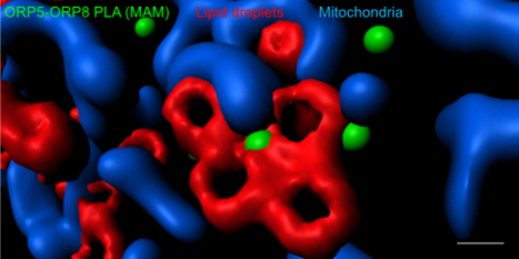
Visualization and Quantification of Endogenous Intra-Organelle Protein Interactions at ER-Mitochondria Contact Sites by Proximity Ligation Assays
|
|
Scooped by
I2BC Paris-Saclay
December 27, 2023 7:59 AM
|
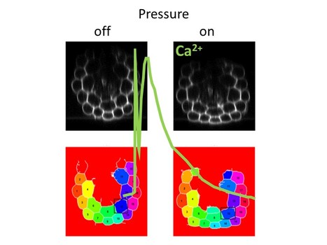
Straining the root on and off triggers local calcium signaling
Combining microfluidics and imaging, reveal that a pressure exerted on the root induces an elastic deformation and a calcium signal both at the onset and at the offset of the stimulation. How plant roots perceive mechanical cues encountered in the soil remains elusive. Combining microfluidics and imaging, we have shown that a pressure exerted on the root induces an elastic deformation and a calcium signal both at the onset and at the offset of the stimulation. Although the intensity of the calcium response increased with the pressure applied, successive stimuli led to a remarkable attenuation of the calcium signal. The calcium elevation was restricted to the tissue under pressure without propagation. This study published in Proceedings of the Royal Society B (https://doi.org/10.1098/rspb.2023.1462) contributes to elucidate the mechanisms of root adaptation to the mechanical cues generated by the soil.
This work was supported by the Région Ile de France through the DIM ELICIT program and by Saclay Plant Sciences through the DYNANO project. More information: doi: 10.1128/msphere.00401-23 Contact: Jean-Marie Frachisse – Sébastien Thomine <jean-marie.frachisse@i2bc.paris-saclay.fr> <sebastien.thomine@i2bc.paris-saclay.fr>
|
|
Scooped by
I2BC Paris-Saclay
December 27, 2023 7:55 AM
|
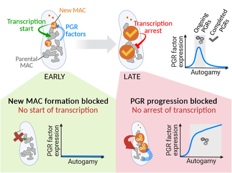
Inter-generational nuclear crosstalk controls the progression of the Paramecium developmental program
Perturbing the Paramecium transcriptome during somatic development reveals the existence of internuclear feed-forward and feedback transcriptional regulatory loops in a multinucleate unicellular eukaryote. The single-celled ciliate Paramecium tetraurelia harbors multiple nuclei within its cytoplasm: two germline micronuclei (MIC) dedicated to sexual reproduction and a somatic macronucleus (MAC) responsible for gene expression. During the sexual cycle, the parental MAC undergoes progressive degradation while, in the same cell, new MACs develop from copies of the zygotic MIC. Throughout this process, gene expression primarily occurs in the parental MAC, and clusters of developmentally regulated genes are successively switched on and off. Concomitantly, the genome of the new MACs is extensively rearranged and cleansed of its transposons and other DNA repeats, to become competent for gene expression. New MAC development has long been known to be controled by the parental MAC, which expresses key genes involved in programmed genome rearrangement (PGR). In this study, RNA deep sequencing and bioinformatic analysis of differential gene expression reveal a reciprocal relationship between the two generations of MACs: the formation of the new MACs is essential for the proper induction of a subset of developmental genes. Later in the sexual cycle, if PGR is hindered in the new MACs, a group of genes partially overlapping the former subset is upregulated, and no longer switched off. Thus, the progression of new MAC development regulates the gene expression program in the parental MAC at different stages. The set of co-expressed genes identified during the course of this work establishes a list of potential novel actors, whose roles in PGR will be explored in future studies. More information: https://doi.org/10.1093/nar/gkad1006 Contact: Olivier Arnaiz – Mireille Bétermier <olivier.arnaiz@i2bc.paris-saclay.fr> <mireille.betermier@i2bc.paris-saclay.fr>
|
|
Scooped by
I2BC Paris-Saclay
December 27, 2023 7:50 AM
|
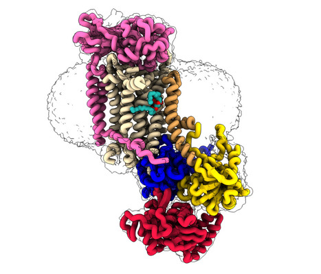
CryoEM studies of the human lipid transporter ATP8B1 involved in intrahepatic cholestasis
The structural characterization of the catalytic cycle of the lipid transporter ATP8B1 by cryoEM reveals fundamental aspects of its substrate specificity and of its regulation. Lipids are the main constituents of our cell membranes, which are formed as lipid bilayers. The distribution of lipids is far from uniform; it is asymmetric, with different lipid compositions in the inner and outer leaflet. This asymmetry is essential for a variety of cellular functions, from maintaining membrane homeostasis to enabling cell signaling and numerous other physiological processes including apoptosis and coagulation. P4-ATPases, also known as flippases, are key players in creating and maintaining this lipid asymmetry. These enzymes actively transport lipids from the outside (exoplasmic) leaflet to the inside (cytosolic) leaflet coupled to ATP hydrolysis and ensure the proper distributions of lipids. The ATP8B1-CDC50A flippase complex in particular, has been the subject of the current study, is involved in a rare inherited liver disease called intrahepatic cholestasis. The function of ATP8B1 lipid flippase is critical for the regulation of bile production, a vital substance in our digestive system, but the direct link within bile producing liver cells remains unknown. Additionally, recent studies have spotlighted the relevance of genetic variants in the regulatory segment of the ATP8B1 gene as a strong genetic marker for Alzheimer’s resilience. In the new study, the research team employed state-of-the-art cryo-electron microscopy techniques to capture nine different states associated with the lipid transport and determine structures at 2.4 to 3.1 Å overall resolution for these conformations. These structural insights, combined with functional and computational studies, reveal the inner workings of the human flippase ATP8B1-CDC50A complex and also resolves earlier discrepancies about the ATP8B1 transport substrates. Additionally, this work also provides key understanding about fine regulation by specific regulatory lipids known as phosphoinositides and by its autoinhibition mechanism. This work was recently awarded ”article of the month” by the Société Française de Biochimie et Biologie Moléculaire (SFBBM) in December 2023. More information: https://www.nature.com/articles/s41467-023-42828-9 Contact: Thibaud DIEUDONNE <thibaud.dieudonne@i2bc.paris-saclay.fr>
|
|
Scooped by
I2BC Paris-Saclay
December 1, 2023 8:47 AM
|
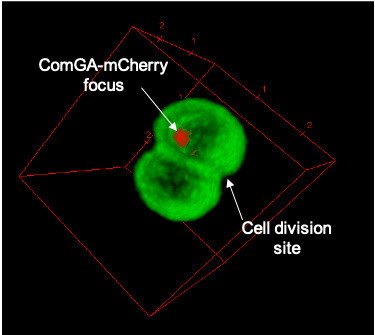
Natural transformation and cell division delay in competent Staphylococcus aureus
We hypothesize that S. aureus competent cells would initiate and then block cell division to ensure the success of natural transformation before the final constriction of the cytokinetic ring.. Genetic competence for natural transformation, considered one of the three main mechanisms leading to horizontal gene transfer in bacteria, is able to promote evolution, through genomic plasticity, and foster antibiotic resistance and virulence factors spreading. Conserved machinery and actors required to perform natural transformation have been shown to accumulate at different cellular localizations depending on the model organism considered. In this study, we investigate the transformation apparatus composition, localization, and dynamics in the human pathogen Staphylococcus aureus. We particularly show that most of the natural transformation actors co-localize in clusters. We also reveal that the localization of natural transformation proteins is dynamic, following the cell cycle. Ultimately, the natural transformation apparatus is preferentially established in the vicinity of the division septum. All these results demonstrate that DNA binding, uptake, and recombination are spatially and temporally coordinated to ensure S. aureus natural transformation. Finally, we hypothesize that S. aureus competent cells would initiate and then block cell division to ensure the success of natural transformation before the final constriction of the cytokinetic ring. More information: https://journals.asm.org/doi/10.1128/spectrum.02807-23 Contact: Nicolas MIROUZE <nicolas.mirouze@i2bc.paris-saclay.fr>
|
|
Scooped by
I2BC Paris-Saclay
December 1, 2023 8:44 AM
|
IP6 : an endogenous small molecule which promotes DNA repair.
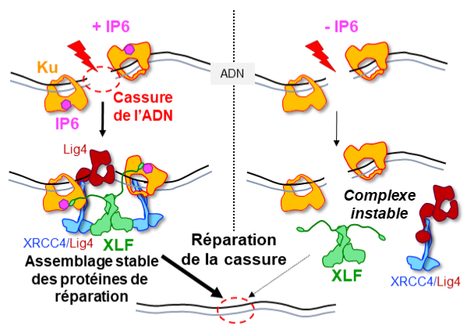
Structural studies by X-ray cristallography and CryoEM combined with cellular analyses show the stabilizing role of IP6 in assembly of DNA double-strand break repair in human. IP6, inositol hexaphosphate or phytic acid, is a product of cellular metabolism that binds to numerous proteins whose functions it regulates. In an article published in the journal Nucleic Acids Research, scientists from I2BC (UMR 9198, CNRS/CEA/UPSaclay, Gif-sur-Yvette), from lPBS (CNRS/Université Paul Sabatier) and from Institut for Structural and Chemical Biology (University of Leicester, UK) reveal how IP6 stabilizes the assembly of a complex responsible for DNA break repair in humans. Many cancer therapies induce DNA double-strand breaks. A DNA double-strand break represents the most serious type of DNA damage to a cell, since it amounts to splitting a chromosome in two. The resulting chromosome fragments can be lost or lead to translocations if they are not quickly rejoined. Unrepaired breaks most often lead to cell death, a property exploited to eradicate tumor cells during radiotherapy or certain chemotherapies. Yet DNA double-strand break repair pathways exist, and their performance in tumors determines the efficacy of these therapies. The dominant system for repairing DNA double-strand breaks in human cells is Non-Homologous End Joining, or NHEJ, initiated by the Ku protein, which rapidly encircles the ends of the break and acts as a hub for the other proteins needed to weld these ends together. Using two techniques for studying proteins at atomic scale (crystallography and cryo-electron microscopy), the scientists discovered how a small molecule produced by the body and sometimes used as a dietary supplement, inositol-hexaphosphate (IP6) or phytic acid, binds to the Ku protein. The scientists then analyzed the effect of IP6 on break repair in human cells. They modified the Ku protein to block its binding to IP6, and thus understand how the latter acts. The Ku mutants revealed that the presence of IP6 on the Ku protein stimulates its binding to the XLF protein, an essential step in efficiently welding the DNA break (see the model below). IP6 thus plays a key role in stabilizing the large complex of NHEJ proteins required for DNA break repair. This fundamental work resolves the question raised two decades ago regarding the role of IP6 in DNA double-strand break repair. It also opens up therapeutic prospects by identifying a new region on the Ku protein that could be targeted by small molecules to block DNA break repair in tumor cells. More information: https://academic.oup.com/nar/advance-article/doi/10.1093/nar/gkad863/7327077?login=false Contact: Jean-Baptiste CHARBONNIER <jb.charbonnier@i2bc.paris-saclay.fr>
|
|
Scooped by
I2BC Paris-Saclay
October 23, 2023 7:12 AM
|
New arrival at the BIOI2 bioinformatics plateform
Fadwa EL KHADDAR is working with us part time at the BIOI2 plateform since the beginning of October 2023 and has brought with her leading-edge experience in omics data analysis that will greatly enrich the range of services offered by the plateform. We welcome Fadwa EL KHADDAR, an engineer in bioinformatics who will be helping out part time (20%) at the BIOI2 plateform, as well as integrating two research teams of the I2BC (BIM and SSFA of the Genomes department) to which she will be lending her expertise in Omics data analysis. Fadwa first discovered bioinformatics during her Master's degree in Medical biotechnology of Rabat, Morocco, where she worked on phylogeny, synteny et pangenome analysis of L. monocytogenes bacteria as an intern in the Biotechnology & bioresource valorisation laboratory (Meknes, Morocco). She more recently followed a Master's degree in Bioinformatics in Montpellier, where she integrated the AGAP Institute (CIRAD, Montpellier) as a trainee and developed an identification pipeline for gene resistance families (NBS-LRR) in common grape vine, V. vinifera. She then completed her Master thesis in the IRD (Montpellier) working on raw long-read nanopore sequencing data to detect whole genome cytosine methylation in model and orphan plant species. With her know-how, Fadwa will be nicely completing the range of expertise offered by the BIOI2 plateform. BIOI2 provides access to bioinformatics resources and activities developed and of use at the I2BC, organises focused training session on bioinformatics tools, and is an official contributing plateform of the nationwide French Institute of Bioinformatics (IFB).
|