Your new post is loading...

|
Scooped by
I2BC Paris-Saclay
Today, 9:28 AM
|
The final of the interuniversity 3-Minute Thesis (3MT®) competition
Linnéa Strandberg, PhD student from Photobiology, Photosynthesis, Photocatalysis team in I2BC, will present her thesis project “When breaking the heart of a plant”. The Institut Polytechnique de Paris will host the final of the interuniversity 3-Minute Thesis (3MT®) competition. Developed by The University of Queensland, the 3MT® is more than a competition—it’s a platform for PhD students to strengthen their communication skills in english and share their research with a non-specialist audience in a clear and engaging manner.
Join on June 25, from 2:00 pm to 4:00 pm at Télécom Paris (Thévenin lecture hall) to support the finalists and vote for the Audience Award (free registration required): https://www.ip-paris.fr/form/3-minute-thesis-registration.
|
|
Scooped by
I2BC Paris-Saclay
June 8, 9:13 AM
|
3R 2026: A Great Edition in La Grande-Motte
The 3R Congress brings together researchers working on DNA Replication, Repair and Recombination, from fundamental molecular mechanisms to clinical applications, promoting scientific exchange within the community. Twenty-two participants from the B3S and Genome departments of I2BC attended the 16th 3R Congress, which took place in La Grande-Motte from May 18 to 21, 2026. High-quality science, lively discussions and a friendly atmosphere once again made this edition a great success. Many thanks to all our sponsors, and especially to I2BC, for their valuable support.
Rencontre-Dédicace avec Frédéric Boccard Sarah Lambert et Bernard Dujon Le 25 Juin 2026 de 19h00 à 20h00 Librairie Liragif 15 Square de la Mairie , 91190 Gif-sur-Yvette Pour Génomes, la construction du vivant paru aux éditions CNRS Une présentation des génomes, de leur séquençage et des innovations permises par leur analyse en biologie. Les auteurs exposent comment l'étude de cette information génétique permet de dévoiler les mécanismes de l'évolution, de retracer l'histoire adaptative des espèces ou encore de décrypter le fonctionnement intime des virus. Frédéric Boccard est directeur de recherche CNRS, directeur de l’Institut de Biologie Intégrative de la Cellule – I2BC (CNRS/CEA/UPSaclay, Gif-sur-Yvette). -> Contact : frederic.boccard@i2bc.paris-saclay.fr
Via Life Sciences UPSaclay
|
|
Scooped by
I2BC Paris-Saclay
May 13, 4:22 AM
|
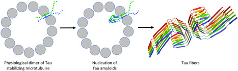
A coherent structural picture of the interaction of Tau with tubulin provides a link to its aggregation.
Tau is a protein regulating microtubule dynamics which also forms neurofibrillary tangles in pathological conditions. Recent results suggest that a physiological dimer of Tau could serve as a nucleus for its aggregation. Tauopathies are a group of neurodegenerative diseases characterized by the presence of insoluble filaments of the Tau protein in the brain. In physiological conditions, Tau is involved in the regulation of microtubule dynamics. The study of its interaction with different tubulin assemblies, using various experimental approaches, leads to a seemingly disparate picture. In this opinion-type article, we integrate this information into a model of how Tau participates in microtubule assembly and stabilization. Related to its intrinsically disordered nature, the binding of Tau to microtubules involves both specific interactions, along protofilaments, and non-specific ones, with the C-terminal region of tubulin subunits. In addition, a Tau:tubulin structure that we recently determined leads to a model of a functional Tau dimer targeting a microtubule aperture between protofilaments. This model also provides a framework for a Tau aggregation that would be initiated on the microtubule. More information : https://www.jbc.org/article/S0021-9258(26)01958-7/fulltext Contact : Benoît Gigant benoit.gigant@i2bc.paris-saclay.fr
|
|
Scooped by
I2BC Paris-Saclay
May 11, 4:50 AM
|
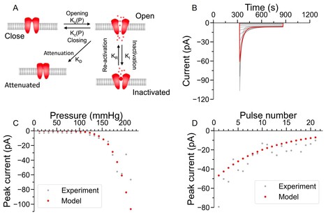
The Rapid Mechanically Activated channel transduces increases in plasma membrane tension into transient calcium influx
In Arabidopsis, RMA ion channel is a candidate for mediating cytosolic calcium signaling in response to high frequency mechanical stimulation. Plants respond to mechanical stimuli by a rapid increase in cytosolic calcium. The intensity and kinetics of the calcium changes define calcium signatures important for biological responses. In this study carried in the BioCell department of I2BC , we determine the properties of a calcium-permeable force-gated channel localized at the plasma membrane called Rapid Mechanically Activated (RMA).
Using patch clamp and pressure clamp, we characterized the kinetics of the Arabidopsis thaliana RMA channel upon stimulation by pressure pulses applied onto the plasma membrane. Combining pressure pulse protocols at different frequencies with modeling, we investigated the channel's capacity to transduce high frequency mechanical stimuli.
The RMA channel rapidly activates in response to membrane tension, then it inactivates during prolonged stimulation. Upon repeated stimulations, the RMA current amplitude decreases irreversibly indicating that it undergoes attenuation. The channel kinetics were modeled with four chemical states and the model predicts that it behaves as a pass band filter in the 10 Hz–1 kHz range.
In conclusion, due to its activation/inactivation, the RMA channel is a candidate for mediating cytosolic calcium signaling in response to mechanostimulation. Its attenuation and filtering properties suggest its involvement in the transduction of high frequency mechanical stimulation, such as those produced by insects' vibrations.
More info: http://doi.org/10.1111/nph.71241 Contact: jean-marie.frachisse@i2bc.paris-saclay.fr
|
|
Scooped by
I2BC Paris-Saclay
May 7, 7:30 AM
|
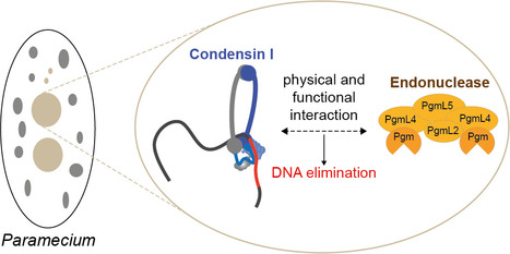
Programmed DNA Elimination in Paramecium: towards the third dimension
A condensin I complex, known for shaping chromosome organization in eukaryotes, is essential to DNA elimination in Paramecium During development, many organisms eliminate specific germline DNA sequences to shape their somatic genome. But how do cells target which sequences to remove? The team “Programmed Genome Rearrangements” has addressed this question in the ciliate Paramecium tetraurelia. Because of its nuclear dimorphism, this unicellular eukaryote provides a powerful model to study somatic differentiation at the genomic scale. At each sexual cycle, a new somatic macronucleus (MAC), specialized for gene expression, is formed from a copy of the germline micronucleus (MIC), inherited from the previous generation. This process involves the elimination of one third of germline sequences, including transposable elements and satellite DNA, to ensure proper gene function and progeny survival.
In collaboration with scientists from the Institut Jacques Monod, the authors have uncovered a surprising new role for a development-specific condensin I, a complex from the SMC family (Structural Maintenance of Chromosomes) generally known for organizing chromosomes in three dimensions. Using TurboID and protein co-immunoprecipitation, combined with quantitative mass spectrometry, we found that the developmental condensin I complex interacts with the PiggyMac (Pgm) endonuclease, an enzyme essential for cutting and removing specific germline DNA sequences during MAC development. High-throughput sequencing of the DNA extracted from purified new MACs revealed that when cells are depleted of condensin I, the germline genetic material is massively retained in the somatic genome. This demonstrates that condensin I plays a key part in programmed DNA elimination.
This discovery suggests that 3D genome organization may help target which sequences are removed. The condensin complex localizes to the developing new MAC, where it stabilizes Pgm and its partners. However, the exact level at which spatial genome architecture influences PDE remains an open question. More info: https://doi.org/10.1093/nar/gkag351 Conatct: Mireille Bétermier mireille.betermier@i2bc.paris-saclay.fr
|
|
Scooped by
I2BC Paris-Saclay
April 29, 4:26 AM
|
How does it end ? Helitrons cap germline chromosomes of Paramecia
The numerous tiny germline chromosomes of Paramecia end in DNA segments produced over evolutionary time by the activity of Helitron mobile elements. These chromosome ends are eliminated during development of the individual by a novel mechanism. Many eukaryotes are able to eliminate chromosome segments or even entire chromosomes during somatic differenciation. This programmed DNA elimination process illustrates eukaryotic genome plasticity and raises questions about the underlying molecular mechanisms. Among the unicellular ciliated protozoans, which include paramecia, the somatic and germline fonctions of chromosomes are separated in two distinct nuclei.
The diploid germline nucleus (MIC, ~ 100 Mb in Paramecium) undergoes meiosis and transmits the genetic information between generations. The polyploid somatic nucleus (MAC, ~ 70 Mb) is responsible for gene expression, but is destroyed at each generation, when a new MAC develops from a copy of the MIC.
Identification of the sequences that undergo programmed DNA elimination requires knowledge of the MIC genome sequence. More than 10 years of research involving several University Paris-Saclay teams and platforms has provided chromosome-scale assemblies of MIC DNA for 7 members of the Paramecium aurelia
species complex and a telomere-to-telomere assembly for one of them, P. tetraurelia. The authors show that the MIC genome consists of around 160 tiny chromosomes (300 kb – 1.2 Mb) with the highest meiotic recombination rate ever reported (420 cM/Mb). The assembly revealed that the germline chromosome ends
are eliminated very early during MAC development by a echanism different from that known to be involved in elimination of other sequences. This terminal genomic compartment contains a new clade of transposable elements (TE) that belong to the Helitron superfamily. These TE are very ancient (having diverged from other Helitron-like elements at the base of the eukaryotic tree) but have remained active in Paramecia. They appear to preferentially insert into telomeric repeats. One exciting perspective of the study is the elucidation of a novel DNA elimination pathway.
Contact: linda.sperling@i2bc.paris-saclay.fr & olivier.arnaiz@i2bc.paris-saclay.fr More information: https://link.springer.com/article/10.1186/s12915-026-02584-w
|
|
Scooped by
I2BC Paris-Saclay
April 23, 5:11 AM
|
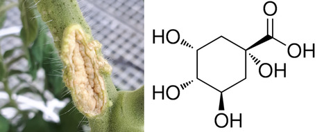
How bacterial pathogens hijack host carbon: Insights from TnSeq metabolic network analysis
We uncovered how Agrobacterium tumefaciens exploits host carbon resources to colonize tomato, revealing quinic acid catabolism as the critical driver of its gall-specific fitness. Agrobacterium tumefaciens is a facultative plant pathogen that forms galls on a wide range of hosts and persists in soil, roots and galls through extensive metabolic versatility. Here, we combined genome-wide transposon sequencing (TnSeq), metabolomics and reverse genetics to identify carbon utilisation pathways supporting A. tumefaciens fitness in tomato roots and galls. TnSeq screening across 21 carbon sources, representative of rhizosphere exudates, root metabolites and gall-derived compounds, identified conserved and substrate-specific fitness determinants, including central metabolic enzymes, transporters and previously uncharacterized catabolic genes. Comparison with in planta TnSeq data revealed environment-dependent metabolic requirements, highlighting fluxes through the Entner–Doudoroff pathway, the TCA cycle and gluconeogenesis. Quinic acid catabolism emerged as a major determinant of fitness specifically in galls, where this compound accumulated. Deletion of pcaC (ATU_RS21295/atu4541) impaired growth on quinic and protocatechuic acids and reduced competitiveness during gall colonisation, with no effect in roots. Together, this work provides a system-level framework for understanding how A. tumefaciens exploits plant-derived nutrients in host-associated environments. More information: https://doi-org.insb.bib.cnrs.fr/10.1111/1462-2920.70311 https://www.i2bc.paris-saclay.fr/equipe-interactions-of-bacteria-with-plants-and-insects/ https://www.i2bc.paris-saclay.fr/sequencing/ Contact: Denis Faure denis.faure@i2bc.paris-saclay.fr
|
|
Scooped by
I2BC Paris-Saclay
April 17, 5:00 AM
|
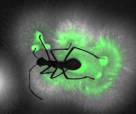
A wolf in sheep’s clothing
Distinguishing beneficial from harmful among closely related bacteria, a crucial question for organisms in nature and for biotechnology Many organisms crucially depend on the services of specific microorganisms and need to maintain these symbionts in large numbers in their body. Hosts dispose of highly specific symbiont selection mechanisms and have symbiotic organs to host safely these symbionts. It is believed that these sophisticated adaptations evolved to counter the thread of exploitation of the symbiotic association by opportunistic pathogens, although such cases are rarely reported. The association of the bean bug Riptortus pedestris with caballeronia bacteria is a model of a highly evolved symbiotic system. In a study published in PNAS, Ishigami et al report on an opportunistic pathogen of the bean bug that is phylogenetically related to symbionts. Initially, this Burholderia pathogen behaves as a true symbiont, efficiently passing all symbiont filtering mechanisms of the host, inducing the development of the symbiotic organ, and infecting and colonizing it. However, once settled, the pathogen throws off its disguise and shows its true nature: it breaks out from its confinement in the symbiotic organ, causes deadly sepsis and then escapes from the dead animal to recolonize the environment (figure). This case of a pathogen exploiting an intricate mutualistic association highlights the evolutionary pressures predicted by theory that have shaped the stringent partner-choice mechanisms usually observed in symbioses and at the same time reveals the fragility of these mutualisms despite their sophistication. Caballeronia and Burkholderia species belong both to the Burkholderia sensu latu clade of betaproteobacteria and were before phylogenetic revisions included in the single genus Burkholderia. In the 1990s, several Burkholderia strains were registered as biocontrol agents for agricultural applications but later withdrawn and a moratorium was placed on the registration of Burkholderia-containing products, after opportunistic human infections were reported with closely related strains. Thus, the Burkholderia has become a showcase of the potential health risks associated with the use of microorganisms in commercial applications. Because of the many known Burkholderia strains with potential applications in agriculture, it is important to dispose of criteria that distinguish friend from foe. A separate study by Agnoli et al, published in the ISME Journal, was conducted on a panel of 76 Burkholderia, Paraburkholderia and Caballeronia strains of the Burkholderia sensu lato. The work identified phenotypic traits and genetic markers that enable improved, strain-level evaluation of pathogenic potential and biocontrol capacity, supporting the rational selection of Burkholderia sensu latu strains for safe agricultural applications. More information :https://academic.oup.com/ismej/advance-article/doi/10.1093/ismejo/wrag081/8650978 Contact : Peter Mergaert peter.mergaert@i2bc.paris-saclay.fr
|
|
Scooped by
I2BC Paris-Saclay
April 15, 4:06 AM
|
The I2BC was well represented on Tuesday, April 14, at the Science Outreach Day co-organized by the French Society for Developmental Biology, the French Society for Cell Biology and the city of Ivry-sur-Seine. Seven members of the I2BC led workshops throughout the day. Nearly 1,200 elementary school students participated in workshops on cell, plant, and animal biology, as well as on the scientific method. Let’s hope we’ve inspired a few future scientists… one thing is certain: the children were thrilled! https://sbcf.fr/newsletter/mediation-scientifique-sbcf/ https://sfbd.fr/ressources/mediation/#1558951904425-8b59e54f-fdb5
|
|
Scooped by
I2BC Paris-Saclay
April 1, 5:18 AM
|
As part of the “Des Plantes et des Hommes” project led by the EUR Saclay Plant Science, a senior high school class visited the I2BC on March 25, 2026. Catherine Grandclément presented the Institute and a lecture on the symbiosis between legume plants and Rhizobium bacteria. The students visited the I2BC greenhouse to search for nodules on the roots of legumes plants before observing them with a macroscope. Finally, the microscopy platform and Amanda Edling de Barros introduced them to a confocal microscope and its use in studying symbiosis. With their interest in the subject and their fascination with the equipment, we hope we’ve inspired a passion for research! More information : Legume-rhizobia Symbiosis : https://www.i2bc.paris-saclay.fr/equipe-interactions-of-bacteria-with-plants-and-insects/ "Des Plantes et des Hommes” project : Des Plantes et des Hommes” project
Vous développez un projet ambitieux en biologie moléculaire et cellulaire, à l’interface de plusieurs disciplines, ou en biologie computationnelle ? Rejoignez l’I2BC, un institut de recherche de premier plan affilié au CEA, au CNRS et à l’Université Paris-Saclay. Nous recherchons de nouveaux responsables d’équipes autour de projets originaux, excellents scientifiquement, et capables de créer des synergies avec les recherches en cours à l’I2BC. Une attention particulière sera portée aux projets multidisciplinaires et aux approches en biologie computationnelle, notamment sur la modélisation des processus biologiques et l’intégration de données multi-échelles. L’I2BC offre un environnement multidisciplinaire dynamique, des plateformes technologiques de pointe, ainsi qu’un accompagnement fort, en particulier pour les profils en début de carrière. Les candidatures de profils plus avancés disposant déjà d’un poste permanent en France sont également bienvenues. 📅 Date limite : 22 avril 2026 📩 Contact : call2026@i2bc.paris-saclay.fr Présélection et notification aux candidats retenus : début juin 2026 - Entretiens sur place avec les candidats retenus : les 2 et 3 juillet. Les candidats doivent s'assurer d'être disponibles ces deux jours-là
- Annonce de la sélection finale d'ici la mi-juillet 2026
- Date de prise de fonction négociable à partir de l'automne 2026, en fonction du poste obtenu et de l'obtention du financement.
🔗 Toutes les informations sur notre site web
Via Life Sciences UPSaclay
La plateforme conjointe de Cryo-Microscopie Électronique de l’Institut de Biologie Intégrative de la Cellule (I2BC, Gif-sur-Yvette, Institut Joliot, CEA, Saclay) et du Synchrotron SOLEIL renforce son expertise avec l’arrivée de Heddy Soufari, recruté en tant qu’ingénieur-chercheur CEA. Spécialisé en biologie structurale et en cryo-microscopie électronique (cryo-EM), il consacre l’essentiel de son activité au développement méthodologique et au soutien scientifique des utilisateurs régionaux et nationaux sur les sites de SOLEIL et de l’I2BC. Docteur en biologie structurale de l’Université de Bordeaux, Heddy Soufari a orienté ses recherches vers la compréhension de la structure et de la dynamique des complexes macromoléculaires. Après un parcours académique entre la France et le Canada, il a acquis une solide expertise en cryo-microscopie électronique appliquée à l’étude de systèmes biologiques complexes. Il a notamment contribué à la caractérisation de ribosomes de mitochondries et de complexes protéiques chez différents modèles eucaryotes, avant de rejoindre la société NovAliX où il dirige le pôle Cryo-EM et met la microscopie au service du structure-based drug design (SBDD) sur des cibles thérapeutiques. Depuis son recrutement au CEA en septembre 2024, il accompagne les chercheurs de l’I2BC et de la région dans leurs projets de cryo-EM, de la préparation d’échantillons à l’analyse des reconstructions tridimensionnelles. Il développe parallèlement une activité méthodologique centrée sur la cryo-tomographie électronique (cryo-ET) et l’imagerie corrélative cryo-CLEM, avec pour objectif d’étudier l’organisation et la dynamique de complexes macromoléculaires in situ. Sa vision scientifique repose sur une approche multi-échelle, allant de la molécule unique à la cellule entière, afin d’explorer la tridimensionnalité des systèmes biologiques dans toute leur complexité. Avec l’arrivée d’Heddy Soufari, la plateforme renforce sa capacité à développer de nouvelles méthodologies et pipelines pour la tomographie cellulaire, l’analyse d’images 3D et la corrélation optique-électronique. Ces efforts visent à faire de la plateforme un pôle de référence pour la visualisation in situ des architectures cellulaires et des complexes macromoléculaires à haute résolution. -> Contact : Stéphane Bressanelli (plt-cryoem@i2bc.paris-saclay.fr) Plug In Labs Université Paris-Saclay : cliquer ICI Envie de (re)lire leurs précédents FOCUS PLATEFORME ? I2BC / Plateforme de cryo-microscopie électronique (CRYO-EM). La plateforme pour la cryo-microscopie électronique I2BC met à disposition une large gamme d’instruments et de services pour la biologie structurale : vitrification d’échantillons (Vitrobot, Leica GP2), microscope Tecnai Spirit (120 kV) pour les expériences préliminaires, microscopes de dernière génération Glacios (200 kV) pour les expériences à haute résolution, ainsi qu’un microscope confocal cryogénique Stellaris pour les approches corrélatives optique-électronique. La plateforme a la capacité de préparer et d’imager des échantillons biologiques de niveau de sécurité 2. Elle s’est associée au mésocentre de calcul Paris-Saclay pour l’hébergement des serveurs de stockage et d’analyse des images produites. La plateforme CryoEM de l’I2BC est associée à SOLEIL dans le cadre de la plateforme conjointe CryoEM@Paris-Saclay. Les images obtenues en cryo-microscopie électronique sur le microscope Glacios, ainsi que les moyens de calcul disponibles sur la plateforme, permettent le crible de grilles et la détermination de structures 3D à des résolutions dépassant fréquemment 3 Å et pouvant atteindre 2 Å. Ces premières structures permettent si nécessaire d'accéder aux microscopes plus puissants, notamment le Titan Krios G4 (300 kV) à SOLEIL, mais aussi les microscopes Titan Krios de l'ESRF, de l’IBS (Grenoble) ou de l'IGBMC (Strasbourg), menant aux résolutions atomiques. La plateforme CryoEM de l’I2BC permet aussi l'observation de complexes in situ (protéines à la surface d'organites purifiés ou de virus enveloppés, protéines membranaires reconstituées dans des liposomes...) par cryo-tomographie électronique. La plateforme accueille des projets issus de la recherche académique, institutionnelle et industrielle, et s’inscrit dans le réseau des infrastructures nationales en biologie structurale et intégrative (FRISBI). Ses missions couvrent la préparation d’échantillons, la collecte de données, le traitement d’images, la formation et l’accompagnement des utilisateurs. Cette plateforme fait partie du pôle des plateformes de Biologie Structurale de l'I2BC qui comprend six plateformes : cristallographie, RMN, CryoEM, mesures d'interactions macromoléculaires (PIM), expression de protéines recombinantes en systèmes eucaryotes (Prot-Ex) et sélection sur mesure de protéines artificielles comme ligands spécifiques de toute protéine d’intérêt (AlphaRep). D’autre part, les plateformes CryoEM, cristallographie, PIM et AlphaRep sont aujourd’hui labélisées IBISA sous la bannière BioStruct@UPSAY. A propos de l’Institut de Biologie Intégrative de la Cellule (I2BC - UMR 9198). L’I2BC est une Unité Mixte de Recherche (CEA, CNRS, Université Paris-Saclay), accueillant une soixantaine d’équipes de recherche et hébergeant 17 plateformes technologiques, réparties en 6 pôles. 2025 a aussi été une année clé pour l’I2BC : cette unité a fêté ses 10 ans !
Via Life Sciences UPSaclay
|
|
|
Scooped by
I2BC Paris-Saclay
June 8, 9:21 AM
|
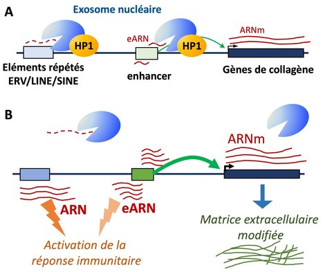
HP1 targets the RNA exosome to chromatin
A novel role for the HP1 proteins in targeting the RNA-exosome to chromatin for the turnover of repetitive RNAs and enhancer RNAs. Heterochromatin protein 1 (HP1), a hallmark of pericentromeric heterochromatin, is a chromatin-bound regulator of co-transcriptional processes including alternative splicing, but its role in RNA degradation remains unexplored. Here, we uncover a direct interaction between HP1 and nuclear RNA exosome complexes, major RNA decay machineries. In mouse embryonic liver cells, inactivation of all three HP1 isoforms leads to accumulation of retrotransposon-derived RNAs and stabilization of enhancer RNAs. These changes coincide with increased activity at a subset of liver enhancers particularly sensitive to reduced exosome activity, many of which regulate genes encoding extracellular matrix components such as collagen genes. Stratifying hepatocellular carcinoma samples by HP1 expression further reveal that tumors with low HP1 are marked by reduced RNA degradation, and increased expression of a similar subset of genes encoding extracellular matrix components and possibly contributing to tumor stiffness. These results suggest that HP1’s impact on RNA turnover contributes to its function in cancer biology. More information: https://www.nature.com/articles/s41467-026-72504-7 Contact : Carl Mann carl.mann@i2bc.paris-saclay.fr
|
|
Scooped by
I2BC Paris-Saclay
June 5, 3:49 AM
|
New Release: Génomes: la construction du vivant
From DNA sequencing and studies of genome function and organization—covering viruses, bacteria, archaea and eucaryotes, this collective book explores genomes and their role in life, evolution, and diversity. A collective book that traces the history of genetics and the major discoveries surrounding genomes. The genome, the complete set of genetic information, builds, operates, and reproduces all living beings, serving as the engine of their evolution. Thanks to DNA, genetic information is stored, decoded, duplicated, and transmitted. But the study of the genome goes much further: it allows us to understand life, trace the history of species, and identify non-coding DNA regions that are nonetheless essential for life, the protection of genetic material, and its diversity. From DNA sequencing and genome function to the mechanisms of evolution—covering viruses, bacteria, archaea and eucaryotes, —the book explores how scientists today are striving to understand genome function and evolution. Coordinated by Frédéric Boccard, Director of the I2BC, this book brings together chapters written by several French scientists, including Mireille Bétermier, Group Leader at the I2BC. More information: https://www.cnrseditions.fr/catalogue/biologie-et-sante/genomes/ Contact: Frédéric Boccard frederic.boccard@i2bc.paris-saclay.fr
Le pôle des plateformes de biophysique de l’Institut de Biologie Intégrative de la Cellule (I2BC, CNRS/CEA/UPSaclay, Gif-sur-Yvette) a le plaisir d’accueillir Viola Caroline D’mello, qui a rejoint le CEA en mars 2025 en tant qu’ingénieure-chercheuse. Après une licence en chimie à l’université de Mumbai et un master en chimie analytique à l’université de Mangalore, elle a réalisé sa thèse au Tata Institute of Fundamental Research (TIFR) à Mumbai. Ses travaux portaient sur l’étude en phase gazeuse de liaisons hydrogène dans des molécules aromatiques azotées par spectroscopies dans les domaines de l’ultraviolet (UV) et de l’infrarouge (IR) nanoseconde – ces liaisons hydrogène étant similaires à celles présentes dans l’ADN et l’ARN. En 2019, elle a rejoint en tant que chercheuse postdoctorante le groupe de recherche LIDYL de l’institut IRAMIS au CEA Saclay, où elle a étudié les paires d’ions et le repliement de peptides en phase gazeuse. Après un court passage dans l’industrie à Bangalore (Inde), elle est revenue à la recherche académique comme postdoctorante au Département de Physique de l’Université de Göteborg, où elle a contribué à la mise en service d’un spectromètre de masse haute résolution couplé à une source à jet supersonique. Depuis son arrivée au sein du Pôle de biophysique de l’I2BC, elle est responsable des plateformes de spectroscopies électroniques, de spectroscopie Raman de résonance et de spectroscopie infrarouge : elle supervise l’accès des utilisateurs et veille à ce que les expériences soient conçues et réalisées dans des conditions optimales. Elle accompagne et conseille régulièrement les utilisateurs sur un large éventail de techniques, incluant l’absorption UV-Visible, l’absorption transitoire ultrarapide (de la femtoseconde à la milliseconde), la spectroscopie infrarouge à transformée de Fourier (FTIR) et la spectroscopie Raman, dont la spectroscopie Raman femtoseconde (plateforme LUMA). L’étendue de son expertise en méthodes photophysiques, associée à sa fiabilité et à son attitude ouverte et collaborative, fait d’elle un pilier central du Pôle de biophysique. -> Contact : Viola-Caroline D'mello (Viola.Dmello@cea.fr) Plug In Labs Université Paris-Saclay : cliquer ICI I2BC / Plateforme de spectroscopies électroniques. La plateforme de Spectroscopies Électroniques (Institut de Biologie Intégrative de la Cellule) offre ses services appliqués aux biomolécules à des équipes de recherche françaises et internationales. Nous sommes capables de suivre des changements spectroscopiques au niveau de la protéine dans des cellules intactes. La plateforme est équipée de plusieurs spectromètres d'absorption et de fluorescence (y compris un certain nombre de spectromètres PAM spécialisés) ainsi que des spectromètres à thermoluminescence. Pour certaines configurations, des cryostats sont disponibles pour les études à basse température, jusqu'à 77K ou 4K. La plateforme a développé (et continue à améliorer) un montage unique de spectroscopie optique résolue dans le temps (ca. 300 ps), surpassant les montages conventionnels (commerciaux) en sensibilité et en résolution temporelle. Ce type de méthodologie est particulièrement déterminant pour l'élucidation de processus irréversibles et/ou de processus qui se produisent dans des fenêtres temporelles allant de quelques centaines de picosecondes à des dizaines de nanosecondes, où les montages conventionnels performent mal ou ne peuvent pas être utilisées du tout. Cette plateforme fait partie du pôle des plateformes de Biophysiques de l'I2BC qui comprend les plateformes de RPE, FTIR, Résonance Raman, Spectroscopies Electroniques et Microscopie de fluorescence à super-résolution. I2BC / Plateforme de spectroscopie RAMAN de résonance. Cette plateforme met à disposition des équipements de spectroscopies avancées Raman et FLN (7 spectromètres, avec plusieurs accessoires, large gamme de température possible (thermostats 273-320 K, cryostats 4-250 K)). Analyses faisables sur échantillons de toutes formes physiques, en particulier ceux qui contiennent des molécules pigmentées (voir ci-dessous). Le laboratoire se spécialise sur les propriétés physico-chimiques des cofacteurs pigmentés en biologie (caroténoïdes, chlorophylles, hèmes, flavines, …), y compris des études in vivo des réactions biochimiques, photo-induites et régulatrices. L'état de l'échantillon (liquide, poudre, gel, solide, …) limité seulement par la taille du signale (présence de molécules pigmentées nécessaires pour des mesures en milieux complexes). Exemples récents : processus régulatoires dans des membranes photosynthétiques in vivo (feuilles entières et micro-organismes), structure moléculaire des caroténoïdes et opsines dans la rétine humaine ex vivo. Cette plateforme fait partie du pôle des plateformes de Biophysiques de l'I2BC qui comprend les plateformes de RPE, FTIR, Résonance Raman, Spectroscopies Electroniques et Microscopie de fluorescence à super-résolution. I2BC / Plateforme de spectroscopie IRTF. La plateforme de spectroscopie IRTF est située au Laboratoire des Mécanismes Fondamentaux en Bioénergétique (UMR 9198). Elle met à disposition des utilisateurs des spectromètres IRTF avancés et elle est équipée pour répondre à la plus grande partie des besoins des analyses IRTF. La plateforme comprend 4 spectromètres avec plusieurs accessoires : cellule à transmission, accessoires ATR, cellule électrochimique, thermostats, cryostats pour expériences à basse température... Elle permet l'étude d'échantillons sous différentes formes (liquide, solide, poudre..). Le laboratoire est spécialisé dans la spectroscopie IRTF différentielle, résolue dans le temps, à basse température, et possède une bonne expertise dans l'étude de réactions biochimiques et photo-induites. La plateforme de spectroscopie IRTF fait partie du pôle des plateformes de Biophysiques de l'I2BC qui comprend les plateformes de RPE, FTIR, Résonance Raman, Spectroscopies Electroniques et Microscopie de fluorescence à super-résolution. A propos de l’Institut de Biologie Intégrative de la Cellule (I2BC - UMR 9198). L’I2BC est une Unité Mixte de Recherche (CEA, CNRS, Université Paris-Saclay), accueillant une soixantaine d’équipes de recherche et hébergeant 17 plateformes technologiques, réparties en 6 pôles. 2025 a aussi été une année clé pour l’I2BC : cette unité a fêté ses 10 ans !
Via Life Sciences UPSaclay
|
|
Scooped by
I2BC Paris-Saclay
May 11, 7:04 AM
|
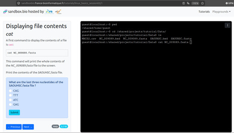
A new training reference for Unix skills in bioinformatics
Master Unix for bioinformatics with a free, progressive skills sheet and interactive Sandbox.bio tutorials—no install needed! Check it out here: https://sandboxbio.france-bioinformatique.fr/ As data generation in life and health sciences accelerates, mastering core bioinformatics skills—especially Unix command-line proficiency—has become essential. In response, the e-learning working group of the IFB (Institut Français de Bioinformatique), in which Claire Toffano-Nioche from the BIOI2 facility of I2BC participates, developed a progressive Unix skill sheet tailored for life scientists with little or no prior experience, recently published in PLoS Comput Biol. This skills sheet summarises the most commonly used Unix commands in the field of bioinformatics. Skills are organised according to increasing levels of difficulty and thematic groups and are cumulative, meaning that skills acquired at a given level are required at the next level. Each skill has been defined operationally by following Bloom's taxonomy of learning objectives. This framework can be freely reused (CC BY 4.0) - by learners as well as tutors - and serves as inspiration for defining the competencies or prerequisites of a training course, or for evaluating the level of a student. Following this sheet, the e-learning working group directly implemented progressive, interactive sessions that can be used for self-paced learning or in classroom settings. These sessions are deployed via Sandbox.bio, an open-source platform leveraging WASM (WebAssembly) technology. This ensures seamless access from any browser—no installation required—while enabling computations to run locally on the user’s machine. To further support self-paced learning, BIOI2 also offers autotraining course materials on their website (https://bioi2.i2bc.paris-saclay.fr/training). These resources draw inspiration from—and contribute back to—multiple open courses, including those from the IFB, creating a collaborative cycle of shared knowledge and continuous improvement. More information: Contact: contact-bioi2@i2bc.paris-saclay.fr
|
|
Scooped by
I2BC Paris-Saclay
May 11, 2:59 AM
|
Yves Gaudin de l'équipe Rhabdovirus à L'i2BC apporte un éclairage scientifique sur l'hantavirus dans Les Matins de France Culture de Guillaume Erner.
|
|
Scooped by
I2BC Paris-Saclay
May 4, 4:38 AM
|
PizzaTech - Workshop: the 3rd technical and scientific event organised by the I2BC’s Imagerie-Gif facility in collaboration with Leica Biosystems.
In March, the 3rd PizzaTech-Workshop took place—a technico-scientific event organized by the Imagerie-Gif facility of the I2BC. Benoit Maury from Leica Biosystems presented the brand-new THUNDER Imager Cell photonic microscope equipped with a Spinning Disk (CICERO), a setup designed for live cell imaging. This allows the observation of rapid cellular biological phenomena in real-time and 3D, thanks to two key components: - Optics: The spinning disk provides optical sectioning and reduces phototoxicity.
- Image Processing: The THUNDER application eliminates out-of-focus residual light, enhancing contrast and resolution to reveal finer details.
PizzaTech & Presentation: After enjoying pizza, researchers explored the technical and functional features of the system, including: - Confocal spinning disk and widefield modalities, both compatible with THUNDER image processing.
- Key applications: live cells, 3D cultures, organoids, spheroids, thick tissues, and model organisms.
Demo Week on the Photonics Microscopy Facility – Imagerie-Gif: Researchers, PhD students, and engineers tested the THUNDER Imager Cell / Spinning Disk CICERO with their own samples for a week. The setup included: - Spinning Disk CICERO module (pinhole size: 50 µm; pinhole spacing: 250 µm; disk rotation speed: ~15,000 rpm; field of view: 22 mm).
- Excitation lasers (405, 470, 555, 640 nm).
- Two sCMOS cameras (Hamamatsu Fusion; Photometrics Kinetix22).
- Inverted microscope (Leica DMi8) with motorized AFC (Adaptive Focus Control).
The system was fully equipped with: - THUNDER 2D/3D (opto-digital clarification).
- Adaptive Immersion (stabilized water objectives).
- SmartCORR (automatic aberration correction).
- Sample Finder (automatic sample navigation).
- Incubator for live cell imaging.
Participating Research Teams: - Institut des Neurosciences Paris-Saclay
- I2BC (teams: Giordano / Lipid trafficking and membrane contact sites, Le Clainche / Cytoskeleton dynamics and motility, Kühl / Mammalian mitochondrial gene expression in health and disease, Urvoas / B3S Protein Engineering and Modeling, Imagerie-Gif facility engineers)
- Institut Curie Orsay
- Genethon
Image credits: - Esther GIL HERNANDEZ, Equipe de GIORDANO Francesca, ERL INSERM. Lipid trafficking and membrane contact sites
- Sharbatanu CHATTERJEE & Md Amit HASAN, Institut NeuroPSI - UMR9197, Circuits neuronaux & comportement
- Aleksandr BALATSKII, Genethon
- Light Microscopy facility, Imagerie-Gif.
More about the THUNDER Imager Cell Spinning Disk: https://www.leica-microsystems.com/products/light-microscopes/p/thunder-imager-cell-spinning-disk/ More information about the facility: https://www.i2bc.paris-saclay.fr/bioimaging/
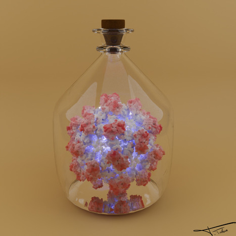
Portrait Jeune Chercheur – Thibault Tubiana, Chercheur en bioinformatique structurale
Thibault Tubiana est chargé de recherche (CRCN) au CNRS depuis la fin de l'année 2024. Il exerce ses fonctions au sein de l’Institut de Biologie Intégrative de la Cellule - I2BC (CNRS/CEA/UPSaclay, Gif-sur-Yvette), dans l'équipe dirigée par le Dr Stéphane Bressanelli. Son parcours illustre une volonté constante de faire le pont entre la bioinformatique structurale, la virologie moléculaire et l'étude des membranes cellulaires. Après avoir obtenu une licence et un master en bioinformatique à l'Université Paris Diderot, où il nourrit déjà un fort attrait pour la virologie, il réalise sa thèse de doctorat sous la co-direction des Drs Stéphane Bressanelli et Yves Boulard. Ses travaux portent alors sur la dynamique d'assemblage de la capside du norovirus, lui permettant d'acquérir une solide expertise en modélisation moléculaire et en approches intégratives (combinant dynamique moléculaire, données SAXS et Cryo-EM). Il poursuit sa carrière par un premier post-doctorat au sein de l'Institut de Recherche Servier (IdRS). Cette immersion dans le monde de l'industrie pharmaceutique lui permet d'acquérir de solides compétences en drug design. Afin d'approfondir ensuite ses connaissances sur les interactions protéines-membranes, il rejoint le groupe de la Pr Nathalie Reuter à l'Université de Bergen (Norvège). Il y mène un vaste projet de cartographie biostatistique et bioinformatique visant à décrypter et redéfinir les interfaces de liaison des protéines membranaires périphériques. Fort de ces expertises pluridisciplinaires, il choisit de revenir en France au sein de son ancienne équipe à l'I2BC. Soutenu dans un premier temps par des financements postdoctoraux de l'ANRS-MIE, il y déploie des approches innovantes en bioinformatique structurale, contribuant activement à l'étude des virus à ARN positif simple brin. Ce travail structurant conduit à son recrutement au CNRS fin 2024. Dans le contexte de la révolution de l'intelligence artificielle appliquée à la biologie (AlphaFold), ses recherches actuelles portent sur la modélisation des complexes de réplication des virus de l'hépatite E (HEV) et de l'hépatite C (HCV). Ses travaux visent à comprendre à l'échelle atomique l'organisation de ces protéines virales et leurs interactions avec les cellules hôtes. Très impliqué dans la communauté scientifique nationale sur ces pathogènes, il a récemment intégré le bureau de l'Action Coordonnée 42 sur les hépatites virales de l'ANRS-MIE. Parallèlement à ses recherches, Thibault Tubiana a toujours eu à cœur de transmettre ses connaissances. Au sein de l'I2BC, en plus de l'encadrement d'étudiants, il s'investit fortement dans la formation continue de son institut. Il a notamment créé et mis en place, en lien avec la plateforme BioI2, une nouvelle formation dédiée à la modélisation et à la visualisation moléculaires, offrant ainsi un outil précieux pour l'ensemble des chercheurs de l'université. « Nous sommes tous des poussières d'étoile. » - Pr. André Brahic -> Contact : thibault.tubiana@i2bc.paris-saclay.fr
Via Life Sciences UPSaclay
|
|
Scooped by
I2BC Paris-Saclay
April 17, 5:04 AM
|
Iron Sulfides Produced by Thermococcales: An Iron Detoxification Mechanism
Thermococcales, hyperthermophilic archaea from hydrothermal vents, promote iron sulfide precipitation, enabling survival in iron-rich environments. Some cells become encrusted in pyrite and do not survive mineralization, while surviving cells activate metal detoxification genes. Thermococcales, sulfur-reducing archaea inhabiting the hottest parts of hydrothermal vents, have evolved to thrive in environments rich in iron and sulfide species. In this study, using experimental analogues of sulfur-rich hydrothermal chimneys, we confirm previous suggestions that the precipitation of iron sulfide minerals promoted by Thermococcales contributes to a population-wide adaptation to reactive species induced by the presence of high levels of iron. In parallel with mineral phases identification, cellular metabolic activity was monitored during mineralization, revealing a mechanism in which a subpopulation of cells does not survive mineralization and becomes encrusted in pyrite, while the remaining living cells exhibit a gene expression profile focused on DNA repair and metal excess associated detoxification. Compared to abiotic conditions, Thermococcales induce a faster precipitation of dissolved iron, immobilising excess metal. Our results clarify the role of mineralizing cells in this survival mechanism, suggesting that this biomineralization process allows resilience to extreme chemical stress. Upon drastic levels of toxic dissolved iron, thanks to a population of mineralizing cells, the surviving Thermococcales are thus more likely to endure those still harsh environments. This complex mechanism is likely a key factor in the adaptation of microorganisms to the hottest environments of hydrothermal vents. More information : https://enviromicro-journals.onlinelibrary.wiley.com/doi/10.1111/1462-2920.70242 Contact : Aurore Gorlas aurore.gorlas@i2bc.paris-saclay.fr
|
|
Scooped by
I2BC Paris-Saclay
April 17, 4:52 AM
|
The tRNA moieties of both aminoacyl-tRNA substrates of a cyclodipeptide synthase share a common binding site, as revealed by RNA microhelices mimicking tRNA acceptor arms
Cyclodipeptide synthases (CDPSs) utilize two aminoacyl-tRNAs as substrates to produce diverse natural products. Here, we demonstrate that CDPSs efficiently recognize aminoacylated microhelices (miHxs) that mimic the tRNA acceptor arm. Structural and enzymological analyses using unacylated, misacylated, and engineered miHxs reveal a shared RNA-binding mode for both substrates. These findings establish miHxs as versatile tools to investigate CDPS function and, more broadly, other aminoacyl-tRNA–dependent enzymes. Two teams from the I2BC, in collaboration with the ICSN, combined enzymological and structural approaches to investigate cyclodipeptide synthases (CDPSs), enzymes involved in natural product biosynthesis. CDPSs sequentially use two aminoacyl-tRNAs (AA-tRNAs) to catalyse cyclodipeptide formation. We previously showed that microhelices (miHxs), mimicking the tRNA acceptor arm, are as efficient as full-length AA-tRNAs when aminoacylated by flexizymes.
Here, we generated a diverse set of miHxs (acylated, unacylated, misacylated, mutated, or shortened) and analysed their interactions with CDPSs. We focused on the Nocardia brasiliensis CDPS (Nbra-CDPS), which synthesizes cyclo(L-Ala–L-Glu) from Ala-tRNAAla and Glu-tRNAGlu. Crystal structures of Nbra-CDPS bound to analogues of its first substrate, including unacylated and acylated miHxAla, were determined. Cryo-EM analysis confirmed that miHxs mimic the acceptor stem of full-length tRNAs.
We also solved the structure of Nbra-CDPS bound to unacylated miHxGlu, an analogue of the second substrate, and found that it superimposes well with miHxAla despite sequence differences. Together with results obtained using misacylated substrates, these data reveal a shared RNA-binding mode for both substrates. Our findings establish miHxs as powerful tools to dissect CDPS function and to study other AA-tRNA–dependent enzymes. More information: https://academic.oup.com/nar/article-abstract/doi/10.1093/nar/gkag307/8625897?utm_source=authortollfreelink&utm_campaign=nar&utm_medium=email Contacts: Muriel Gondry muriel.gondry@i2bc.paris-saclay.fr https://www.i2bc.paris-saclay.fr/enzymology-and-non-ribosomal-peptide-biosynthesis/ Jean-Baptiste Charbonnier jb.charbonnier@i2bc.paris-saclay.fr https://www.i2bc.paris-saclay.fr/nuclear-enveloppe-telomeres-and-dna-repair/
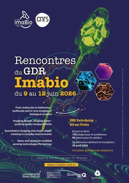
Les 3èmes journées de rencontre du GDR Imabio se tiendront à l’ENS Paris-Saclay du 9 au 12 juin 2026. - Registration is free, but seats are limited (approximately 150 for conferences, 50 for workshops).
- Abstract submission deadline: 26th April 2026
- Registration deadline: 11th May 2026
- Inscription et programme
Via Life Sciences UPSaclay
|
|
Scooped by
I2BC Paris-Saclay
March 25, 5:30 AM
|
Inspiring the Next Generation of Scientists at Collège A. Fournier, Orsay
Researchers from the I2BC recently visited Collège A. Fournier in Orsay to introduce students to various scientific career paths. Magali Noiray presented the institute’s state-of-the-art facilities, while Olivier Namy showcased the exciting work of its research teams. Science is for everyone, and the journey can start at any age. Let’s inspire the next generation of innovators! More information about I2BC: https://www.i2bc.paris-saclay.fr/ A great thanks to Magali & Olivier for their time and commitment!
L’autophagie est un mécanisme de dégradation des composants cellulaires, indispensable à la survie de la cellule. Mais ses fonctions vont au-delà du nettoyage ou de la survie en conditions de stress : elle est impliquée dans l’immunité, les maladies neurodégénératives, le vieillissement, le cancer… Avec - Audrey Esclatine, professeure à l’Université Paris-Saclay, co-responsable de l’équipe Autophagy and antiviral immunity de l’I2BC (CNRS/CEA/UPSaclay, Gif-sur-Yvette), présidente du Club Francophone de l'Autophagie
- Pierre-Emmanuel Joubert, maître de conférences à Sorbonne Université en immunologie
- Flavie Strappazzon, chargée de recherche CNRS à l’Institut NeuroMyoGène
L'autophagie ("se manger soi-même") est un processus d'autodigestion essentiel au maintien de l'homéostasie cellulaire. Il s'agit d'une dégradation de constituants cellulaires par la voie lysosomale en composants recyclés. Réécouter le podcast de l'émission du 3 mars 2026 -> Contact : audrey.esclatine@i2bc.paris-saclay.fr
Via Life Sciences UPSaclay
|