Your new post is loading...

|
Scooped by
I2BC Paris-Saclay
July 25, 2022 4:56 AM
|
Emergence of a new function after duplication of a metal transporter gene in poplar
A combination of detailed phylogenetic and functional analyses of a pair of tandem duplicated metal transporter encoding genes uncovers a rare example of neofunctionalization caught in the act. Gene duplication is an important mechanism in evolution because it opens the possibility to generate proteins with new functions through mutations in one of the copies. However, there are only few examples clearly illustrating this process of neofunctionalization in the literature. Poplar is important economically and as a model tree species. It is also grown in polluted areas in the context of phytoremediation. However, little is known about the mechanisms of metal homeostasis in poplar and in trees in general. The authors have taken advantage of the availability of genomic sequences for a wide array of Populus and Salix species to establish a detailed phylogeny of NRAMP3 gene in Salicaceae. This revealed a duplication of NRAMP3 metal transporter gene coinciding with the emergence of the genus Populus. Synonymous codon analysis pointed at strong purifying selection on each of the copies, suggesting that both copies encode functional proteins. In parallel, the functional analysis of the two copies in yeast and Arabidopsis demonstrated that both copies encode functional metal transporters but that only one retained the integrated function of the Arabidopsis thaliana homologues. Combining cellular imaging and expression in poplar highlighted a new and unexpected function in cell-to-cell transport of manganese for the other copy, associated with a change in subcellular localization. This work provides a rare example of neofunctionalization of a metal transporter encoding gene after a tandem duplication specific of the genus Populus.
|
|
Scooped by
I2BC Paris-Saclay
June 30, 2022 11:03 AM
|
Plant mechanotransduction in the spotlight of Mechano-Sensitive channels
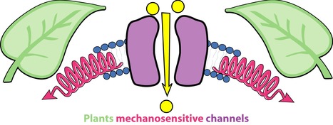
Mechanosensitive channels provide to the plant microprobe which convert instantaneously force into biological signals. The study of mechanosensitive channels (MS) in living organisms has progressed considerably over the past two decades. The understanding of their roles in mechanosensation and mechanotransduction was consecrated by the awarding the Nobel Prize in 2021 to A. Patapoutian for his discoveries on the role of MS channels in mechanoperception in humans. In plant, identified MS channels belong to MSL, MCA, Piezo, OSCA and TPK families. They convert instantaneously (ms range) membrane tension variations into biological signals. Upon activation, MS channels mediate Ca2+ signals, electrical signals or osmotic signals. The next challenge in plant mechanotransduction is to map forces at the cellular scale in order understand under which conditions and at which locations MS channels activate “in cellulo”. This article was published in Current Opinion in Plant Biology.
|
|
Scooped by
I2BC Paris-Saclay
June 15, 2022 11:49 AM
|
Discovery of the first inhibitors of the bacterial undecaprenyl-pyrophosphate phosphatase BacA
The first inhibitors of the undecaprenyl-pyrophosphate phosphatase BacA, showing BacA-targeted antibacterial activity, were discovered by high-troughput virtual screening, molecular dynamics and biological activity measurements. The peptidoglycan is an essential heteropolymer of the bacterial envelope that guarantees the integrity and the shape of all bacterial cells. This is also their Achilles’s heel as underlined by the extent of compounds that impede its structure or biogenesis. The biosynthesis of peptidoglycan relies on a lipid carrier, the undecaprenyl phosphate, which is used as a cargo for the export of the subunits across the plasma membrane. As a byproduct of peptidoglycan polymerization, the lipid carrier is released in the undecaprenyl pyrophosphate form, which must be recycled. To this end, it undergoes a dephosphorylation catalyzed by the membrane enzyme BacA, which is specific and highly conserved in bacteria. In the present study, we identified the first small antibacterial compounds displaying inhibitory activity towards BacA using high-throughput virtual screening (HTVS). We prepared a virtual library of 8 million commercial compounds, followed by HTVS through molecular docking using the 3D structure of BacA, that we recently resolved (El Ghachi et al., Nat Commun, 2018), and molecular dynamics snapshots to account for the flexibility of the protein. Of 83 best compounds computationally selected, one hit compound inhibited BacA activity in vitro. Subsequently, an additional 33 scaffold analogs were selected, of which 6 compounds exhibited BacA inhibition. The half-maximal inhibitory concentration (IC50) values of these compounds ranged from 42 to 366 μM. Importantly, the best compounds also displayed an antibacterial activity against Escherichia coli that was further demonstrated to be dependent on BacA. This overall strategy and the identified structural scaffold of these inhibitors provide a solid foundation for the further development of potent BacA-targeted inhibitors and the design of novel antibiotics. More information: https://www.sciencedirect.com/science/article/pii/S2001037022001660?via%3Dihub Contact: Thierry Touzé <thierry.touze@i2bc.paris-saclay.fr>
|
|
Scooped by
I2BC Paris-Saclay
June 15, 2022 11:40 AM
|
The CTCF protein: a misguided jack-of-all-trades in cancer cells
The CTCF protein shapes our chromosomes to make sure that genes are correctly activated in our cells. In this review, Segueni and Noordermeer summarize how the functions of this protein can be perturbed in numerous ways in cancer cells. Human cells need to precisely regulate the activity of their genes, making sure that in our bodies, each gene is active at the right place, at the right time and in the right amount. Incorrect gene activity is a leading cause of diseases, including cancers. The three-dimensional folding of human chromosomes is an important contributor to gene activity. A key factor in this process is a DNA-binding protein called CTCF, which acts as a “jack-of-all-trades” to organize chromosomes into different levels of folding. All these levels differently contribute to gene activity, thereby maintaining the essential balance for correct cell functions. Unsurprisingly, numerous perturbations of CTCF functions have been implied in different types of cancers. In this literature review, Segueni and Noordermeer have summarized recent insights into how diverse CTCF dysfunctions lead to oncogenesis. Incorrect binding of CTCF to chromosomes is the most common cause of perturbed gene regulation. While such changes in binding can be caused by changes to the CTCF protein itself, they are mostly due to changes in the DNA sequence or DNA methylation at the CTCF binding sites in the chromosomes. These differences in binding can lead to “enhancer hijacking” where disrupted chromosome folding permits unwanted contacts between oncogenes and activating elements in the genome. A number of non-canonical functions of CTCF in the emergence of cancers is discussed as well. To provide guidance for the analysis of CTCF perturbations, a computational strategy for genomics data is introduced to link changes in CTCF binding with reorganized chromosome folding in cancer cells. Based on its diversity of functions, a better understanding of CTCF perturbations can ultimately open up new opportunities for the development of treatments and therapies in different cancers. More information: https://doi.org/10.1016/j.csbj.2022.05.044 Contact: Daan Noordermeer <daan.noordermeer@i2bc.paris-saclay.fr>
|
|
Scooped by
I2BC Paris-Saclay
June 15, 2022 11:32 AM
|
Interdisciplinary access call to Structural biology, Biological imaging and Proteomics IBISA facilities
The 3 national infrastructures ProFi, FBI and FRISBI along with the GIS IBiSA proposed a call for a funded access to IBiSA-labelled facilities. This 1st call was a success with 7 projects selected among which one using the Imagerie-Gif facility (@I2BC). The three national infrastructures ProFi, FranceBioImaging and FRISBI along with the GIS IBiSA proposed a call for a funded access to IBiSA-labelled facilities. The aim was to promote IBiSA facilities networking through transdisciplinary research projects. This 1st call was a success with 26 proposals submitted. Seven projects were selected among which one using using the Imagerie-Gif facility.
One project coordinated by Mireille Bétermier in collaboration with JB Charbonnier was selected. It includes access to the Cytometry platform of Imagerie-Gif at I2BC (France BioImaging), the Proteopole Biophysics platform of Institut Pasteur and the ProteoSeine Mass spectrometry platform of Institut Jacques Monod. The project aims at using approaches from these three IBiSA platforms to integrate complementary data on programmed genome rearrangement and double-strand break repair in Paramecium Contact: Julie Ménétrey <julie.menetrey@i2bc.paris-saclay.fr>
|
|
Scooped by
I2BC Paris-Saclay
June 15, 2022 11:22 AM
|
Towards understanding the mechanisms responsible for intrahepatic cholestasis: biochemical and structural study of the human ATPase ATP8B1
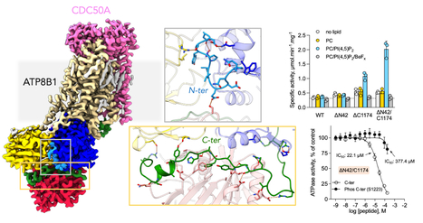
A study involving the LPSM unveils the mechanism of the human flippase ATP8B1 autoinhibition, as well as its regulation by lipids and phosphorylation. The study provides a better understanding of the impact of flippase mutations responsible for intrahepatic cholestasis on its enzymatic activity. Biological membranes are dynamic objects whose protein and lipid composition varies over time to ensure many cellular processes. In particular, there is a difference in lipid composition between the two layers of the membrane (lipid asymmetry) which is finely regulated by proteins, including flippases. These proteins catalyze the transport of lipids from the outer (exoplasmic) layer to the inner (cytosolic) layer of the membranes. In humans, mutations in the ATP8B1 flippase are responsible for several forms of intrahepatic cholestasis (familial progressive intrahepatic cholestasis, intrahepatic cholestasis of pregnancy and benign recurrent intrahepatic cholestasis).
Significant efforts are made to understand the lipid transport mechanisms used by ATP8B1 family proteins (P4 ATPase family). In 2019, a European collaboration involving a team from the Protein and Membrane Systems Laboratory (I2BC) unveiled three high-resolution structures (obtained by cryo-electron microscopy) of the yeast homolog (Drs2p) interacting with its Cdc50p subunit. This work revealed the mechanism by which the C-terminus of Drs2p inhibits the activity of the complex and the first steps in the removal of this auto-inhibition by PI4P.
In collaboration with the Universities of Aarhus, Bochum and Copenhagen, the I2BC researchers have this time studied the mechanism of ATP8B1 autoinhibition, as well as its regulation by lipids and phosphorylation. Using cryo-electron microscopy, they established the structure of the ATP8B1-CDC50A complex in an autoinhibited state. They also measured the enzymatic activity of the wild-type protein and of mutants truncated at its N- and/or C-terminus. The researchers show that both ends, and particularly the C-terminal end, play an essential role in the auto-inhibition of the enzyme activity. The efficiency of the inhibition is dependent on the phosphorylation of residue S1223 located in the C-terminal segment. In addition, phosphoinositides, primarily phosphatidylinositol-3,4,5-phosphate (PI(3,4,5)P3), are activators of the enzyme. The use of a synthetic peptide mimicking the C-terminal segment of the protein restores the inhibition of truncated forms of ATP8B1 but differently depending on whether the truncated form contains the N-terminal segment or not. These data suggest a molecular communication between the N- and C-terminal segments in autoinhibition. This result also demonstrates that it is possible to design compounds capable of regulating the activity of the complex. The resolution of the structure of ATP8B1/CDC50A in an autoinhibited state allows to localize the mutations of ATP8B1 responsible for the different forms of intrahepatic cholestasis. The mutations are uniformly distributed over the sequence but most are located in key areas that directly impact the catalytic properties of the enzyme. The presence of the (G/A)(Y/F)AFS motif in the auto-inhibitory C-terminal segment, already identified in other ATPases of the same family and directly involved in the auto-regulation of the enzymatic activity, suggests that this mechanism is widely employed among the flippases of the P4 ATPase family.family and directly involved in the auto-regulation of the enzymatic activity, suggests that this mechanism is widely used among the flippases of the P4 ATPase family. More information: https://doi.org/10.7554/eLife.75272 Contact: Guillaume Lenoir <guillaume.lenoir@i2bc.paris-saclay.fr >
|
|
Scooped by
I2BC Paris-Saclay
June 15, 2022 11:09 AM
|
Scientific day of Memory in Mike Dubow September 30th
The morning will be devoted to tributes and testimonials from people who have been fortunate enough to work with Mike Dubow during his teaching and research activities. The afternoon will be devoted to a symposium of the Department of Microbiology, which will focus on three scientific themes dear to Mike: extremophiles, biofilms and environmental phages. Each theme will include a presentation by a member of the Department and an external collaborator. Registration: https://evento.renater.fr/survey/hommage-a-michael-dubow-vendredi-30-septembre-2022-iary7xq2 Contact: hommage.mike@i2bc.paris-saclay.fr
|
|
Scooped by
I2BC Paris-Saclay
June 3, 2022 10:16 AM
|
French Days of Photosynthesis: June 9th-10th - Klaus Brettel's Portait
Klaus Brettel studied physics and biophysics at the universities of Stuttgart and Gießen (Germany) and obtained his PhD in 1985 for spectroscopic work on primary reactions in photosynthesis with Prof. H. T. Witt at the Max-Volmer-Institute of the Technische Universität Berlin (TUB). After a postdoctoral stay in the bioenergetics lab at CEA Saclay (France), he habilitated in physical chemistry at the TUB in 1990. He accepted a permanent researcher position at CEA Saclay in 1991. Twenty years ago, he shifted his center of interest from photosynthesis to DNA photolyases and cryptochrome blue light receptors. More recently, he also contributed to the elucidation of the reaction mechanism of another light-driven enzyme, fatty acid photodecarboxylase (FAP).
|
|
Scooped by
I2BC Paris-Saclay
May 19, 2022 4:11 AM
|
A Continuous Assay Set to Screen and Characterize Novel Protein N-Acetyltransferases Unveils Rice General Control Non-repressible 5-Related N-Acetyltransferase2 Activity
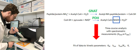
A new, robust continuous high-trough put test to screen and characterize new acetyltransferases. Protein N-acetyltransferases (NATs) belong to the general control non-repressible 5 (Gcn5)-related N-acetyltransferases (GNATs) superfamily. GNATs catalyze the transfer of acetyl from acetyl-CoA to the reactive amine moiety of a wide range of acceptors. NAT sequences are difficult to distinguish from other members of the GNAT superfamily and there are many uncharacterized GNATs. To facilitate the discovery and characterization of new GNATs, we have developed a new continuous, non-radioactive assay. This assay is virtually independent of the substrate and can be used to get substrate specificity hints. We validated first the assay with the well-characterized Schizosaccharomyces pombe NatA (SpNatA). The SpNatA kinetic parameters were determined with various peptides confirming the robustness of the new assay. We reveal that the longer the peptide substrate the more efficient the enzyme. As a proof of concept of the relevance of the new assay, we characterized a NAA90 member from rice (Oryza sativa), OsGNAT2. We took advantage of an in vivo medium-scale characterization of OsGNAT2 specificity to identify and then validate in vitro several specific peptide substrates. With this assay, we reveal long-range synergic effects of basic residues on OsGNAT2 activity. Overall, this new, high-throughput assay allows better understanding of the substrate specificity and activity of any GNAT. More information: https://pubmed.ncbi.nlm.nih.gov/35273627/ Contact: Carmela Giglione <carmela.giglione@i2bc.paris-saclay.fr>
|
|
Scooped by
I2BC Paris-Saclay
May 19, 2022 3:38 AM
|
Starting the engine of the powerhouse: mitochondrial transcription and beyond
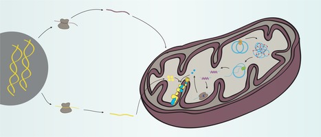
Mitochondria are the powerhouses of cells thanks to the oxidative phosphorylation system partially encoded by a small mitochondrial genome (mtDNA). Miranda et al review our current knowledge on mtDNA expression, associated disease phenotypes, and how mtDNA and nuclear gene expression are coordinated. Mitochondria are central hubs for cellular metabolism, coordinating a variety of metabolic reactions crucial for human health. Mitochondria provide most of the cellular energy via their oxidative phosphorylation (OXPHOS) system, which requires the coordinated expression of genes encoded by both the nuclear (nDNA) and mitochondrial genomes (mtDNA). Transcription of mtDNA is not only essential for the biogenesis of the OXPHOS system, but also generates RNA primers necessary to initiate mtDNA replication. Like the prokaryotic system, mitochondria have no membrane-based compartmentalization to separate the different steps of mtDNA maintenance and expression and depend entirely on nDNA-encoded factors imported into the organelle. Our understanding of mitochondrial transcription in mammalian cells has largely progressed, but the mechanisms regulating mtDNA gene expression are still poorly understood despite their profound importance for human disease. Here, we review mechanisms of mitochondrial gene expression with a focus on the recent findings in the field of mammalian mtDNA transcription and disease phenotypes caused by defects in proteins involved in this process.
|
|
Scooped by
I2BC Paris-Saclay
April 15, 2022 9:55 AM
|
Oxydative stress is a major trigger of the biosynthesis of some antibiotics in Streptomyces.
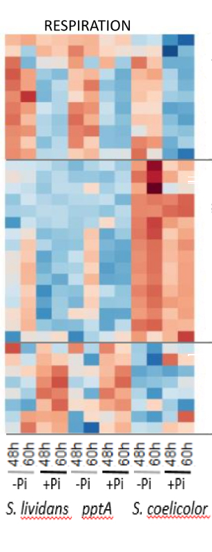
A proteomic study indicated that the ability of the pptA/sco4144 mutant strain of S. lividans to produce antibiotics is linked to a deficit in the generation of NADP(H) leading to high oxidative stress, a powerful trigger of a specific class of antibiotics. Streptomyces coelicolor and Streptomyces lividans, are closely related strains possessing the same biosynthetic clusters directing the biosynthesis of various specialized bio-active metabolites including that of the blue polyketide, actinorhoddin (ACT), that bears anti-oxidant properties. However most of these pathways are expressed in S. coelicolor but not in S. lividans. Interestingly, the disruption of a single gene (pptA) of S. lividans conferred to this strain the ability to produce ACT. A comparative quantitative proteomic analysis of these strains was carried out in order to define the potentially common features of the ACT producing strains. The abundance of proteins of numerous pathways differed greatly between S. coelicolor and the S. lividans strains, especially those of central carbon metabolism and respiration. S. coelicolor is characterized by a weak glycolytic activity resulting into a low carbon flux through the Pentose Phosphate Pathway, involved in the generation of NADPH, a co-factor of thioredoxin reductases necessary to combat oxidative stress. Furthermore, the complex I of the respiratory chain thought to generate reactive oxygen/nitrogen species was highly abundant in S. coelicolor. Oxidative stress was thus predicted to be high in S. coelicolor. In contrast, the S. lividans strains had rather similar proteins abundance for most pathways except for that involved into NAD(P)(H) metabolism. PptA being an accessory protein forcing polyphosphate into a conformation allowing their efficient use by various enzymes taking polyphosphate as a donor of phosphate and energy, including the ATP/Polyphosphate-dependent NAD kinase SCO1781, this suggested that the phosphorylation of NAD into NADP, would be inefficient in the pptA mutant resulting in low NADP(H) levels and thus high oxidative stress. Our results thus indicated that high oxidative stress is the common feature triggering ACT biosynthesis in S. coelicolor and in the pptA mutant of S. lividans. More information: https://www.frontiersin.org/articles/10.3389/fmicb.2021.813993/full contact: Marie-Joëlle VIROLLE <marie-joelle.virolle@i2bc.paris-saclay.fr>
|
|
Scooped by
I2BC Paris-Saclay
April 15, 2022 9:19 AM
|
Essential role of hyperacetylated microtubules in innate immunity escape orchestrated by the EBV-encoded BHRF1 protein
BHRF1, a multifunctional viral protein expressed during Epstein-Barr virus reactivation, modulates mitochondrial dynamics and induces MT hyperacetylation to escape innate immunity. Moreover, the loss of MT hyperacetylation impedes BHRF1 to initiate mitophagy, which is essential to inhibit the signaling pathway. Innate immunity constitutes the first line of defense against viruses, in which mitochondria play an important role in the induction of the interferon (IFN) response. BHRF1, a multifunctional viral protein expressed during Epstein-Barr virus reactivation, modulates mitochondrial dynamics and disrupts the IFN signaling pathway. Mitochondria are mobile organelles that move through the cytoplasm thanks to the cytoskeleton and in particular the microtubule (MT) network. MTs undergo various post-translational modifications, among them tubulin acetylation. In this study, we demonstrated that BHRF1 induces MT hyperacetylation to escape innate immunity. Indeed, the expression of BHRF1 induces the clustering of shortened mitochondria next to the nucleus. This "mito-aggresome" is organized around the centrosome and its formation is MT-dependent. We also observed that the α-tubulin acetyltransferase ATAT1 interacts with BHRF1. Using ATAT1 knockdown or a non-acetylatable α-tubulin mutant, we demonstrated that this hyperacetylation is necessary for the mito-aggresome formation. Similar results were observed during EBV reactivation. We investigated the mechanism leading to the clustering of mitochondria, and we identified dyneins as motors that are required for mitochondrial clustering. Finally, we demonstrated that BHRF1 needs MT hyperacetylation to block the induction of the IFN response. Moreover, the loss of MT hyperacetylation blocks the localization of autophagosomes close to the mito-aggresome, impeding BHRF1 to initiate mitophagy, which is essential to inhibiting the signaling pathway. Therefore, our results reveal the role of the MT network, and its acetylation level, in the induction of a pro-viral mitophagy. More information: https://journals.plos.org/plospathogens/article/authors?id=10.1371/journal.ppat.1010371 contact: Audrey ESCLATINE <audrey.esclatine@i2bc.paris-saclay.fr>
|
|
Scooped by
I2BC Paris-Saclay
April 15, 2022 8:56 AM
|
ComFC mediates transport and handling of single-stranded DNA during natural transformation
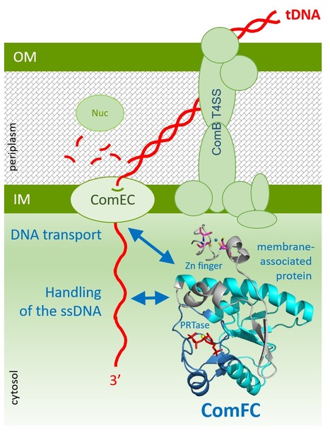
ComFC, a protein essential for natural transformation, is a membrane-associated protein with affinity for ssDNA. This dual-specificity allows us to propose that it provides the link between the transport of the transforming DNA into the cytoplasm and its handling by the recombination machinery. Natural transformation plays a major role in the spreading of antibiotic resistance genes and virulence factors. Whilst bacterial species display specificities in the molecular machineries allowing transforming DNA capture and integration into their genome, the ComFC protein is essential for natural transformation in all Gram- positive and -negative species studied. Despite this, its role remains largely unknown. Here, we show that Helicobacter pylori ComFC is not only involved in DNA transport through the cell membrane, but is required for the handling of the ssDNA once it is delivered into the cytoplasm. ComFC crystal structure revealed the presence of a zinc-finger motif and a putative phosphoribosyl transferase domain, both necessary for the protein's in vivo activity. ComFC is a membrane-associated protein with affinity for single-stranded DNA. Collectively, our results suggest that ComFC provides the link between the transport of the transforming DNA into the cytoplasm and its handling by the recombination machinery. More information: https://doi.org/10.1038/s41467-022-29494-z contact: Sophie QUEVILLON-CHERUEL <sophie.quevillon-cheruel@i2bc.paris-saclay.fr>
|
|
|
Scooped by
I2BC Paris-Saclay
July 25, 2022 4:53 AM
|
Training in Transmission electron microscopy for cell Biology
To acquire the theoretical basis of transmission electron microscopy (TEM) necessary for a functional exploration of the cell
Learn the different sample preparation techniques associated with TEM
Learn how to prepare your own sample Deadline registration September 27th. More information: here
|
|
Scooped by
I2BC Paris-Saclay
June 15, 2022 11:54 AM
|
Save the dates: MICROBES Symposium September 7-8, 2022
The OI MICROBES is pleased to invite you to the 1st MICROBES Symposium that will be held on September 7-8, 2022 in the Auditorium Blandin of the Laboratoire de Physique des Solides, Orsay, 1 rue Nicolas Appert, Bâtiment 510. The interdisciplinary MICROBES symposium is a yearly internal meeting where the teams of the Paris-Saclay MICROBES community present their research. The Symposium will have invited speaker presentations, posters and selected flash talks of posters. The symposium PROGRAM has five Plenary and Poster Sessions: Session 1 - Health and Disease (wed 7/9/22 morning) Session 2 - Ecology and Evolution (wed 7/9/22 afternoon) Session 3 - Fundamental Microbiology (wed 7/9/22 afternoon & thu 8/9/22 morning) Session 4 - Systems Biology and Engineering (thu 8/9/22 morning & afternoon) Session 5 - Agriculture and Food (thu 8/9/22 morning) Confirmed Speakers of the symposium are (to be completed): Mathieu Almeida (MGP) Harold Auradou (FAST) Théodore Bouchez (PROSE) Amandine Cornille (GQE) Bernard Delmas (VIM) Marina Elez (MICALIS) Denis Faure (I2BC) Tatiana Giraud (ESE) Amélie Leforestier (LPS) Bruno Le Pioufle (Institut d'Alembert) Virginia Lioy (I2BC) Filipa Lopes (LGPM) Florent Malloggi (NIMBE) Françoise Rul (MICALIS) Guillaume Tresset (LPS) David Vallenet (GM) INSCRIPTIONS are free but mandatory. Note that places are limited. Inscription deadline: August 31, 2022. Flash talk submission deadline: July 15, 2022. REGISTER HERE
For more information: oi.microbes@universite-paris-saclay.fr
|
|
Scooped by
I2BC Paris-Saclay
June 15, 2022 11:44 AM
|
Vibrio cholerae Chromosome Partitioning without Polar Anchoring by HubP.
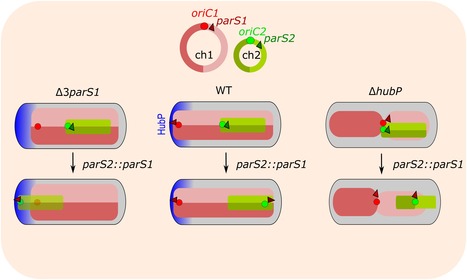
The polar anchoring HubP involved in partition system of Vibrio cholerae chromosome I (ch1) is dispensable for an active positioning of ch1 replication origin but is required for the normal longitudinal organization of ch1. Partition systems are widespread among bacterial chromosomes. They are composed of two effectors, ParA and ParB, and cis acting sites, parS, located close to the replication origin of the chromosome (oriC). ParABS participate in chromosome segregation, at least in part because they serve to properly position sister copies of oriC. A fourth element, located at cell poles, is also involved in some cases, such as HubP for the ParABS1 system of Vibrio cholerae chromosome 1 (ch1). The polar anchoring of oriC of ch1 (oriC1) is lost when HubP or ParABS1 are inactivated. Here, we report that in the absence of HubP, ParABS1 actively maintains oriC1 at mid-cell, leading to the subcellular separation of the two ch1 replication arms. We further show that parS1 sites ectopically inserted in chromosome 2 (ch2) stabilize the inheritance of this replicon in the absence of its endogenous partition system, even without HubP. We also observe the positioning interference between oriC1 and oriC of ch2 regions when their positionings are both driven by ParABS1 (parS2::parS1). Altogether, these data indicate that ParABS1 remains functional in the absence of HubP, which raises questions about the role of the polar anchoring of oriC1 in the cell cycle. More information: https://www.mdpi.com/2073-4425/13/5/877 Contact: Christophe Possoz <christophe.possoz@i2bc.paris-saclay.fr>
|
|
Scooped by
I2BC Paris-Saclay
June 15, 2022 11:36 AM
|
New insights into the mechanism of iron transport through the bacterial Ftr-system present in pathogens
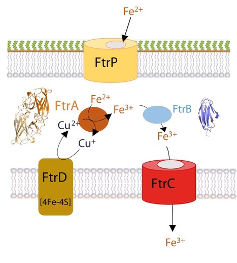
FtrA within the ferrous transport system (Ftr-type) found in pathogenic bacteria is a ferrous iron oxidase able to transform the ferrous ion into its ferric form by reducing a bound copper. Iron is an essential nutrient in bacteria. Its ferrous form, mostly present in low oxygen and acidic pH environments, can be imported using the specific Ftr-type transport system, which encompasses the conserved FtrABCD system found in pathogenic bacteria such as Bordetella, Brucella and Burkholderia. The non-pathogenicity and versatile metabolism of Rubrivivax gelatinosus make it an ideal model to study the FtrABCD system. Here, we report a new aspect of its regulation and the role of the periplasmic proteins FtrA and FtrB using in vivo and in vitro approaches. We investigated the metal binding mode and redox state of copper and iron to FtrA by crystallography and biophysical methods. An ‘as isolated’ FtrA protein from the bacterial periplasm contained a copper ion (Cu+) identified by electron paramagnetic resonance (EPR). Copper is coordinated by four conserved side chains (His and Met) in the primary metal site. Structural analysis of R. gelatinosus FtrA and FtrA homologues revealed that copper binding induces a rearrangement of the His95 imidazole ring, releasing thereafter space, as well as both Asp45 and Asp92 side chains, for iron binding in the secondary metal site. EPR highlighted that FtrA can oxidize the bound ferrous ion into the ferric form by reducing the bound Cu2+ into Cu+, both metal sites being separated by 7 Å. Finally, we showed that FtrB binds iron and not copper. These results provide new insights into the mechanism of ferrous iron utilization by the conserved FtrABCD iron transporter for which we propose a new functional model. More information: https://febs.onlinelibrary.wiley.com/doi/10.1111/febs.16476 Contact: Solange Morera <solange.morera@i2bc.paris-saclay.fr >
|
|
Scooped by
I2BC Paris-Saclay
June 15, 2022 11:27 AM
|
After two years, the whole Genome Biology Department meet in person to celebrate the VI Genome Biology Day
After two years, the whole Genome Biology Department meet in person to celebrate the VI Genome Biology Day. Last May 20th the Genome Biology Department met during a whole day to present and discuss the research projects carried out by the researchers of the department. Great short talks and posters covering studies on bioinformatics and genomics; gene expression and regulation; and dynamics and stability of genomes were presented during the day. In addition, two keynote talks were presented by Cynthia Sharma (University of Würzburg, Germany) and Ian Henderson (University of Cambridge, UK). Cynthia’s talk covered novel aspects of gene regulation by non-coding RNAs in Helicobacter pylori and Campilobacter jejeuni. Ian’s presentation focused into how the the Arabidopsis pan-centromere revealed the contribution of transposable elements on centromere evolution. At the end of the day, the winners of the ‘Best Talk Awards’ (James Provan, Barre’s lab & Kevin André, Souturina’s lab) and of the ‘Best Poster Awards’ (Amelie Besombes, Barre’s Lab; Kam Pou Ha, Bouloc’s lab and Estelle Leroy, Rougmaille’s lab) were announced. Altogether, this was a great day in which the GB department could meet and discuss about their favorite research topic: The Biology of Genomes. Contact: organizing commeette <virginia.lioy@i2bc.paris-saclay.fr>,<yoshiharu.yamaichi@i2bc.paris-saclay.fr> & <angelique.deleris@i2bc.paris-saclay.fr>
|
|
Scooped by
I2BC Paris-Saclay
June 15, 2022 11:13 AM
|
Top match between shade and wounding on leaf growth and movement: an easy win for shade?
Studying the plant growth-defense tradeoff, the authors show that shade suppresses wound-induced leaf repositioning and identify key players in signal integration. Plants face different types of stressful situations without the ability to relocate to favorable environments. For example, increasing plant density reduces access to sunlight as plants start to shade each other. Foliar shading represents a stress that many plants cope with by changing their morphology. This includes elongation of stem-like structures and repositioning of leaves to favor access to unfiltered sunlight. Plants also defend themselves against various pathogens including herbivores. Defense mechanisms include the production of deterrent chemical and morphological adaptations such as stunted growth and downwards leaf repositioning.
Here we studied the morphological response of plants when simultaneously facing shade and herbivore stress. When facing both stresses petiole growth was intermediate between the shade-enhanced and wound-repressed response. In contrast, the shade cue overrides the wounding cue leading to a similar upwards leaf repositioning in the combined treatments or in the response to shade alone. Using gene expression analyses and genetics we identified two members of the Phytochrome Kinase Substrate family as playing a signal integration role when plants simultaneously faced both stresses. This contributes to our understanding of the mechanisms underlying plant morphological adaptations when facing multiple stresses.
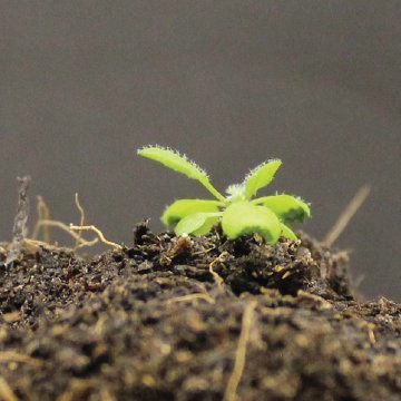
This work was led by Anne-Sophie Fiorucci during her post-doctoral studies in the lab of Christian Fankhauser (University of Lausanne). It sets the basis for her projects as Assistant Professor in the group of Sébastien Thomine at I2BC. In the frame of the ANR JCJC NitReST she got in 2021, she is now focusing on plant adaptations to shade and temperature in relationship with nitrogen nutrition. More information: https://journals.plos.org/plosgenetics/article?id=10.1371/journal.pgen.1010213 Contact: Anne-Sophie Fiorucci <anne-sophie.fiorucci@i2bc.paris-saclay.fr>
|
|
Scooped by
I2BC Paris-Saclay
June 15, 2022 10:59 AM
|
The apo-form of the Vibrio cholerae replicative helicase DnaB is a labile and inactive planar trimer of dimers
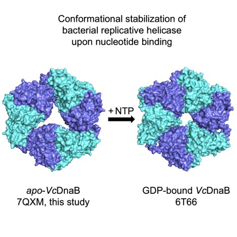
The bacterial replicative helicase DnaB is an hexameric ring-shaped ATPase molecular motor that unwinds dsDNA in front of the replisome during replication. Here, we provided a novel structure of the apo-DnaB from Vibrio cholerae which could be an intermediate state in DnaB formation. To enable chromosomal replication, DNA is unwound by the ATPase molecular motor replicative helicase. The bacterial helicase DnaB is a ring-shaped homo-hexamer whose conformational dynamics are being studied through its different 3D structural states adapted along its functional cycle. Our findings describe a new crystal structure for the apo-DnaB from Vibrio cholerae, forming a planar hexamer with a pseudo-symmetry, constituted by a trimer of dimers in which the CTD domains delimit a triskelion-shaped hole. This hexamer is labile and inactive. We suggest it represents an intermediate state allowing the formation of the active NTP-bound hexamer, from dimers. More information: http://doi.org/10.1002/1873-3468.14403 Contact: Claire Cargemel <claire.cargemel@i2bc.paris-saclay.fr>
|
|
Scooped by
I2BC Paris-Saclay
June 3, 2022 10:12 AM
|
French Days of Photosynthesis: June 9th-10th - Diana Kirilovsky's Portait
Diana Kirilovsky was born in Buenos Aires. She obtained her Ph.D. in Biochemistry at the Hebrew University of Jerusalem (Israel). After post-doctoral training in France at CNRS and CEA laboratories, she obtained a Senior Research Scientist position at the CNRS in 1991. She worked at several CNRS laboratories in Gif sur Yvette, l’ENS and the CEA Saclay. She finished her scientific career as Research Director “Classe Exceptionnelle (DRCE1, CNRS) and as Group Leader in the Institute of Integral Biology of the Cell (I2BC, CNRS). Diana Kirilovsky has studied the molecular biology and physiology of cyanobacteria for over 35 years focusing on light induced processes, in particular, the role of light as a source of stress and as a regulator. She principally studied the mechanisms of photoinhibition, photoprotection and light acclimation in cyanobacteria. A major contribution of her group was the discovery and characterization of a new mechanism of photoprotection in cyanobacteria involving a unique type of photoactive carotenoid protein. She has more than 100 peer-rewieved publications (including Science, PNAS, Plant Cell and JACS). She gave more than 50 invited conferences in international meetings and workshops including 7 Gordon Conferences (photosynthesis, carotenoids and photoreceptors). She was a member of the steering committees of the French and International Photosynthesis Societies during several years and of the French Photobiology Society .
|
|
Scooped by
I2BC Paris-Saclay
May 19, 2022 4:04 AM
|
Caveolin 1β promotes the production of active human microsomal glutathione S-transferase in induced intracellular vesicles in Spodoptera frugiperda 21 insect cells
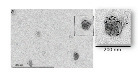
Caveolin-1β kept its capacity to induce intracellular vesicles in the host when co-expressed with hMGST1 (human microsomal glutathione S-transferase 1) in Spodoptera frugiperda 21 (Sf21) insect cells. Moreover, hMGST1 increased its specific activity. For a better understanding of membrane proteins cellular role, a detailed knowledge of their function and structure at a molecular level is required. For such studies, especially when combining site-directed mutagenesis and activity test experiments, membrane proteins are efficiently produced using overexpression in heterologous cell systems, among which insect cells. In mammalian cells, caveolin-1, an integral membrane protein of 21 kDa, is the main and characteristic membrane protein component of caveolae membranes. The heterologous expression in Spodoptera frugiperda 21 (Sf21) insect cells of the β isoform of canine caveolin-1 (caveolin-1β), using a baculovirus-based vector, resulted in intracellular vesicles enriched in caveolin-1β. We showed that caveolin-1β kept its capacity to induce intracellular vesicles in the host when co-expressed with hMGST1 (human microsomal glutathione S-transferase 1), and that hMGST1 is in part addressed to these vesicles. Remarkably, a fourfold increase in the amount of active hMGST1 was found in the most enriched membrane fraction, along with an increase of its specific activity by 60% when it was co-expressed with caveolin-1β. Thus, heterologously expressed caveolin-1β was able to induce cytoplasmic vesicles in which a co-expressed exogenous membrane protein is diverted and sequestered, providing a favorable environment for this cargo. More information: https://doi.org/10.1016/j.bbamem.2022.183922 Contact: Nadège Jamin <nadege.jamin@i2bc.paris-saclay.fr>
|
|
Scooped by
I2BC Paris-Saclay
May 19, 2022 3:24 AM
|
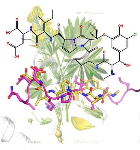
Structural convergence between inhibitors and a regulator of microtubule dynamics
A region of CPAP, a protein regulating the length of the centrioles, is found to have a tubulin binding mode similar to that of bacterial and fungal peptide-like metabolites which inhibit cell division. Microtubules are dynamic assemblies of αβ-tubulin that are involved in critical cellular functions in Eukaryotes, including cell division, ciliogenesis and intracellular transport. To fulfill these different functions, microtubules rearrange to form different types of networks, like the microtubule aster of interphasic cells or the mitotic spindle of dividing cells. They also form the architecture of well-defined structures like the centrioles. Relatedly, microtubule dynamics is regulated in the cell by different families of proteins. It is also inhibited by small molecules, some of which being used in oncology. The extent to which these compounds target the binding sites of cellular partners of tubulin remains poorly characterized. We have determined the structure of tubulin bound to the PN2-3 domain of CPAP, a protein controlling the length of the centrioles. Our results show in particular that the PN2-3 N-terminal region lies in a β-tubulin binding site known as the vinca domain. This site is also targeted by fungal and bacterial peptide-like inhibitors of tubulin, which share a very similar binding mode with CPAP. Therefore, our work identifies a structural convergence for tubulin binding between inhibitors and a regulator of microtubule dynamics.
|
|
Scooped by
I2BC Paris-Saclay
April 15, 2022 9:31 AM
|
6th edition of the Paris-Saclay Physics & Biological Systems conference PhysBio2022 - June 27-29 2022 - ENS Paris-Saclay.
Early-bird registrations are now open until the 15th of May at a discount rate, and free for students.
The PhysBio conference series brings together international scientists working at the interface between Physics and Biology. The scope of the conference is intentionally broad, comprising cell and tissue mechanics, biomedical engineering, morphogenesis and self-assembling, but also instrumental and methodological developments.
This breadth of topics goes together with a focus on excellent speakers, to whom we provide long-format talks that allow them to make their latest research accessible to all attendants, non-specialists and students included.
As discoveries of the coming decades will come from young generations, PhysBio conference series is endowed with a student committee that selected a dozen of short talks from outstanding PhD students.
Finally, we encourage all attendees to submit a poster contribution to initiate the discussion on their research topics with all the other participants.
Registration until May 15th: http://lptms.u-psud.fr/physbio2022/registration/ The organizing committee,
Christophe Le Clainche, Martin Lenz, Karen Perronet, Marie-Claire Schanne-Klein, and François Treussart.
|
|
Scooped by
I2BC Paris-Saclay
April 15, 2022 9:04 AM
|
Intensive week of training at Imagerie-Gif
|