Your new post is loading...

|
Scooped by
Gilbert C FAURE
September 12, 2020 4:20 AM
|
Morning Session Morning Chair: Dr Tom Marrs 09:00 Welcome and Introduction Dr Tom Marrs Consultant in Paediatric Allergy St Thomas' Hospital, London 09:10 Remote managment of eczema and urticaria in primary care Dr Alya Abdul-Wahab Consultant Dermatologist St George's Hospital, London 09:50 How to fix common gastro-intestinal symptoms in children Dr Rakesh Vora Paediatric Gastroenterology Consultant St Thomas' Hospital, London 10:35 << BREAK >> 11:00 How to manage IgE and non-IgE food allergies Dr Tom Marrs Consultant in Paediatric Allergy St Thomas' Hospital, London 11:40 How to recomment avoidance of egg and nuts Rebecca Brocklehurst Paediatric Allergy Dietitian St Thomas' Hospital, London 12:20 << LUNCH >> Afternoon Session Afternoon Chair: Dr Rosy Wells 13:00 Preventing food allergy: what to tell parents Dr Michael Perkin Consultant Allergist and Reader in Clinical Epidemiology St George's Hospital, London 13:30 Priorities to Tell Families Living With Food Allergy Professor Adam Fox Professor in Paediatric Allergy St Thomas' Hospital, London 14:00 QUICK FIRE: Adrenaline auto-injector guide IN PRACTICE: Spare pens in schools Alia Boardman Clinical Nurse Specialst in Paediatric Allergy St George's Hospital, London 14:30 << BREAK >> 15:00 Managing Long-Term Asthma in a Virtual World Dr Richard Iles Paediatric Respirology Consultant St Thomas' Hospital, London 15:40 How to recognise and treat allergic rhinitis Dr Anne Chsitopher Consultant Paediatrician and Allergist St George's Hospital, London 16:10 New inhalers and nasal sprays Katherine Knight Clinical Nurse Specialist in Paediatric Allergy St Thomas' Hospital, London 16:40 Webinar feedback and Close of meeting
|
|
Scooped by
Gilbert C FAURE
July 24, 2020 3:24 AM
|
Food allergy is an emerging epidemic that affects all age groups, with the highest prevalence rates being reported amongst Western countries such as the United States (US), United Kingdom (UK), and Australia.
|
|
Scooped by
Gilbert C FAURE
May 24, 2020 11:57 AM
|
Abstract Background A few studies have investigated the antecedents and outcomes of infants who demonstrate IgE sensitization to foods that they clinically tolerate. Improved understanding of this ...
|
|
Scooped by
Gilbert C FAURE
April 18, 2020 10:38 AM
|
Food allergies can be scary. Here how to reduce your baby’s risk, understand allergy tests and respond to a reaction if it happens.
|
|
Scooped by
Gilbert C FAURE
April 4, 2020 8:25 AM
|
|
|
Scooped by
Gilbert C FAURE
March 1, 2020 6:59 AM
|
Food Allergy research collection and call for papers.
|
|
Scooped by
Gilbert C FAURE
February 5, 2020 4:45 AM
|
New research shows that more than 25% of food samples tested by Suffolk Trading Standards were found to have hidden allergies from 2016 to 2019, with no indication that an allergen was present. Out of 187 food samples taken, 47 contained traces of allergenic substances that could prove harmful or...
|
|
Rescooped by
Gilbert C FAURE
from Immunology and Biotherapies
December 8, 2019 10:29 AM
|
Although trends have been reported, a common limitation of these biomarkers is that none are able to reproducibly predict prognosis during AIT. Further studies are needed to expand the currently available biomarker repertoire to provide personalized approaches to AIT.
Keywords
Food allergy Allergen Immunotherapy Biomarkers Basophils Tregs IgG4
Via Krishan Maggon
|
|
Rescooped by
Gilbert C FAURE
from Immunology and Biotherapies
November 25, 2019 4:28 AM
|
Abstract
Allergen Immunotherapy (AIT) was introduced in clinical practice on an empirical basis more than 100 years ago. Since the first attempts, AIT was administered subcutaneously. Indeed, other routes of administration were proposed and studied, in particular to improve the safety, but only the sublingual route (SLIT) achieved a credibility based on evidence, and was then accepted as a viable “alternative” option to the subcutaneous route. SLIT was largely used in clinical trials and clinical practice in this last 30 years. Thus, a large amount of data is available, coming from either controlled trials and post‐marketing surveillance studies. It is clear that SLIT is overall effective, but it is also clear that the efficacy is not “class‐related”, as derived from meta‐analyses, but restricted to each specific product. The 30‐year lasting use of SLIT allowed to clarify many clinical aspects, such as efficacy, safety, use in asthma, regimens of administration, optimal doses. In parallel, the mechanisms of action of AIT were elucidated, and new indications were proposed (e.g. food allergy, atopic dermatitis). In addition, the introduction of molecular‐based diagnosis, allowed to better refine the prescription of SLIT, based on specific sensitization profiles.
The present article will describe the origin and evolution of SLIT for respiratory allergy, taking into account the clinical context that suggested this form of treatment, the recently developed aspects, the future perspectives and unmet needs, This is not, therefore, a systematic review, rather a narrative historical description of the past history, and a look forward to the future opportunities.
Via Krishan Maggon
|
|
Scooped by
Gilbert C FAURE
November 4, 2019 4:48 AM
|
Grains in Relation to Celiac (Coeliac) Disease Donald D. Kasarda U.S. Department of Agriculture Agricultural Research Service Western Regional Research Center Albany, CA 94710 Here, I present my assessment of celiac disease in relation to cereal grains. What I have to say is based on many years of research in the area of gluten proteins as they relate to celiac disease, but because of the complexity of the subject, I do not claim definitive knowledge. My conclusions do not necessarily represent those of the Agricultural Research Service, United States Department of Agriculture (USDA), and are not intended to define USDA policy. There is much to be learned about celiac disease and some of my conclusions based on current knowledge may be modified as new information develops. In other words, some things I say here might turn out to be incorrect. Finally, I am a research chemist, not a physician, and do not intend this essay to be taken as medical advice in any legal sense. Celiac disease (coeliac is the usual spelling in Europe and Australia) is a condition that may develop in certain genetically susceptible individuals. People with celiac disease cannot eat wheat, rye, or barley. Proteins in these grains (and peptides derived from the proteins during digestion) initiate pathophysiological processes that may eventually lead to severe damage to the absorptive epithelium lining the small intestine. It appears likely that celiac disease is initiated by a mechanism involving immune response, but this has not been proved beyond any question. Certainly, immune reactions become involved after initiation. Because almost all nutrients, vitamins, minerals, amino acids, carbohydrates, and so on are absorbed by way of the small intestine, malabsorption resulting from damage to the absorptive lining of the small intestine can have wide ranging consequences; weight loss, osteoporosis, neuropathy, and so on. There is a wide range in response among those with celiac disease-some may have only minimal changes in the intestinal epithelium and no obvious symptoms, others may have severe damage to the lining of the intestine and severe symptoms. Although poor digestion of food usually leads to diarrhea, one of the most common symptoms in celiac disease, patients presenting with constipation have been reported. Although there is a definite genetic component, celiac disease is apparently a multigene disease, and its inheritance is not completely understood. It has been strongly associated with European populations and may be rare in African blacks or Asians of Chinese or Japanese descent. There is a strong correlation with certain histocompatibility antigens, but some people with the suspect antigens show no evidence of celiac disease. Although some people manifest evidence of celiac disease in the first year of life shortly after the introduction of gluten into the diet, others experience the onset of disease manifestations later in life-even very late in life. Consequently, it has been hypothesized that some environmental factor is likely to be involved in triggering the disease. Candidates for this environmental factor are viral infection, parasite infection (Giardia?, Cryptosporidium? Eimeria?), surgery, childbirth, even the stress of giving up smoking-these suggestions are highly speculative. A-GLIADIN PEPTIDES (SYNTHETIC) FOR WHICH THERE IS IN VIVO EVIDENCE OF ACTIVITY IN COELIAC DISEASE 31 L-G-Q-Q-Q-P-F-P-P-Q-Q-P-Y-P-Q-P-Q-P-F 49 31 L-G-Q-Q-Q-P-F-P-P-Q-Q-P-Y 43 44 P-Q-P-Q-P-F-P-S-Q-Q-P-Y 55 P = PROLINE Q = GLUTAMINE F = PHENYLANANINE L = LEUCINE Y = TYROSINE G = GLYCINE The manifestations of celiac disease are initiated (and re-initiated) in susceptible individuals upon eating wheat, rye, or barley, or any products from these grains that contain the main storage proteins of these grains. Both the proteins themselves and relatively small peptides derived from the proteins by enzymatic digestion are active in celiac disease. Accordingly, testing for intact proteins rather than peptides derived from then will often be ineffective. Celiac disease may be signalled by the presence of antibodies to gliadins or to endomysium in the blood serum and there are a number of commercial testing centers that provide testing for these antibodies. These tests are valuable, but do not provide complete certainty-either in indicating celiac disease or in indicating its absence. The presence of antigliadin or anti-endomysium antibodies will frequently result in a recommendation by the diagnosing physician to proceed to the most generally accepted test, intestinal biopsy. In severe cases, the biopsy will show mucosal damage, indicated especially by a flattening of the surface and loss of villous structure. Even this latter test is not entirely specific. A flattened mucosa may be the consequence of a few other diseases and damage may be patchy. The tiny tissue sample excised from the intestine in the biopsy procedure might by chance be taken from a relatively normal patch. To eliminate false negative histological results, the latest recommendation is to obtain 4-5 biopsies from different spots in the small intestine. The earliest stages of the disease may be subtle, perhaps indicated only by lymphocyte infiltration of the epithelium. Nevertheless, the finding of a flattened mucosa by way of the biopsy, followed by a marked improvement in symptoms and healing of the intestine upon initiation of a wheat, rye, and barley free diet are a pretty good indication of celiac disease. Because antibody levels decline and the intestinal mucosa recovers on such a diet, it is best for tests to be carried out before the potential celiac patient initiates the appropriate diet, thereby making testing impossible or difficult without a new challenge. This is especially so because the time for a challenge to take effect may vary considerably from person-to-person and too short a challenge might fail to bring about changes sufficient for diagnosis. The only plants demonstrated to have proteins that damage the small intestines of people with celiac disease are those from wheat, rye, and barley, (and the man-made wheat-rye cross called triticale). Until recently, oats have been considered harmful on the basis of early studies. Several recent studies of very high quality involving testing approaches that were not available to earlier workers, indicate that oats are not harmful to celiac patients or to those with dermatitis herpetiformis, but these findings have not been accepted by all physicians. There is also a practical problem with oats in that they tend to be grown in rotation with wheat or in nearby fields, the same machinery and storage bins might be used for both. Consequently, oats can be contaminated with small amounts of wheat. Wheat, rye, and barley are members of the grass family and are quite closely related to one another according to various schemes of plant classification (taxonomy). However, not all members of the grass family have proteins capable of damaging the intestines of celiac patients. Rice and corn, for example, are apparently harmless. Many other grains have not been subjected to controlled testing or to the same scrutiny as wheat, rye, barley, oats, rice, and corn in relation to celiac disease. If we accept corn and rice as safe, then members of the grass family that are more closely related to these species (on the basis of taxonomy) than to wheat are likely to be safe. Such grasses include sorghum, millet, teff, ragi, and Job's tears, which appear to be reasonably closely related to corn, and wild rice, which is closely related to cultivated rice. In some cases, there are protein structure studies that support of this conclusion, although the studies are not sufficiently complete to provide more than guidance. Scientifically controlled feeding studies with celiac patients would provide a better answer. However, such studies are not likely to be carried out in the forseeable future because of high costs and the difficulty of obtaining patient participation (such studies would be very likely to involve intestinal biopsy and patients are reluctant to undergo challenge once they are well). The scientific name for bread wheat is Triticum aestivum--the first part of the name defines the genus (Triticum) and the second part, the species (aestivum). Species falling in the genus Triticum are almost certain to be harmful to celiac patients. Grain proteins of these species include the various types characteristic of the gluten proteins found in bread wheats (including the alpha-gliadins) that cause damage to the small intestine in celiac disease. Some Triticum species of current concern include Triticum spelta (common names include spelt or spelta), Triticum polonicum (common names include Polish wheat, and, recently, Kamut), and Triticum monococcum (common names include einkorn and small spelt). I recommend that celiac patients avoid grain from these species. Rye (Secale cereale) and barley (Hordeum vulgare) are also toxic in celiac disease even though these two species are less closely related to bread wheat than spelta and Kamut. They belong to different genera, Secale and Hordeum, respectively, and lack alpha-gliadins, which may be an especially toxic fraction. There have been anecdotal reports suggesting a lack of toxicity in celiac disease for spelta and Kamut. Controlled tests would be necessary to draw a firm conclusion, but I don't consider anecdotal reports as reliable for the following reasons. The diagnosis, sometimes self-diagnosis, of celiac disease is occasionally made without benefit of reasonably rigorous medical or clinical tests, especially intestinal biopsy. Individuals who are "diagnosed" in this way without rigorous testing may not actually have celiac disease. Claims that particular foods cause this latter group no problems in relation to their celiac disease could cause confusion. Furthermore, celiac patients who report no problems in the short run with spelta or Kamut will very likely relapse later. There is now adequate evidence that when celiac patients on a "gluten-free" diet (that is, a diet free of any proteins or peptides from wheat, rye, barley, and oats) have wheat reintroduced to their diets, times-to-relapse vary enormously among individuals, ranging from hours to months, or even years. And this is for wheat, presumably the most toxic of all cereal grains to celiac patients. Additionally, the relapse may not be accompanied by obvious symptoms, but could be recognized only by physicians through observation of characteristic changes in the small intestinal tissues obtained by biopsy. The reasons for the enormous variability of response times are not known. It may be speculated that they have something to do with the degree of recovery of the lining of the small intestine on a gluten-free diet, the degree of stress that the patient had been experiencing (including infections), and individual genetic differences. As I have indicated, all known grain species that cause problems for celiac patients are members of the grass family. In plant taxonomy, the grass family belongs to the Plant Kingdom Subclass known as monocotyledonous plants (monocots). The only other grouping at the Subclass level is that of dicotyledonous plants (dicots). Some other species about which celiac patients have questions actually are dicots, which places them in very distant relationship to the grass family. Such species include buckwheat, amaranth, quinoa, and rape. The seed of the last plant listed, rape, is not eaten, but an oil is pressed from the seeds that is becoming commonly used in cooking. This oil is being marketed as canola oil. Because of their very distant relationship to the grass family and to wheat, it is highly unlikely that dicots will contain the same type of protein sequence found in wheat proteins that causes problems for celiac patients. Of course, some quirk of evolution could have given rise in these dicot plants to proteins with the harmful amino acid sequence found in wheat proteins. But if such concerns were carried to a logical conclusion, celiac patients would have to exclude all plant foods from their diets. It may be in order to caution celiac patients that they may have undesirable reactions to any of these foods--reactions that are not related to celiac disease. Allergic reactions may occur to almost any protein, but there is a great deal of individual variation in allergic reactions, and there are possibly non-allergic food reactions, such as to the sulfites used to preserve certain foods, which further complicates the situation. Also, buckwheat, for example, has been claimed to contain a photosensitizing agent that will cause some people who have just eaten it to develop a skin rash when they are exposed to sunlight. Such reactions, apparently rare, should be looked for, but for most people, buckwheat eaten in moderation apparently does not cause a problem. (Buckwheat is sometimes found in mixture with wheat, which of course would cause a problem for celiac patients.) It seems no more necessary for all people with celiac disease to exclude buckwheat from their diets because some celiac patients react to it than it would be for all celiac patients to exclude milk from their diets because some celiac patients have a problem with digestion of milk sugar (lactose) or are allergic to milk proteins, such as lactalbumin. Buckwheat, quinoa, and amaranth have been reported to have relatively high levels of oxalic acid, almost as much as in spinach, and may not be suitable for very young children because the oxalic acid may cause gatrointestinal problems. Some celiac patients may exhibit allergic reactions to gluten proteins or non-gluten proteins of wheat (and rye and barley), the alpha-amylase inhibitors being an example of the non-gluten proteins that can cause allergic reactions. Related inhibitor proteins can be found in rice as well. Alpha-amylase inhibitors might also interfere with starch digestion, causing symptoms similar to lactose intolerance in people with a weakened digestive capability. Celiac disease is thought to involve delayed immunoreaction and patients would not generally be expected to have an immediate and violent reaction to eating wheat whereas allergic reactions of the immediate hypersensitivity type might be both immediate and violent. It is also possible that both immediate hypersensitivity and delayed reactions might be present in the same person. There is a considerable potential then for confusion between allergy and celiac disease. It may be difficult to distinguish immediate hypersensitivity reactions or allergies from celiac disease as traditionally defined, but more research on this problem is needed. In conclusion, scientific knowledge of celiac disease, including knowledge of the proteins that cause the problem, and the grains that contain these proteins, is in a continuing state of development. There is much that remains to be done. Nevertheless, steady progress has been made over the years. As far as I know, the following statements regarding various grains are a valid discrimination of the state of our knowledge: Spelt or Spelta and Kamut are wheats. They have proteins toxic to celiac patients and should be avoided just as bread wheat, durum wheat, rye, barley, and triticale should be avoided. Rice and corn (maize) are not toxic to celiac patients. Certain cereal grains, such as various millets, sorghum, teff, ragi, and Job's tears are close enough in their genetic relationship to corn to make it likely that these grains are safe for celiac patients to eat. American wild rice is sufficiently closely related to normal rice that it is likely also to be safe. Significant scientific studies with celiac patients have not been carried out, however, for these grains. There is no reason for celiac patients to avoid plant foods that are very distantly related to wheat. These include buckwheat, quinoa, amaranth, and rapeseed oil (canola). Some celiac patients might suffer allergies or other adverse reactions to these grains or foodstuffs made from them, but there is currently no scientific basis for saying that these allergies or adverse reactions have anything to do with celiac disease. A celiac patient may be lactose intolerant or have an allergy to milk proteins, but that does not mean that all celiac patients will have an adverse reaction to milk. related web sites: http://www.celiac.com/ http://www.panix.com/~donwiss/ http://www.dietitian.com/gluten.html http://www.gretmar.com/webdoctor/celiac.html http://cpmcnet.columbia.edu/dept/gi/celiac.html http://www.vtmednet.org/~naspgn/celiac.html http://www.nutrimed.com/SPRUE.HTM A list of my publications with pertinence to celiac disease follows. Cross-references to the literature for most of the points discussed above can be found in these publications. Kasarda, D.D. In press. Gluten and gliadin: precipitating factors in coeliac disease. Proceedings of the 7th International Symposium on Coeliac Disease, September 5-7, 1996, Tampere, Finland. Srinivasan, U., Leonard, N., Jones, E., Kasarda, D.D., Weir, D.G., O'Farrelly, C., and Feighery, C. 1996. Absence of oats toxicity in coeliac disease. British Medical Journal 313:1300-1301. Tatham, A.S., Fido, R.J., Moore, C.M., Kasarda, D.D., Kuzmicky, D.D., Keen, J.N., and Shewry, P.R. Characterization of the major prolamins of tef (Eragrostis tef) and finger millet (Eleusine coracana). J. Cereal Sci. 24:65-71. 1996. Kasarda, D.D. 1994. Defining cereals toxicity in coeliac disease. In: Gastrointestinal Immunology and Gluten-Sensitive Disease. Feighery, C., and O'Farrelly, F., Eds., Oak Tree Press, Dublin, pp. 203-220. Shewry, P.R., Tatham, A.S., and Kasarda, D.D.1992. Cereal proteins and coeliac disease, pp. 305-348. In Marsh, M.N. (ed.) Coeliac Disease, Blackwell Scientific Publications, Oxford, U.K. De Ritis, G., Auricchio, S., Jones, H.W., Lew, E.J.-L., Bernardin, J.E. and Kasarda, D.D. 1988. In vitro (organ culture) studies of the toxicity of specific Agliadin peptides in celiac disease. Gastroenterology 94:41-49. Kagnoff, M.F., Patterson, Y.J., Kumar, P.J., Kasarda, D.D., Carbone, F.R., Unsworth, D.J. and Austin, R.K. 1987. Evidence for the role of a human intestinal adenovirus in the pathogenesis of celiac disease. Gut 28:995-1001. Levenson, S.D., Austin, R.K., Dietler, M.D., Kasarda, D.D. and Kagnoff, M.F. 1985. Specificity of antigliadin antibody in celiac disease. Gastroenterology 89: 1-5. Kagnoff, M.F., Austin, R.K., Hubert, J.J., Bernardin, J.E. and Kasarda, D.D. 1984. Possible role for a human adenovirus in the pathogenesis of celiac disease. J. Exp. Med. 160: 1544-1557. Kagnoff, M.F., Austin, R.K., Johnson, H.C.L., Bernardin, J.E., Dietler, M.D. and Kasarda, D.D. 1982. Celiac sprue: correlation with murine T cell responses to wheat gliadin components. J. Immunology 82: 1296-2693. Kasarda, D.D. 1981 Toxic proteins and peptides in celiac disease: relations to cereal genetics, In: Food, Nutrition, and Evolution: Food as an Environmental Factor in the Genesis of Human Variability. Walcher, D. and Kretchmer, N. (eds.),. Masson Publishing, USA, NY, pp 201-216. Falchuk, S.M., Nelson, D.L., Katz, A.J., Bernardin, J.E., Kasarda, D.D., and Strober, W. 1980. Gluten-sensitive enteropathy: influence of histocompatibility type on gluten sensitivity in vitro. J. Clin. Invest. 66: 227-233. Kasarda, D.D. 1978. The relationship of wheat proteins to celiac disease. Cereal Foods World 23: 240-244, 262. 1978. Kasarda, D.D., Qualset, C.O., Mecham, D.K., Goodenberger, D.M., and Strober, W. 1978. A test of the toxicity of bread made from wheat lacking ocgliadins coded for the 6A chromosome. In: Perspectives in Coeliac Disease-Proc. 3rd Int. Symposium on Coeliac Disease. McNicholl, B., McCarthy, C.F., and Fottrell, P.F. (eds.), MTP Press Ltd.,Lancaster, U.K, University Park, Press, Baltimore, MD, USA, pp. 55-61. Bernardin, J.E., Saunders, R.M., and Kasarda, D.D. 1976. Absence of carbohydrate in celiac-toxic A-gliadin. Cereal Chem. 53: 612-614. Kasarda, D.D. 1975. Celiac disease. In: Protein Nutritional Quality of Foods and Feeds. Part 2. Friedman, M. (ed.), Marcel Dekker, New York, pp. 565-593. Kasarda, D.D., Nimmo, C.C., and Bernardin, J.E. 1974. Structural aspects and genetic relationships of gliadins. In: Coelic Disease: Proc. 2nd Int. Conference. Hekkens, W.Th.J.M. and Pena, A.S. (eds.), Stenfert Kroese, Leiden, pp. 25-36. Kasarda, D.D. 1972. Celiac disease: malabsorption of nutrients induced by a toxic factor in gluten. Baker's Digest 46(6): 25-31. back to the GrainGenes Homepage
|
|
Rescooped by
Gilbert C FAURE
from Immunology and Biotherapies
October 30, 2019 3:51 PM
|
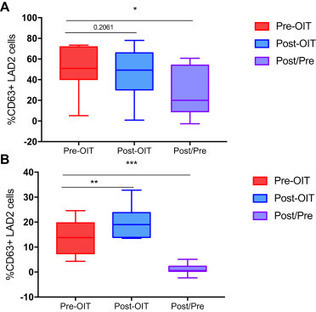
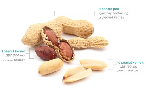
Peanut allergy is one of the most common and severe food allergies, affecting about
2% of school-aged children in the United Kingdom and United States. Current management of peanut allergy consists of strict allergen avoidance and carrying emergency medication to treat acute allergic reactions...
Via Krishan Maggon
|
|
Rescooped by
Gilbert C FAURE
from Immunology and Biotherapies
September 15, 2019 1:22 PM
|
BRISBANE, Calif.--(BUSINESS WIRE)--Sep. 13, 2019-- Aimmune Therapeutics, Inc. (Nasdaq:AIMT), a biopharmaceutical company developing treatments for potentially life-threatening food allergies, today announced that the Allergenic Products Advisory Committee (APAC) convened by the U.S. Food and Drug Administration (FDA) voted to support the use of AR101 (proposed trade name PALFORZIA™) in children and teens with peanut allergy. PALFORZIA is a complex, biologic oral immunotherapy (OIT) candidate designed to reduce the incidence and severity of allergic reactions, including anaphylaxis, after accidental exposure to peanut in patients aged 4 through 17 years with a confirmed diagnosis of peanut allergy.
The APAC voted 7 to 2 that the efficacy data and 8 to 1 that the safety data, in conjunction with additional safeguards, are adequate to support the use of PALFORZIA.
Via Krishan Maggon
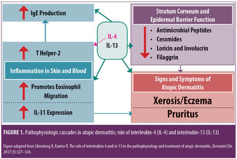
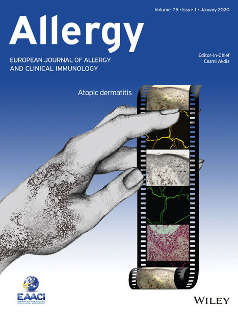
Monoclonal Antibody Therapies for Atopic Dermatitis: Where Are We Now in the Spectrum of Disease Management? JCAD Online Editor | February 1, 2019 This ongoing column explores emerging treatment options, drug development trends, and pathophysiologic concepts in the field of dermatology. J Clin Aesthet Dermatol. 2019;12(2):39–43 by James Q. Del Rosso, DO Dr. Del Rosso is Research Director of JDR Dermatology Research in Las Vegas, Nevada; is with Thomas Dermatology in Las Vegas, Nevada; and is Adjunct Clinical Professor (Dermatology) with Touro University Nevada in Henderson, Nevada. FUNDING: There was no funding related to the development, writing, or publication of this article. DISCLOSURES: Dr. Del Rosso is a consultant, speaker, and/or researcher for several companies who market products used in the management of atopic dermatitis or have compounds under development. These include Almirall, Dermira, Galderma, Genentech, LaRoche Posay, Leo Pharma, Loreal, Ortho Dermatologics, Pfizer, Promius, Regeneron, Sanofi-Genzyme, Skinfix, Sonoma, Sun Pharma, and Taro. Abstract: Atopic dermatitis (AD) is a chronic disorder that requires thorough patient education and a therapeutic management strategy designed to control flares, decrease recurrences, and reduce pruritus. In cases that cannot be controlled by proper skin care and barrier repair, topical therapy, and avoidance of triggers, systemic therapy is often required to control flares and maintain remission. It is important for clinicians to avoid becoming overly dependent on the intermittent use of systemic corticosteroid therapy to control flares, without incorporating other treatment options that might more optimally control AD over time. This article provides an overview of systemic therapies, including conventional oral therapy options and injectable biologic agents, that modulate the immune dysregulation in AD. Major emphasis is placed on the monoclonal antibodies currently available (e.g., dupilumab) for the treatment of AD, as well as those in latter stages of development, with a focus on agents targeting IL-4 and/or IL-13. KEYWORDS: Atopic dermatitis, calcineurin inhibitors, phosphodiesterase-4 inhibitors, immunosuppressants, interleukin-4, interleukin-13 Many patients with atopic dermatitis (AD) are able to control their disease primarily with topical agents, including corticosteroids, calcineurin inhibitors, phosphodiesterase-4 (PDE4) inhibitors, moisturizers/barrier repair agents, wet wraps, and the avoidance of triggers.1,2 However, it is important to better define the word “control,” as AD is a chronic disorder characterized by marked flares of eczema and pruritus, variable periods of persistent eczema of lesser severity with itching, and complete remission, all of which vary in intensity, frequency, and duration among each individual affected by AD. Marked flares can often be mitigated with topical agents of adequate potency and duration, and, in selected cases, in conjunction with short courses of systemic corticosteroid (CS) therapy. The most difficult therapeutic challenges in AD are effective control of eczematous dermatitis (eczema) and pruritus, both of which are persistent but of a lesser overall severity, and the maintenance of remission after control of disease flares.1–5 Many patients with AD, including the parents/guardians of children with AD, deserve a discussion of what options exist beyond topical management alone and intermittent systemic CS therapy. This discussion often needs to be initiated by the clinician, as patients with AD or other chronic disorders depend on their clinician to direct them toward what is likely to be the most effective treatment for them at any given point in time. There are only so many oral CS courses or intramuscular CS injections a clinician can prescribe to help control AD flares without tipping the benefit versus risk balance toward too much risk. This same principle also applies to repeated use of topical CS therapy, which can progress to use so frequent that the risk for adverse effects is increased significantly. Skin barrier repair agents and steroid-sparing topical agents (e.g., pimecrolimus, tacrolimus, crisaborole) provide marked benefit in some cases of AD, especially on certain anatomic sites or when the affected body surface area (BSA) is not too extensive.1–3 However, most patients with AD would benefit from systemic therapies that are designed to achieve optimal suppression of AD, including eczematous dermatitis and/or pruritus. Daily diffuse application of a well-formulated moisturizer for skin barrier maintenance and the application of prescription topical therapies to persistent AD lesions remain part of the standard therapeutic regimen, especially for localized refractory and lichenified sites.1–6 Finding the optimal balance of therapeutic choices varies among individual patients and requires careful consideration of the overall clinical situation and specific patient-related factors, such as age, severity of AD signs and symptoms, and patient and clinician comfort levels with the treatments selected. Ultimately, the clinician should identify what is most likely to achieve an optimal level of control and express their treatment recommendations to the patient with realistic confidence and a proper benefit versus risk discussion. The time has come for clinicians treating AD to consider moving from a rescue approach for flares to treating AD as a chronic, inflammatory, cutaneous and systemic disorder by using therapies that more selectively suppress the underlying disease pathophysiology, effectively treat eczema and pruritus, mitigate flares, and sustain long-term control of the disease. While topical therapies to manage epidermal barrier dysfunction and inflammation of AD should remain an important component of the total management approach for patients with AD, clinicians would be prudent to also consider therapies with better short-term and long-term safety profiles than the conventional oral agents that are currently available. In this article, an overview of the current conventional oral systemic therapeutic options for atopic dermatitis are presented, followed by an overview of the new systemic therapeutic options for AD, namely monoclonal antibody agents, including the currently available agent, duplimab, and other agents in latter stages of development, with a focus on compounds targeting IL-4 and/or IL-13. Other monoclonal antibodies that have been studied and/or are currently under evaluation for treatment of AD, such mepolizumab (anti-IL-5), nemolizumab (anti-IL-31), and omalizumab (anti-IgE), as well as other drug classes, will be discussed in future installments of “What’s New in the Medicine Chest.” Conventional Systemic Therapeutic Options for Atopic Dermatitis—Oral Agents When patients with moderate-to-severe AD and their clinicians are considering systemic therapy for AD, a variety of treatment options are available.3,5–12 Prior to 2018, available systemic therapies for AD were primarily oral agents, such as cyclosporin, methotrexate, azathioprine, and mycophenolate mofetil, all of which appear to modulate the underlying pathophysiologic pathways that contribute to AD.3,6–8 Each of these agents has variable amounts of data available regarding its use in children and adults for treatment of AD.3,6–12 However, none of these oral agents are approved by the United States Food and Drug Administration (FDA) for the treatment of AD, and all exhibit immunosuppressant properties.3,8 Oral antihistamines have also been used as part of the treatment regimen for AD, primarily as an adjunctive therapy to help reduce pruritus and/or decrease interference with sleep (i.e., sedating antihistamines).12 It is important to note that chronic or frequent use of systemic CS is best avoided in children and adults due to the risk of several significant AEs.6,10–12 Cyclosporin. Among the conventional systemic oral agents used in the management of AD, cyclosporin appears to exhibit the fastest onset of efficacy, but its use is limited by its safety profile, which includes risks of nausea, cephalgia, hypertension, nephrotoxicity, sequelae of chronic immunosuppression, gingival hyperplasia, and drug interactions.6,8,10 Cyclosporin is primarily recommended for treatment-resistant and/or uncontrolled AD, after which patients are usually transitioned to a safer, long-term approach; continuous use of cyclosporin beyond 12 to 24 months generally is not advisable.6,8,10 Methotrexate. Methotrexate therapy, another conventional systemic oral treatment for AD, can exhibit efficacy in as little as 4 to 8 weeks, but, like cyclosporin, warrants careful monitoring due to potential adverse events (AEs); these include nausea, bone marrow suppression (including pancytopenia), hepatotoxicity, pulmonary fibrosis, potential sequelae of immunosuppression, drug interactions, and the need to avoid alcohol intake.6,8,10 As with cyclosporin, long-term use of methotrexate should likely be avoided. Azathioprine. Azathioprine is another conventional systemic oral treatment option for AD, but it is not usually considered an initial systemic option due to its slower onset of efficacy and potential toxicities. Potential AEs include bone marrow suppression, increased malignancy risk, other sequelae of immunosuppression, severe nausea/vomiting, abdominal pain, hepatotoxicity, drug hypersensitivity syndrome, and risk for drug-drug interactions (e.g., allopurinol).6,8,10 Mycophenolate mofetil. Finally, although data for use of mycophenolate mofetil as a treatment option for AD are more limited than cyclosporin data, mycophenolate mofetil appears to be the safest oral agent, when compared with cyclosporin, methotrexate, and azathioprine; it has an efficacy onset range of 4 to 12 weeks, making it a logical choice when transitioning patients to longer-term oral maintenance therapy after initial use of cyclosporin for treatment-refractory or severe AD. Potential AEs include gastrointestinal side effects, fatigue, hematologic changes, and potential sequelae of immunosuppression.6,8,10 Biologics for Treatment of Atopic Dermatitis Research is in progress evaluating a variety of injectable and/or oral agents, including PDE4 inhibitors, Janus kinase (JAK) inhibitors, cannabinoid receptor agonists, kappa-opioid receptor agonists, and agents that target thymic stromal lymphopoietin (TSLP).14–17 A systematic review and meta-analysis of published studies evaluating the efficacy of biologics in AD treatment (published in April 2018) reported good evidence, to date, regarding agents that inhibit IL-4 and/or IL-13; a relative lack of evidence supporting efficacy in AD was noted thus far in studies with biologics modulating other targets, such as omalizumab (anti-IgE), infliximab ((anti-tumor necrosis factor), ustekinumab (anti-IL-12/23), and rituximab (anti-B-cell).19 IL-4 and IL-13 are reported to play prominent roles in AD with inflammation in skin and/or blood, epidermal barrier impairment, pruritis, and susceptibility to infection (Figure 1).18 Monoclonal antibodies that inhibit the effects of various ILs (i.e., IL-4, IL 13, IL-5, IL-17, IL-22, IL-31, IL-33) are showing therapeutic promise for the treatment of AD. Monoclonal Antibody Interleukin-4 and Interleukin-13 Inhibitor Dupilumab. Dupilumab is an injectable human IgG4 monoclonal antibody that inhibits IL-4 and IL-13 cytokine responses, including the expression and/or release of proinflammatory cytokines, chemokines, and IgE; binding of dupilumab occurs with both Types I and II IL-4 alpha receptors, found on hematopoietic cells and keratinocytes, respectively.13,20,21 In March 2017, duplimab was FDA-approved for the treatment of moderate-to-severe AD in adult patients (aged ?18 years) in whom the disease has not been adequately controlled with prescription topical therapies or in cases where such therapies are not advisable. In October 2018, duplimab was also approved as an add-on maintenance treatment in adolescent and adult patients (aged ?12 years of age) for moderate-to-severe asthma with an eosinophilic phenotype or oral–corticosteroid-dependent asthma.13 13 The dosing regimens for AD and asthma might differ between patients; however, the common regimen includes a 600mg loading dose (2×300mg2/mL injections), followed by a single 300mg injection every two weeks; with regard to asthma, dupilumab is not indicated or recommended for relief of acute bronchospasm or status asthmaticus.13 Clinical response. In the pivotal randomized, controlled trials (RCTs) evaluating dupilumab for AD, which included a Phase II, dose-ranging study, two 16-week monotherapy RCTs versus placebo, and a 52-week RCT that allowed for combination use with a topical CS, 1,472 subjects received dupilumab, with 739 treated for more than 52 weeks.13,20–22 Efficacy was substantiated by improvements in several assessment parameters versus placebo, both clinically and statistically, including positive changes in Investigator Global Assessment (IGA), marked reductions in Eczema Area Severity Index (EASI) scores, and significant decreases in pruritus, with clinical improvements sustained in the 52-week study without any loss of efficacy.13,20,21 Many patients reported a definite improvement in eczema and pruritus within the first few injections of dupilumab; however, onset of efficacy occurred later in some individuals (within 2 to 3 months after starting therapy). In patients currently undergoing other systemic therapies for severe AD (e.g., cyclosporin, methotrexate) who are starting dupilumab, researchers recommended that therapy be bridged without abrupt discontinuation of the patients’ previous therapy in order to avoid rebound exacerbation of AD while waiting for the clinical effects of dupilumab to manifest. Clinicians should then determine, on a case-by-case basis, the optimal approach to take when tapering patients off previous systemic therapy. 13,20–22 Safety. During the RCTs, no significant changes occurred in laboratory test results of the study subjects; thus, laboratory monitoring was not required by the FDA to be included in the approved product labeling for dupilumab.13 The most common AEs observed in the RCTs were injection site reactions and conjunctivitis (10–16% in active arms vs. 2–9% in placebo arms); separately, hypersensitivity reactions (e.g., urticaria, serum sickness-type reactions) were observed in less than one percent of the active-treatment study subjects.13,20–22 Most cases of conjunctivitis did not require stopping dupilumab, and were treated with topical ophthalmic lubricants and anti-inflammatory agents, and appeared to resolve or markedly improve despite continued use of the drug; however, some cases were severe enough to require discontinuation of dupilumab therapy.13,20–23 New onset or worsening ocular symptoms warrant referral to an ophthalmologist for evaluation.13,23 Ocular abnormalities inherent to AD that are unrelated to dupilumab use, including conjunctivitis and blepharitis, are not uncommon; the cause of the conjunctivitis that occurs related to use of dupilumab is not fully understood.24 Dupilumab and concomitant systemic therapy. A complete review of publications on dupilumab are beyond the scope of this article; however, a few articles provide information on the effective and safe use of dupilumab in a subpopulation of patients previously treated with cyclosporin. In a 16-week RCT study of adults with AD (N=390), responses to dupilumab therapy in conjunction with a medium-potency topical CS were assessed in subjects with inadequate response to or intolerance of oral cyclosporin or those in whom it was clinically inadvisable to use cyclosporin.25 Researchers reported that, following individual clinical assessment, topical CS therapy was safely tapered and/or stopped in many patients. Results of the study indicate that dupilumab with concomitant topical CS therapy (when needed) might signi?cantly improve signs and symptoms of AD and patient quality of life, with no new safety signals noted by the investigators.25 Infection risk. Eight RCTs that assessed outcomes with dupilumab versus placebo in patients with AD were analyzed by meta-analysis, with an emphasis on the incidence of AEs.26 Regarding infection rate risks, dupilumab had a lower risk of skin infection (risk ratio: 0.54), compared with placebo, with similar to negligible risks noted for nasopharyngitis, urinary tract infection, upper respiratory tract infection, and herpes virus infection. These observations further support the concept that dupilumab is immunomodulatory through the mitigation of IL-4 and IL-13 signaling, without a significant increased risk of infection, which can occur with immunosuppressive agents. It is important to note that by counteracting certain immune dysfunctions that lead to epidermal barrier impairment and cascades of Th2-driven humoral and cutaneous inflammation, dupilumab might help to normalize certain immunologic processes that are dysregulated in AD. Continued research and pharmacovigilance will help elucidate the efficacy and safety factors associated with dupilumab in greater detail. Monoclonal Antibody Interleukin-13 Inhibitors Lebrikizumab. Lebrikizumab is an injectable monoclonal antibody that exhibits high-affinity binding to soluble IL-13, thus preventing pro-inflammatory signaling by inhibiting heterodimerization of the IL-13 alpha/IL-4 alpha complex.27 In a preliminary Phase II, dose/frequency-ranging 12-week RCT, 209 adults with moderate-to-severe AD were treated with one of three dosing regimens of active drug versus placebo. Following a two-week “run in” with medium-potency topical CS therapy (triamcinolone acetonide 0.1% applied twice daily with lower potency hydrocortisone 2.5% allowed for facial AD), patients were randomized to receive lebrikizumab 125mg every four weeks, a single dose of lebrikizumab (125mg or 250mg), or placebo. Primary efficacy endpoint was the percent of subjects achieving a 50-percent reduction in EASI at Week 12.27 Investigators reported that patients in the lebrikizumab 125mg every four weeks achieved markedly superior results compared with those in the single-dose lebrikizumab group and those in the control group. Superiority to placebo was also observed in other parameters (e.g., SCORAD-50, reduction in BSA). An increasing trajectory of favorable response based on the EASI-50 results was noted at the end of the study (12 weeks) in the group receiving lebrikizumab 125mg every four weeks. Overall, the safety profile was favorable in all study arms.27 Data from this early study in AD suggest that lebrikizumab for AD shows promise as a treatment for AD. Additional research is needed on whether further increases in the dose per injection or treatment frequency (i.e., interval between doses) and use of a loading dose improve lebrikisumab’s efficacy, without affecting safety, for initial and maintenance therapy for AD. Tralokinumab. Tralokinumab, an IgG4 human monoclonal antibody that specifically neutralizes IL-13, was evaluated in a Phase IIb, dose-ranging, 12-week RCT of adult subjects (N=202) with moderate-to-severe AD.28,29 Patients were randomized to receive a 45mg (n=50), 150mg (n=51), or 300mg (n=51) subcutaneous injection of tralokinumab or placebo (n=50) every two weeks after a two-week “run in” with a mid-strength topical CS.29 Several efficacy parameters were assessed, with the coprimary endpoints being the change from baseline in total EASI score at Week 12 and the percent of IGA responders at Week 12 versus baseline (IGA score of clear/almost clear + at least a 2-grade reduction). Overall, AEs were generally similar among all study arms. Interestingly, six of the 204 subjects (2.9%) exhibited treatment-emergent conjunctivitis during the study (placebo, n= 2 [3.9%], tralokinumab 45mg, n =1 [2.0%], and tralokinumab 150mg, n=3 [5.9%]). Another important observation was that the serum level of dipeptidyl peptidase 4 might serve as a predictive biomarker for patients who could benefit from tralokinumab therapy.29 As with lebrikizumab, initial results with this agent for AD are encouraging and hopefully will be further supported by additional RCTs. Summary Points AD is a chronic disorder that, from the outset, requires a management strategy designed to control flares, decrease recurrences, and reduce pruritus. Cases of AD that are not adequately controlled with conventional measures and topical therapy can usually be effectively treated with incorporation of systemic therapy. It is important to assess the benefits versus the risks of various options in each case. It is also important to avoid becoming dependent on the intermittent use of intramuscular and/or oral corticosteroid therapy to control flares. Incorporation of other treatment options that can more optimally control AD over time are recommended. With the use of oral immunosuppressive agents such as cyclosporin, methotrexate, mycophenolate mofetil, and azathioprine, baseline and periodic laboratory and clinical monitoring are very important. Each of these agents carries its own significant “side effects baggage” to keep track of with relevant testing. Dupilumab is a newer option shown to be effective in markedly decreasing signs and symptoms of AD. In the opinion of the author, based on the available data and experiences thus far, dupilumab therapy offers a more favorable overall safety profile in comparison with the available oral systemic agents. Lebrikizumab and tralokinumab, both inhibitors of IL-13, are currently under development and show promise based on preliminary studies in adult patients with moderate-to-severe AD. References Eichenfield LF, Tom WL, Berger TG, et al. Guidelines of care for the management of atopic dermatitis: section 2—management and treatment of atopic dermatitis with topical therapies. J Am Acad Dermatol. 2014;71(1):116–132. Sidbury R, Tom WL, Bergman JN, et al. Guidelines of care for the management of atopic dermatitis: section 4—prevention of disease flares and use of adjunctive therapies and approaches. J Am Acad Dermatol. 2014;71(6):1218–1233. Del Rosso JQ, Harper J, Kircik L, et al. Consensus recommendations on adjunctive topical management of atopic dermatitis. J Drugs Dermatol. 2018;17(10):1070–1076. Czarnowicki T, Krueger JG, Guttman-Yassky E. Novel concepts of prevention and treatment of atopic dermatitis through barrier and immune manipulations with implications for the atopic march. J Allergy Clin Immunol. 2017;139(6):1723–1734. Thomson J, Wernham AGH, Williams HC. Long-term management of moderate-to-severe atopic dermatitis with dupilumab and concomitant topical corticosteroids (LIBERTY AD CHRONOS): a critical appraisal. Br J Dermatol. 2018;178(4):897–902. Prezzano JC, Beck LA. Long-term treatment of atopic dermatitis. Dermatol Clin. 2017;35(3):335–349. Admani S, Eichenfield LF. Atopic dermatitis. In: Lebwohl MG, Berth-Jones J, Heymann WR, Coulson I, Eds. Treatment of Skin Disease: Comprehensive Therapeutic Strategies. 4th edition. Philadelphia, PA: Elsevier-Saunders; 2014: 52–60. Akhavan A, Rudikoff D. Systemic agents for the treatment of atopic dermatitis. In: Rudikoff D, Cohen SR, Scheinfeld N (eds). Atopic Dermatitis and Eczematous Disorders. Boca Raton, FL: CRC Press/Taylor & Francis Group; 2014:187–199. Dhadwal G, Albrecht L, Gniadecki R, et al. Approach to the assessment and management of adult patients with atopic dermatitis: a consensus document. section IV: treatment options for the management of atopic dermatitis. J Cutan Med Surg. 2018;22(1 Suppl):21S–29S. Mayba J, Gooderham M. Oral agents for atopic dermatitis: current and in development. In: Yamauchi PS (ed). Biologic and Systemic Agents in Dermatology. Cham, Switzerland: Springer International Publishing; 2018:319–330. Wolverton SE. Systemic corticosteroids. In: Wolverton SE (ed). Comprehensive Dermatologic Drug Therapy, 3rd edition. Philadelphia, PA: Elsevier-Saunders; 2013:143–168. Thomas K, Bath-Hextall F, Ravenscroft J, et al. Atopic eczema. In: Williams H. Bigby M, Diepgen T, et al (eds). Evidence-Based Dermatology, 2nd edition. Malden, MA: Blackwell Publishing; 2008: 128–163. Regeneron Pharmaceuticals and Sanofi-Genzyme. Dupixent (dupilumab) Injection, Full Prescribing Information. October 2018. Kusari A, Han AM, Schairer D, et al. Atopic dermatitis: new developments. Dermatol Clin. 2019;37(1):11–20. Patel N, Strowd LC. The future of atopic dermatitis treatment. Adv Exp Med Biol. 2017;1027:185–210. Edwards T, Patel NU, Blake A, et al. Insights into future therapeutics for atopic dermatitis. Expert Opin Pharmacother. 2018;19(3):265–278. Napolitano M, Marasca C, Fabbrocini G, et al. Adult atopic dermatitis: new and emerging therapies. Expert Rev Clin Pharmacol. 2018;11(9):867–878. Silverberg JI, Kantor R. The role of interleukins 4 and/or 13 in the pathophysiology and treatment of atopic dermatitis. Dermatol Clinic. 2017;35(3):327–334. Snast I, Reiter O, Hodak E, et al. Are biologics efficacious in atopic dermatitis: a systematic review and meta-analysis. Am J Clin Dermatol. 2018;19(2):145–165. Simpson EL, Bieber T, Guttman-Yassky E, et al. Two Phase 3 trials of dupilumab versus placebo in atopic dermatitis. N Engl J Med. 2016;375(24):2335–2348. Gooderham MJ, Hong HC, Eshtiaghi P, et al. Dupilumab: a review of its use in the treatment of atopic dermatitis. J Am Acad Dermatol. 2018;78(3S1):S28–S36. Hajar T, Hill E, Simpson E. Biologics for treatment of atopic dermatitis. In: Yamauchi PS (ed). Biologic and Systemic Agents in Dermatology. Cham, Switzerland: Springer International Publishing; 2018: 309–317. Treister AD, Kraff-Cooper C, Lio PA. Risk factors for dupilumab-associated conjunctivitis in patients with atopic dermatitis. JAMA Dermatol. 2018;154(10):1208–1211. Thyssen JP, Toft PB, Halling-Overgaard AS, et al. Incidence, prevalence, and risk of selected ocular disease in adults with atopic dermatitis. J Am Acad Dermatol. 2017;77(2):280–286. de Bruin-Weller M, Thaci D, Smith CH, et al. Dupilumab with concomitant topical corticosteroid treatment in adults with atopic dermatitis with an inadequate response or intolerance to ciclosporin A or when this treatment is medically inadvisable: a placebo-controlled, randomized phase III clinical trial (LIBERTY AD CAFE). Br J Dermatol. 2018;178(5): 1083–1101. Ou Z, Chen C, Chen A, et al. Adverse events of Dupilumab in adults with moderate-to-severe atopic dermatitis: a meta-analysis. Int Immunopharmacol. 2018;54:303–310. Simpson E, Flohr C, Eichenfield LE, et al. Efficacy and safety of lebrikizumab (an anti-IL-13 monoclonal antibody) in adults with severe moderate-to-severe atopic dermatitis inadequately controlled by topical corticosteroids: a randomized, placebo-controlled phase II trial (TREBLE). J Am Acad Dermatol. 2018;78(5):863–871. May RD, Monk PD, Cohen ES, et al. Preclinical development of CAT-354, an IL-13 neutralizing antibody, for the treatment of severe uncontrolled asthma. Br J Pharmacol. 2012;166(1):177–193. Wollenberg A, Howell MD, Guttman-Yassky E, et al. A Phase 2b dose-ranging efficacy and safety study of tralokinumab in adult patients with moderate to severe atopic dermatitis (AD). Poster presentation. Orlando, FL: American Academy of Dermatology Meeting. 3–7 Mar 2017. Tags: Atopic Dermatitis, calcineurin inhibitors, immunosuppressants, interleukin-13, interleukin-4, phosphodiesterase-4 inhibitors Category: Atopic Dermatitis, Past Articles, What's New in the Medicine Chest
Via Krishan Maggon
|

|
Suggested by
LIGHTING
September 11, 2020 3:20 PM
|
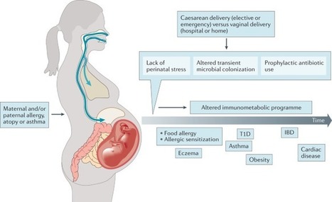
Allergies, including asthma, food allergy and atopic dermatitis, are increasing in prevalence, particularly in westernized countries. Although a detailed mechanistic explanation for this increase is lacking, recent evidence indicates that, in addition to genetic predisposition, lifestyle changes owing to modernization have an important role. Such changes include increased rates of birth by caesarean delivery, increased early use of antibiotics, a westernized diet and the associated development of obesity, and changes in indoor and outdoor lifestyle and activity patterns. Most of these factors directly and indirectly impact the formation of a diverse microbiota, which includes bacterial, viral and fungal components; the microbiota has a leading role in shaping (early) immune responses. This default programme is markedly disturbed under the influence of environmental and lifestyle risk factors. Here, we review the most important allergy risk factors associated with changes in our exposure to the microbial world and the application of this knowledge to allergy prevention strategies. Here, the authors explore how the modern way of life increases the risk of allergy and asthma, in particular by affecting the formation and diversity of the microbiota in early life. Understanding these changes highlights strategies for allergy prevention.
|
|
Scooped by
Gilbert C FAURE
May 24, 2020 12:26 PM
|
The gut-associated lymphoid tissue (GALT) faces a considerable challenge. It encounters antigens derived from an estimated 1014 commensal microbes and greater than 30 kg of food proteins yearly. It...
|
|
Scooped by
Gilbert C FAURE
May 13, 2020 7:00 AM
|
Food allergy is a condition with significant social and economic impact and a topic of intense concern for scientists and clinicians alike. Worldwide, over 220 million people suffer from some form of food allergy, but the number reported is just the tip ...
|
|
Scooped by
Gilbert C FAURE
April 9, 2020 7:59 AM
|
PubMed comprises more than 30 million citations for biomedical literature from MEDLINE, life science journals, and online books. Citations may include links to full-text content from PubMed Central and publisher web sites.
|
|
Scooped by
Gilbert C FAURE
March 5, 2020 2:12 PM
|
Some people produce immunoglobulin E (IgE) antibodies to proteins in common foods. As a result, these foods can trigger severe allergic inflammation (anaphylaxis). There are several structurally and functionally distinct antibody isotypes (IgM, IgD, IgG, IgA, and IgE), and which isotype binds to a target molecule (antigen) influences what happens next. For example, IgG that binds peanut proteins is harmless, but IgE bound to the same proteins can induce anaphylaxis and death. Therefore, how, where, and why allergen-reactive IgE is made are decades-old questions. Hoh et al. (1) found that gut tissue is a likely place for IgE development in peanut-allergic individuals. In addition, despite vast sequence possibilities, they found that many individuals share similar peanut-reactive IgE DNA sequences. This suggests that IgE antibodies in different individuals recognize peanut proteins in a similar manner, which could inform strategies for pharmacological interventions. Antibodies are produced by cells of the B lymphocyte lineage and consist of four Ig polypeptide chains—two identical heavy (H) chains and two identical light (L) chains—and each chain has a variable (V) region and a constant (C) region. The V region forms the surface that physically binds to antigens such as peanut proteins. The C region of IgH (CH) dictates antibody function largely by triggering processes that lead to neutralization, elimination, or induction of inflammation. Assembly of multiple IGH gene segments through the process of V(D)J recombination results in enormous sequence diversity, particularly in the exon encoding the V region, which comprises much of the antigen binding surface. Next to the V exon are a variety of C-region exons arranged in tandem (e.g., Cµ, Cγ, Cε, and Cα), which define the different antibody isotypes (2). Newly assembled Ig is produced as IgM. However, during an immune response, the same V region of IgH (VH) can be expressed in the context of another C-region isotype through a process called class switch recombination (CSR). This positions an alternative CH region next to the VH exon by permanent excision of the intervening DNA and associated CH-encoding exons. The position of CH exons relative to each other thus determines which IgH isotypes are available for secondary switch events. For example, an IgM-expressing B cell can switch to IgG1, and then that same IgG1+ B cell can switch to IgE because Cε is 3′ (downstream) to Cγ1 in the IGH locus. However, IgE-expressing B cells cannot switch to IgG1 (or any other isotype whose CH is positioned 5′ to Cε). Somatic hypermutation (SHM) further diversifies V regions by introducing mutations that can enhance affinity to antigen (3). The V(D)J recombination–, CSR-, and SHM-mediated processing of IGH enable phylogenetic mapping of how B-lineage cells and the antibodies they produce are related to one another. To gain insights into the origins of IgE production and the relationships between antibody-producing cells, Hoh et al. sequenced the IGH genes from B-lineage plasma cells in upper digestive tract tissues of 19 peanut-allergic individuals and compared them with those of nonallergic controls. They found more IgE-expressing cells in gut tissue in food-allergic individuals, confirming previous findings (4). Multiple clonally related VH-encoding sequences in IgE antibodies were also shared with other IgH isotypes. These findings suggest that B cells undergo CSR to IgE in the gut tissue as opposed to undergoing CSR to IgE elsewhere before migrating to the gut. This is different to the case of bone marrow, which is a major destination for antibody-producing cells after CSR elsewhere. This raises an important question: What features of the gut environment favor CSR to IgE? Moreover, because the bone marrow is a major location of antibody production, including IgE in allergic disease (5), the degree to which gut-derived versus bone marrow–derived IgE affects clinical disease, prognosis, and treatment approaches remains to be determined (see the figure). Hoh et al. identified antibody sequences that are reactive to the peanut protein Ara h 2 (Arachis hypogaea allergen 2) and found groups of similar sequences among multiple individuals. Similar sequences were also found in analyses of IgE+ B cells from peripheral blood in individuals with peanut allergy (6), further validating the concept of convergent IgE development to peanut proteins. Nonallergic individuals also had Ara h 2–reactive sequences, but only in non-IgE isotypes such as IgM, IgG, and IgA. These findings highlight how antibodies that induce a food-allergic response are generated. The production of antibodies that bind peanut proteins does not seem to be the problem per se; instead, the switching of that antibody to the IgE isotype appears to be key. This is consistent with observations that humans make IgG to a variety of dietary proteins without correlation to food allergy (7). In addition, it is possible that IgG to food antigens may be protective from food allergy by either blocking IgE binding or otherwise interfering with IgE function (8, 9). Perhaps an intervention that discourages gut IgE CSR could prevent food allergy. The convergence of IgE sequences in multiple peanut-allergic individuals suggests that immune recognition may occur through antibody binding to a few finite regions on key proteins. In this regard, drugs that block IgE binding to these regions holds promise as a therapy in allergic disease. There is proof of principle: Two therapeutic monoclonal IgG antibodies against a cat allergen inhibited IgE binding, and treatment with the combination of these two antibodies alone was sufficient to reduce allergic symptoms in 34 cat-allergic individuals in a clinical trial (10). Blocking IgE to peanut antigens may be similarly efficacious. Although seemingly innocuous, food allergens may influence the gut environment to generate conditions that induce CSR to IgE. In this regard, allergenic foods may have properties that induce allergic inflammation, as has been proposed (11, 12). Understanding the influences of the gut microbiota, age of exposure, and environment on the regulation of allergic responses to food (13, 14) promises to provide clues to elucidating how IgE CSR is regulated. http://www.sciencemag.org/about/science-licenses-journal-article-reuse This is an article distributed under the terms of the Science Journals Default License. References and Notes ↵ R. A. Hoh et al., Sci. Immunol. 5, eaay4209 (2020). ↵ P. Tong, D. R. Wesemann, Curr. Top. Microbiol. Immunol. 388, 21 (2015).OpenUrl ↵ L. Mesin, J. Ersching, G. D. Victora, Immunity 45, 471 (2016).OpenUrlPubMed ↵ C. Caffarelli, E. Romanini, P. Caruana, M. E. Street, G. De' Angelis, Pediatr. Res. 44, 485 (1998).OpenUrlPubMed ↵ S. Asrat et al., Sci. Immunol. 5, eaav8402 (2020). ↵ D. Croote, S. Darmanis, K. C. Nadeau, S. R. Quake, Science 362, 1306 (2018). ↵ J. Gocki, Z. Bartuzi, Postepy Dermatol. Alergol. 33, 253 (2016).OpenUrl ↵ O. T. Burton et al., J. Allergy Clin. Immunol. 134, 1310 (2014).OpenUrlCrossRef ↵ O. T. Burton, J. M. Tamayo, A. J. Stranks, K. J. Koleoglou, H. C. Oettgen, J. Allergy Clin. Immunol. 141, 189 (2018).OpenUrl ↵ J. M. Orengo et al., Nat. Commun. 9, 1421 (2018).OpenUrlCrossRef ↵ M. Profet, Q. Rev. Biol. 66, 23 (1991). ↵ N. W. Palm, R. K. Rosenstein, R. Medzhitov, Nature 484, 465 (2012). ↵ O. I. Iweala, C. R. Nagler, Annu. Rev. Immunol. 37, 377 (2019).OpenUrl ↵ D. R. Wesemann, C. R. Nagler, Immunity 44, 728 (2016).OpenUrl Acknowledgments: D.R.W. is supported by the NIH (AI121394, AI139538, and AI137940), the Burroughs Wellcome Fund, and an anonymous donor. C.R.N. is supported by the NIH (AI106302, AI134923, and AI146099) and the Sunshine Charitable Foundation. C.R.N. is president and co-founder of ClostraBio, Inc.
|
|
Rescooped by
Gilbert C FAURE
from Immunology and Biotherapies
February 17, 2020 1:07 PM
|
Abstract IgE mediated food allergy remains a significant and growing problem across the globe. Of the various treatment modalities, oral immunotherapy (OIT) and epicutaneous immunotherapy (EPIT) ha...
Via Krishan Maggon
|
|
Scooped by
Gilbert C FAURE
December 26, 2019 1:05 PM
|
Among children with other food allergies, 17% are also allergic to sesame, and antibody testing can accurately identify them, according to a recently published...
|
|
Scooped by
Gilbert C FAURE
November 25, 2019 6:39 AM
|
The 2020 Gordon Research Conference on Food Allergy will be held in Ventura, CA. Apply today to reserve your spot.
|
|
Scooped by
Gilbert C FAURE
November 8, 2019 1:54 PM
|
Food industry news, voices and jobs. Optimized for your mobile phone.
|
|
Scooped by
Gilbert C FAURE
November 4, 2019 4:47 AM
|
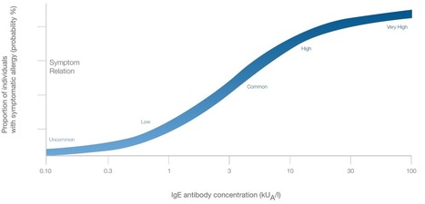
Patient history, clinical evaluation, and specific IgE tests are valuable tools in diagnosing food allergies.
|
|
Scooped by
Gilbert C FAURE
September 20, 2019 4:51 AM
|
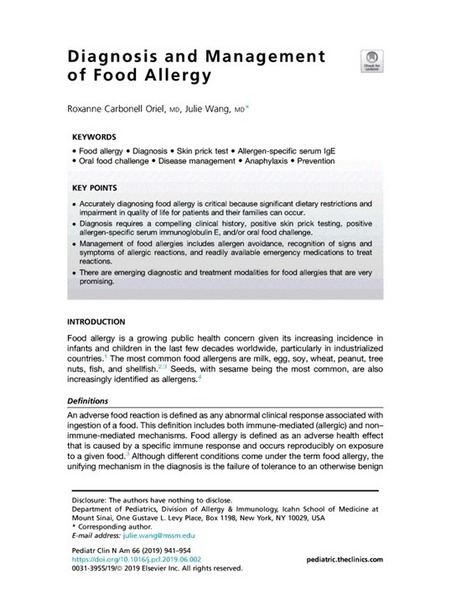
JavaScript is disabled on your browser. Please enable JavaScript to use all the features on this page. Pediatric Clinics of North America Volume 66, Issue 5, October 2019, Pages 941-954 Diagnosis and Management of Food Allergy Author links open overlay panel Show more Keywords Food allergyDiagnosisSkin prick testAllergen-specific serum IgEOral food challengeDisease managementAnaphylaxisPrevention First page preview Open this preview in PDF Disclosure: The authors have nothing to disclose. © 2019 Elsevier Inc. All rights reserved.
|
|
Scooped by
Gilbert C FAURE
August 13, 2019 4:57 AM
|
Tolle Totum Lisa Ghent, ND Atopic diseases are a collection of problems that arise when the immune system has an exaggerated IgE-mediated response to what are normally harmless antigens.1 Atopic diseases include atopic dermatitis, allergic rhinitis, asthma, and food allergies, and are one of the...
|