Your new post is loading...

|
Scooped by
Gilbert C FAURE
August 25, 8:26 AM
|
Functional beverages are having a moment. But what’s really driving the conversation? 🥤
From prebiotic sodas to kombucha, the functional beverage category is becoming increasingly visible across social media.
But looking at mentions alone only tells part of the story.
We analysed functional beverage conversations across LinkedIn, TikTok and Instagram to understand the trends, communities and brands driving attention online.
Here’s what stood out:
🔎 Gut health is a major conversation driver
#GutHealth, #Prebiotics and #Probiotics are closely intertwined with functional beverage conversations.
📱 Products are becoming part of creator culture
Brands such as Poppi are appearing in shopping, lifestyle and entertainment content — with some posts generating millions of views.
📊 The category is fragmented
Consumers aren't talking about “functional beverages” as one category. They're entering the conversation through wellness, low sugar, fermentation, gut health and other interests.
💡 Context matters
The most useful insight isn't simply how many times a brand is mentioned, but who is talking about it, what they're talking about and where those conversations are happening.
👉 Read the full analysis:
https://lnkd.in/eugKNDdP
|
|
Scooped by
Gilbert C FAURE
December 3, 2025 3:48 AM
|
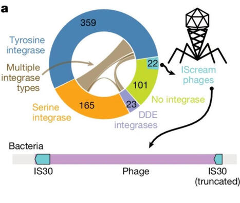
Gut Phageome: A New Frontier for Clinicians
A new Nature study using long-read metagenomics reveals that the human gut microbiome is far more dynamic than we thought
https://lnkd.in/dnVnetzX
Most gut bacteriophages (phages) are integrated in bacterial genomes, but a meaningful fraction are gained, lost, or even jump across bacterial species over time.
This means the gut microbiome is not just a community of bacteria - it’s an evolving genetic ecosystem shaped by phage–bacteria interactions.
Why it matters for clinical practice
Emerging evidence suggests that phage dynamics can influence:
• microbiome stability and dysbiosis
• inflammation and immune tone
• responses to antibiotics, diet, and microbiome-targeted therapies
Phage-aware diagnostics and therapeutics (including targeted phage cocktails) are already entering early clinical and translational research.
Take-home for clinicians:
1. The virome matters: Gut phages may become essential in interpreting microbiome results and understanding dysbiosis.
2. Therapies will evolve: Next-generation probiotics, FMT, and phage therapy will likely consider phage–bacteria interactions.
3. Expect personalization: Phage patterns may soon help stratify patients and guide individualized microbiome interventions.
Understanding the phageome is becoming a key part of understanding our gut
|
|
Scooped by
Gilbert C FAURE
September 9, 2025 7:38 AM
|
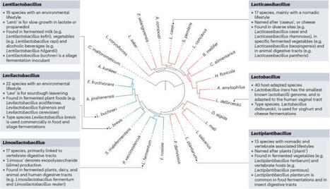
Lactobacilli encompass more than 300 species, spanning 25 genera, found in the microbiomes of humans, animals and plants with relevance in agriculture, foods and medicine. Lactobacilli comprise all bacteria previously assigned to the Lactobacillus genus and, similar to other lactic acid bacteria, are characterized by their saccharolytic, fermentation-energy metabolism and diverse enzymatic pathways that support redox balance and maintain intracellular pH. Some lactobacilli are pervasive in dairy, meat and plant foods, where they either contribute to spoilage and food waste or are desired and necessary for the production of fermented foods and animal feed. Strains of lactobacilli are the most applied probiotics tested in clinical studies. The study of host-associated intestinal and vaginal microbiomes has demonstrated that lactobacilli drive epithelial and immune cell responses, resulting in mainly beneficial effects on host health. This Review explores both established and emerging concepts related to this group of microorganisms. It highlights central tenants of their genetic diversity, metabolism, stress tolerance and distribution across host-associated microbiomes, as well as their importance in fermented foods and in health modulation as probiotics. With this accumulated knowledge, there remain substantial opportunities for expanded application of lactobacilli across different domains relevant to food production and health. Lactobacilli are important members of human, animal and insect microbiomes and are prominent in food fermentations. In this Review, Mejía-Caballero and Marco explore the diversity of lactobacilli, focusing on their fundamental traits and their applications in foods and medicine.
|
|
Scooped by
Gilbert C FAURE
November 14, 2021 5:25 AM
|
Viral respiratory tract infection (RTI) is the most frequent cause of infectious illnesses including the common cold. Pharmacological solutions for treating or preventing viral RTIs are so far limited and thus several self-care products are available in the market.
|
|
Scooped by
Gilbert C FAURE
August 27, 2020 7:43 AM
|
Nutr Clin Pract. Author manuscript; available in PMC 2016 Dec 1. Published in final edited form as: Published online 2015 Oct 8. doi: 10.1177/0884533615609899 PMCID: PMC4838018 NIHMSID: NIHMS777222 PMID: 26449893 The Gut Microbiome: What we do and don’t know Gail A. Cresci, PhD, RD, LD, CNSC, Associate Staff and Emmy Bawden, RD The publisher's final edited version of this article is available at Nutr Clin Pract See other articles in PMC that cite the published article. Introduction Interest in and knowledge of the gut microbiome has increased exponentially in the past decade. An internet search using key words such as “gut microbiome” or “gut microbiota” generates between 1 and 2.2 million results. Once overlooked, this component of the gastrointestinal (GI) tract is now gaining appreciation for its importance in optimal health. The food industry has taken this to full heights and a plethora of food and supplement products touting “probiotic” or “fermented foods” populate store shelves; and advertisements for them can be found in magazines, the internet and television commercials. Never before has the discussion of one’s bowel habits and GI symptoms been more mainstream. Interestingly, this rapid growth has left both consumers and many clinicians confused as the data, while growing, still leave many unanswered questions. Dating back to the early 1900’s Eli Metchnikoff, a Russian scientist of the Pasteur Institute in Paris, associated longevity of rural Bulgarians to their consumption of fermented milk products. He postulated that the lactic acid bacteria in the fermented milk products ingested by these peasants living in harsh climate and poverty provided an anti-aging effect which contributed to their greatly outliving wealthier Europeans. He named the organism “Lactobacillus bulgaricus.” As Metchnikoff researched, he hypothesized that seeding the gut with healthy bacteria by drinking fermented milk products could fight off harmful bacteria and prolong life. He was the first scientist to suggest that it was possible to modify the gut microbiome by replacing bad bacteria with good bacteria, and he earned a Nobel Prize in 1908 for his work in immunity. The discovery of penicillin in 1928 by Sir Alexander Fleming, a Scottish biologist, turned the attention of researchers away from using bacteria to assist with healing to that of using soil fungi derivatives to kill bacteria. During the golden age of antibiotic discovery from 1940–1960, most antibiotics were originally isolated by screening soil-derived actinomycetes (1). However as returns diminished on this discovery platform, new discoveries were based upon synthetic compounds. However this target-focused screening process of large libraries also failed, partly due to the inability of these synthetic compounds to penetrate the bacterial envelope (1). Unfortunately the deceleration of antibiotic discovery is accompanied with the spread of resistant bacteria, and a major public health threat of untreatable infections. With advances in micro- and molecular biology techniques, the culture is poised to revisit past successes with bacterial therapy for gut health and immunity in order to develop future treatments. One of several international efforts, the Human Microbiome Project (HMP) utilizes high through multi ‘omics analyses to identify and study the microbiome in human health (2,3). Funded by the NIH common fund in 2008, the HMP has resulted in the isolation and sequencing of over 1,300 reference strains thus far from the human body (2,3). The HMP Consortium has reported the structure and function of the human microbiome in 300 healthy adults (18–40 yrs) at 18 body sites from a single time point. This has led to an unprecedented amount of data about the complexity of the human microbiome allowing for a baseline for further research into the impacts of the microbiome on health and disease. As a precursor to the Human Microbiome Project, the Human Gut Microbiome Project has widened our appreciation for the bacterial ecosystem that resides within the human intestinal tract. This system is comprised of microorganisms such as bacteria, archaea, fungus and viruses that are distributed throughout the entire gastrointestinal tract (4). Ongoing investigations are revealing the importance of the gut microorganisms in exerting beneficial effects on human health. The typical healthy person is inhabited with trillions of microbes (Figure 1). But to confuse matters, two healthy people may have very different microbiomes. Analysis of HMP samples with lifestyle and history questionnaires has revealed associations between life-history characteristics and microbiome composition (5). Ding and Schloss analyzed a 16S rRNA gene sequence data set from the HMP Consortium. As a significant difficulty in analyzing microbiome data has been the considerable intra- and interpersonal variation in the composition of the human microbiome, this study took the approach of clustering samples into bins based on their taxonomic similarity. Using bacterial community structures collected from18 body sites and up to three time points, they applied community typing analysis to better understand the factors that affect the structure of the microbiome and contribute to human health. Using Dirichlet multinomial mixture models to partition the data into community types for each body site, their analysis of simulated data and the HMP data suggest that the community types represent clusters of relative abundance profiles. Three important observations were made with this approach. Firstly, there were strong associations between whether someone was breastfed, their gender and education level with their community types at several body sites. Secondly, the community types of the oral cavity and gut microbiome were predictive of each other even though the specific taxonomic compositions were different. Lastly, stability of community types was least stable in the oral cavity and most stable in the vagina and gut communities over the course of the sampling period. Therefore the authors concluded that despite substantial intra- and interpersonal variation in the human microbiome, by partitioning this variation intocommunity types, the types became predictive of each other and are likely the result of life-history characteristics (5). The gut microbiome is influenced by multiple factors including mode of infant delivery and feeding, aging process, diet composition, geography, medications, and stress (Figure 2). The following sections provide a brief overview of current knowledge regarding variances in the gut microbiota related to these factors. Our first colonization In utero It was previously believed that the GI tract was sterile in utero, and that during the birthing process was when first colonization and initiation of the gut microbiome occurred (6). However, recent studies have challenged this belief by demonstrating the presence of microorganisms in the placenta, amniotic fluid and umbilical cord (7–9). This work is still preliminary but deserves further study as it highlights the importance of maternal gut microbiota health. It is hypothesized that by swallowing amniotic fluid and its bacteria in utero, the fetus begins to colonize the developing GI tract (10). The meconium, the first infant stool sample, has also been shown to contain microorganisms (11). Notably, the meconium from preterm infants shows a different microbiota composition from that seen in a sample acquired after the first week of life, demonstrating how normal gut colonization takes place throughout the course of fetal development (10). Absence of this progressive colonization could place preterm infants at risk for various gastrointestinal infections, including necrotizing enterocolitis (NEC) (10). As a means to mimic the natural inoculation of the gut, multiple studies have evaluated probiotic supplementation as a means to influence the incidence and severity of NEC. A recent Cochrane review, which included 24 randomized controlled trials (n=5529), assessed the role of probiotic supplementation in preterm neonates. The review confirmed previous findings that probiotic supplementation significantly reduces the risk of stage II or greater necrotizing enterocolitis and all cause mortality in this patient population (12). As probiotics are known to have potentially beneficial effects on gut function and maturity, Athalye-Jape et al conducted a meta-analysis that included 25 randomized controlled trials (n=5895) to determine if probiotic supplementation influenced the time to establish first full enteral feeds in preterm neonates (13). Overall, the subjects supplemented with probiotics took less time to achieve full feeds (mean difference: −1.54 d; 95% CI: −2.75, −0.32d; p=0.01; I2 = 93%). The type and/or number of probiotic strains (e.g., Bifidobacterium or non-Bifidobacterium) did not influence this improvement. Additionally, probiotic supplementation reduced hospital length of stay, feeding intolerance, and duration of indirect hyperbilirubinemia as well as increased weight gain and growth velocity. None of the trials reported adverse effects of probiotics on outcomes. Delivery Mode The birthing process exposes newborns to a wide range of microorganisms that also contribute to the colonization of the gut microbiome. The mode of delivery affects the composition of the infant’s gut microbiota, and interestingly, the gut microbiota of the newborn will closely resemble the microbiota with which they encountered during birth. During a vaginal birth, the infant is inoculated with vaginal microbiota, which differs from skin microbiota encountered during a caesarian section (14). At the phylum level, studies have shown that infants delivered vaginally have a larger population of Bacteroidetes than Firmicutes when compared to infants delivered via cesarean section (15). Despite exposure in utero, the majority of microbes that will colonize the infant gut are acquired postpartum. The initial colonization pattern is thought to be chaotic, and increasing evidence suggests that environmental exposures early in life, including diet, are responsible for these variations (16). Gut colonization in an infant occurs in a succession of stages. Early on, the gut becomes colonized by primarily aerobic organisms, such as enterobacteria, staphylococci, and streptococci, many of which have the potential to be pathogenic. These early colonizers begin to change the gut environment, paving the way for colonization by an increasingly anaerobic community of microbes (17). The structure of the gut community continues to change over the first year of life and thereafter in response to external factors such as diet and antibiotic use (18, 19). Weaning, breastfeeding status, and the successive introduction of different types of food all correspondingly affect the infant gut microbiome and immune system (20). Infant Feeding Method Breast milk is the optimal food for infants as it meets all their nutritional and physiologic requirements. Human milk contains protein, fat and carbohydrate, as well as immunoglobulins and endocannabinoids (21). Breast milk is not sterile as it contains as many as 600 different species of bacteria including beneficial Bifidobacterium breve, B. adolescentis, B. longum, B. bifidum, and B. dentium (22). In addition to lactose, the carbohydrate component of human milk also contains oligosaccharides which comprise the third largest solid component (23). Human milk oligosaccharides are indigestible polymers formed by a small number of different monosacchrides that serve as prebiotics by selectively stimulating growth of members of the genus Bifidobacterium(23). Some of the beneficial effects of breast feeding over formula feeding are attributed to the effects of oligosaccharides on the beneficial bacteria as an increased proportion of Bifidobacteria is noted in breast-fed infants compared to those fed formula (24). Bifidobacteria have been linked with strengthening gut mucosal protection through activities against pathogens (25). Bifidobacteria have been shown to increase the production of immunoglobulin A which is correlated with modulation of the intestinal immune system (26). Schwartz et al studied breast-fed and formula-fed infants and their mothers. Stool samples from each infant were collected, and microbial DNA was extracted and sequenced; and mRNA was isolated from stool containing host gut exfoliated epithelial cells and processed for microarray analysis (20). They found that the microbiome of breast-fed infants to be significantly enriched in genes associated with virulence functionality, and demonstrated a multivariate correlation between the gut flora genes associated with bacterial pathogenicity and the expression of host genes associated with immune and defense mechanisms. Interestingly, the operational taxonomic unit (OTU) composition and genetic potential of the microbiota differed between breast-fed and formula-fed infants. The researchers suggested their findings indicate human milk promotes the mutualistic crosstalk between the mucosal immune system and the microbiome in the maintenance of intestinal homeostasis. More research is needed to better determine the mechanisms by which Bifidobacteria produce these effects. Aerotolerance of the intestinal microbiota also seems to differ between breast-fed and formula-fed infants. Aerobic organisms are more common in the feces of breast-fed infants, whereas anaerobic and facultatively anaerobic organisms, which preferentially use anaerobic glycolysis, are more frequently identified in feces of formula-fed infants (27). Bacteroides and Clostridia colonization differs between the two types of feeding, with breast-fed infants characterized with lower concentrations of both (28–30). With a better understanding of the composition of human breast milk, developments of complex infant formulas have attempted to mimic its nutritional value making them an acceptable alternative for mothers unable to breast-feed. Infant formulas are not a perfect substitute for human milk since bioactive compounds contained in breast milk known to affect nutrient absorption and digestion, immune protection, and defense against potentially pathogenic microbes are lacking (21). Unfortunately it is difficult to mimic the actions of these bioactive compounds. While infants fed a formula enriched with oligosaccharides have been shown to harbor more Bididobacteria in the feces, more evidence is needed for affirmation that infant formulas designed to mimic breast milk are beneficial (31). Changes in the Gut Microbiome with Age Revealed by metagenomic analysis, gut microbiota composition transforms throughout early stages of human development and is influenced by the diet (32). As an infant’s diet comprises breast milk and formula, this is reflective in that the microbiome has minimal diversity and is enriched in genes to facilitate lactate utilization (33). A shift in the functional capacity to preferentially utilize plant-derived glycans occurs prior to the introduction of solid foods. Around 3-years of age, the bacterial composition resembles that of an adult and remains stable until old age when variability in community composition increases (34). In terms of ecological succession, the Bifidobacterium-dominated microbiota of the infant changes over time into the Bacteroidetes- and Firmicutes-dominated microbiota of the adult (35). This distribution remains fairly stable throughout adulthood in the absence of perturbations, such as long-term dietary changes or repeated antibiotic usage. Declines in dentition, salivary function, digestion and intestinal transit time may affect the gut microbiota upon ageing (36). There are notable differences in the microbiota in elderly people compared to young adults, with relative proportions of Bacteroidetes predominating in the elderly compared to higher proportions of Firmicutes in young adults (37). The elderly are also noted to have significant decreases in Bifidobacteria, Bacteriodes, and Clostridium cluster IV (38). Variability varies greatly among individuals ranging from 3 to 92% for Bacteroidetes and 7 to 94% for Firmicutes (34). However the microbiota are less variable within individual subjects (34). Alterations in gut microbiome are associated with health concerns pertaining to the elderly, such as frailty. A significant reduction in microbial diversity with reduced composition of lactobacilli, Bacteroides/Prevotella and Faecalibacterium prausnitzii, and increased proportions of Ruminococcus, Atopobium and Enterobacteriaceae was observed in people with high frailty scores (39). Claesson et al investigated the links between diet, environment, health and microbiota in 178 older people (>65 years) subjects and found an association between gut microbiome diversity and functional independence (39). Decreased microbial diversity was noted in individuals living in short- or long-term residential care compared to those living in the community, and this difference was associated with increased frailty, decreased diet diversity and health parameters as well as increased inflammatory markers (serum TNF-α, IL-6, IL-8 and C-reactive protein) (38). Dietary patterns in residential location correlated with separations based on microbiota composition with the most discriminating food types being vegetables, fruit and meats. Complete linkage clusterings revealed four dietary groupings: low fat/high fiber and moderate fat/high fiber included 98% of the community and day hospital subjects; and moderate fat/low fiber and high fat/low fiber included 83% of long-stay subjects (39).Significant associations between several health/frailty measurements were found with minimum variability amongst community dwellers, but within the long-stay subjects the most significant associations were related to functional independence, Barthel index (functional assessment) and nutrition, followed by blood pressure and calf circumference. The authors speculated that the later may be attributable to the influence of diet and/or the microbiota on muscle mass and sarcopenia and hence frailty. Influences on the Gut Microbiome Geography Geographical location and ethnicity have been shown to be determinants of the diversity and overall composition of microbiota. A 2013 study performed by Prideaux (40) et al looked at Caucasian and Chinese subjects in the United States and Hong Kong and found that microbial composition differed between countries and between ethnicities within the same country. In an elegant study, Yatsunenko et al characterized bacterial species in fecal samples from 531 individuals comprised of healthy children and adults from the Amazonas of Venezuela, rural Malawi and US metropolitan areas and included mono- and dizygotic twins (41). All populations studied showed shared features of gut microbiome development during the first three years of life including age-associated changes in the genes involved in vitamin biosynthesis and metabolism. Phylogenetic composition of fecal microbiota was significantly altered between individuals living in the different countries, with pronounced separation occurring between the US and the Malawian and Amerindian gut communities; this was true for individuals aged 0–3 years, 3–17 years, and for adults. However there was also separation between the non-US populations. Bacterial diversity increased with age in all three populations with the microbiome of the US population having the least diversity. Microbiome datasets from breastfed children (n=110: (24 babies (0.6–5 months old), 60 children and adolescents (6 months to 17 years old) and 26 adults) were analyzed to determine which bacterial taxa changed monotonically with increasing age within and between the three sampled populations. In all babies 16S rRNA sequences mapped to members of the Bifidobacterium genus, and while Bifidobacteria dominated fecal communities during the first year of life, they proportionally declined during this period. Also noted were age and population-related changes in metabolism. A total of 476 enzymes were identified as being significantly different in US versus Malawian and Amerindian breastfed babies (p<.0.001). The most prominent differences involved pathways related to vitamin biosynthesis and carbohydrate metabolism. Malawian and Amerindian babies, but not adults, had higher representation of enzymes that were components of the vitamin B2 (riboflavin) biosynthetic pathway. Compared with adults, baby microbiomes were enriched in enzymes involved in the scavenging of glycans represented in breastmilk and the intestinal mucosa (mannans, sialylated glycans, galactose and fucosyloligosaccharides), with several of these genes significantly overrepresented in Amerindian and Malawian baby microbiomes compared with US. Interestingly, urease gene representation was significantly higher but decreased with age in Malawian and Amerindian baby microbiomes, as opposed to the United States, where it was low from infancy to adulthood. Urea, which comprises up to 15% of the nitrogen present in human breast milk, is broken down by urease to ammonia which can then be used for microbial biosynthesis of essential and nonessential amino acids. Urease also has a crucial role in nitrogen recycling, particularly when diets are deficient in protein making urease potentially advantageous to both the microbiome and host when dietary nitrogen supply is suboptimal (42). A typical US diet is rich in protein, whereas diets in Malawian and Amerindian populations are dominated by corn and cassava. The differences between US and Malawian/Amerindian microbiomes can be related to these differences in diet. The enzymes that were the most significantly enriched in US fecal microbiomes parallel differences observed in carnivorous versus herbivorous mammals (43). Interestingly, several enzymes involved in the degradation of amino acids were overrepresented in adult US fecal microbiomes including aspartate, proline, ornithine and lysine as were enzymes involved in the catabolism of simple sugars (glucose-6-phosphate dehydrogenase and 6-phosphofructokinase), sugar substitutes (L-iditol 2-dehydrogenase, which degrades sorbitol), and host glycans (α-mannosidase, β-mannosidase and α-fucosidase). By contrast Malawian and Amerindian microbiomes had an over-representation of α-amylase which participates in the degradation of starch reflecting their corn-rich diet. Perhaps reflecting a fat-rich diet, the US microbiomes had an over-representation of enzymes involved with vitamin biosynthesis (cobalamin, biotin and lipoic acid), in the metabolism of xenobiotics (phenylacetate CoA ligase), which participates in the metabolism of aromatic compounds, and mercury reductase, and in bile salt metabolism (choloylglycine hydrolase). To obtain insights into commonalities and differences between gut microbiomes across different populations, Arumugam et al sequenced 22 European metagenomes from Danish, French, Italian and Spanish individuals that were selected for diversity and combined them with previous sequencing results from Japanese and Americans to total 39 individuals (44). They found that despite the vast number of species residing in the gut and their inter-individual variability, the microbiota composition can be classified into at least 3 distinct groups, or enterotypes. The enterotypes contain functional markers that correlate with individual features such as age and body mass index (BMI). For example, starch degradation enzymes such as glycosidases and glucan phosphorylases increase with age, which could be a reaction to decreased efficiency of host breakdown of dietary carbohydrates with age (45). Three marker modules, two of which were ATPase complexes, correlated strongly with the hosts’ BMI supporting the link found between the gut microbiota’s capacity for energy harvest and obesity in the host (46). The authors concluded that these functional markers might be utilizable for diagnostic and perhaps even prognostic tools for numerous human disorders, for instance colorectal cancer and obesity-linked co-morbidities such as metabolic syndrome, diabetes and cardiovascular pathologies. Food Supply Diet has emerged as one of the most relevant factors in influencing the gut microbiome. Significant and meaningful changes in the gut microbiota have been associated with dietary alterations, primarily consumption of dietary fiber from fruits, vegetables and other plants. A diet that is varied and complex is associated with a more diversified microbiome. Globally the microbiome composition is noted to be different amongst different populations and cultures. De Filippo et al compared the gut microbiota of children aged 1–6 years living in a village of rural Africa in an environment that still resembles that of Neolithic subsistence farmers with the gut microbiota of western European children (Florence, Italy) of the same age, with dietary habits and living conditions typical of the developed world (47). Traditionally the African children are breast-fed until the age of 2 after which their diet is predominantly vegetarian; low in fat and animal protein and rich in starch, plant polysaccharides and fiber. All food resources are completely produced locally, cultivated and harvested nearby the village. The dietary content of carbohydrate, fiber and non-animal protein is very high. Animal protein intake is low and consists of mainly chicken and termites during the rainy season. The European children were breast-fed for up to 1 y of age after which they ate a typical Western diet high in animal protein, sugar, starch, and fat and low in fiber. Average caloric intake differed between the two populations (African children: 1–2 y old, 672 kcal/d; 2–6 y old, 996 kcal/d; European children: 1–2 y old, 1,068 kcal/d; 2–6 y old, 1,512 kcal/d). Multiplex pyrosequencing of the 16S rRNA gene revealed that 94.2% of the sequences in all of the African and European samples were found to belong to the four most populated bacterial phyla, namely Actinobacteria, Bacteroidetes, Firmicutes,and Proteobacteria, in agreement with previous studies describing such phyla as those contributing to the majority of human gut microbiota (48). Relevant differences were found in the proportions of four phyla: Actinobacteria and Bacteroidetes were more represented in African than in European children’s microbiota (10.1% versus 6.7% and 57.7% versus 22.4%, respectively), whereas Firmicutes and Proteobacteria were more abundant in European than in African children (63.7% versus 27.3% and 6.7% versus 0.8%, respectively). Firmicutes were twice as abundant in the European children as evidenced by the different ratio between Firmicutes and Bacteroidetes (F/B ratio ± SD, 2.8 ± 0.06 in European and 0.47 ± 0.05 in African). While short-chain fatty acid (SCFA)-producing bacteria were found in both populations, bacteria (Xylanibacter, Prevotella, Butyrivibrio, and Treponema) which use xylane, xylose, and carboxymethylcellulose to produce high levels of SCFAs and have a protective role against gut inflammation (49) were found exclusively in the African children. Notably, the African children had higher levels of total SCFA than the European children, with 4 times higher levels of butyrate and propionate. The authors concluded a correlation exists between polysaccharide-degrading microbiota and the calories that the host can extract from his/her diet, potentially influencing the survival and fitness of the host, suggesting that the microbiome of the African children co-evolved with their diet to assist with energy harvest by producing high levels of SCFA. Microbial enrichment has been associated with diets high in fruits, vegetables and fiber compared to a Western diet rich in fat, sugars and animal protein and depleted of fiber. Zimmer et al analyzed the fecal flora of a large group of healthy volunteers on a strict vegetarian or vegan diet with classical microbiological culture and compared them with age and gender matched subjects consuming an omnivorous diet (50). The faecal microbiota of vegetarian and vegan subjects showed significantly lower microbial counts of Bifidobacterium, Bacteroides, E. coli and Enterobacteriaceae species and lower stool pH compared with omnivores. Compared to an omnivore diet, a vegetarian/vegan diet is associated with a higher carbohydrate and fiber content in which the undigestible polysaccharides can be fermented into SCFA by the gut microbiota. Production of SCFA is associated with decreasing luminal pH. The fact that E. coli and Enterobacteriacea do not thrive in lower pH ranges (5.5–6.5) and that they prefer proteins as their energy source, may explain the lower counts in the subjects consuming a vegan/vegetarian diet. Depleted microbial biodiversity of the gut microbiota in people consuming a Western diet is associated with increasing incidence of obesity, coronary vascular disease, metabolic syndrome and certain malignancies. Detailed discussion of each of these associations is beyond the scope of this paper. At a minimum, the diversity of the gut microbiome may be a future biomarker of long-term consumption of a “healthy” versus “unhealthy” diet which may be linked to potential for disease development. Stress Stress is defined as an organism’s total response to environmental demands or pressures. There are several different types of stressors, such as acute or chronic, acute on chronic, or repetitive acute. Stress can be predictable and controllable as well as unpredictable and uncontrollable, mild or severe, and occur in or out of context (51). Both the perception of stress and the persistence of its consequences vary between people. Stress contributes to susceptibility to disease and disabilities, therefore represents a severe economic burden. Psychological and physical stressors activate the hypothalamic-pituitary-adrenal (HPA) axis. This results in a series or hormonal responses including release of corticotrophin-releasing hormone which induces the release of adrenocorticotrophic hormone systemically with then stimulates glucocorticoid synthesis (cortisol) in the adrenal cortex (52). Additionally, catecholamines (noradrenaline and adrenaline) are also released following psychological and physical stressors. The GI tract, and more recently known, the gut microbiota, is sensitive to stress and stress mediators. Enteric bacteria respond to the release of stress-related neurochemical mediators by the host which can influence the response to a bacterial infection (53). Recent theory suggests that bacteria act as delivery vehicles for neuroactive compounds, and therefore can affect host physiology by providing neurochemicals (54). Physiological Stress-Exercise High intensity exercise is a physiological stressor that can lead to gastrointestinal distress. There are some reports that between 30–90% of distance runners have experienced intestinal problems related to exercise (55). The degree of intestinal distress can range from mild to severe, and symptoms include nausea, vomiting, abdominal angina, and bloody diarrhea. High intensity training has been associated with reduced GI blood flow, tissue hyperthermia and hypoxia leading to possible alterations of microbiota and gut barrier. Athletes often have different dietary pattern from non-athletes depending upon the type of activity they in which they participate. In a study of professional rugby players during a regulated environment of preseason training, Clarke et al demonstrated the impact of exercise and associated dietary changes on the gut microbiota (56). Enhanced gut microbial diversity was significantly higher and positively correlated with increased exercise and dietary protein intake in athletes as compared with size matched (high Body Mass Index – BMI ~30 kg/m2) and age/gender matched (lower BMI <25) non-athletic control groups, with fewer differences seen between the two control groups. Interestingly, when compared to controls, the athletes also exhibited lower inflammatory markers and improved metabolic markers. Athletes consumed more calories, with a higher percentage of protein, and grazed eating throughout the day compared to non-athletes. Microbiota diversity measures were positively correlated with protein intake and plasma creatine kinase levels, a marker of extreme exercise, suggesting that both diet and exercise were influencing changes in microbial diversity. The athlete’s increased microbial diversity was reflected by the presence of representatives of 22 phyla of bacteria in contrast to 11 and 9 phyla in the low and high BMI controls, respectively. There are several potential mechanisms by which physical activity and fitness might modify the microbiota. Abrupt exercise produces multiple metabolites and inflammatory mediators whereas habitual exercise and fitness leads to suppression of basal inflammatory cytokines suggesting a regulatory loop between exercise biology and host immunity (57). Regular exercise has an anti-inflammatory effect which improves the immunological profile in type 2 diabetes mellitus, coronary artery disease, peripheral arterial disease and obesity (58–61). In animal models, repeated exercise results in reduced pro-inflammatory cytokine expression and increased anti-inflammatory IL-10 expression (62, 63). Regular exercise also decreases colonic oxidative insult in a rat model of colitis (64). Contrary to the benefits of regular exercise, prolonged excessive exercise can negatively affect intestinal function. As high-intensity exercise can lead to prolonged intestinal hypo-perfusion, intestinal ischemia may result. Increased intestinal permeability can result making the gut susceptible to endotoxin translocation (65–67). Probiotic supplements, in conjunction with other dietary strategies, have been studied in athletes as a means to improve gut health, as characterized by decreased symptoms of nausea, cramping, bloating and diarrhea,as well as improved immunity. Primarily supplements containing Lactobacillus and/or Bifidobacterium species have been utilized. Products have been provided from 1 to 6 months, before and/or after exercise or a competition, and at varying doses (109–1012 CFU/day). Some studies report clinical outcomes of improved upper respiratory tract illness (64–69), gastrointestinal illness (70–72)), as well as immunological measures and outcomes (67, 68, 73–76)). Physiological Stress-Critical Illness As seen in high intensity exercise, intestinal hypoperfusion resulting from redistribution of splanchnic circulation severe enough to cause gut ischemia and mucosal injury, often occurs in the critically ill. In the critically ill, the gut has an important role in promoting infectious complications and multiple organ dysfunction syndrome (MODS). This is due to deteriorated intestinal epithelium, altered gut immune system, and dysfunctional metabolic activities of commensal bacteria (77). Shimizu et al, evaluated the gut microbiota and gut environment (fecal pH and presence of organic acids) in patients with systemic inflammatory response syndrome (SIRS) (78). In comparison to healthy controls, patients with severe SIRS had significantly lower total anaerobic bacterial counts (especially 2–4 log fewer commensal Bifidobacterium and Lactobacillus) and 2 log higher potentially pathogenic Staphylococcus and Pseudomonas group counts. Concentrations of total organic acids, in particular the short-chain fatty acids (SCFA) acetate, propionate and butyrate, were significantly decreased in the patients, whereas pH was markedly increased. This group of researchers further investigated the impact of fecal pH in critical illness in 138 trauma patients (79). Patients with acidic or alkaline feces were noted to have decreased Bacteroide and Bifidobacterium species. The incidence of bacteremia in patients with an acidic or alkaline fecal pH was significantly higher than those with a fecal pH in the normal range (P < 0.05 versus normal range). The incidence of both bacteremia and mortality were associated with an increased pH of 6.6. When the pH level was increased or decreased by one, the incidence of bacteremia more than tripled and mortality more than doubled. Total SCFA (propionate, butyrate) concentrations decreased with pH >6.6; lactic, succinic, and formic acids were increased in acidic feces which is notable as these are produced by Enterobacteriaceae (80). Whether these changes are a cause or consequence of SIRS is yet to be determined. While this study lends potential that fecal pH could be a risk factor marker, it does have limitations because these data were made with the use of culture-based interrogation of the microbiota and because the pH was not specified by gastrointestinal region. Alterations in the gut microbiome have been shown to occur within 6 hours of a metabolic insult and the microbiome fails to return to microbial patterns seen in healthy controls (81). Numerous studies attempting to modify the gut microbiome and improve clinical outcomes in the critically ill by providing probiotics, prebiotics or synbiotics have been performed (82–89). Details of these studies are beyond the scope of this review. Methods of dosing, supplement duration and supplements utilized vary amongst studies making general recommendations difficult. Several meta-analyses and systematic reviews have been performed evaluating probiotics in the critically ill (89–91)). The choice of the studies included in the analysis will determine the overall outcome of the study. Generally, it seems probiotic supplementation in the critically ill favorably improves outcomes. However, the issue of heterogeneity and lack of good quality studies is a highlight of systematic reviews of probiotic use in critically ill patients. Knowing that unrecoverable gut dysbiosis occurs early after metabolic insult and knowing the potential mechanisms of action of certain probiotic strains, further rigorous study on probiotic provision in the critically ill deserves attention. Future studies should focus on 1) clarification of viable probiotic strain(s) provided, and their dosage, route and timing of delivery; 2) clear definitions for the outcomes attempting to modify (ventilator associated pneumonia, diarrhea); and 3) focus effect of probiotic supplementation on meaningful clinical endpoints (mortality, duration of mechanical ventilation, other infections, length of stay). Psychological Stress The discovery of hormonal regulation of digestion initiated the concept of the gut-brain axis (92). The interaction of psychological factors and altered gut physiology via the gut-brain axis, where brain and gut symptoms are reciprocally influencing each other’s expression, is a conceptual framework of functional GI disorders (irritable bowel syndrome, functional dyspepsia). Early life stressors (e.g., psychological, sexual and/or physical abuse) have been suggested as important contributors to the pathogenesis of functional GI disorders (93–96)). This is a crucial developmental period when the gut microbiota are diversifying, thus making it particularly vulnerable to these stressors. Animal models of maternal separation, a model commonly used for early life stress, induces prolonged HPA-axis hyperactivity (97–103), anxiety-like behavior (100,,104–106), visceral hypersensitivity (107, 108–110), and altered cholinergic activity in the gut (111, 112), along with increased intestinal permeability (107, 113–116). This model results in a dysfunctional gut-brain axis and produces phenotypes found in irritable bowel syndrome patients (106, 116– 118). This animal model also yields gut dysbiosis in addition to other physiological and behavioral features of irritable bowel syndrome (118–120)). Various means to restore gut dysbiosis with probiotics have been investigated and appear to improve the negative stress-induced effects (107, 111, 120– 122).). Morphologic changes also result from maternal separation with increased goblet cells in the crypts of proximal colon with a subsequent increase in mucus secretion and a thinner mucosal layer (112). Thus changes in the gut microbiota in maternally separated animals could be resultant from changes to gut physiology (111– 114)) and morphology (112). Although the exact mechanisms are unknown, data thus suggest that stress, whether acute or chronic, creates a dysbiotic gut microbiome which then may induce anxiety and depression (52). It seems that metabolites produced by the gut microbiota might modulate brain biochemistry and behavior (54, 123–125). Through regulation of the vagus nerve, behavior can be altered by the gut microbiota which then affects neurotransmitter metabolism (126, 127)) or other undefined pathways (128)). Thus futuristic therapy for patients with irritable bowel syndrome with co-morbid depression or anxiety may be targeted probiotic and/or synbiotic either alone or as an adjuvant to traditional therapy. Pharmaceuticals There is substantial variation in the pharmacokinetics of most commonly used drugs, and large differences in the responses of ill persons to therapeutics. The gut microbiota assists in the conversion of inactive therapeutics (e.g., prodrugs) and dietary bioactives into their active forms (129–131). For example, a medication prescribed for ulcerative colitis, sulfasalazine remains inactive until it reaches the distal gut. This prodrug consists of an ant-iinflammatory 5-aminosalicylic acid (5-ASA) molecule connected to a sulfapyridine molecule through an N-N double bond. Gut microbiota encoded azoreductases cleave the N-N double bond to release active 5-ASA (131). Likewise, foods such as fruits, vegetables, cereals and coffee contain conjugated hydroxycinnamates, antioxidant and anti-inflammatory compounds which are activated following microbial biotransformation (132). Therefore, a question arises whether variation in medication response amongst people is due to alterations in their gut microbiome? The host possesses many protective mechanisms to protect against ingested pathogens including an acidic gastric environment, optimal bile flow, peristalsis and the gut microbiota. Current data suggest that the gut microbiota protects the host from pathogens by competing for binding sites, competing for requirements, and by direct inhibition through release of inhibitory molecules (133). When these protective mechanisms are disrupted, an imbalance in the gut microbiota can occur. Gastric Acid Suppression Several conditions such as intestinal dysmotility, altered GI anatomy, immune deficiencies, and hypochlorhydria (134) are predisposing factors for the development of small intestinal bacterial overgrowth (SIBO). While hypochlorhydria can be a result of Helicobacter pylori colonization and aging, many people also take medications to reduce their gastric acidity for stress ulcer prophylaxis or gastric esophageal reflux disease. The pH of gastric acid in the stomach lumen is normally 1.5–3.5; maintaining intragastric pH above 3.5–5.0 prevents gastric mucosal injury and may be facilitated with histamine 2 receptor antagonists (H2RAs), sucralfate, and proton pump inhibitors (PPIs) (135). Previous studies show that PPIs are known to alter the GI bacterial population in 50% of patients on long-term treatment with any type of effective antisecretory drug (135, 136). Lombardo et al, conducted a study in 200 patients with gastroesophageal reflux disease (GERD) receiving PPI treatment, 200 patients with irritable bowel syndrome (IBS) not receiving PPI treatment, and 50 healthy control subjects not receiving PPI treatment greater than 10 years and showed that SIBO occurs more frequently in long-term PPI users than patients with IBS or control subjects (137). They found the incidence of SIBO was significantly different (P< .001) in patients using PPIs (50%) as compared to patients with IBS not using PPIs (24.5%), and healthy control subjects (6%). According to a multivariate analysis including 5,387 elderly subjects, the presence of diarrhea was significantly associated with use of PPIs (odds ratio [OR] 2.97, 95% confidence interval [CI] 2.03–4.35) and antibiotics (OR 4.58, 95% CI 1.95–10.73) (138). Theisen et al, found that the suppression of gastric acid with omeprazole led to a high prevalence of SIBO, leading to a markedly increased concentration of unconjugated bile acids (139). Conversely a retrospective chart review completed in 1,191 patients with glucose hydrogen breath testing (GHBT) evaluated whether this value positively correlated with patients on PPI therapy (134). Interestingly, GHBT was positively associated with older age and antidiarrheal use, but not with PPI use. Therefore, the authors concluded that PPI use was not associated with the presence of SIBO as measured by GHBT. However, Lombardo (140) challenged these results making a point that PPI use has become very common, both prescription and over-the-counter, and many patients may not consider non-prescription use as a medication and not disclose it to their physician. The authors were unable to confirm actual use of PPI due to the nature of a retrospective chart review. Additionally, as PPI duration is related to incidence of SIBO, the lack of knowledge regarding the duration the patients were taking a PPI leads one to speculate that if they only took it for a short time would it have an influence in GHBT results? Knowing the protective role gastric acidity has in regards to protecting the host against ingested pathogens, it is plausible that prolonged use of gastric acid suppressants contributes to the incidence of SIBO and that clinicians should be judicious in recommending their use. Antibiotics Antibiotic therapies not only target pathogenic microorganisms, but also the host-associated microbial communities in the gut. Most antibiotics have broad-spectrum activity so they can be used to treat many diseases. Thus, although antibiotics are designed to target pathogenic organisms, related members of the microbiota are also affected, leaving a lasting negative effect on the gut microbial community long after the antibiotics have ceased (141). Antibiotics can also promote the expansion of antibiotic-resistant strains which can act as a reservoir for resistance genes in the gut microenvironment (142). Decreased diversity in the microbiome typically follows antibiotic treatment, and even though most of the microbiota return to pretreatment levels, some members are lost from the community indefinitely (142). As microorganisms can be dependent upon other colonizers for the provision of nutrition, secondary metabolites or removal of toxic waste products, alterations in microbial co-dependence can leave detrimental effects (143). The antibiotic spectrum of activity will influence the shift in gut microbiota composition. Additionally, the administration dose of the antibiotic is also important in determining the ecological impact it will have on the microbiota. While the sub-therapeutic antibiotic dosing often used by the agriculture industry prophylactically and to promote animal growth does not reduce the total bacterial mass, this use is criticized as it shifts the composition of the microbiota and promotes dangerous levels of antibiotic resistance (144). Similarly in humans, antibiotic therapeutic doses are designed to minimize these effects, but despite these efforts, subsets of the microbiota can shift to that of increased colonization by opportunistic pathogens such as Clostridium difficile and Candida albicans. Noteworthy is that these effects of antibiotic delivery are not limited to oral delivery. Intravenously delivered antibiotics can have an effect on the gut microbiota as they become incorporated into bile and secreted into the intestine via the biliary system (144). Gut microbial community alterations can result in dysregulation of host immune homeostasis and an increased susceptibility to disease. Host-microbial interactions are very specific and antibiotic therapy causes alterations or loss of highly co-evolved processes (145, 146). Antibiotic-induced changes that are important to microbial regulation of host immunity include loss of bacterial ligands that are recognized by the host, loss of specific bacterial signals, and alterations in the metabolites produced by the microbiota such as short-chain fatty acids (147). Short-chain fatty acids (SCFA) are beneficial for gut health as they serve as a primary fuel source for the colonocyte, are involved with water and electrolyte absorption, help to maintain the intestinal barrier, and modulate cell proliferation, differentiation, growth and apoptosis (147). Provision of butyrate during broad-spectrum antibiotic therapy mitigated the negative effects of antibiotics on gut health, notably the mRNA and protein expression for several anion exchangers, butyrate receptor and transports as well as protein expression of intestinal tight junction proteins (148). Changes in gut immunity increase host susceptibility to infection by pathogens. For instance, treatment with metronidazole, an antibiotic that targets anaerobic bacteria, reduces the integrity of the mucus layer and accelerates mucosal attachment of C. rodentium (149). Changes in gut microbiota structure and function after antibiotic treatment create a metabolic environment that favors C. difficile germination and colonization and associated infectious diarrhea (150). While antibiotic therapy is the major risk factor for C. difficile colonization, the accepted treatment for C. difficile infection is further antibiotic therapy, initially with metronidazole and then vancomycin, if C. difficile persists (151). Repeated antibiotic use is often necessary in the treatment of recurrent Clostridium difficile infections which unfortunately causes further disruption of an imbalanced microbiota (152). Chang et al, identified that patients with recurrent C. difficile infection possessed a microbiota characterized by markedly reduced overall diversity compared to controls and to patients with an initial episode of the infection (152). C. difficile infection is debilitating to patients and extremely costly, with symptoms ranging from diarrhea to fulminant colitis, toxic megacolon and death (153). Fecal Microbiota Transplantation (FMT) is the process by which a homogenized stool sample from a healthy donor is administered into the GI tract of an individual. This therapy is now becoming a sought-out treatment to eradicate C. difficile colonization and correct gut dysbiosis. FMT has been shown to be a successful treatment option to eliminate recurrent and refractory C. difficile infection and to re-establish a healthy colony in certain patients (154). With its 91% primary and 98% secondary cure rates, FMT is gaining increased interest among researchers as a treatment option for other gastrointestinal disorders (154). Perspective and future directions With technological advances, knowledge regarding the gut microbiome has expanded from earlier beliefs (155, 156). The topics covered in this review are examples of what is currently understood regarding how the human gut microbiome is first established and how certain life encounters alter the composition, but they are by no means inclusive. The influence of the gut microbiome on health and disease is currently one of the most exciting areas of science. Learning more about how breaches upon the gut microbiome early in life coupled with further insults throughout life influences health may provide new insights for treatments of many challenging syndromes and diseases (e.g., fibromyalgia, irritable bowel disease, IBD, autism). We are just beginning to uncover the impact of how things we ingest can influence pathology. Future research should direct its focus to the gut microbiome as a biomarker for dietary intake and disease development, the effect of probiotic supplementation on clinical endpoints, and bacterial therapy for gut health, immunity and therapeutic treatments. New available and developing technologies to uncover the metabolon opens the path of exploration to this exciting frontier. The horizons are broad with exciting developments sure to come. Supplementary Material Copyright Click here to view.(14K, docx) Contributor Information Gail A. Cresci, Department of Gastroenterology/Hepatology, Cleveland Clinic, Cleveland, OH 44195, Ph: 216-445-8317, Fax: 216-444-9401. Emmy Bawden, Dietetic Intern, Cleveland Clinic, Cleveland, OH 44195. References 1. Lewis K. Platforms for antibiotic discovery. Nature Reviews. 2013;12:371–387. [PubMed] [Google Scholar] 2. Human Microbiome Project Consortium. A framework for human microbiome research. Nature. 2012;486:215–221. [PMC free article] [PubMed] [Google Scholar] 3. Human Microbiome Project Consortium. Structure, function and diversity of the healthy human microbiome. Nature. 2012;86:207–214. [PMC free article] [PubMed] [Google Scholar] 4. Backhed F, Ley RE, Sonnenburg JL, Peterson DA, Gordon JI. Host-bacterial mutualism in the human intestine. Nature. 2005;307:1915–1920. [PubMed] [Google Scholar] 5. Ding T, Schloss PD. Dynamics and associations of microbial community types across the human body. Nature. 2014;509(7500):357–60. [PMC free article] [PubMed] [Google Scholar] 6. Salazar N, Arboleya S, Valdés L, et al. The human intestinal microbiome at extreme ages of life. Frontiers in Genetics. 2014;5:1–9. [PMC free article] [PubMed] [Google Scholar] 7. DiGiulio DB. Diversity of microbes in amniotic fluid. Semin Fetal Neonatal Med. 2012;17:2–11. [PubMed] [Google Scholar] 8. Jiménez E, Fernándes L, Marín M, et al. Isolation of commensal bacteria from umbilical cord blood of healthy neonates born by cesarean section. Curr Microbiol. 2005;51:270–274. [PubMed] [Google Scholar] 9. Aagard K, Ma J, Antony K, et al. The placenta harbors a unique microbiome. Sci Transl Med. 2014;6:237–265. [Google Scholar] 10. Moles L, Gomez M, Heilig H, Bustos G, Fuentes S, de Vos W, Fernández L, Rodríguez JM, Jiménez E. Bacterial diversity in meconium of preterm neonates and evolution of their fecal microbiota during the first months of life. PLoS ONE. 2013;8:1–13. [PMC free article] [PubMed] [Google Scholar] 11. Jimenez E, Marín ML, Martín R, et al. Is meconium from healthy newborns actually sterile? Res Microbiol. 2008;159:187–193. [PubMed] [Google Scholar] 12. Alfaleh K, Anabrees J. Probiotics for prevention of necrotizing enterocolotis in preterm infants. The Cochrane Database Syst Rev. 2014;4:CD005496. [PubMed] [Google Scholar] 13. Athalye-Jape G, Deshpande G, Rao S, Patole S. Benefits of probiotics on enteral nutrition in preterm neonates: a systematic review. Am J Clin Nutr. 2014;100:1508–19. [PubMed] [Google Scholar] 14. Clemente JC, Ursell LK, Parfrey LW, et al. The impact of the gut microbiota on human health: An integrated view. Cell. 2012;148:1258–1270. [PMC free article] [PubMed] [Google Scholar] 15. Jakobsson HE, Abrahamsson TR, Jenmalm MC, et al. Decreased gut microbiota diversity, delayed Bacteriodetes colonisation and reduced Th1 responses in infants delivered by caesarean section. Gut. 2014;63:559–566. [PubMed] [Google Scholar] 16. Koenig JE, Spor A, Scalfone N, et al. Succession of microbial consortia in the developing infant gut microbiome. Proc Natl Acad Sci U S A. 2011;108(Suppl 1):4578–85. [PMC free article] [PubMed] [Google Scholar] 17. Pop M. We are what we eat: how the diet of infants affects their gut microbiome. Genome Biology. 2012;13:152. [PMC free article] [PubMed] [Google Scholar] 18. Palmer C, Bik EM, DiGiulio DB, Relman DA, Brown PO. Development of the human infant intestinal microbiota. PLoS Biol. 2007;5:e177. [PMC free article] [PubMed] [Google Scholar] 19. Morowitz MJ, Denef VJ, Costello EK, Thomas BC, Poroyko V, Relman DA, Banfield JF. Strain-resolved community genomic analysis of gut microbial colonization in a premature infant. Proc Natl Acad Sci U S A. 2011;108:1128–1133. [PMC free article] [PubMed] [Google Scholar] 20. Schwartz S, Friedberg I, Ivanov I, Davidson LA, Goldsby JS, Dahl DB, et al. A metagenomic study of diet-dependent interaction between gut microbiota and host in infants reveals differences in immune response. Genome Biology. 2012;13:r32. [PMC free article] [PubMed] [Google Scholar] 21. Le Huerou-Luron I, Blat S, Boudry G. Breast- v. formula-feeding: impacts on the digestive tract and immediate and long-term health effects. Nutr Res Rev. 2010;23:23–36. [PubMed] [Google Scholar] 22. Martín R, Jiménez E, Heilig H, Fernández L, Marín ML, Zoetendal EG, Rodríguez JM. Isolation of Bifidobacteria from breast milk and assessment of the bifidobacterial population by pcr-denaturing gradient gel electrophoresis and quantitative real-time pcr. Applied and Environmental Microbiology. 2009;75:965–969. [PMC free article] [PubMed] [Google Scholar] 23. German JB, Freeman SL, Lebrilla CB, Mills DA. Human Milk Oligosaccharides: Evolution, Structures and Bioselectivity as Substrates for Intestinal Bacteria. Nestle Nutr Workshop Ser Pediatr Program. 2008;62:205–222. [PMC free article] [PubMed] [Google Scholar] 24. Balmer SE, Wharton BA. Diet and faecal flora in the newborn: breast milk and infant formula. Arch Dis Child. 1989;64:1672–7. [PMC free article] [PubMed] [Google Scholar] 25. Fukuda S, Toh H, Hase K, et al. Bifidobacteria can protect from enteropathogenic infection through production of acetate. Nature. 2011;469:543–7. [PubMed] [Google Scholar] 26. Ouwehand A, Isolauri E, Salminen S. The role of the intestinal microflora for the development of the immune system in early childhood. Eur J Nutr. 2002;41(Suppl 1):I32– 7. [PubMed] [Google Scholar] 27. Stark PL, Lee A. The microbial ecology of the large bowel of breast-fed and formula-fed infants during the first year of life. J Med Microbiol. 1982;15:189–203. [PubMed] [Google Scholar] 28. Harmsen HK, Wildeboer-Veloo AC, Raangs GC, Wagendorp AA, Klijn N, Bindels JG, Welling GW. Analysis of intestinal flora development in breast-fed and formula-fed infants by using molecular identification and detection methods. J Pediatr Gastroenterol Nutr. 2000;30:61–7. [PubMed] [Google Scholar] 29. Stark PL, Lee A, Parsonage BD. Colonization of the large bowel by Clostridium difficile in healthy infants: quantitative study. Infect Immun. 1982;35:895–9. [PMC free article] [PubMed] [Google Scholar] 30. Marcobal A, Barboza M, Sonnenburg ED, et al. Bacteroides in the infant gut consume milk oligosaccharides via mucus-utilization pathways. Cell Host Microbe. 2011;10:507–14. [PMC free article] [PubMed] [Google Scholar] 31. Beereman-Wauters G, Staelens S, Van de Broek H, et al. Physiological and bifidogenic effects of prebiotic supplements in infant formulae. J Pediatr Gastroenterol Nutr. 2011;52:763–71. [PubMed] [Google Scholar] 32. Koenig JE, Spor A, Scalfone N, Fricker AD, Stombaugh J, Knight R, Angenent LT, Ley RE. Succession of microbial consortia in the developing infant gut microbiome. Proc Natl Acad Sci USA. 2011;108(Suppl 1):4578–4585. [PMC free article] [PubMed] [Google Scholar] 33. Salazar N, Arboleya S, Valdés L, et al. The human intestinal microbiome at extreme ages of life. Frontiers in Genetics. 2014;5:1–9. [PMC free article] [PubMed] [Google Scholar] 34. Claesson MJ, Cusack S, O’Sullivan O, Greene-Diniz R, de Weerd H, Flannery E, Marchesi JR, Falush D, Dinan T, Fitzgerald G, et al. Composition, variability, and temporal stability of the intestinal microbiota of the elderly. Proc Natl Acad Sci USA. 2011;108(Suppl 1):4586– 4591. [PMC f
|
|
Scooped by
Gilbert C FAURE
November 11, 2019 3:23 AM
|
Probiotics are routinely administered to hospitalized patients for many potential indications1 but have been associated with adverse effects that may outweigh their potential benefits2–7. It is particularly alarming that probiotic strains can cause bacteremia8,9, yet direct evidence for an ancestral link between blood isolates and administered probiotics is lacking. Here we report a markedly higher risk of Lactobacillus bacteremia for intensive care unit (ICU) patients treated with probiotics compared to those not treated, and provide genomics data that support the idea of direct clonal transmission of probiotics to the bloodstream. Whole-genome-based phylogeny showed that Lactobacilli isolated from treated patients’ blood were phylogenetically inseparable from Lactobacilli isolated from the associated probiotic product. Indeed, the minute genetic diversity among the blood isolates mostly mirrored pre-existing genetic heterogeneity found in the probiotic product. Some blood isolates also contained de novo mutations, including a non-synonymous SNP conferring antibiotic resistance in one patient. Our findings support that probiotic strains can directly cause bacteremia and adaptively evolve within ICU patients. Patients in intensive care units administered a bacterial probiotic had an elevated risk of bloodstream infection due to the probiotic strain.

|
Suggested by
Société Francaise d'Immunologie
May 7, 2019 6:57 AM
|
The use and promotion of probiotics is widespread, but debatable in many cases. Prospective large-scale randomized studies that assess their effectiveness in promoting health and curing disease and take into account personalized responses of discrete human subpopulations will help clarify specific...
|
|
Scooped by
Gilbert C FAURE
January 28, 2019 1:38 PM
|
Summary The immune system is essential for maintaining a delicate balance between eliminating pathogens and maintaining tolerance to self‐tissues to avoid autoimmunity. An enormous and complex community of gut microbiota provides essential health benefits to the host, particularly by regulating immune homeostasis. Many of the metabolites derived from commensals can impact host health by directly regulating the immune system. Many autoimmune diseases arise from an imbalance between pathogenic effector T cells and regulatory T (Treg) cells. Recent interest has emerged in understanding how cross‐talk between gut microbiota and the host immune system promotes autoimmune development by controlling the differentiation and plasticity of T helper and Treg cells. At the molecular level, our recent study, along with others, demonstrates that asymptomatic colonization by commensal bacteria in the gut is capable of triggering autoimmune disease by molecular mimicking self‐antigen and skewing the expression of dual T‐cell receptors on T cells. Dysbiosis, an imbalance of the gut microbiota, is involved in autoimmune development in both mice and humans. Although it is well known that dysbiosis can impact diseases occurring within the gut, growing literature suggests that dysbiosis also causes the development of gut‐distal/non‐gut autoimmunity. In this review, we discuss recent advances in understanding the potential molecular mechanisms whereby gut microbiota induces autoimmunity, and the evidence that the gut microbiota triggers gut‐distal autoimmune diseases. Introduction The mammalian gastrointestinal tract is home to an enormous and complex community of commensal bacteria.1-3 This gut microbial community (microbiota) has co‐evolved with its host over millennia, suggested by the fact that out of 52 bacterial phyla, two dominant phyla, Bacteroidetes and Firmicutes, represent 98% of the bacteria in the gut.4, 5 These gut microbes provide benefits to their host in many ways, including, but not limited to, digestion, production of nutrients, detoxification, protection against pathogens and regulation of the immune system.1-3, 6, 7 As a result of its collective metabolic activity, and necessity for human health, the gut microbiota is often referred to as the ‘forgotten’ organ.8 Interestingly, many of the metabolites derived from commensal bacteria have recently been found to directly impact the immune system. Commensal‐mediated immunomodulation includes the promotion of T helper cell subset differentiation and T helper cell plasticity, the ability of a differentiated CD4+ T cell to take on characteristics of other T‐cell subsets simultaneously or at different times.9 Plasticity is an especially important subject for mucosal immunity, as mucosal tissues are major sites where T‐cell plasticity has been observed.10-14 Therefore, the mucosa harbors the frontline immune response to commensal bacteria, and their interactions may subsequently control health versus disease status through T‐cell differentiation and plasticity. An autoimmune condition develops when the immune system mistakenly attacks our own self‐tissues. A long‐standing question in the field of microbe–host communication is how interaction of microbes with T cells promotes autoimmune development. Molecular mimicry is one prominent hypothesis, theorizing that microbes trigger autoimmunity by a shared immunogenic epitope between microbes and a self‐peptide, which leads to the activation of autoreactive T cells.15, 16 Another less well‐known theory is that expression of dual T‐cell receptors (TCRs) on T cells promotes autoimmunity by allowing autoreactive T cells to escape thymic clonal deletion.17-19 Alterations in the composition of the gut microbiome, known as dysbiosis, have been implicated in many diseases and disease models, including those that show clear association with the gut such as inflammatory bowel disease, colorectal cancer, enteric infections, and obesity.20-27 Dysbiosis has also been implicated in immune disorders that occur outside the gut, such as asthma, eczema, allergies, and gut‐distal autoimmune diseases (autoimmune arthritis, type 1 diabetes, experimental autoimmune encephalomyelitis).20, 28-37 Although it is easy to see how the gut microbiota can influence immune responses within the gut, over the last decade an appreciation for the impact of the gut microbiome on gut‐distal autoimmunity disease has developed. For example, a strong interest has emerged to characterize the impact of gut microbiota on health and disease status in the lung and brain, namely ‘gut–lung axis’ and ‘gut–brain axis’, respectively.38, 39 This article aims to review the influence of microbiota on the immune system by producing key commensal‐derived metabolites as well as controlling T‐cell subsets and plasticity. Additionally, we discuss the recent findings on how gut commensals affect autoimmunity by molecular mimicry and skewing the expression of dual TCRs. Finally, we discuss the role of dysbiosis in the pathogenesis of common autoimmune diseases. As dysbiosis in inflammatory bowel disease has been extensively reviewed elsewhere (see ref. 40-42), this review will focus on the impact of gut microbiota dysbiosis in the development of gut‐distal autoimmune diseases. Understanding the mechanisms of how gut commensals affect the immune system will be crucial for us to elucidate autoimmune pathogenesis and to generate novel therapies. This is an urgent subject, as dysbiosis‐related diseases have emerged as new epidemics in the industrialized world.43-45 Commensal bacteria‐mediated metabolites and immunity Gut commensals are well‐known for their function in digestion, a process involving extraction and synthesis of many metabolites, some of which are produced only by commensal bacteria and are crucial for host health.46 Recently, several advancements have been made determining how the metabolites generated by commensal bacteria can directly influence the development and function of the immune system, directly impacting health and diseases (Table 1). Here, we review the most recent reports in this field. Commensal Metabolite Disease phenotype Immunomodulation References Clusters IV and XIVa of Clostridium ↑ IDO, enzyme involved in tryptophan catabolism, on IEC ↓ Colitis ↑ Colon Treg ↓ Th1 63, 66 Lactobacillus reuteri ↑ Indole derivatives by metabolizing tryptophan ↑ Differentiation of DP IEL 69 Clostridium sporogenes ↑ IPA derivatives by metabolizing tryptophan, phenylalanine, and tyrosine ↓ Permeable intestines ↓ Neutrophils, monocytes, and memory T cells 72 General microbiota signal through MyD88 ↑ RALDH, enzyme able to catalyze the synthesis of RA from Vitamin A ↓ EAE ↓ Autoimmune arthritis ↑ Treg ↓ Th17 ↑ Gut‐homing integrin α4β7 ↑ Maintenance of peripheral lymph nodes 78, 79, 92 Ruminococcaceae eubacterium ↑ SCFAs signal through GPR43 ↓ EAE ↓ Type 1 diabetes ↑ Lamina propria Treg ↑ Antimicrobial peptides 57, 53, 55 Abbreviations: DP IEL, double‐positive intraepithelial T lymphocytes; EAE, experimental autoimmune encephalomyelitis; IDO, indoleamine 2,3‐dioxygenase; IEC, intestinal epithelial cells; IPA, indolepropionic acid; RA, rheumatoid arthritis; RALDH, retinal dehydrogenase; SCFA, short‐chain fatty acids; Th1, T helper type 1; Treg, regulatory T. Short‐chain fatty acids Short‐chain fatty acids (SCFAs) including butyric acid, propionic acid, and acetic acid are the main metabolic products of undigested carbohydrates by gut commensal bacteria and have broad effects on the host immune system.47 Among the fatty acid receptors, two orphan G protein‐coupled receptors, GPR41 and GPR43, are activated by SCFAs.48, 49 SCFAs are significantly reduced in the colon of germ‐free mice indicating that the gut microbiota is essential for their production.50 Recently, one group reported that long‐chain fatty acids enhanced the differentiation and proliferation of T helper type 1 (Th1) and/or Th17 cells, whereas SCFAs expanded gut regulatory T (Treg) cells.51, 52 Using experimental autoimmune encephalomyelitis (EAE) as a model for multiple sclerosis, Haghikia et al. showed that long‐chain fatty acids decreased SCFAs in the gut, leading to exacerbated EAE by expanding pathogenic Th1 and/or Th17 cells in the small intestine.51 Treatment with SCFAs ameliorated EAE by inducing lamina‐propria‐derived Treg cells. Consistent with the anti‐inflammatory properties of SCFAs, treatment of germ‐free mice with acetate, a type of SCFA, is sufficient to reduce intestinal inflammation in the dextran sulfate sodium model of colitis.53 The protective effect of acetate was lost in GPR43−/− mice, indicating that signaling of acetate through GPR43 is necessary for the anti‐inflammatory effect of SCFAs. Remarkably, administration of the SCFA butyrate ameliorates inflammation in patients with ulcerative colitis.54 SCFAs also indirectly control type 1 diabetes through the production of antimicrobial peptides. Mechanistically, SCFAs produced by the gut microbiota have been shown to stimulate the production of antimicrobial peptides by pancreatic beta cells.55 Furthermore, systemic administration of these antimicrobial peptides induce Treg cells in the pancreatic islets of pre‐diabetic mice, reducing the incidence of autoimmune diabetes. Additionally, it has been recently demonstrated that administration of the SCFA propionate significantly attenuated HLA‐B27‐associated intestinal inflammation in Fischer 344 HLA‐B27/β2m transgenic rats.56 Interestingly, this propionate‐mediated inhibition of inflammation is independent of its role in regulating the Treg cell response. Amino acids An early study gives an indication that gut bacteria may play an important role in host amino acid homeostasis and health by showing that germ‐free mice had an altered distribution of free amino acids along the gastrointestinal tract compared with conventionalized mice.58 More recent reports suggest that the most abundant amino‐acid‐fermenting bacteria in the human small intestine are bacteria belonging to the Clostridia, the Bacillus–Lactobacillus–Streptococcus groups, Proteobacteria, and Peptostreptococcus.59-61 These bacteria are therefore likely to be important for amino acid absorption in the gastrointestinal tract. Among the 20 amino acids, tryptophan belongs to one of the nine ‘essential’ amino acids that humans and higher vertebrates cannot synthesize, and that must be supplied in their diet.62 Numerous studies suggest that tryptophan metabolites generated by gut commensal bacteria serve as important signaling molecules in host–microbe cross‐talk. For example, the colonic intestinal epithelial cells from mice colonized with clusters IV, XIVa, and XVIII of Clostridium express high levels of indoleamine 2,3‐dioxygenase,63 an enzyme involved in the initial and rate‐limiting step of tryptophan catabolism, which has been implicated in Treg cell induction.64 Specifically, it has been demonstrated that kynurenine, the first product in the indoleamine 2,3‐dioxygenase‐dependent tryptophan degradation pathway, activates the aryl hydrocarbon receptor, leading to aryl hydrocarbon receptor‐dependent Treg cell generation.65 Indeed, an induction of colonic Treg cells was observed in mice colonized with Clostridium species.63 The small intestinal epithelium contains a unique population of CD4+ CD8αα+ double‐positive intraepithelial T lymphocytes (DP IELs) which exhibit anti‐inflammatory properties.67, 54 It has been shown that upon migration to the epithelium, Treg cells lose Foxp3 and convert to DP IELs in a microbiota‐dependent manner, as microbiota depletion by treating mice with broad‐spectrum antibiotics prevents Foxp3 loss.68 Whereas all Treg cells express the CD4‐lineage transcription factor T helper‐inducing POZ/Krüppel‐like factor (ThPOK), CD8αα+ CD4+ and > 50% of Foxp3– CD8α– CD4+ cells in the small intestinal epithelium lack ThPOK expression. Later, it was found that loss of ThPOK corresponds to an IEL‐like behavior in CD4+ T cells. Interestingly, supplement of TCR stimulation can overcome the microbiota requirement for the DP IEL differentiation, as demonstrated by the relatively normal DP IEL differentiation in antibiotics‐treated Rag1−/− OT‐II (ova‐specific) mice exposed to oral ovalbumin. By comparing the microbiota profile between mice housed at two vivaria, with mice at one vivarium displaying significant numbers and another displaying negligible or absent DP IELs, Cervantes‐Barragan et al. discovered that the commensal Lactobacillus reuteri was responsible for the differentiation of DP IELs.69 Lactobacillus reuteri generated indole derivatives by metabolizing tryptophan, which activated the aryl‐hydrocarbon receptor in CD4+ T cells, leading to their ThPOK down‐regulation and differentiation into DP IELs. Hence, L. reuteri together with a tryptophan‐rich diet can promote gut immune homeostasis by allowing the differentiation of intraepithelial CD4+ T cells into anti‐inflammatory DP IELs. Only a few species of gut commensals, including Clostridium sporogenes, can break down tryptophan and produce the metabolite indolepropionic acid (IPA), a deamination product of tryptophan.70 Bacterial tryptophanase catalyzes the conversion of dietary tryptophan to indole and subsequently to IPA.71 A recent study identified a total of 12 compounds that can be produced in this process, nine of which can accumulate in the blood and three of which are produced exclusively by bacteria.72 Specifically, the C. sporogenes‐expressed gene fldC is necessary for the production of IPA. In fact, fldC is essential for the reductive metabolism of all three aromatic amino acids (tryptophan, phenylalanine, and tyrosine). Germ‐free mice receiving wild‐type C. sporogenes exhibit high levels (~80 μm) of serum IPA whereas germ‐free mice receiving the mutated version of C. sporogenes lacking fldC, had undetectable serum IPA. Importantly, mice with undetectable IPA had higher levels of immune cells, including neutrophils, classical monocytes, and memory T cells. In addition, the mice with the engineered version of C. sporogenes had increased intestinal permeability, a defect that is often seen in inflammatory bowel disease. In addition to having a direct impact on the immune system, undigested amino acids have the potential to become supplemental precursors for SCFA generation by the gut microbiota, in addition to indigestible carbohydrates.73 Therefore, amino acids could have indirect impact on the immune system through SCFAs as we discussed earlier. Numerous amino acids including glycine, threonine, glutamate, and ornithine can be metabolized by anaerobic bacteria to generate acetate, whereas threonine, lysine, and glutamate can be used for butyrate synthesis.74 Moreover, at the molecular level, it has been shown that intracellular leucine concentrations can be sensed by the multiprotein complex leucyl‐tRNA synthetase,75, 76 which activates the mechanistic target of rapamycin kinase, proving to be a vital link between immune function and metabolism.77 Retinoic acid Retinoic acid, a metabolite of vitamin A, is one of the most active physiological retinoid metabolites and has a wide range of biological activity including regulating immune responses.78 A major part of retinoic acid's anti‐inflammatory effects depends on the inhibition of Th17 cells and promotion of Foxp3+ Treg cell responses.78, 79 In addition, retinoic acid has been shown to be important for the expression of the gut homing receptor integrin α4β7 on T cells.80-82 The α4β7 integrin receptors are imprinted on lymphocytes by dendritic cells (DCs) from Peyer's patches (PPs), and mesenteric lymph nodes.82, 83 In the absence of microbial toll‐like receptor signaling in Myd88−/− mice, gut DCs express low levels of retinal dehydrogenase (RALDH) required for retinoic acid biosynthesis, so these Myd88−/− mice are unable to generate gut‐homing lymphocytes.84 AM80 is a synthetic retinoic acid that is characterized by higher stability and fewer potential adverse effects compared with retinoic acid.85, 86 It has been reported that both retinoic acid and AM80 ameliorate many autoimmune responses, including experimental autoimmune myositis, experimental autoimmune encephalitis, and collagen‐induced arthritis.87-90 We recently showed that oral administration of AM80 inhibits autoimmune disease in the joints as well as in the lung.91 We elucidated a novel mechanism whereby AM80 suppresses the autoimmune pathology in both the lung and joints by inhibiting T follicular helper (Tfh) cells in addition to inhibiting Th17 responses. Specifically, AM80 increased the expression of the gut‐homing integrin α4β7 on Tfh cells, which diverted Tfh cells from systemic (non‐gut) inflamed sites such as the lung draining lymph nodes into the gut (the non‐immunopathological site) and so reduced systemic autoantibody production. Moreover, the impact of retinoic acid can go beyond the cellular level and impact the development of whole lymphoid tissues.92 It has been reported that cellular expansion in peripheral lymphoid tissues is controlled by gut microbiota in a retinoic acid‐dependent manner.93 The mucosal addressin MAdCAM‐1 is the receptor for the gut‐homing integrin α4β7, and peripheral node addressin PNAd is the receptor for CD62L.94 In neonatal mice, MAdCAM‐1 expression in lymph nodes is elevated shortly after birth,95 followed by a switch to a PNAd over a course of 2–3 weeks.93 Zhang et al. demonstrated that commensal fungi drive a wave of RALDH+ DCs to migrate to the peripheral lymph nodes after birth.92 The arrival of these cells increases the amounts of retinoic acid in situ, mediates the neonatal MAdCAM‐1 to adult PNAd addressin switch on endothelial cells, and directs the homing of lymphocytes to gut‐associated lymphoid tissues. Finally, the authors found that a diet deficient in vitamin A causes reduced homing of RALDH+ DCs into peripheral lymph nodes and a lack of maintenance of peripheral lymph node structures, suggesting a dependence on retinoic acid signaling for structural and functional maintenance of peripheral immune tissues. Microbiota and T‐cell subset determination Understanding the role of microbiota in T‐cell subset commitment and plasticity holds the key for unveiling the pathogenesis and therapeutic options for autoimmune diseases, as well as diseases associated with unbalanced immune responses such as cancers and infections. Not surprisingly, addressing this question has become a focused area of research in recent years. One study using transgenic mice expressing a limited but diverse TCR repertoire, through fixed TCR‐β expression, showed that the TCR repertoire of colonic Treg cells is unique compared with thymically derived Treg cells.96 Furthermore, these colonic Treg cells were shown to react with bacterial isolates, suggesting that encountering gut microbes in the intestines leads to peripheral Treg cell induction, and hence to tolerance to the gut microbiota.96 A more recent study, also using transgenic mice expressing a constrained TCR repertoire, through fixed TCR‐β expression and limited TCR‐α rearrangement (TCRmini mice97) showed that thymic and intestinal Treg cells expressed overlapping TCR repertoires, many of which recognize microbial antigens.98 Alteration of the gut microbiota through treatment of TCRmini mice with a cocktail of antibiotics concurrently altered the colonic and thymic Treg TCR repertoire.98 This suggests that the repertoire of thymically derived Treg cells is heavily influenced by the microbiota. Although these studies have differing results regarding the initial site of gut microbiota influence, in either case it is clear that direct recognition of microbial antigens by T cells can promote Treg cell lineage commitment. Beyond Treg cells, the gut microbiota can also influence T helper subsets. In the presence of a normal microbiota, CX3CR1+ intestinal antigen‐presenting cells act to limit Th1 cell development and expansion.99 However, antibiotic‐induced dysbiosis leads to a shift in the function of CX3CR1+ antigen‐presenting cells, allowing them to promote pathogenic Th1 cell development.99 Our laboratory and others have also demonstrated that a single type of commensal bacteria, segmented filamentous bacteria (SFB), can promote Th17 cell responses. In C57BL/6 mice, SFB specifically induce intestinal Th17 cells.25 More recent studies, however, have demonstrated that SFB can also influence T helper subsets outside the gut. In 2011, Lee et al. discovered that SFB alone was able to promote Th17 cells specifically in the spinal cord of EAE mice.33 Similarly, we have shown that in the K/BxN mouse model of autoimmune arthritis, SFB specifically promotes Th17 cell responses in the lung.100 Additionally, SFB can also drive the differentiation of Tfh cells and promote their migration to systemic sites, leading to exacerbated autoimmune arthritis in K/BxN mice.101 However, the exact molecule(s) employed by SFB to promote the differentiation of T helper cell subsets remain elusive. Although many microbiome studies focus on the microbes residing in the intestines, the entire digestive tract is colonized with bacteria. Hence, the oral microbiome has recently become an area of interest with regards to mucosal immunity. Data from Dutzan et al. showed that oral homeostatic Th17 responses, unlike gut Th17 responses, were unaffected by shifts in the oral or gut microbiomes.25, 102 This suggests that not all mucosal T cells are influenced by the microbiome, and that other environmental cues such as chewing activity in the oral cavity and chewing‐associated damage can maintain the Th17 cell population. Interestingly, a more recent study from this same group found that inflammatory Th17 cells isolated directly from periodontal lesions were in fact heavily influenced by oral microbiome dysbiosis.103 By focusing on Th17 isolated from periodontal lesions of both mice and humans, they found that increased abundance of Th17 cells and enhanced production of inflammatory cytokine IL‐17 was associated with shifts in the oral microbiome. Specific outgrowth of Enterococcus and Actinobacteria along with loss of Streptococcus was associated with periodontitis and enhanced Th17 cell responses in the ligature‐induced periodontitis mouse model. Together the studies suggest that only pathogenic but not homeostatic Th17 cell responses are influenced by dysbiosis. We recently addressed how both age and gut microbiota affect T‐cell subsets in the context of autoimmunity.104 Our results show an augmented autoimmune disease phenotype in both the joints and the lung of middle‐aged compared with young mice. Mechanistically, we saw a soaring accumulation of Tfh, but surprisingly not Th17 cells with age. Our data suggest exposure to immunomodulatory commensals such as SFB may allow the young host to develop an overactive immune system similar to that found in middle‐aged hosts. Our study also shows that age can independently increase the Tfh cell response without the help of immunomodulatory commensals. CD4 T helper cell plasticity and the microbiota The development of specific transcription factor reporter mice has proven to be an invaluable tool in dissecting the plasticity of T‐cell subsets. An early study using adoptive transfer of cells from Foxp3eGFP reporter mice showed that 80% of Treg cells lost GFP expression in the PPs compared with 50% in spleen.12 Within the GFP‐negative PP population, > 60% of the cells expressed CXCR5, consistent with a Tfh phenotype,12 suggesting that PP, but not spleen, is the preferred site for a Treg to Tfh reprogramming. Two other studies found that retinoic‐acid‐receptor‐related orphan receptor γ t (Rorγt), the hallmark Th17 transcription factor, was preferentially expressed by colonic Treg cells, and furthermore, that in the absence of gut bacteria, the Rorγt+ Treg cell population was substantially diminished.10, 14 Interestingly, several bacterial species were able to induce Rorγt+ Treg cells, which were indispensable for maintaining gut homeostasis as determined by enhanced IL‐17, interferon‐γ (IFN‐γ), and colitis in their absence.10 Another study showed that the pathogenic bacterium Helicobacter hepaticus promoted expansion of Rorγt+ Treg cells in a c‐MAF‐dependent manner, which selectively suppressed pro‐inflammatory Th17 cells in the intestines.105 However, in the absence of Treg cells (in Il10−/− mice), H. hepaticus predominantly induced Th17 cells, suggesting that pathobionts can have an impact on T‐cell plasticity by expanding potent suppressive T cells, which in turn, inhibit the inflammatory T helper cells necessary for pathogen elimination. Plasticity among T helper subsets has also been observed. Using cell fate reporter mice in which Th17 cells are permanently marked by YFP (Il17creR26eYFP), Hirota et al. found that selectively in the PP, but not spleen, Th17 cells were reprogramed to a Tfh (PD‐1hi CXCR5hi) phenotype.13 Furthermore, these ex‐Th17‐Tfh cells localized in germinal centers where they assisted in T‐cell‐dependent IgA production. In a Th17 adoptive transfer model of colitis, it was demonstrated that Th17 cell conversion to a Th1 phenotype was necessary for colitis development as Th17 cells unable to produce IFN‐γ (Ifng−/−) were incapable of inducing colitis.106 Interestingly, not only did Th17 cells convert to Th1 and contribute to colitis development directly, but also Th17 were capable of providing help to naive T cells for Th1 lineage commitment. Together these studies suggest that gut microbiota is able to influence T‐cell plasticity within the gut microenvironment and influence the development of autoimmunity and other inflammatory diseases. Gut microbiota induces autoimmunity by TCR‐mediated mechanisms Central to T‐cell function is the TCR, through which T cells recognize their cognate antigen and become activated. An important feature of the TCR is its ability to recognize multiple antigens, known as TCR cross‐reactivity. Sequencing‐based methods estimate that humans and mice express 107 and 106 productive unique αβ TCRs, respectively, yet this is insufficient to cover the possible foreign antigen repertoire.107, 108 Therefore, TCRs possess the ability to recognize multiple antigens in order to recognize the plethora of possible foreign antigens (see ref. 109). One potential mechanism through which pathogenic autoreactive T cells may become activated is through weak recognition of a microbial antigen for which their TCR is cross‐reactive. This is also known as molecular mimicry, a phenomenon in which a foreign antigen shares significant structural or sequence similarities with a self‐antigen.15, 16 Later, this now activated autoreactive T cell can home to the tissue where its cognate self‐antigen is expressed and elicit autoimmune pathology. In fact, cross‐reactive TCR recognition of microbial peptides by autoreactive T cells has been seen in several autoimmune settings (Fig. 1). In 2015, Horai et al. showed that T cells restricted to expressing only the retina‐specific TCR (R161H), became activated in the intestines in response to an unidentified microbial antigen.110 T‐cell activation was in response to TCR‐signaling to a non‐cognate microbial antigen, as T cells in the small intestines of R161H‐Tcrα−/− mice had clear co‐localization of phosphorylated Zap‐70 with CD4, known to be downstream of TCR signaling.110-112 Later, it was shown that Islet‐specific glucose‐6‐phosphate catalytic subunit‐related protein (IGRP)‐specific CD8+ T cells in non‐obese diabetic (NOD) mice were activated through recognition of a peptide from Fusobacteria and elicited autoimmune diabetes.113 Interestingly, the Fusobacteria peptide shared significant homology with the IGRP peptide targeted by the IGRP‐specific transgenic TCR,113 suggesting that molecular mimicry in microbial peptides may pose a threat because they can activate pathogenic autoreactive T cells. Most recently, it was shown that gut commensal bacteria expressing an ortholog to human Ro60, a nuclear RNA‐binding protein and primary targeted self‐antigen in lupus, were able to activate human Ro60‐specific CD4+ T cells through cross‐reactive TCR recognition.114 Furthermore, germ‐free mice monocolonized with Ro60‐ortholog‐expressing commensal bacteria spontaneously developed lupus‐like disease, as indicated by glomerular immune complex deposits. Together, these studies suggest that molecular mimicry is a mechanism through which the intestinal microbiota is able to propagate autoimmune responses leading to disease. In addition to molecular mimicry, it has long been thought that pathogenic autoreactive T cells may arise from a subset of thymocytes expressing two unique TCRs, one of which allows them to evade negative selection along with one that has a high affinity for a self‐antigen.115-117 Approaching from a different angle, outside the central immune system, we found that in peripheral tissues, SFB selectively expand dual TCR‐expressing T cells, leading to the augmentation of lung autoimmune pathology (Fig. 1).100 This is the first study to show that dual TCR T cells can be activated through the recognition of the cognate commensal‐derived antigen and migrate to elicit autoimmune pathology in the lung, a gut‐distal site. At the molecular level, we demonstrate that SFB selectively expand autoimmune T cells co‐expressing SFB‐specific TCRs in addition to their self‐reactive TCRs. This additional SFB‐specific TCR provides a proliferative advantage for autoreactive Th17 cells in SFB(+) hosts. Our data suggest that the induction of robust SFB‐specific Th17 response by transferring SFB(+) recipients with SFB‐specific 7B8 T cells could not rescue the autoimmune activity of Rag−/−.KRN T cells through bystander activation. Instead, our data suggest that the same T cells need to specifically recognize self‐ as well as SFB‐antigen to induce IL‐17‐expressing pathogenic T cells to cause autoimmunity. This is supported by previous data showing that in healthy mice, cognate TCR recognition of SFB‐antigen is crucial for SFB‐mediated Th17 induction.118, 119 Taken together, our study suggests that the same T cell needs to recognize both self‐antigen and be activated by SFB to undergo preferential Th17 cell expansion and so enhance autoimmunity in an SFB‐dependent manner. Beyond molecular mimicry, our study suggests an alternative, dual TCR‐based mechanism for commensal‐mediated autoimmunity. Role of gut microbiota in human and murine gut‐distal autoimmunity Autoimmune disorders have been sharply increasing worldwide in recent decades.26 The dramatic changes in the disease onset rate cannot be explained by genetic basis as it occurs in such a short period of time. On the contrary, these data suggest that environmental factors play a key role in the recent surge of autoimmune diseases. In recent years, we have begun to appreciate that the gut microbiota provides environmental cues controlling human health and disease, including its potential impact on autoimmune responses. Under healthy conditions, the human intestinal microbiota is composed primarily of bacteria of the phylum Firmicutes, with Bacteroidetes and Actinobacteria also represented.120 On the family and species level, the gut microbiota varies widely among individuals, although it remains largely stable within an individual over the course of several years.121 Studies using mouse models or samples from human subjects transplanted into mice have shown that age, gender, diet, smoking, autoimmunity, and other factors can alter the microbiota, and that the altered microbiota can be transferred and exert influence over the metabolism and immune system of the recipient.122 These studies demonstrate that the host–microbiota relationship is not a one‐way street, but rather a dynamic conversation that can have long‐lasting impacts. Many studies have investigated a correlation between gut microbiome composition and autoimmune disease in humans, but these studies are limited in their ability to determine causality. Exploring this field using animal models allows for perturbation of the gut microbiome to better distinguish cause from effect, permit mechanistic dissection and allow pre‐clinical evaluation of suggested therapeutic strategies. Throughout this section, we will highlight the important observations made in both human patients and mouse models. Multiple sclerosis (MS) is an autoimmune disease in which pathogenic CD4+ T cells penetrate the blood–brain barrier and cause damage to the central nervous system.123 Numerous studies have shown that patients with MS display intestinal microbiome dysbiosis.124-126 Two independent studies comparing patients with relapsing–remitting MS and healthy controls found significant alterations in the composition of the microbiome from patients with MS.124, 127 Perhaps even more intriguing, when compared with treatment‐naive patients, those receiving treatment displayed some restoration in their microbiome composition.127 Untreated MS patients displayed increased relative abundance of Methanobrevibacter and Akkermansia, which were lower in healthy controls and treated MS patients. MS patients receiving treatment had increased Sutterella, which has been previously observed to be increased in healthy controls compared with treatment‐naive patients with inflammatory bowel disease (Fig. 2).128 This suggests that although dysbiosis may contribute to a predisposition for developing MS, perhaps treatment may act to normalize pro‐inflammatory microbiota. A recent study by Cekanaviciute et al. showed that intestinal microbiome samples from MS patients displayed specific outgrowth of Acinetobacter and Akkermansia.129 Importantly, they further demonstrated that in the presence of Acinetobacter calcoaceticus, human peripheral blood mononuclear cells preferentially shifted towards an inflammatory phenotype (IFN‐γ) and away from a regulatory phenotype (Foxp3+ CD25+). This suggests that the specific dysbiosis observed in patients with MS favors an inflammatory T‐cell response. More definitive studies using the EAE mouse model of MS have shown that in germ‐free mice lacking gut microflora, EAE disease severity is reduced to an almost undetectable level, due to a more than twofold reduction in IFN‐γ and IL‐17 production from CD4+ T cells.33 Importantly, if CD4+ T cells from germ‐free mice were transferred to specific pathogen‐free mice harboring a diverse intestinal microflora, these cells gained the ability to mediate EAE disease development, suggesting that the gut microbiota is necessary to elicit the pathogenic Th1 and Th17 responses seen in EAE and perhaps MS.33 Importantly, it was recently demonstrated that colonization of germ‐free Myelin oliodendrocyte glycoprotein (MOG)‐specific TCR transgenic mice (RR mice) with gut microbiota from patients with MS resulted in increased incidence of spontaneous EAE development.130 This suggests that the microbiota of patients is sufficient to precipitate autoimmune disease. Together these studies suggest a gut–brain axis of communication that allows gut microbes to elicit autoimmune responses in the brain. Rheumatoid arthritis (RA) is an autoimmune disease that primarily affects joints but may also affect other parts of the body including the lungs and heart.131-133 Several studies have suggested that many patients with RA display antigen–antibody complexes with complement fixation in joints that can cause tissue damage and tumor necrosis factor‐α production.134-139 Recent studies also suggest that anti‐citrullinated protein antibodies are involved in RA pathogenesis.140, 141 Tfh cells are a T effector cell type specialized in helping B cells.142-144 Over‐reactive Tfh responses pose a threat of triggering excessive autoantibody production and autoimmune disease.145-149 In that regard, the presence of circulating CXCR5+ Tfh (cTfh) cells in the blood has been observed in patients with different autoimmune diseases, including RA.20 In a recent study assessing 77 treatment‐naive patients with RA, 21 treated patients with RA, and 80 healthy controls, the gut microbiome was found to be altered in patients with RA.150 In particular, Haemophilus spp. were depleted in individuals with RA and negatively correlated with serum autoantibodies titers, whereas Lactobacillus salivarius was over‐represented in individuals with RA and abundance correlated with disease severity (Fig. 2). Interestingly, similar to what has been observed in patients with MS, patients with RA undergoing treatment displayed partial normalization of their intestinal microflora compared with treatment‐naive patients. By using a photo‐labeling mouse model to track cell migration, our laboratory has demonstrated that a commensal bacterium, SFB, is able to promote the differentiation and migration Tfh cells from the PPs in the small intestine to systemic sites where they elicit autoimmune arthritis development.101 At the molecular level, SFB induce PP Tfh cell differentiation by limiting the access of IL‐2 to PP CD4+ T cells and DCs are required for SFB‐mediated IL‐2Rα suppression and up‐regulation of Bcl‐6, a master regulator of Tfh cells, in PPs. Furthermore, we have recently shown that SFB can also promote RA‐associated autoimmune lung disease through propagation and mobilization of gut‐derived Th17 cells.100 Importantly, this highlights a gut‐lung axis of communication through which specific gut commensal bacteria are able to promote pathogenic T‐cell responses leading to gut‐distal autoimmune pathology. In the collagen‐induced arthritis model of RA, dysbiosis characterized by outgrowth of Lachnospiraceae and Lactobacillaceae was detected before arthritis development.151 Furthermore, antibiotic‐induced depletion of the microbiota before induction of collagen‐induced arthritis reduced disease severity by 40% and was accompanied by reduction in the inflammatory cytokines IL‐17, IL‐22, and IL‐23 in the intestines along with reduced anti‐collagen antibodies. This suggests that the gut microbiota is necessary for autoimmune disease development and that alterations in the composition of the gut microbiota precipitate autoimmune disease. Systemic lupus erythematosus (SLE) is an autoimmune disease affecting almost all organs of the body, in which both autoreactive T cells and autoantibody‐producing B cells contribute to the pathogenesis of SLE.152 Like many gut‐distal autoimmune diseases, recent studies have linked gut microbiota dysbiosis with SLE.153 In a 2016 study from Lopez et al., it was shown that intestinal microbiota isolates from patients with SLE promoted naive T‐cell differentiation into Th17 cells.154 Furthermore, peripheral blood mononuclear cells from patients with SLE were enriched for both Th17 and Foxp3+ IL‐17+ cells compared with healthy controls. As described earlier, due to T cell plasticity, Treg cells are able to shift between regulatory and potentially pathogenic phenotypes. Hence, the presence of Foxp3+ IL‐17+ cells in the peripheral blood of patients with SLE suggests that the shift observed in the gut microbiome of these patients may promote inflammatory T‐cell responses and may negatively impact Treg cell stability. More recently, in a study by Luo et al. which assessed the intestinal microbiota dynamics of both SLE patients and the NZB/W F1 mouse model of SLE, it was shown that alteration in the gut microbiota resulted in decreased diversity with increased representation of Gram‐negative bacteria (Fig. 2).155 Interestingly, as NZB/W F1 mice progressed to overt disease, the relative abundance of Lactobacillaceae increased from 0·1% to 10%. Furthermore, there was a positive correlation between Lactobacillaceae abundance and disease severity in female NZB/W F1 mice. This suggests that reduction in α diversity, a measure of the number of different species present in a given site, and outgrowth of specific commensal microbes may promote autoreactive immune responses in susceptible individuals. Another report found a high frequency of Th17 cells in the kidneys of patients with glomerulonephritis, a form of SLE.156 By using Kaede mice to track intestinal T‐cell migration during glomerulonephritis induction, they observed that Th17 cells egress from the intestine and subsequently migrate to the kidney through CCR6 recognition of CCL20. Furthermore, depletion of gut Th17 cells in germ‐free and antibiotics‐treated mice ameliorated the autoimmune glomerulonephritis, suggesting that targeting the intestinal Th17 cells may offer a therapeutic strategy for autoimmune diseases. Hidradenitis suppurativa (HS) is a chronic inflammatory skin condition of the hair follicles thought to be mediated in large part by Th17 cells (Fig. 2).157 With respect to perturbations in the microbiota, most studies assess changes in the cutaneous microbiota as this is the site of the disease manifestation. Recently, HS has been identified to have a strong concurrence rate with inflammatory bowel disease, with over 17% of Crohn's patients also having HS.158 Hence, while there are definite changes in the cutaneous microbiome of patients with HS,159 taking a closer look at the gut microbiome may prove fruitful in the quest for identifying a causal mechanism. Type 1 diabetes (T1D) is a T‐cell‐mediated autoimmune disease characterized by the selective destruction of insulin secreting β‐cells in the pancreatic islets.160 Many patients with T1D present with increased intestinal permeability or ‘leaky gut’, which has been shown to precede the onset of clinical disease.161 In addition, patients with T1D also display an altered intestinal microbiome characterized by decreased α diversity and increased abundance of inflammatory species compared with healthy controls (Fig. 2).162 Hence, it is tempting to assume that in combination with increased intestinal permeability, dysbiosis could promote an inflammatory environment in the gut–proximal pancreatic tissue leading to activation of an autoimmune response. In a 2016 study from Costa et al., it was determined that autoimmune diabetes development in the streptozotocin‐induced model of T1D is dependent on translocation of gut microbiota to the pancreatic lymph node.163 Interestingly, islet‐infiltrating T cells seem to be of gut origin as they often express α4β7 integrin.83, 164, 165 We now know that the effect of commensal bacteria on autoimmune disease can be model dependent. For example, SFB is pathogenic in animal models of arthritis and multiple sclerosis32, 33 but protective in the NOD mouse model of type1 diabetes.166, 167 The beneficial and detrimental effects of SFB could depend on whether commensal‐mediated immunomodulation is enhancing or inhibiting the pathogenesis of each disease. For example, autoantibodies are a key pathogenic factor and diseases can be induced by passive transfer of autoantibodies in the K/BxN and EAE models.168-170 In contrast, type 1 diabetes in NOD mice is a T‐cell‐mediated autoimmune disease, and although B cells of NOD mice produce autoantibodies, these are not thought to play a diabetogenic role.171, 172 Hence, SFB, with their Tfh and autoantibody boosting effect, are more likely to play a pathogenic role in the K/BxN model and other antibody‐mediated autoimmune diseases than in T‐cell‐mediated autoimmune diseases such as in NOD mice. One feature that many of the autoimmune diseases have in common is evidence of gut‐derived T cells participating in the autoimmune response.83, 101, 156, 164, 165 This would suggest that perhaps one mechanism through which the gut microbiota modulates autoimmune responses is through mobilization of T cells from the tolerogenic environment of the gut to systemic sites where alleviated tolerogenic pressure allows these cells to become active and initiate autoimmune disease development.173-175 However, more studies are required to determine whether the altered microbiome drives changes in immunity or conversely the onset of immunological disease induces changes in the gut microflora in human patients. It is likely a combination, as changes in the gut microbiota precede onset of clinical autoimmune manifestations,162 yet some evidence suggests that perhaps T cells, Foxp3+ Treg cells in particular, regulate gut microbiota diversity.176 Conclusions and future directions There is a surge of recent studies investigating the molecular mechanisms of dysbiosis‐related autoimmune diseases. In both molecular mimicry and dual TCR theories, until recently, only infectious pathogens including viruses and bacteria have been implicated as the primary culprits,15-17 and little is known about the molecular mechanism by which asymptomatic commensal colonization could trigger autoimmunity. Our recent study along with others demonstrates a previously unknown condition that asymptomatic colonization by commensal bacteria in the gut is capable of triggering systemic autoimmune disease by molecular mimicking self‐antigen and skewing the dual TCR expression in the host. Notably, dual TCR expression is not limited to transgenic mouse models. In wild‐type mice and in humans, up to 15% and 33%, respectively, of peripheral T cells have been reported to express dual TCRs, due to incomplete allelic exclusion at the Tcra locus.177, 178 Future investigation is required to determine whether dual TCR expression is involved in dysbiosis‐related immune disorders in human patients. Another important, but poorly understood, aspect of microbiota–T‐cell interaction is how different microbial signals impact T‐cell stability and plasticity at the mucosal frontline. Understanding the plasticity and stability of T‐cell subsets in mucosal tissues is highly relevant for future therapeutic strategies as we will not be able to manipulate T cells for therapeutic purposes without understanding the mechanisms by which T cells shift between alternative programs because of exposure to gut microbes. As illustrated in this review, the use of animal models provides a powerful tool for mechanistic studies as it allows us to establish the causative effect of microbiota in disease development. By using germ‐free and specific pathogen‐free mouse models, numerous studies unravel the crucial roles of gut commensals in immune regulation in the context of health and disease. Although these studies provide promising clinical implications, various challenges need to be overcome by the field to harness these findings for future diagnostic and therapeutic approaches. One of the challenges is that the reductionist experiments in mice often do not reflect important features of the immune system in the adult human. In this regard, Beura et al. demonstrated that laboratory mice – like neonates, but not adult humans – lack effector‐differentiated and mucosal memory T cells.179 These cell populations are present in feral mice and pet store mice with constant and diverse microbial exposure. Furthermore, laboratory mice co‐housed with pet store mice display profound changes in immune systems, resulting in an immune signature that is more closely reflected by adult humans than by neonates. Hence, the ‘dirty’ mouse model may provide a unique advantage over the current reductionist mouse models and should be considered for studies with translational purposes in human disease. One of the biggest dilemmas facing the field of gut microbiota research is how to apply laboratory findings to clinical therapies. Currently, microbiota‐based therapy relies on three major strategies: complete fecal microbiota transplants (FMTs), administering one or more species of bacteria orally (probiotics), or administering substrates to favor the expansion of certain kinds of bacteria or a shift in metabolite production, a strategy known as prebiotics.180 Each of these approaches has limitations; for example, probiotics often colonize only transiently because of ineffective competition with the existing microbiota.181 Additionally, despite the focus on taxonomic composition analyzed by 16S rRNA sequencing of microbial communities in many studies, the functional relevance of microbial communities is more likely to be revealed by their metagenomic gene expression and metabolomics profiles. This is further proved by the fact that bacterial isolates of the same strain can display very different immunoregulatory capabilities.182 FMTs depend on severe depletion of the constituent microbiota through antibiotics and bowel flushing before administration of the transfer.183, 184 Other researchers are working to find alternative methods to circumvent the difficulties of FMTs, such as filtering donor material to the point of removing intact cells, leaving only bacterial byproducts such as metabolites.185 An additional approach is to identify bacterial metabolites able to modulate human immune responses and artificially administer the identified metabolites as novel therapeutics.186 This approach would be appealing from a pharmaceutical perspective, because it would allow the development of drugs that would need to be taken regularly. It would also minimize the factors that need to be controlled, since administering metabolites would not depend on the engraftment of a foreign bacterial species. However, a careful determination on how to achieve a comparable delivery of the metabolite that mimics the one produced by the intestinal microbiota will be crucial for the effectiveness of the treatment. In conclusion, the gut microbiota is a complex community that engages in significant cross‐talk with the host. A better understanding of host–microbe interaction and the underlying microbiota‐derived molecules that modulate the immune system and disease development may help to pave the way for better patient‐tailored interventions and microbial molecule‐based therapies for immune disorders. Disclosures The authors have no competing interests to declare. References
|
|
Scooped by
Gilbert C FAURE
November 1, 2018 1:39 PM
|
Many products claim to contain probiotics, but what are probiotics, and what do they actually do. Here, we explore the answers to these questions.

|
Suggested by
LIGHTING
September 9, 2018 10:46 AM
|
A study suggests "good bacteria" have little or no effect inside the body.
|
|
Scooped by
Gilbert C FAURE
September 6, 2018 12:17 PM
|
Probiotics perturb rather than aid in microbiota recovery back to baseline after antibiotic
treatment in humans.
|
|
Scooped by
Gilbert C FAURE
February 16, 2018 10:49 AM
|
In the last decades, probiotics have been widely used as food supplements because of their putative beneficial health effects. They are generally considered safe but rare reports of serious infections caused by bacteria included in the definition of probiotics raise concerns on their potential pathogenic role in patients with particular predisposing factors. Patients with hereditary hemorrhagic telangiectasia (HHT) are exposed to infections because of telangiectasias and arteriovenous malformations (AVMs). We describe what is, to our knowledge, the first case of infective endocarditis (IE) caused by Lactobacillus rhamnosus in a patient with HHT. A systematic review of the relevant medical literature is presented. A patient with HHT and an aortic bioprosthesis was admitted because of prolonged fever not responding to antibiotics. The patient had a history of repeated serious infections with hospitalizations and prolonged use of antibiotics, and used to assume large amounts of different commercial products containing probiotics. Weeks before the onset of symptoms the patient had been treated with nasal packings and with surgical closure of a nasal bleeding site because of recurrent epistaxis. A diagnosis of IE of the aortic bioprosthesis was made. All blood coltures were positive for L. rhamnosus. The patients responded to a cycle of 6 weeks of amoxicillin/clavulanate plus gentamicin. A systematic review of IE linked to consumption of probiotics, and of infective endocarditis in patients with HHT was conducted. 10 cases of IE linked to probiotics consumption and 6 cases of IE in patients with HHT were found. Consumption of probiotics can pose a risk of serious infections in patients with particular predisposing factors. Patients with HHT can be considered at risk because of their predisposition to infections. Prophylaxis with antibiotics before nasal packings in patients with HHT can be considered.
|
|
Scooped by
Gilbert C FAURE
June 29, 2017 12:59 PM
|
|
|
|
Scooped by
Gilbert C FAURE
May 26, 4:56 AM
|
The probiotics field, a historically popular yet scientifically debated discipline, is moving beyond a decades-long promotion of ‘first-generation’ food-derived strains towards the development of ‘next-generation probiotics’ (NGP) or ‘precision probiotics’, natural and engineered strains featuring improved human colonization, clinical efficacy and safety profiles. In this Review, we outline the evolution of NGP and means by which their development is designed to tackle challenges of live bacterial therapy related to colonization resistance, in-host evolution, long-term safety and insufficient understanding of therapeutic and off-target mechanisms of activity. We showcase how a variety of emerging strategies enable the identification of NGP strains and define consortia featuring therapeutic potentials in metabolic, immune and oncological diseases. Finally, we discuss how computational and artificial intelligence (AI) advances can reshape NGP development, including AI-based discovery of strains and bioactive compounds; computational-driven design of engineered microorganisms and multi-kingdom consortia; and AI-assisted structural and metabolic network-based modelling predicting personalized NGP function, interactions and therapeutic impacts. In this Review, Kern, Tofield, Frame and Elinav discuss recent advances in the design of next-generation probiotics, from identification of candidates to therapeutic applications across diverse disease contexts, and highlight major challenges and the potential of artificial intelligence to develop effective, personalized probiotics with therapeutic functions.
|
|
Scooped by
Gilbert C FAURE
November 20, 2025 9:40 AM
|
I believe this is one of the most important papers in microbiome science.
This new review in Neuron (Mitchell et al, 2025) systematically dismantles the gut microbiome-autism hypothesis through a critical assessment of published papers.
I was honestly shocked by the numbers: Despite 100+ papers per year and $20-25M in annual NIH funding since 2018, the most cited studies have tiny sample sizes (n=20 for some), uncorrected multiple testing, and contradictory findings. Some report higher microbial diversity in autism, others lower, many find no difference. The taxa they report are inconsistent. When properly analyzed with sibling controls and adequate statistical power, associations largely disappear.
The mouse model data is equally troubling. For example, a 2019 Cell paper claimed fecal transplants from autistic donors induced "autism-like behaviors" in mice. But when reanalyzed with proper statistics, the effects vanished. And it is really not clear how mouse behavior can model human autism.
And these are not obscure papers - they are published in the most high profile journals and have thousands of citations each.
Maybe it is not surprising that randomized controlled trials of probiotics show no consistent benefit for autism.
As a result, we're watching well-intentioned science fuel a billion dollar "wellness" industry selling unproven interventions to vulnerable families.
As a microbiome scientist, this really troubles me. Excellent research IS being done, and the microbiome holds real promise as a diagnostic and therapeutic tool. But these overhyped, methodologically weak studies cast a shadow over our entire field.
The solution, in my opinion – and completely I agree with the authors on this – is to adopt the rigor that transformed human genomics: pre-specified hypotheses, adequate power, standardized protocols and pipelines, large sample sizes, statistical robustness, replication, and above all: resisting the hype machine.
#microbiome #autism #scientificrigor #reproducibility #clinicaltrials | 36 comments on LinkedIn
|
|
Scooped by
Gilbert C FAURE
April 13, 2025 7:59 AM
|
🦷 Your Mouth: More Than a Smile—It’s a Microbial Powerhouse 😁
Did you know your oral microbiome is home to over 700 species of bacteria—and it’s not just about cavities or fresh breath?
This complex ecosystem:
• Regulates inflammation throughout the body: Oral pathogens can enter the bloodstream and trigger chronic low-grade inflammation—a known driver of many modern diseases. For example, chronic gum disease is associated with elevated C-reactive protein (CRP) levels, a marker of systemic inflammation.
• Acts as the first line of immune defense: A healthy oral microbiome helps prevent colonization by harmful microbes. When balance is lost, immune dysregulation can occur. Studies show that oral dysbiosis weakens the immune barrier, increasing vulnerability to infection.
• Impacts gut health, brain function, and the cardiovascular system: Swallowed oral bacteria influence the gut microbiome. Fusobacterium nucleatum, for instance, is linked to colorectal cancer. In the brain, P. gingivalis has been found in Alzheimer’s plaques. And in the arteries, oral bacteria contribute to atherosclerosis.
🔬 The problem isn’t the presence of bacteria—it’s the loss of harmony between beneficial and harmful species, known as oral dysbiosis. This imbalance has been linked to:
• Systemic inflammation and chronic diseases
• Increased risk of heart disease, Alzheimer’s, and diabetes
• Poor gut health via microbial cross-talk and immune signaling
How to support your oral microbiome?
🚫Avoid aggressive mouthwash (especially those with chlorhexidine)—they wipe out good microbes too
🪥 Brush and floss regularly, but don’t overdo it—balance matters
🫐 Eat fiber-rich, polyphenol-rich foods ( berries, green tea, dark chocolate, extra vergin olive oil, dark leafy greens...)
🍩 Limit refined sugars and alcohol
👃 Breathe through your nose, not your mouth—dryness disrupts microbial balance
🧫 Consider oral probiotics or fermented foods to support microbial diversity
Your oral microbiome isn’t just about a clean mouth—it’s about whole-body health. Support your oral ecosystem, and your whole body will thank you. 🙌
#OralHealth #Microbiome #FunctionalMedicine #ChronicDiseasePrevention #kPNI #IntegrativeHealth #Inflammation
www.savourhealth.nl
|
|
Scooped by
Gilbert C FAURE
October 6, 2021 2:50 AM
|
Inflammatory bowel disease (IBD) is a group of multifactorial and inflammatory infirmities comprised of two main entities: Ulcerative colitis (UC) and Crohn’s disease (CD). Classic strategies to treat IBD are focused on decreasing inflammation besides inducing and extending disease remission. However, these approaches have several limitations such as low responsiveness, excessive immunosuppression, and refractoriness. Despite the multifactorial causality of IBD, immune disturbances and intestinal dysbiosis have been suggested as the central players in disease pathogenesis. Hence, therapies aiming at modulating intestinal microbial composition may represent a promising strategy in IBD control. Fecal microbiota transplantation (FMT) and probiotics have been explored as promising candidates to reestablish microbial balance in several immune-mediated diseases such as IBD. These microbial-based therapies have demonstrated the ability to reduce both the dysbiotic environment and production of inflammatory mediators, thus inducing remission, especially in UC. Despite these promising results, there is still no consensus on the relevance of such treatments in IBD as a potential clinical strategy. Thus, this review aims to critically review and describe the use of FMT and probiotics to treat patients with IBD.
|
|
Scooped by
Gilbert C FAURE
January 15, 2020 5:23 AM
|
Probiotics are over-the-counter products marketed for enhancing human health. Online information has been key in promoting probiotics worldwide. However, only few rigorous clinical studies have met the stringent criteria required to establish the efficacy and safety of probiotics. The present study was undertaken to assess the information quality of webpages referring to probiotics and to compare the recommendations available online with the information collected from trusted scientific sources. We evaluated 150 webpages returned by Google searching “probiotics” in terms of typology of website, health information quality based on the JAMA score and the HONcode certification, as well as completeness of the information based on the presence of four criteria: (1) links to scientific references supporting health claims, (2) cautionary notes about level of evidence for alleged benefits, (3) safety considerations, and (4) regulatory status. We then enumerated the health claims mentioned online and the corresponding clinical trials and reviews registered in the Cochrane library. Finally, the conclusions of Cochrane reviews were used to assess the level of scientific evidence of the information available through Google search. HON-certified websites were significantly more frequent in the top 10 websites than in the remaining websites. In terms of completeness of information, only 10% of webpages met all four criteria, 40% had a cautionary note on benefits, 35% referred t
|
|
Suggested by
LIGHTING
July 17, 2019 9:03 AM
|
|
|
Scooped by
Gilbert C FAURE
March 19, 2019 3:39 PM
|
Recently, the risk of viral infection has dramatically increased owing to changes in human ecology such as global warming and an increased geographical movement of people and goods. Howev-er, the efficacy of vaccines and remedies for infectious diseases ...
|
|
Scooped by
Gilbert C FAURE
November 3, 2018 12:21 PM
|
The modern techniques of microbiome science can be applied to the development and evaluation of all microbiota-directed products, including probiotics and fecal microbiota transplantation.
|
|
Scooped by
Gilbert C FAURE
September 12, 2018 2:46 PM
|
Citation: Sullender ME, Baldridge MT (2018) Norovirus interactions with the commensal microbiota. PLoS Pathog 14(9): e1007183. https://doi.org/10.1371/journal.ppat.1007183 Editor: Richard C. Condit, University of Florida, UNITED STATES Published: September 6, 2018 Copyright: © 2018 Sullender, Baldridge. This is an open access article distributed under the terms of the Creative Commons Attribution License, which permits unrestricted use, distribution, and reproduction in any medium, provided the original author and source are credited. Funding: MTB was supported by NIH grant K22 AI127846-01, DDRCC grant P30 DK052574, and the Global Probiotics Council’s Young Investigator Grant for Probiotics Research. The funders had no role in study design, data collection and analysis, decision to publish, or preparation of the manuscript. Competing interests: The authors have declared that no competing interests exist. Introduction Human norovirus (HNoV) is the leading cause of epidemic nonbacterial gastroenteritis worldwide, causing an acute diarrheal infection and occasionally chronic infection in immunocompromised individuals. Mouse and tissue culture models utilizing murine norovirus (MNoV) have allowed for interrogation of viral mechanisms of infection and pathogenesis. Here, we outline the interactions between the commensal microbiota of the intestine and norovirus and their implications (Fig 1). Download: PPT PowerPoint slide PNG larger image TIFF original image Fig 1. Norovirus pathogenesis is affected by many factors in the enteric environment. a) The presence of commensal bacteria allows for efficient MNoV infection, with tuft cells being one rare cell population infected. b) Absence of commensal bacteria reduces MNoV titers by depleting tuft cell populations and potentially altering innate immune responses during persistent MNoV infection. c) Binding of HNoV to HBGA-positive enteric bacteria has been found to facilitate infection of B cells. d) Sialic acid moieties on the cell surface have been found to act as coreceptors for MNoV infection of macrophages, while bile acids have been found to be important for the establishment of HNoV infection of enterocytes. e) MNoV infection has been found to trigger the expression of IFN-λ in infected cells, up-regulating interferon simulated genes that restrict viral replication and subsequent spread of infection—the mechanism of this process has yet to be characterized, but commensals are believed to play a major regulatory role. HBGA, histo-blood group antigen; HNoV, human norovirus; IFN-λ, interferon lambda; MNoV, murine norovirus. https://doi.org/10.1371/journal.ppat.1007183.g001 Question 1: Is norovirus infection in mice a good model for norovirus infection in humans? Due to the fact that HNoV cannot readily grow in mice and, until recently, has not been culturable in vitro, the use of MNoV has provided robust animal and tissue culture model systems, which allow for mechanistic studies of an orthologous pathogen [1–4]. The MNoV model system allows for the merging of basic mechanistic principles of infection and replication from cell culture systems to pathogenesis in a host system that is both genetically malleable and affordable. Thus far, MNoV studies have allowed for the elucidation of a species-specific proteinaceous receptor (CD300LF) and viral tropism for a rare intestinal epithelial cell population called tuft cells during persistent infection as well as macrophages, dendritic cells, and lymphocytes during acute infection in vivo [5–7]. Many external factors outside of the narrow window of host cell–virus interactions that affect NoV pathogenicity have also been identified, such as bile, sialic acid, and intestinal bacteria [2,5,6,8]. Additionally, host mucosal cytokine interferon lambda (IFN-λ) has recently been identified as a potent anti-NoV molecule, opening the possibility of its therapeutic use for HNoV in the future [9]. Multiple strains of MNoV allow for the study of both chronic (MNV.CR6, MNV-3) and acute (MNV-1, MNV-1.CW3) infection, further adding to the strengths and complexity of this model system [10]. Despite some differences in symptom presentation and species-specific receptors, HNoV and MNoV exhibit many similarities in cellular tropism, requirement of carbohydrate attachment factors, and potential for persistent viral shedding after symptom resolution [3,10]. However, some clinical symptoms do differ between MNoV and HNoV infections, most notably the lack of vomiting and inability to produce more than statistically significant mild diarrhea in mice [11]. Question 2: Do all strains of norovirus depend on the commensal microbiota for infection? A large and diverse population of commensal microbes, consisting of bacteria, viruses, fungi, and parasites, reside within the intestinal lumen. NoV, being an enteric pathogen, encounters and interacts with members of this community, resulting in outcomes beneficial or detrimental to the host. HNoV has been found to interact directly with commensal (Enterobacter cloacae) and pathogenic (Clostridium difficile) bacterial species via the viral capsid and histo-blood group antigen (HGBA)-like carbohydrates expressed on bacterial surface membranes [12,13]. In addition, both HNoV and MNoV have been reported to bind sialic acid residues, which can be expressed on bacteria, suggesting that MNoV could also interact directly with the enteric microbiota [4,14]. Experimental alteration of the enteric microbiota with oral antibiotics drastically depletes the intestinal bacterial population. This in turn reduces the severity of acute MNoV infection (reduced MNV-1 titers) and also prevents or reduces persistent infections (drastically reduced MNV.CR6 and MNV-3 titers) in the ileum and colon [15]. Additionally, infection by MNV.CR6 can be rescued by fecal microbiota transplant (FMT) from nonantibiotic-treated to antibiotic-treated mice, highlighting the importance of commensal bacteria for MNoV infection [15]. While all murine NoV strains tested and reported thus far exhibit a dependence upon commensal bacteria for infection, further studies will be needed to determine whether this is a phenomenon affecting all HNoV and MNoV strains and to define strain-specific mechanisms. One putative explanation by which bacteria can promote MNoV infection comes from the recent description of tuft cells as the physiologic target cell of persistent MNoV infection and propagation. Within the mouse intestine, tuft cells are regulated by commensal bacteria such that antibiotic treatment correlates with reduced numbers of tuft cells and leads to reduced viral titers [6]. In addition to commensal bacteria, parasitic worms (such as Trichinella spiralis) also exhibit a proviral effect in the context of MNoV infection [16,17] via induction of tuft cells by type 2 immune responses (IL-4, IL-25 cytokines) [6]. Thus, both bacteria and enteric metazoans can regulate NoV infection. In addition to microbe–NoV and microbe–host interactions impacting viral pathogenesis, NoV infection itself can also alter the enteric microbial communities of the host. This virus-induced dysbiosis is characterized by an enhanced Firmicutes to Bacteroidetes ratio. This alteration is seen both in a subset of HNoV infections and early acute MNoV infections (MNV-1) [18,19]. However, this effect was not detected in longitudinal studies of both acute and persistent strains of MNoV (MNV-1, MNV.CR6, and MNV-4), suggesting potential temporal or facility-based effects [20]. Question 3: How do commensal bacteria regulate enteric virus infections via immune skewing? The host intestinal immune system is highly regulated by a complex interplay of various lymphoid tissues, immune cells, cytokines, and their receptors [21–23] and possesses three distinct layers: mucus, epithelia, and lamina propria. In the small intestine, mucus-secreting goblet cells and antimicrobial peptide-secreting Paneth cells form the mucosal barrier that segregates commensal bacteria from the intestinal epithelia [24]. Intestinal epithelial cells directly interact with and survey the gut environment in coordination with innate lymphoid cells, which communicate with the immune system via secretion of cytokines and chemokines [24–26]. Dendritic cells ferry antigen from the lumen across the epithelial barrier to draining lymph nodes and mucosal lymphoid tissues in the lamina propria [27], and innate inflammatory signals and other luminal signals activate T- and B-cell responses [26]. These interacting layers play a large role in maintaining the microbiota and host immune system in homeostasis as well as regulating infection, inflammation, and autoimmunity. While the interactions between commensal bacteria, enteric viruses, and the intestinal immune system are still poorly understood, several recent studies have suggested important interplay between these factors. Innate immune responses are primed via commensal bacterial recognition by the enteric epithelium, which activates antiviral intestinal responses after a secondary viral-induced signal within the gut [22]. In contrast, mouse mammary tumor virus (MMTV) has evolved to evade innate immune responses by binding bacterial lipopolysaccharide, inducing the immunosuppressive cytokine IL-10 via Toll-like receptor signaling pathways [21]. This effect is entirely dependent upon the microbiota; mice receiving parenteral administration of MMTV do not experience a suppressed immune response, and antibiotic-treated or germ-free mice receiving MMTV orally fail to pass MMTV to their offspring. Microbial modulation of innate immunity also offers a second explanation of the preventive effect of antibiotics on MNoV infection: it is the result of the bacterial microbiota hindering a yet-to-be-identified immune pathway, which limits the antiviral efficacy of IFN-λ during persistent MNoV infection [23]. Evidence for this comes from experiments demonstrating that mice lacking IFN-λ signaling no longer require commensal bacteria for successful MNoV infection [23]. This triangle of interactions—commensals, viral pathogen, and host—produce anti- or proviral environments through many disparate and yet-to-be-characterized mechanisms. Question 4: Does a dependence on the commensal microbiota apply to other viruses? Other enteric viruses, including rotavirus and poliovirus, have been found to depend on enteric bacteria to infect, similar to MNoV [28,29]. Commensal bacteria act as a proviral factor during poliovirus infection, as antibiotic treatment results in mice being less susceptible to infection and a reduced viral load in the intestine [28]. The mechanism underlying this involves viral particles binding to bacterial lipopolysaccharide, causing enhanced host cell receptor binding and virion stability [28,30]. Paradoxically, microbial depletion was found to increase antibody responses against rotavirus, which may contribute to enhanced viral clearance during antibiotic treatment [29]. In contrast, nonenteric viral infections are enhanced in mice depleted of commensal bacteria. For neurotropic flavivirus (West Nile, dengue, Zika) infections, depletion of the enteric microbiota significantly increased viral susceptibility, viral burden, disease severity, and lethal outcomes in mice [31]. Additionally, respiratory influenza A virus (IAV) and lymphocytic choriomeningitis virus (LCMV) infection have been found to be intensified (sustained, high viral titers in lung tissue and serum, respectively) due to impaired immune responses secondary to depletion of gram-positive bacteria in the gut [32,33]. In these cases, antibiotic treatment reduced virus-specific cluster of differentiation 8+ (CD8+) T-cell responses, though there is apparent variation in the manifestation of defects. Antibiotic treatment resulted in decreased numbers of dendritic cells (DCs) for antigen presentation in the case of flavivirus infection, whereas in the case of IAV and LCMV, there was a defect in DC migration to lymph nodes attributed to reduced inflammasome activation upon infection [31,32]. The bacterial metabolite desminotyrosine was also found to regulate type I IFN signaling in the lung to control IAV infection [32,34]. These findings suggest that bacteria interact with both innate and adaptive immune systems to control both local and systemic antiviral responses, leading to distinct outcomes for enteric and nonenteric viruses. Question 5: Does microbiome modulation have therapeutic potential for infectious diseases in humans? While it is clear that the microbiome plays a significant role in both infectious and noninfectious diseases alike, much remains unknown about the exact mechanisms of action. FMTs have proven to be an effective treatment for Clostridium difficile (C. diff) infection and treatment-resistant irritable bowel syndrome (IBS) and may have potential in inflammatory bowel diseases [35]. It is likely that different underlying mechanisms contribute to the efficacy of these treatments; for example, specific bile acids regulated by intestinal bacteria are critical for resistance to C. diff infection [36,37]. Targeted administration of efficacious microbes would be ideal to prevent disease, and we are just beginning to identify the specific bacterial species that may regulate diseases from multiple sclerosis to diabetes to norovirus. While it may appear that treating HNoV patients with antibiotics would prove beneficial since antibiotic treatment reduces MNoV titers in mice, it may actually cause more harm than good due to the overall beneficial impact of the microbiome on human health and the potential for increased susceptibility to other viral, fungal, or bacterial infections. Probiotics may be a better therapeutic option, such as adding helpful microbes to a patient’s microbiome to fight infection or as a form of biological vaccine adjuvant. And in the case of NoV, development of drugs that temporarily mitigate the effect of commensal microbes and their metabolites without clearing bacterial populations could prove to be a viable treatment option in the future. Acknowledgments We apologize to all colleagues whose work could not be cited due to length restrictions. We also wish to thank Mudra Hegde, Dylan Lawrence, and Elizabeth Kennedy for their helpful critiques of this review. References 1. Jones MK, Grau KR, Costantini V, Kolawole AO, de Graaf M, Freiden P, et al. Human norovirus culture in B cells. Nat Protoc [Internet]. 2015 Dec [cited 2018 Apr 30];10(12):1939–47. Available from: http://www.ncbi.nlm.nih.gov/pubmed/26513671 pmid:26513671 View Article PubMed/NCBI Google Scholar 2. Ettayebi K, Crawford SE, Murakami K, Broughman JR, Karandikar U, Tenge VR, et al. Replication of human noroviruses in stem cell-derived human enteroids. Science (80-) [Internet]. 2016 Sep 23 [cited 2018 Apr 30];353(6306):1387–93. Available from: http://www.ncbi.nlm.nih.gov/pubmed/27562956 pmid:27562956 View Article PubMed/NCBI Google Scholar 3. Wobus CE, Thackray LB, Virgin HW. Murine norovirus: a model system to study norovirus biology and pathogenesis. J Virol [Internet]. 2006 Jun 1 [cited 2018 Apr 30];80(11):5104–12. Available from: http://www.ncbi.nlm.nih.gov/pubmed/16698991 pmid:16698991 View Article PubMed/NCBI Google Scholar 4. Bartnicki E, Cunha JB, Kolawole AO, Wobus CE. Recent advances in understanding noroviruses. F1000Research [Internet]. 2017 [cited 2018 May 1];6:79. Available from: http://www.ncbi.nlm.nih.gov/pubmed/28163914 pmid:28163914 View Article PubMed/NCBI Google Scholar 5. Orchard RC, Wilen CB, Doench JG, Baldridge MT, McCune BT, Lee Y-CJ, et al. Discovery of a proteinaceous cellular receptor for a norovirus. Science [Internet]. 2016 [cited 2018 Apr 30];353(6302):933–6. Available from: http://www.ncbi.nlm.nih.gov/pubmed/27540007 pmid:27540007 View Article PubMed/NCBI Google Scholar 6. Wilen CB, Lee S, Hsieh LL, Orchard RC, Desai C, Hykes BL, et al. Tropism for tuft cells determines immune promotion of norovirus pathogenesis. Science [Internet]. 2018 Apr 13 [cited 2018 Apr 30];360(6385):204–8. Available from: http://www.ncbi.nlm.nih.gov/pubmed/29650672 pmid:29650672 View Article PubMed/NCBI Google Scholar 7. Grau KR, Roth AN, Zhu S, Hernandez A, Colliou N, DiVita BB, et al. The major targets of acute norovirus infection are immune cells in the gut-associated lymphoid tissue. Nat Microbiol [Internet]. 2017 Dec 6 [cited 2018 Jun 19];2(12):1586–91. Available from: http://www.ncbi.nlm.nih.gov/pubmed/29109476 8. Taube S, Perry JW, Yetming K, Patel SP, Auble H, Shu L, et al. Ganglioside-linked terminal sialic acid moieties on murine macrophages function as attachment receptors for murine noroviruses. J Virol [Internet]. 2009 May [cited 2018 May 1];83(9):4092–101. Available from: http://www.ncbi.nlm.nih.gov/pubmed/19244326 pmid:19244326 View Article PubMed/NCBI Google Scholar 9. Lee S, Wilen CB, Orvedahl A, McCune BT, Kim K-W, Orchard RC, et al. Norovirus Cell Tropism Is Determined by Combinatorial Action of a Viral Non-structural Protein and Host Cytokine. Cell Host Microbe [Internet]. 2017 Oct 11 [cited 2018 May 9];22(4):449–459.e4. Available from: https://www.sciencedirect.com/science/article/pii/S1931312817303542?via%3Dihub pmid:28966054 View Article PubMed/NCBI Google Scholar 10. Baldridge MT, Turula H, Wobus CE. Norovirus Regulation by Host and Microbe. Trends Mol Med [Internet]. 2016 [cited 2018 Apr 30];22(12):1047–59. Available from: http://www.ncbi.nlm.nih.gov/pubmed/27887808 pmid:27887808 View Article PubMed/NCBI Google Scholar 11. Liu G, Kahan SM, Jia Y, Karst SM. Primary high-dose murine norovirus 1 infection fails to protect from secondary challenge with homologous virus. J Virol [Internet]. 2009 Jul [cited 2018 Jun 19];83(13):6963–8. Available from: http://www.ncbi.nlm.nih.gov/pubmed/19403675 pmid:19403675 View Article PubMed/NCBI Google Scholar 12. Li D, Breiman A, le Pendu J, Uyttendaele M. Binding to histo-blood group antigen-expressing bacteria protects human norovirus from acute heat stress. Front Microbiol [Internet]. 2015 Jul 1 [cited 2018 May 1];6:659. Available from: http://journal.frontiersin.org/article/10.3389/fmicb.2015.00659 13. Miura T, Sano D, Suenaga A, Yoshimura T, Fuzawa M, Nakagomi T, et al. Histo-blood group antigen-like substances of human enteric bacteria as specific adsorbents for human noroviruses. J Virol [Internet]. 2013 Sep 1 [cited 2018 May 1];87(17):9441–51. Available from: http://www.ncbi.nlm.nih.gov/pubmed/23804639 pmid:23804639 View Article PubMed/NCBI Google Scholar 14. Almagro-Moreno S, Boyd EF. Bacterial catabolism of nonulosonic (sialic) acid and fitness in the gut. Gut Microbes [Internet]. 2010 Jan [cited 2018 May 1];1(1):45–50. Available from: http://www.ncbi.nlm.nih.gov/pubmed/21327116 pmid:21327116 View Article PubMed/NCBI Google Scholar 15. Baldridge MT, Nice TJ, McCune BT, Yokoyama CC, Kambal A, Wheadon M, et al. Commensal microbes and interferon-λ determine persistence of enteric murine norovirus infection. Science [Internet]. 2015 Jan 16 [cited 2018 Apr 30];347(6219):266–9. Available from: http://www.ncbi.nlm.nih.gov/pubmed/25431490 pmid:25431490 View Article PubMed/NCBI Google Scholar 16. Hayes KS, Bancroft AJ, Goldrick M, Portsmouth C, Roberts IS, Grencis RK, et al. Exploitation of the intestinal microflora by the parasitic nematode Trichuris muris. Science [Internet]. 2010 Jun 11 [cited 2018 Apr 30];328(5984):1391–4. Available from: http://www.ncbi.nlm.nih.gov/pubmed/20538949 pmid:20538949 View Article PubMed/NCBI Google Scholar 17. Osborne LC, Monticelli LA, Nice TJ, Sutherland TE, Siracusa MC, Hepworth MR, et al. Coinfection. Virus-helminth coinfection reveals a microbiota-independent mechanism of immunomodulation. Science [Internet]. 2014 Aug 1 [cited 2018 May 9];345(6196):578–82. Available from: http://www.ncbi.nlm.nih.gov/pubmed/25082704 pmid:25082704 View Article PubMed/NCBI Google Scholar 18. Nelson AM, Walk ST, Taube S, Taniuchi M, Houpt ER, Wobus CE, et al. Disruption of the human gut microbiota following Norovirus infection. PLoS ONE [Internet]. 2012 [cited 2018 May 1];7(10):e48224. Available from: http://www.ncbi.nlm.nih.gov/pubmed/23118957 pmid:23118957 View Article PubMed/NCBI Google Scholar 19. Hickman D, Jones MK, Zhu S, Kirkpatrick E, Ostrov DA, Wang X, et al. The effect of malnutrition on norovirus infection. MBio [Internet]. 2014 Mar 4 [cited 2018 May 1];5(2):e01032–13. Available from: http://www.ncbi.nlm.nih.gov/pubmed/24595373 pmid:24595373 View Article PubMed/NCBI Google Scholar 20. Nelson AM, Elftman MD, Pinto AK, Baldridge M, Hooper P, Kuczynski J, et al. Murine norovirus infection does not cause major disruptions in the murine intestinal microbiota. Microbiome [Internet]. 2013 Feb 18 [cited 2018 Apr 30];1(1):7. Available from: http://www.ncbi.nlm.nih.gov/pubmed/24451302 pmid:24451302 View Article PubMed/NCBI Google Scholar 21. Kane M, Case LK, Kopaskie K, Kozlova A, MacDearmid C, Chervonsky A V, et al. Successful transmission of a retrovirus depends on the commensal microbiota. Science [Internet]. 2011 Oct 14 [cited 2018 May 9];334(6053):245–9. Available from: http://www.ncbi.nlm.nih.gov/pubmed/21998394 pmid:21998394 View Article PubMed/NCBI Google Scholar 22. Sansone CL, Cohen J, Yasunaga A, Xu J, Osborn G, Subramanian H, et al. Microbiota-Dependent Priming of Antiviral Intestinal Immunity in Drosophila. Cell Host Microbe [Internet]. 2015 Nov 11 [cited 2018 May 9];18(5):571–81. Available from: http://www.ncbi.nlm.nih.gov/pubmed/26567510 pmid:26567510 View Article PubMed/NCBI Google Scholar 23. Baldridge MT, Nice TJ, McCune BT, Yokoyama CC, Kambal A, Wheadon M, et al. Commensal microbes and interferon-λ determine persistence of enteric murine norovirus infection. Science [Internet]. 2015 Jan 16 [cited 2018 May 9];347(6219):266–9. Available from: http://www.ncbi.nlm.nih.gov/pubmed/25431490 pmid:25431490 View Article PubMed/NCBI Google Scholar 24. Shi N, Li N, Duan X, Niu H. Interaction between the gut microbiome and mucosal immune system. Mil Med Res [Internet]. 2017 [cited 2018 Apr 30];4:14. Available from: http://www.ncbi.nlm.nih.gov/pubmed/28465831 pmid:28465831 View Article PubMed/NCBI Google Scholar 25. Ivanov II, Honda K. Intestinal commensal microbes as immune modulators. Cell Host Microbe [Internet]. 2012 Oct 18 [cited 2018 May 9];12(4):496–508. Available from: http://www.ncbi.nlm.nih.gov/pubmed/23084918 pmid:23084918 View Article PubMed/NCBI Google Scholar 26. Hepworth MR, Monticelli LA, Fung TC, Ziegler CGK, Grunberg S, Sinha R, et al. Innate lymphoid cells regulate CD4+ T-cell responses to intestinal commensal bacteria. Nature [Internet]. 2013 Jun 6 [cited 2018 May 9];498(7452):113–7. Available from: http://www.ncbi.nlm.nih.gov/pubmed/23698371 pmid:23698371 View Article PubMed/NCBI Google Scholar 27. Karst SM. The influence of commensal bacteria on infection with enteric viruses. Nat Rev Microbiol [Internet]. 2016 Apr [cited 2018 May 9];14(4):197–204. Available from: http://www.ncbi.nlm.nih.gov/pubmed/26853118 pmid:26853118 View Article PubMed/NCBI Google Scholar 28. Kuss SK, Best GT, Etheredge CA, Pruijssers AJ, Frierson JM, Hooper L V, et al. Intestinal microbiota promote enteric virus replication and systemic pathogenesis. Science [Internet]. 2011 Oct 14 [cited 2018 May 9];334(6053):249–52. Available from: http://www.ncbi.nlm.nih.gov/pubmed/21998395 pmid:21998395 View Article PubMed/NCBI Google Scholar 29. Uchiyama R, Chassaing B, Zhang B, Gewirtz AT. Antibiotic treatment suppresses rotavirus infection and enhances specific humoral immunity. J Infect Dis [Internet]. 2014 Jul 15 [cited 2018 May 9];210(2):171–82. Available from: http://www.ncbi.nlm.nih.gov/pubmed/24436449 pmid:24436449 View Article PubMed/NCBI Google Scholar 30. Robinson CM, Jesudhasan PR, Pfeiffer JK. Bacterial lipopolysaccharide binding enhances virion stability and promotes environmental fitness of an enteric virus. Cell Host Microbe [Internet]. 2014 Jan 15 [cited 2018 Jun 22];15(1):36–46. Available from: http://www.ncbi.nlm.nih.gov/pubmed/24439896 pmid:24439896 View Article PubMed/NCBI Google Scholar 31. Thackray LB, Handley SA, Gorman MJ, Murphy KM, Virgin HW, Correspondence MSD, et al. Oral Antibiotic Treatment of Mice Exacerbates the Disease Severity of Multiple Flavivirus Infections. CellReports [Internet]. 2018 [cited 2018 Apr 30];22:3440–3453.e6. Available from: pmid:29590614 View Article PubMed/NCBI Google Scholar 32. Ichinohe T, Pang IK, Kumamoto Y, Peaper DR, Ho JH, Murray TS, et al. Microbiota regulates immune defense against respiratory tract influenza A virus infection. Proc Natl Acad Sci U S A [Internet]. 2011 Mar 29 [cited 2018 Apr 30];108(13):5354–9. Available from: http://www.ncbi.nlm.nih.gov/pubmed/21402903 pmid:21402903 View Article PubMed/NCBI Google Scholar 33. Abt MC, Osborne LC, Monticelli LA, Doering TA, Alenghat T, Sonnenberg GF, et al. Commensal Bacteria Calibrate the Activation Threshold of Innate Antiviral Immunity. Immunity [Internet]. 2012 Jul 27 [cited 2018 Apr 30];37(1):158–70. Available from: http://www.ncbi.nlm.nih.gov/pubmed/22705104 pmid:22705104 View Article PubMed/NCBI Google Scholar 34. Steed AL, Christophi GP, Kaiko GE, Sun L, Goodwin VM, Jain U, et al. The microbial metabolite desaminotyrosine protects from influenza through type I interferon. Science [Internet]. 2017 [cited 2018 May 9];357(6350):498–502. Available from: http://www.ncbi.nlm.nih.gov/pubmed/28774928 pmid:28774928 View Article PubMed/NCBI Google Scholar 35. Holleran G, Scaldaferri F, Ianiro G, Lopetuso L, Mc Namara D, Mele MC, et al. Fecal microbiota transplantation for the treatment of patients with ulcerative colitis and other gastrointestinal conditions beyond Clostridium difficile infection: an update. Drugs Today (Barc) [Internet]. 2018 Feb [cited 2018 May 9];54(2):123–36. Available from: http://www.ncbi.nlm.nih.gov/pubmed/29637938 36. Theriot CM, Bowman AA, Young VB. Antibiotic-Induced Alterations of the Gut Microbiota Alter Secondary Bile Acid Production and Allow for Clostridium difficile Spore Germination and Outgrowth in the Large Intestine. mSphere [Internet]. 2016 [cited 2018 May 9];1(1). Available from: http://www.ncbi.nlm.nih.gov/pubmed/27239562 37. Buffie CG, Bucci V, Stein RR, McKenney PT, Ling L, Gobourne A, et al. Precision microbiome reconstitution restores bile acid mediated resistance to Clostridium difficile. Nature [Internet]. 2015 Jan 8 [cited 2018 May 9];517(7533):205–8. Available from: http://www.ncbi.nlm.nih.gov/pubmed/25337874 pmid:25337874 View Article PubMed/NCBI Google Scholar
|
|
Scooped by
Gilbert C FAURE
September 8, 2018 3:16 PM
|
Probiotics transiently colonize the human gut mucosa in highly individualized patterns,
thereby differentially impacting the indigenous microbiome and host gene-expression
profile, a trait which is predictable by baseline host and microbiome features, but
not by stool shedding.
|
|
Scooped by
Gilbert C FAURE
June 17, 2018 1:35 PM
|
Two different types of bacteria—one genetically engineered and one from cheese—defend animal intestines from Vibrio cholerae infection.
|
|
Scooped by
Gilbert C FAURE
October 27, 2017 11:50 AM
|
The intestinal microbial ecosystem is actively regulated by Paneth cell–derived antimicrobial peptides such as α-defensins. Various disorders, including graft-versus-host disease (GVHD), disrupt Paneth cell functions, resulting in unfavorably altered intestinal microbiota (dysbiosis), which further accelerates the underlying diseases. Current strategies to restore the gut ecosystem are bacteriotherapy such as fecal microbiota transplantation and probiotics, and no physiological approach has been developed so far. In this study, we demonstrate a novel approach to restore gut microbial ecology by Wnt agonist R-Spondin1 (R-Spo1) or recombinant α-defensin in mice. R-Spo1 stimulates intestinal stem cells to differentiate to Paneth cells and enhances luminal secretion of α-defensins. Administration of R-Spo1 or recombinant α-defensin prevents GVHD-mediated dysbiosis, thus representing a novel and physiological approach at modifying the gut ecosystem to restore intestinal homeostasis and host–microbiota cross talk toward therapeutic benefits.
|