Your new post is loading...

|
Scooped by
Gilbert C FAURE
April 21, 2019 2:11 PM
|
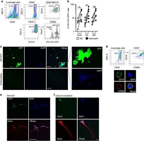
The increased survival of white blood cells called neutrophils is associated with alterations in the intestinal microbiome of HIV-infected individuals, according to a study published April 11 in the open-access journal PLOS ...
|
|
Scooped by
Gilbert C FAURE
March 25, 2019 3:26 PM
|
Mucosal-associated invariant T (MAIT) cells are nonconventional T lymphocytes that recognize bacterial metabolites presented by MR1. Whereas gut bacterial translocation and the loss/dysfunction of peripheral MAIT cells in HIV infection is well described, MAIT cells in nonhuman primate models are...
|
|
Scooped by
Gilbert C FAURE
November 25, 2018 1:21 PM
|
Abstract Mucosa‐associated invariant T (MAIT) cells are unconventional T lymphocytes defined by their innate‐like characteristics and broad antimicrobial responsiveness. Whether MAIT cells are part of the tissue‐resident defense in the oral mucosal barrier is unknown. Here, we found MAIT cells present in the buccal mucosa, with a tendency to cluster near the basement membrane, and located in both epithelium and the underlying connective tissue. Overall MAIT cell levels were similar in the mucosa compared to peripheral blood, in contrast to conventional T cells that showed an altered representation of CD4+ and CD8+ subsets. The major mucosal MAIT cell subset displayed a tissue‐resident and activated profile with high expression of CD69, CD103, HLA‐DR, and PD‐1, as well as a skewed subset distribution with higher representation of CD4–/CD8– double‐negative cells and CD8αα+ cells. Interestingly, tissue‐resident MAIT cells had a specialized polyfunctional response profile with higher IL‐17 levels, as assessed by polyclonal stimulus and compared to tissue nonresident and circulating populations. Furthermore, resident buccal MAIT cells were low in perforin. Together, these data indicate that MAIT cells form a part of the oral mucosal T cell compartment, where they exhibit a tissue‐resident‐activated profile biased toward IL‐17 production. Introduction Mucosa‐associated invariant T (MAIT) cells are nonclassical innate‐like T cells that recognize microbial antigens presented by the MHC‐Ib‐related protein 1 (MR1) 1, 2. MR1 displays an extraordinary level of evolutionary conservation among mammals 2-4, and was shown to present microbial vitamin B2 (riboflavin) metabolites from a wide range of microbes that carry the riboflavin biosynthesis pathway 5, 6. MAIT cells express a semi‐invariant T cell receptor (TCR), including the Vα7.2 segment coupled with Jα33, Jα12, or Jα20, and limited TCR β‐chain diversity 7, 8. This TCR repertoire endows MAIT cells with the capacity to respond to the MR1‐restricted riboflavin derivatives produced by diverse microbes 9, 10. MAIT cells respond to antigenic stimulus with an innate‐like speed and produce proinflammatory cytokines including TNF, IFN‐γ, and IL‐17 9, 11-14. They can furthermore kill cells infected by microbes expressing the riboflavin pathway 15, 16 and inhibit intracellular microbial growth 17. Human MAIT cells are also defined by their high expression of CD161, the IL‐18 receptor α subunit (IL‐18Rα), and the transcription factor ZBTB16 18, also known as promyelocytic leukemia zinc finger protein (PLZF) 9, 19. The majority of MAIT cells are CD8+, with some being CD4–/CD8– double‐negative (DN), and a minor CD4+ population 9, 11, 12, 19. Human MAIT cells acquire innate‐like antimicrobial activity in the fetal intestinal mucosa prenatally, prior to the establishment of the commensal microflora 14. In adults, MAIT cells are highly abundant in mucosal tissues, liver, and peripheral blood 1, 2, 20-22. Studies of the murine oral immune system revealed diverse populations of DCs 23, 24, and provided insight into oral T‐cell function 25, but the immunology of the human oral mucosa remains relatively little studied. However, Dutzan et al. recently performed a broad characterization of the immune cell network at the gingival interface and identified T cells as the dominant immune cell population in both the buccal and gingival mucosa 26. CD4+ T cells, mainly of the CD45RO+ "memory" phenotype, were found to be the largest subset at around half of CD3+ cells, whereas smaller subsets of CD8+ and γδ T cells were also present 26. The same study also identified tissue‐resident CD4+ T cells as major producers of IL‐17 in periodontitis. IL‐17 is a cytokine central to the regulation of immune activity at mucosal surfaces 27, 28. Although IL‐17 is implicated in a variety of functions, its role in the regulation of mucosal immunity hinges on three core effects: maintenance of mucosal integrity via regulation of tight junction proteins, induction of antimicrobial molecule production by epithelial cells, and recruitment of neutrophils to sites of infection. Given the capacity of MAIT cells to recognize and respond to several commensal and disease causing microbes occurring in the oral cavity, and the ability of these cells to locate to mucosal sites, one can hypothesize a role for MAIT cells in the oral mucosal barrier. However, the immunobiology of MAIT cells at this mucosal site has to date not been explored. In the present study, we therefore investigated the presence, location, characteristics, and function of MAIT cells in the human buccal mucosa. The findings demonstrate that MAIT cells are part of the healthy buccal mucosal immune defense with specialized tissue‐resident and nonresident subpopulations exhibiting distinct functional profiles, where in particular, the CD69+CD103+ MAIT cell population show strong IL‐17 production. We discuss these findings in the context of contemporary knowledge of immune homeostasis and defense at this important barrier site. Results Identification of MAIT cells in the buccal mucosa Given the broad antimicrobial reactivity of MAIT cells, we were interested in investigating the potential role of these cells in oral mucosa, a site continuously exposed to a broad array of microbial stimuli. Buccal biopsies and matched peripheral blood samples were collected from healthy donors (Supporting Information Table 1), and cells isolated after tissue processing were analyzed by flow cytometry. All subjects were examined for their oral health and showed no oral mucosal disease, active dental caries, infection, or periodontitis. Using antibody combinations allowing identification of MAIT cells in blood, buccal MAIT cells were defined as CD45+ CD3+ CD161hi Vα7.2+ cells (Fig. 1A). To ensure that cells defined by this gating strategy were bona fide MAIT cells, their MR1 restriction was confirmed by staining with the MR1 5‐(2‐oxopropylideneamino)‐6‐D‐ribitylaminouracil (5‐OP‐RU) tetramer (Fig. 1B). The percentage of MAIT cells in the mucosa ranged between 0.1 and 7% of total T cells (Fig. 1C). Overall, these levels were not significantly different from those in matched blood, although in any given individual, the levels seen at the two sites were often rather divergent. Interestingly, major conventional T‐cell subsets were present in the buccal mucosa at frequencies different from those in the blood. Levels of CD4+ T cells were lower than in blood, whereas CD8+ T cells and CD4–CD8– T cells were enriched in buccal mucosa as compared to blood (Fig. 1C). These data demonstrate that MAIT cells are part of the buccal mucosa T‐cell compartment. Detection of buccal mucosal MAIT cells in situ To determine MAIT cell location within the buccal mucosa, we performed in situ staining for Vα7.2 and IL‐18Rα in oral mucosal sections (Fig. 1D). In mucosa, staining for IL‐18Rα can be used in lieu of CD161, as we have previously shown for genital mucosa 22. Using the Vα7.2+ IL‐18Rα+ definition, MAIT cells were readily identified in buccal mucosal sections, and they tended to locate close to the basement membrane (Fig. 1D). MAIT cells were present in the epithelial layer, just above the basal membrane (distance from basal membrane: median = 14.6 μm; range = 1.0–75.9 μm), as well as in the underlying connective tissue, just beneath the basal membrane (median = 20.7 μm; range = 2.8–292.2 μm). Non‐MAIT T cells, as well as MR1‐expressing HLA‐DR+ antigen‐presenting cells, were also found in buccal mucosa as determined by immunofluorescence staining (Fig. 1E and F). MAIT cell location around the basement membrane is consistent with a possible role as innate‐like T cell sentinels to detect microbial epithelial barrier breach. Altered subset distribution and Jα‐TCR usage in mucosal MAIT cells In peripheral blood, MAIT cells are predominantly CD8+, with some CD4–CD8– cells and a very small CD4+ subpopulation. In the buccal mucosa, MAIT cells were enriched in CD4–CD8– and CD4+ subsets, and had significantly lower levels of the CD8+ subset (Fig. 2A). Within the CD8+ subset, similar to blood MAIT cells, the mucosal MAIT cells exhibited an increased frequency of CD8αα+ cells compared to non‐MAIT T cells (Fig. 2B and C). Notably, MAIT cells represented almost half of all CD8αα+ T cells in the buccal mucosa, with significant donor variability (Fig. 2D). The distribution of Jα‐TCR usage within the MAIT cell populations in the buccal mucosa and blood was compared using quantitative real‐time PCR (qRT‐PCR; Fig. 2E). We quantified the relative expression of three Jα previously shown to be represented within the MAIT cell TCR repertoire, the Jα12, Jα20, and Jα33 8, 29, 30. As expected, blood MAIT cells displayed a consistent pattern with dominant Jα33 usage, with smaller Jα20 and Jα12 usage. However, in matched mucosa, the pattern was more diverse, with different Jα segments dominating in different donors. Together, these data support a model of compartmentalization where the subset representation and TCR‐Jα usage in buccal MAIT cell population are partly different that of the circulating MAIT cells found in blood. MAIT cells in buccal mucosa express an activated perforinlow phenotype To assess the phenotypic characteristics of the buccal MAIT cells in more detail, we next stained isolated mucosal cells with markers of activation, CD38 and HLA‐DR (Fig. 3A). The staining pattern revealed the presence of an activated HLA‐DR+CD38+ MAIT cell population in buccal mucosa (Fig. 3B). Compared to their blood counterparts, the mucosal MAIT cells expressed higher levels of HLA‐DR, whereas CD38 levels appear to be slightly lower (Fig. 3C). In addition, expression of PD‐1 was common in mucosal MAIT cells, whereas PD‐1 was less expressed in peripheral blood MAIT cells (Fig. 3C). To investigate the phenotype of MAIT cells in more detail, paired blood and mucosal cell samples were stained intracellularly for the expression of the master transcription factor PLZF, as well as the cytolytic effector molecules perforin and granulysin (Fig. 3D). Perforin levels were mostly lower in the buccal MAIT cells than in their blood counterparts (Fig. 3E), whereas expression levels of PLZF and granulysin were similar in both compartments. These findings suggest that MAIT cells in the buccal mucosa are highly activated cells with significant expression of the checkpoint inhibition receptor PD‐1, and display a reduced perforin content as compared to circulating MAIT cells. Characterization of resident and nonresident MAIT cells in the buccal mucosa We next investigated the MAIT cell expression of receptors associated with tissue residency in the mucosa. Resident T cells can be distinguished by coexpression of CD69 and CD103 (αE integrin) in a range of tissues 31-35, including intestinal mucosa 36. Flow cytometric assessment of CD69 and CD103 expression revealed the existence of a major CD69+CD103+ tissue‐resident MAIT cell population in the buccal mucosa (Fig. 4A). However, a minority of MAIT cells were negative for CD69 and CD103, consistent with a tissue nonresident or "passing through" phenotype (Fig. 4A). Compared to peripheral blood MAIT cells, the buccal mucosal MAIT cell population was highly enriched in CD69 and CD103 expression, and CD69 expression was mostly seen in the CD103+ population (Fig. 4B). As the CD69 expression formed a continuum including dim cells of both CD103+ and CD103– character, we compared the four MAIT cell populations defined by CD69 and CD103 with regard to CD4+ and CD8+ expression. CD103+ MAIT cells were mainly CD8+ with a minority CD4–CD8– cells, whereas CD103– MAIT cells had a distinct opposite pattern with a majority CD4–CD8– character (Fig. 4C and D). CD69 expression did not significantly influence this pattern (Fig. 4C). In addition, CD103+ MAIT cells were more activated than their CD103– counterparts, as evidenced by higher expression of HLA‐DR and CD38 (Fig. 4E). In contrast, the CD103+ subpopulation expressed lower levels of the cytolytic effector molecule perforin, whereas PLZF did not vary depending on CD103 (Fig. 4F). These results indicate the existence of two distinct buccal MAIT cell subpopulations where one express the CD103 marker of tissue residency and one lacks CD103 consistent with a nonresident character. Distinct polyfunctional responses of mucosal resident, nonresident, and circulating MAIT cells To investigate the functional profile of MAIT cells in buccal mucosa in comparison with peripheral blood, isolated lymphocytes from the two tissues were activated with PMA/ionomycin and their repertoire of functions evaluated by flow cytometry staining for TNF, IFN‐γ, IL‐2, IL‐17, and granzyme B (Fig. 5A). Overall, both blood and mucosal MAIT cells displayed detectable production of all these effector functions, with some notable tissue‐dependent differences. Peripheral blood MAIT cells produced TNF and IFN‐γ more strongly than their mucosal counterparts, whereas IL‐2 and granzyme B were similar (Fig. 5A and B). In contrast, IL‐17 production was very low in blood MAIT cells, with the exception of one outlier, while this cytokine was clearly more highly expressed in the buccal MAIT cells (Fig. 5A and B). In the mucosal tissue, the resident CD103+ subset and the CD103– nonresident subset were largely similar in their response patterns with two notable exceptions. IL‐17 in particular, and to some extent granzyme B expression, were significantly higher in the CD103+ MAIT cell subset. To understand the functional specialization of mucosal resident, mucosal nonresident, and circulating MAIT cells in more detail, we analyzed the polyfunctional response profiles of these subsets. First, we determined the percentages of MAIT cells in the three locations that responded with at least one of the five functions measured (Fig. 5C). For all three sites, the vast majority of MAIT cells expressed at least one function. However, the mucosal MAIT cells displayed somewhat lower responsiveness, where in particular, the CD103– MAIT cells were less responsive. When the overall polyfunctionality was analyzed, the ability to express 1, 2, 3, 4, or 5 functions was fairly similar between the different tissue locations, although the CD103+ resident MAIT cells tended to be more 4‐ or 5‐functional (Fig. 5D). Finally, we analyzed the data by Boolean gating to assess which specific polyfunctional profiles were expressed by peripheral blood, CD103+ mucosal resident, and CD103– nonresident MAIT cells (Fig. 5E; Supporting Information Table 2). This analysis brought forward the pattern that peripheral blood MAIT cells were focused primarily on TNF production, either alone or in combinations with IFN‐γ and IL‐2. The mucosal CD103+ resident MAIT cells instead primarily focused on IL‐17 production either alone or in combinations with TNF or IL‐2. The CD103– nonresident MAIT cells had a functional profile intermediate between the circulating and resident populations, and were mostly expressing TNF with or without IL‐2, and less IL‐17. Altogether, these findings indicate that the mucosal resident, the mucosal nonresident, and the circulating MAIT cells deploy different effector cytokine response profiles. Discussion Tissue‐resident immune cell populations are increasingly recognized for their importance in defense against invading pathogens. The oral mucosa is continuously exposed to a range of antigens derived from food, microbiota, and pathogens. To maintain barrier immune defenses at this site, while not reacting against nonpathogenic encounters, is a considerable challenge. Recent studies have started to address this research question 37, 38, but no studies to date have addressed the potential role of the MR1‐restricted MAIT cell population in oral mucosa. Here, we observe that MAIT cells are dispersed throughout the buccal mucosa with a preferential location close to the basement membrane. The buccal MAIT cells are biased toward a CD4–/CD8– DN profile, and the CD8+ subset is primarily CD8αα and make up a significant proportion of all CD8αα T cells in the mucosa. The MAIT cell population in this mucosal site is composed of distinct CD69+CD103+ intraepithelial resident and CD69–CD103– nonresident subsets. The CD103+ subset is enriched in CD8+ MAIT cells, is more activated, and has lower cytolytic potential. Finally, the buccal MAIT cells show a distinct functional profile with enhanced IL‐17 production and less TNF and IFN‐γ, as compared to their peripheral blood counterparts, and this pattern was particularly pronounced for the CD103+ resident MAIT cells. The MR1‐presented riboflavin metabolite antigens recognized by MAIT cells are conserved among many prokaryotic and eukaryotic microbes relevant for mucosal health. Thus, MAIT cells may play a unique role in the oral mucosa with broad innate‐like recognition of such microbes in a manner that does not require the kind of priming which conventional adaptive T‐cell responses do. It is interesting that the MAIT cells to at least some extent locate around the basement membrane, where they may function as innate‐like T‐cell sentinels to detect microbial epithelial barrier breach. Around the similar location, MR1+HLA–DR+ cells are residing and those may be able to present riboflavin metabolite antigens from invading microbes. Together, MAIT cells and MR1+ antigen presenting cells may form a rapid innate response immune unit at the mucosal barrier, capable of responding to pathogens to which the host has no preexisting adaptive immunity. The CD103+CD69+ mucosal‐resident MAIT cell population is fairly strongly biased toward IL‐17 production, with less TNF and IFN‐γ expression, as compared to the CD103– MAIT cell subpopulation. Resident MAIT cell recognition of an invading microbial pathogen may thus setoff an IL‐17‐mediated inflammatory response with induction of antimicrobial peptides in epithelium and recruitment of neutrophils. Such a pattern would be reminiscent to the situation described for chronic severe periodontitis by Dutzan et al. 26, where IL‐17 produced by presumably adaptive Th17 cells was associated with increased gingival tissue infiltration of neutrophils. Given their innate‐like response characteristics, MAIT cells may be among the first immune cells to detect microbial infiltration and respond with IL‐17 release. Here, it is interesting to note that IL‐17 has emerged as an important mediator and amplifier of immunity against Candida albicans 38. The dimorphic fungus C. albicans expresses the riboflavin biosynthesis pathway and is recognized by MAIT cells in an MR1‐restricted manner 10. Many cells respond to IL‐17 by upregulation of proinflammatory cytokines, such as IL‐6, and chemokines for recruitment of neutrophils including CXCL1, CXCL2, and CXCL5 39. In addition, IL‐17 stimulates production of β‐defensins in epithelial cells 40. MAIT cells are thus well located to initiate mucosal immune responses in oropharyngeal candidiasis. Peripheral blood MAIT cells are predominantly CD8+ with a minority CD4–CD8– subset. This pattern is reversed in buccal mucosa, such that the CD4–CD8– MAIT cells are more numerous. It is however interesting to note that the CD8+ MAIT subset in buccal mucosa is primarily CD8αα, and that this subset makeup almost half of all CD8αα T cells in the oral mucosa. These CD8αα MAIT cells bear resemblance to the intestinal mucosal CD8αα intraepithelial lymphocytes (IELs) that have been extensively characterized in murine models, but may be less frequent in humans 41, 42. The intestinal IELs of mice are a mix of TCRγδ T cells and TCRαβ T cells with diverse specificities, and the representation of MAIT cells among these IELs in different sites is to our knowledge largely unknown. Our data indicate that the CD103+ MAIT cell population is mostly, but not exclusively, CD8+ and composed of both CD8αα and CD8αβ cells. These findings together suggest that MAIT cells makeup a significant part of the human buccal IEL‐like population. The human oral mucosal barrier retains a commensal bacterial microbiota that is both varied and unique among other sites 43, 44, dominated by the genera Streptococcus, Haemophilus, Prevotella, and Veillonella. In addition to bacteria, the oral mucosal barrier is home to many species of fungi including C. albicans 45. The oral immune system thus has to manage and tolerate a diverse commensal microbiome, and at the same time guard against conditions arising either from dysfunction of normal oral homeostasis or caused by pathogens normally not present in the oral cavity. T cells are believed to play a role in multiple oral mucosa pathologies including aphthous stomatitis, oral leukoplakia, oral reticular or ulcerative lichen planus, celiac disease, and oral psoriasis 46, 47. Whether MAIT cells have a role in any such conditions in humans remains to be explored. In summary, we have shown that MAIT cell populations with resident and nonresident characteristics are part of the buccal mucosal immune system in healthy donors and that they have unique functional profiles. Future studies should aim to investigate how these populations respond to commensal and pathogenic microbes, and how they are affected in different disease conditions. Materials and methods Tissue donor recruitment and sample collection A total of 94 volunteers were recruited in two healthy donor groups A and B (Supporting Information Table 1). Inclusion criteria for both groups were: 20–50 years of age, HIV‐negative, non‐smoking, no antibiotics in the last 3 months, and not pregnant. Ethical permission was obtained from the Regional Ethical Review Board in Stockholm in accordance with the Declaration of Helsinki. All participants gave written informed consent. The oral health of all subjects was evaluated using standard dental examination procedures, including inspection of oral mucosa, teeth, and surrounding soft tissues, to ensure the donors had no visible mucosal lesions, no signs of gingivitis, active dental caries, or periodontitis. All donors were instructed to abstain from food or drink for 1 h before tissue collection. Oral mucosal tissue samples and matched peripheral blood samples were collected from two sets of healthy donors. Donor group A was recruited from the general population. A matched blood sample of 20–30 mL venous blood was taken from each participant on the day of the procedure, prior to the oral sample collection. The blood was stored in heparin tubes at room temperature until analysis. Local anesthesia with xylocain/adrenalin was applied to the left bucca and three punch biopsies (5 mm in diameter, 3–4 mm deep) were taken from the site. For donor group B, tissue samples were taken before wound closure on patients who underwent orthognatic surgery of the mandible. The patients underwent these corrective surgeries due to abnormal position of the mandible in relation to the base of skull, but no buccal disease or inflammation is associated with these conditions. The patients were treated under general anesthesia and local anesthesia was additionally applied. Tissue samples 20 × 10 mm in size were taken bilaterally from the mucosa. Matched blood samples from donor group B were collected at the time of the procedure. After collection, the biopsies were stored at 4°C in serum‐free RPMI (ThermoFisher Scientific, Waltham, MA, USA) supplemented with 50 μg/mL gentamicin (ThermoFisher Scientific) and 100 μg/mL normocin (InvivoGen, San Diego, CA, USA). Biopsies intended for qPCR analysis were stored in RNAlater (QIAgen, Venlo, Netherlands) at −20°C. Biopsies intended for in situ microscopic analysis were submerged in OCT Cryomount (HistoLab, Askim, Sweden), snap‐frozen in liquid nitrogen, and then stored at −80°C. Sample processing for flow cytometry All collected samples were processed within 3 h of collection. Buccal biopsies were placed into serum‐free RPMI supplemented with 50 μg/mL gentamicin and 100 μg/mL normocin and then incubated with 1 mg/mL DNase I and collagenase A (both from Roche, Penzberg, Germany) at 37°C with shaking at 800 rpm for 1 h. After incubation, the reaction was stopped by addition of fetal calf serum (FCS; Sigma‐Aldrich, St Louis, MO, USA) to a final concentration of 10%. The biopsies were then pressed through a 100 μm cell strainer in order to detach loose cells. The cells were then washed in PBS and incubated in RBC lysis buffer for 10 min at RT to remove any erythrocyte contamination. The cells were washed again in PBS and stained for flow cytometry. Donor‐matched PBMCs were isolated from blood samples by Ficoll‐Hypaque density gradient centrifugation using Lymphoprep (Axis‐Shield, Dundee, Scotland). The PBMC layer was washed twice with PBS, and incubated in RBC lysis buffer for 10 minutes at room temperature to remove erythrocyte contamination. The cells were washed again in PBS and 106 cells were stained for flow cytometry. In situ staining In situ staining was performed on 8‐μm‐thick sections of frozen biopsies, as previously described 22. Sections were fixed in 2% formaldehyde and stained sequentially. Antibody reagents used for tissue staining are listed in Supporting Information Table 3. The tissue sections stained for Vα7.2 in combination with IL‐18Rα were scanned into digital images using a Pannoramic 250 Flash Slide Scanner (3DHistech, Budapest, Hungary). The distance measurement was performed using the digital microscope application CaseViewer (3DHistech), where the distance from the surface of the double‐positive cells to the basal membrane was measured. The tissue sections stained for HLA‐DR in combination with MR1, and Vα7.2 in combination with CD3 were visualized using DMR‐X microscope (Leica, Weitzlar, Germany) and images were acquired with a Retiga 2000 R camera (Qimaging, Surrey, Canada). Functional assay One million lymphocytes in suspension were cultured overnight in RPMI +10% FCS, supplemented with 100 μg/mL normocin and 50 μg/mL gentamicin. After overnight incubation, they were activated using Leukocyte Activation Cocktail (PMA/ionomycin) (BD Biosciences, San Jose, CA, USA) in the presence of GolgiPlug (monensin) for 6 h. At the end of the incubation, the cells were stained and analyzed by flow cytometry. Flow cytometry and antibodies Cells in suspension were stained for surface antigens in FACS buffer (PBS with 2 mM EDTA and 2% FCS) for 20 min at 4°C. Afterwards, they were washed in FACS buffer and permeabilized using the transcription factor fixation and permeabilization buffer (BD Biosciences) for 30 min at 4°C. Following permeabilization, cells were washed twice in transcription factor wash and permeabilization buffer (BD Biosciences), and then stained for intracellular antigens in the same buffer for 20 min at 4°C. PE‐conjugated human MR1:5‐OP‐RU tetramer was obtained from the NIH Tetramer Core Facility. For tetramer staining, cells in suspension were incubated with MR1:5‐OP‐RU tetramer for 40 min at room temperature and then washed with FACS buffer prior to surface antibody staining. Monoclonal antibody reagents used in the study are listed in Supporting Information Table 4. After staining, cells were washed and analyzed using an LSRFortessa flow cytometer (BD Biosciences) equipped with 355, 405, 488, 561, and 639 nm lasers. In addition to the antibodies, cells were stained with LIVE/DEAD Fixable Dead Cell Stain (Near‐Infrared or Aqua; ThermoFisher Scientific). Quantitative RT‐PCR qRT‐PCR was performed to determine the mRNA expression of TCRs Vα7.2‐Jα33, Vα7.2‐Jα20, and Vα7.2‐Jα12 as well as the constant Cα chain, in healthy oral mucosa and blood. Briefly, total RNA was extracted from buccal mucosa and PBMCs using TRIzol reagent (ThermoFisher Scientific). For reverse transcription, 250 ng of RNA was used in a 15 μL total reaction volume using the IScript cDNA Synthesis kit (BioRad, Hercules, CA, USA). For amplification, a 20 μL supermix reaction was prepared accordingly: 1 μL cDNA, 5 μL SsoAdvanced™ Universal® SYBR Green (BioRad), 1 μL of each primer at a final concentration of 500 nM, and nuclease free H2O. Primer sequences as well as annealing temperatures for each of the primer sets are listed in Supporting Information Table 5. qPCR was performed using 7500 Fast real time PCR system (Applied Biosystems, Waltham, CA, USA) with the following cycling conditions: 1 cycle at 95°C for 5 min, then 40 cycles at 94°C for 10 s, 58°C or 60°C depending on primer for 30 s, and 72°C for 27 s. The relative abundance of each Vα7.2‐Jα was determined as relative to Cα by the comparative ΔΔCT method. Software and statistical analysis Analysis of flow cytometric data was performed using FlowJo 10 (FlowJo LLC, Ashland, OR, USA), and microscopic images were processed using Image‐Pro Premier 9.3 (Media Cybernetics, Rockville, MD, USA). Statistical tests were performed using GraphPad Prism version 7.0c for Mac OS X (GraphPad Software, La Jolla, CA, USA). Statistical significance was assessed using Wilcoxon matched pairs test for paired data, and Mann–Whitney U‐test for unpaired data. For data presented in Fig. 5, analysis and presentation of distributions were performed using SPICE version 5.1, downloaded from http://exon.niaid.nih.gov 48. Comparison of distributions was performed using a Wilcoxon matched pairs test as described 48. For all statistical analysis, p‐values below 0.05 were regarded as significant. Acknowledgements The authors wish to acknowledge the contribution of Sam Chehrehgosha, Angelica Kroonder, Emelie Molin, and Beatrice Wiberg in recruiting study volunteers and gathering oral biopsies. This research was supported by grants to JKS from the Swedish Research Council (2016‐03052), the Swedish Cancer Society (CAN 2017/777), and the US National Institutes of Health (R01DK108350). Grants to M.S.C. were from the Foundation for Odontological Research (OF11211233), and the Swedish Cancer Society (CAN 2016/731). Further support to E.L. was from the Swedish Research Council (2015‐00174) and Marie Skłodowska Curie Actions, Cofund, Project INCA 600398. J.D. was supported by Fundação para a Ciência e a Tecnologia (SFRH/BD/85290/2012, doctoral fellowship), through program QREN‐POPH‐typology 4.1. The MR1 tetramer technology was developed jointly by Dr. James McCluskey, Dr. Jamie Rossjohn, and Dr. David Fairlie, and the material was produced by the NIH Tetramer Core Facility as permitted to be distributed by the University of Melbourne. The authors wish to thank the healthy volunteers that contributed time and effort to sample donation for this study. Author contributions J.K.S., E.L., and M.S.C. conceived the original research idea and study design. M.J.S., H.D., M.M., A.T., J.D., E.L., M.S.C., and J.K.S. designed experiments. M.J.S., H.D, A.G., and J.E. conducted experiments. M.J.S., H.D., and A.G. analyzed experimental data. R.H., S.A., and C.K.W. recruited study volunteers and collected essential human samples. M.J.S. and J.K.S. wrote the manuscript. All authors reviewed and commented on the manuscript. Conflict of interest The authors declare no commercial or financial conflict of interest. Supporting Information References Abbreviations IEL intraepithelial lymphocyte MAIT mucosal‐associated invariant T MR1 MHC‐Ib‐related protein 1 PLZF promyelocytic leukemia zinc finger protein
|
|
Scooped by
Gilbert C FAURE
October 4, 2018 2:30 AM
|
In a study published today in Science Translational Medicine, Mount Sinai researchers describe for the first time a mechanism that may shrink collections of immune cells in the gastrointestinal (GI) tract, called lymphoid ...
|
|
Scooped by
Gilbert C FAURE
August 9, 2018 4:04 AM
|
Veranstaltungstyp: Gastvortrag;...
|
|
Scooped by
Gilbert C FAURE
July 26, 2018 11:24 AM
|
Increasing drug resistance and the lack of an effective vaccine are the main factors contributing to Mycobacterium tuberculosis (Mtb) being a major cause of death globally. Despite intensive research efforts, it is not well understood why some individuals control Mtb infection and some others develop active disease. HIV-1 infection is associated with an increased incidence of active Tuberculosis (TB), even in virally suppressed individuals. MAIT and iNKT cells are innate T cells that can recognize Mtb-infected cells. Contradicting results regarding the frequency of MAIT cells in latent Mtb infection have been reported. In this confirmatory study, we investigated the frequency, phenotype, and IFNγ production of MAIT and iNKT cells in subjects with latent or active Mtb infection. We found that the frequency of both cell types was increased in subjects with latent Mtb infection compared to uninfected individuals or subjects with active infection. We found no change in the expression of HLA-DR, PD-1, and CCR6, as well as the production of IFNγ by MAIT and iNKT cells, among subjects with latent Mtb infection or uninfected controls. The proportion of CD4-CD8+ MAIT cells in individuals with latent Mtb infection was, however, increased. HIV-1 infection was associated with a loss of MAIT and iNKT cells, and the residual cells had elevated expression of the exhaustion marker PD-1. Altogether, the results suggest a role for MAIT and iNKT cells in immunity against Mtb, and show
|
|
Scooped by
Gilbert C FAURE
June 6, 2018 3:19 PM
|
|
|
Scooped by
Gilbert C FAURE
May 11, 2018 12:04 PM
|
HIV-1 infection is associated with B cell dysregulation and dysfunction. In HIV-1–infected patients, we previously reported preservation of intestinal lymphoid structures and dendritic cell maturation pathways after early combination antiretroviral therapy (e-ART), started during the acute phase of the infection, compared with late combination antiretroviral therapy started during the chronic phase. In this study, we investigated whether the timing of combination antiretroviral therapy initiation was associated with the development of the HIV-1–specific humoral response in the gut. The results showed that e-ART was associated with higher frequencies of functional resting memory B cells in the gut. These frequencies correlated strongly with those of follicular Th cells in the gut. Importantly, frequencies of HIV-1 Env gp140–reactive B cells were higher in patients given e-ART, in whom gp140-reactive IgG production by mucosal B cells increased after stimulation. Moreover, IL-21 release by PBMCs stimulated with HIV-1 peptide pools was greater with e-ART than with late combination antiretroviral therapy. Thus, early treatment initiation helps to maintain HIV-1–reactive memory B cells in the gut as well as follicular Th cells, whose role is crucial in the development of potent affinity-matured and broadly neutralizing Abs.
This article is featured in In This Issue , p.[3315][1]
[1]: /lookup/volpage/200/3315
|
|
Scooped by
Gilbert C FAURE
March 28, 2018 12:36 PM
|
As a rich source of CD4+ T cells and macrophages, the gastrointestinal (GI) tract is a major target site for HIV infection. The interplay between GI resident macrophages and intestinal epithelial cells (IECs) constitutes an important element of GI innate immunity against pathogens. In this study, we investigated whether human IECs have the ability to produce antiviral factors that can inhibit HIV infection of macrophages. We demonstrated that IECs possess functional toll-like receptor 3 (TLR3), the activation of which resulted in induction of key IFN regulatory factors (IRF3 and IRF7), IFN-β, IFN-λ and CC chemokines (MIP-1α, MIP-1β, RANTES), the ligands of HIV entry co-receptor CCR5. In addition, TLR3-activated IECs release exosomes that contained the anti-HIV factors, including IFN stimulated genes (ISGs: ISG15, ISG56, MxB, OAS-1, GBP5, and Viperin) and HIV restriction miRNAs (miRNA-17, miRNA-20, miRNA-28, miRNA-29 family members and miRNA-125b). Importantly, treatment of macrophages with supernatant (SN) from the activated IEC cultures inhibited HIV replication. Further studies showed that IEC SN could also induce the expression of antiviral ISGs and cellular HIV restriction factors (Tetherin and APOBEC3G/3F) in HIV-infected macrophages. These findings indicated that IECs might act as an important element in GI innate immunity against HIV infection/replication.
|
|
Scooped by
Gilbert C FAURE
January 21, 2018 12:09 PM
|
We previously reported that CD8+ T cells in human gastrointestinal mucosa exhibit reduced perforin expression and weak or impaired cytotoxic capacity compared with their counterparts in blood. Nevertheless, these cells degranulate and express cytokines and chemokines in response to cognate Ag. In addition to weak expression of perforin, earlier studies suggested differential regulation of perforin and granzymes (Gzms), with GzmA and B expressed by significantly higher percentages of mucosal CD8+ T cells than perforin. However, this topic has not been fully explored. The goal of this study was to elucidate the expression and coexpression patterns of GzmA, B, and K in conjunction with perforin in rectosigmoid CD8+ T cells during HIV-1 infection. We found that expression of both perforin and GzmB, but not GzmA or GzmK, was reduced in mucosa compared with blood. A large fraction of rectosigmoid CD8+ T cells either did not express Gzms or were single-positive for GzmA. Rectosigmoid CD8+ T cells appeared skewed toward cytokine production rather than cytotoxic responses, with cells expressing multiple cytokines and chemokines generally lacking in perforin and Gzm expression. These data support the interpretation that perforin and Gzms are differentially regulated, and display distinct expression patterns in blood and rectosigmoid T cells. These studies may help inform the development of strategies to combat HIV-1 and other mucosal pathogens.
|
|
Scooped by
Gilbert C FAURE
November 16, 2017 9:34 AM
|
|
|
Scooped by
Gilbert C FAURE
October 5, 2017 7:53 AM
|
The fungal pathogen, Cryptococcus neoformans, causes devastating levels of morbidity and mortality. Infections with this fungus tend to be predominantly in immunocompromised individuals, such as those with HIV. Infections initiate with inhalation of cryptococcal cells and entry of the pathogen into the lungs. The bronchial epithelial cells of the upper airway and the alveolar epithelial cells of the lower airway are likely to be the first host cells that Cryptococcus engage with. Thus the interaction of cryptococci and the respiratory epithelia will be the focus of this review. C. neoformans has been shown to adhere to respiratory epithelial cells, although if the role of the capsule is in aiding or hindering this adhesion is debatable. The epithelia are also able to react to cryptococci with the release of cytokines and chemokines to start the immune response to this invading pathogen. The activity of surfactant components that line this mucosal barrier towards Cryptococcus and the metabolic and transcriptional reaction of cryptococci when encountering epithelial cells will also be discussed.
|
|
Scooped by
Gilbert C FAURE
September 27, 2017 1:33 PM
|
Interleukin-17 (IL-17) is a proinflammatory cytokine produced by adaptive CD4+ T helper cells and innate lymphocytes, such as γδ-T cells and TCRβ+ “natural” Th17 cells. IL-17 activates signaling through the IL-17 receptor, which induces other proinflammatory cytokines, antimicrobial peptides and neutrophil chemokines that are important for antifungal activity. The importance of IL-17 in protective antifungal immunity is evident in mice and humans, where various genetic defects related to the IL-17-signaling pathway render them highly susceptible to forms of candidiasis such oropharyngeal candidiasis (OPC) or more broadly chronic mucocutaneous candidiasis (CMC), both caused mainly by the opportunistic fungal pathogen Candida albicans. OPC is common in infants and the elderly, HIV/AIDS and patients receiving chemotherapy and/or radiotherapy for head and neck cancers. This review focuses on the role of IL-17 in protection against candidiasis, and includes a brief discussion of non-Candida albicans fungal infections, as well as how therapeutic interventions blocking IL-17-related components can affect antifungal immunity.
|

|
Suggested by
Société Francaise d'Immunologie
April 4, 2019 2:17 PM
|
Research ArticleGastroenterologyImmunology Free access | 10.1172/JCI121668 IL-7 receptor influences anti-TNF responsiveness and T cell gut homing in inflammatory bowel disease Lyssia Belarif,1 Richard Danger,2,3 Laetitia Kermarrec,4 Véronique Nerrière-Daguin,2,3 Sabrina Pengam,1 Tony Durand,4 Caroline Mary,1 Elise Kerdreux,5 Vanessa Gauttier,1 Aneta Kucik,6 Virginie Thepenier,1 Jerome C. Martin,7,8,9 Christie Chang,7,8,9 Adeeb Rahman,7,10,11 Nina Salabert-Le Guen,2,12,13,14 Cécile Braudeau,2,12,13 Ahmed Abidi,2,15 Grégoire David,4 Florent Malard,2 Celine Takoudju,4 Bernard Martinet,2,3 Nathalie Gérard,2,3 Isabelle Neveu,4,5 Michel Neunlist,4,5 Emmanuel Coron,4,5 Thomas T. MacDonald,6 Pierre Desreumaux,16 Hoa-Le Mai,2,3 Stephanie Le Bas-Bernardet,2,3 Jean-François Mosnier,2,17 Miriam Merad,7,8,9,11 Régis Josien,2,3,12,14 Sophie Brouard,2,3 Jean-Paul Soulillou,2 Gilles Blancho,2,3 Arnaud Bourreille,4,5 Philippe Naveilhan,4,5 Bernard Vanhove,1 and Nicolas Poirier1 First published April 2, 2019 - More info Abstract It remains unknown what causes inflammatory bowel disease (IBD), including signaling networks perpetuating chronic gastrointestinal inflammation in Crohn’s disease (CD) and ulcerative colitis (UC), in humans. According to an analysis of up to 500 patients with IBD and 100 controls, we report that key transcripts of the IL-7 receptor (IL-7R) pathway are accumulated in inflamed colon tissues of severe CD and UC patients not responding to either immunosuppressive/corticosteroid, anti-TNF, or anti-α4β7 therapies. High expression of both IL7R and IL-7R signaling signature in the colon before treatment is strongly associated with nonresponsiveness to anti-TNF therapy. While in mice IL-7 is known to play a role in systemic inflammation, we found that in humans IL-7 also controlled α4β7 integrin expression and imprinted gut-homing specificity on T cells. IL-7R blockade reduced human T cell homing to the gut and colonic inflammation in vivo in humanized mouse models, and altered effector T cells in colon explants from UC patients grown ex vivo. Our findings show that failure of current treatments for CD and UC is strongly associated with an overexpressed IL-7R signaling pathway and point to IL-7R as a relevant therapeutic target and potential biomarker to fill an unmet need in clinical IBD detection and treatment. Graphical Abstract Introduction Inflammatory bowel disease (IBD) consists of 2 major forms, ulcerative colitis (UC) and Crohn’s disease (CD), which are chronic relapsing gastrointestinal disorders characterized by chronic intestinal inflammation, dysregulated immune responses to intestinal microbiota, and dysfunction of the epithelial barrier (1, 2). The incidence and prevalence of IBD are increasing worldwide, and these diseases are associated with marked morbidity and have a major impact on quality of life and ability to work (3, 4). Current conventional treatments are aimed at dampening inflammation with the gradual use of antiinflammatory agents, steroids, immunosuppressive drugs, and biological agents targeting inflammatory cytokines (mainly TNF) for refractory and severe forms of IBD (5, 6). However, approximately one-third of patients receiving anti-TNF agents do not respond to treatment (primary failure), and a significant proportion (up to 50%) become refractory over time (secondary failure) (7). A key feature of IBD is also the rapid recruitment and prolonged persistence of leukocytes at the site of inflammation, which is facilitated by integrin interaction with cognate receptors expressed by endothelial cells allowing cell adhesion and transmigration (8, 9). Emerging therapies are targeting this entry point to the gut with antiadhesion molecules, specially targeting the gut-specific α4β7 integrin (10, 11). However, these therapies do not induce remission in more than half of patients, and relapse also occurs over time in primary responders. Thus, one major goal is to identify novel mechanisms and signaling networks that underlie the chronicity and relapse of IBD. Interleukin-7 (IL-7) is a limiting and nonredundant cytokine produced mainly by epithelial and stromal cells that regulates T lymphocyte homeostasis (12, 13). Almost all conventional mature T lymphocytes express high IL-7 receptor (IL-7R) levels, with the specific exception of naturally occurring Tregs, constituting a rare opportunity to selectively target pathogenic effectors while preserving natural regulators (14–16). IL-7 signals through the cell surface IL-7R that consists of a specific IL-7R α chain (IL-7Rα, CD127) that dimerizes with the common cytokine receptor γ chain (γc, CD132, IL2RG) (17). IL-7Rα also interacts with the thymic stromal lymphopoietin receptor (TSLPR) chain to form the heterodimeric receptor of the TSLP cytokine (18). Upon activation, IL-7R delivers proliferative and antiapoptotic signals by activating PI3K and JAK/STAT pathways as well as regulating expression of anti- and proapoptotic BCL-2 family members (19). More than 2 decades of research has reproducibly highlighted the crucial systemic influence of the IL-7 pathway and depicted IL-7 as a fuel for chronic autoimmune and inflammatory diseases as well as transplant rejection in diverse rodent models (20–22). Remarkably, the colon is the major source of IL-7 outside of the lymphoid tissues in mice, and commensal microflora promotes steady-state IL-7 production by intestinal epithelial cells (23, 24). IL-7–transgenic mice spontaneously developed chronic colitis with IL-7Rαhi T cells infiltrating the gut mucosa, which, once isolated, transferred colitis to immunodeficient recipient mice, thereby demonstrating their pathogenicity (25–29). More recently, IL-7Rαhi innate lymphoid cells (ILCs) were described as new key effectors of IBD (30–32), and IL-7Rα antagonists suppressed both adaptive and innate inflammatory responses in different experimental models of colitis (26, 27, 33, 34). In humans, however, research on IBD has mainly focused on a proinflammatory and antiinflammatory cytokine balance (35–37), while the IL-7/IL-7Rα axis remains poorly studied. IL-7 production by intestinal epithelial cells and IL7R expression by mucosal T lymphocytes were described in colon biopsies from healthy donors (38). One study reported genetic loci variation of the IL7R gene being associated with susceptibility to UC (39). Lastly, elevated IL-7 signaling pathway gene expression in blood CD8+ T cells at diagnosis was significantly associated with relapsing and a more aggressive IBD disease course (40). Whether IL-7 activities are implicated in the pathogenesis of IBD in humans and determine the fate of treatment is uncertain. Results Colon-specific increased mucosal IL7R signaling pathway signature in refractory IBD. Higher gene expression of the IL-7 signaling pathway members in peripheral blood CD8+ T cells has been previously correlated with a relapsing and aggressive disease course of IBD (40). We therefore sought to address whether the local/mucosal IL-7/IL-7R pathway could be enriched in severe IBD and associated with the failure of conventional therapies. For this purpose, we focused our hypothesis-driven analysis on only the transcriptional IL-7/IL-7R signaling signature of 20 genes (curated from BioCarta by the Molecular Signatures Database [MSigDB]; ref. 40) in colon mucosal biopsies from previously published cohorts of IBD patients with clinically active or inactive disease after immunosuppressive therapy. We examined 2 independent cohorts (UC [n = 23] compared with controls without IBD [n = 13], ref. 41; and UC [n = 97] or CD [n = 8] compared with controls without IBD [n = 11]; ref. 42). We found that this 20-mRNA expression signature was altered according to the clinical status (Figure 1, A and D), with a particularly significant accumulation of IL7R mRNA transcripts in inflamed mucosa from refractory IBD patients relative to those in uninvolved control mucosa from the same patients (P < 0.0001), mucosa from responder patients (P < 0.001), or non-IBD controls (Figure 1, C and F). Principal component analysis (PCA) of the mucosal IL-7R signaling signature after immunosuppressive therapy displayed in both cohorts a clear and distinct separation between active versus inactive disease status and the group of uninvolved mucosa that gathered from non-IBD controls (Figure 1, B and E). Figure 1 IL-7R signaling pathway expression is highly expressed in colonic mucosa of refractory IBD. (A) Heatmap of the expression of the 20 selected genes of the IL-7R signaling pathway (40) in colon biopsies from non-IBD control patients (black; n = 13), inactive UC patients (green; n = 8), and active UC patients with involved (red; n = 15) or uninvolved proximal segments (blue; n = 7) after corticosteroids and/or immunosuppression therapy (data set GSE38713; ref. 41). The heatmap represents median-centered and colorized expression values. (B) PCA of the IL-7R signaling signature and (C) relative IL7R expression (log2 data were normalized to the control median; same patient and color groups as in A). (D) Heatmap of the IL-7R signaling signature in colon biopsies from non-IBD controls (black; n = 11), inactive (green; n = 23) or active (red; n = 74) UC patients, and active CD patients (purple; n = 8) after corticosteroids and/or immunosuppression therapy. (E) PCA of the IL-7R signaling signature and (F) relative IL7R expression (log2 data were normalized to the control median; same patient and color groups as in D). (G) Heatmap of the IL-7R signaling signature in colon biopsies from UC patients at weeks 0, 8, and 30 following anti-TNF therapy (green, stable responders, n = 12–17; orange, relapsing patients, n = 3–8; red, primary nonresponders, n = 3–6). (H) PCA after 8 (top) and 30 (bottom) weeks of anti-TNF treatment of the IL-7R signaling signature and (I) IL7R expression (log2 data were normalized to the median of the stable responder group; same patient and color groups as in G). *P < 0.05; **P < 0.01; ***P < 0.001; ****P < 0.0001 between the indicated groups. #P < 0.05; ###P < 0.001 between responders and nonresponders at a single time point. §P < 0.05; §§§P < 0.001, Kruskal-Wallis with post hoc Dunn’s multiple comparisons test within a single response group across all 3 time points. Similarly, the IL-7R signaling signature after anti-TNF treatment allows distinguishing between biopsies according to disease status in a third cohort of UC patients involved in the phase III ACT-1 clinical trial of infliximab (including long-term responders [n = 17], short-term responders and then relapse [n = 8], and primary nonresponders [n = 6]; ref. 43) (Figure 1G). PCA of the mucosal IL-7R signature clearly separates responder from nonresponder patients 30 weeks after maintenance with anti-TNF treatment (Figure 1H). Furthermore, mucosal IL7R expression was significantly higher in clinically active versus inactive disease in anti-TNF–treated patients at both early (week 8, P < 0.05) and late (week 30, P < 0.001) time points (Figure 1I). IL7R expression significantly decreased after anti-TNF therapy in long-term responders compared with baseline pretreatment levels (P < 0.001); however, no significant difference was measurable before treatment between groups in this clinical trial cohort. In contrast to colon mucosa, the IL-7R signature in ileal biopsies was not associated with clinical status in a fourth cohort (44) of untreated pediatric patients with newly diagnosed ileal (iCD; n = 143) or colon-only (cCD, n = 30) CD or UC (n = 38) as compared with pediatric healthy controls (n = 42) (Supplemental Figure 1, A and B; supplemental material available online with this article; https://doi.org/10.1172/JCI121668DS1). IL7R expression was only higher in ileal biopsies with histological microinflammation versus uninflamed biopsies of cCD; however, no difference was observed in ulcerated or nonulcerated iCD relative to healthy controls (Supplemental Figure 1C). Furthermore, IL7R was not differentially expressed in colon mucosa of different non-IBD types of controls, including in inflammatory non-IBD pathology such as diverticulitis (Supplemental Figure 2). Altogether, the data indicate that mucosal expression of the IL-7R signaling pathway, and specifically IL7R, is strongly and selectively associated with colonic but not ileal IBD inflammation. Expression is related to disease activity in IBD patients (Table 1) and could be one mechanism associated with refractory and severe IBD. Table 1 IL7R expression significance in active IBD mucosa versus healthy controls after treatment with immunosuppression and/or corticosteroids, or before therapy with anti-TNF or anti-α4β7 between responders and nonresponders Colonic IL-7Rα expression predicts resistance to anti-TNF therapy. We then asked whether the mucosal IL-7R signaling signature is associated with responsiveness to anti-TNF before initiation of biologic therapy in patients refractory to corticosteroids and/or immunosuppression. We thus performed a meta-analysis of publicly available transcriptional data sets of 3 cohorts of UC patients with colon mucosa biopsies performed before anti-TNF treatment (within a week) (45–47). In these 3 cohorts, anti-TNF response was defined as histological healing analyzed 4–6 weeks after their first anti-TNF infusion (altogether: n = 18 non-IBD controls, n = 41 UC nonresponders, and n = 28 UC responders). Differential analysis of all expressed genes of the meta–data set (17,037 genes) identified only 85 genes significantly (adjusted P value < 0.05) and differentially (log2 FC > 1) expressed between responders and nonresponders, including IL7R (Supplemental Figure 3), which clustered with a large group of genes associated with adaptive immune response, T cell activation, and leukocyte migration/adhesion (Supplemental Figure 4). Analysis of the transcriptional IL-7R signaling signature (20 genes previously described) revealed that the IL-7R signaling signature, in particular IL7R, was already elevated in colon biopsies of primary UC nonresponder patients before initiation of anti-TNF therapy (Figure 2, A–C). A lasso (least absolute shrinkage and selection operator) regression analysis identified a combination of 10 genes (IL7R, IL2RG, JAK1, PIK3CA, LCK, PTK2B, EP300, NMI, CRLF2, and TSLP) within this IL-7R signaling signature able to discriminate anti-TNF nonresponders from responders. Pretreatment expression of IL7R or the 10-gene IL-7R–restricted signature strongly discriminated primary nonresponsiveness to anti-TNF therapy in this meta-analysis (IL7R receiver operating characteristic [ROC] AUC = 84%, 10-gene IL-7R signature ROC AUC = 93%) (Figure 2D). The absence of IL7R expression in the signature decreased correlation with anti-TNF response (AUC = 88%). These data suggest that the entire pathway is associated with the nonresponse to anti-TNF, from IL-7R to transcriptionally regulated proteins (i.e., ITGA4) to major key signaling components (JAK/STAT, PI3K) (Supplemental Figure 5). Intestinal oncostatin M (OSM) has been also recently reported to be associated with anti-TNF unresponsiveness in IBD (48). OSM was also identified as differentially expressed among 85 genes in our meta-analysis (Supplemental Figure 3). OSM expression correlated in these UC biopsies with IL7R expression but was not superior to IL7R in discriminating primary nonresponse to anti-TNF therapy (Supplemental Figure 6). While combining OSM or OSMR with IL7R expression slightly improved ROC analysis, the 10-gene IL-7R–restricted signature remained more potent. Finally, the abundance of immune cell populations in this meta–data set was estimated using the deconvolution CIBERSORT tool (49). IL7R expression was not correlated with relative abundance of myeloid cells, B cells, or NK cells (data not shown) or with any subsets of effector T lymphocytes (CD8+, CD4+, naive, or memory; or resting, activated, or follicular Th cells) (Figure 2E). In contrast, deconvolution of the meta–data set revealed that IL7R expression was significantly and inversely correlated with the relative fraction of Tregs (r = –0.67, P < 0.0001) in colon mucosa of UC patients before anti-TNF therapy and associated with an imbalanced local ratio of Tregs to effector T lymphocytes (Teffs) (r = –0.66, P < 0.0001) (Figure 2E). IL7R expression also correlated significantly and positively with both IFNG and Tbx21 (T-bet) gene expression, hallmarks of Th1 effector cells, and with expression of some Th17 hallmark genes (such as IL17A, IL22, and IL23, but, surprisingly, in an inverse correlation with the RORC gene), as well as the GATA3 hallmark of Th2 (though no correlation was observed with IL4, IL5, or IL13) (Supplemental Figure 7). Finally, using a recently published gene expression signature of human tonsil CD127+ ILCs characterized by single-cell RNA sequencing (50, 51), we found that signatures of all ILC subsets (ILC1, ILC2, and ILC3) were enriched in UC patients from the meta–data set as compared with non-IBD controls, and that ILC signatures were significantly overexpressed in anti-TNF nonresponder versus responder patients (Supplemental Figure 8). However, these signatures were built using isolated tonsil ILC subsets and not to estimate the relative abundance of ILCs in a bulk RNA data set. Furthermore, the low abundance of ILCs in human colon (52, 53) makes them unlikely to explain transcriptomic differences detected in bulk transcriptomic data. Figure 2 Colonic mucosal IL7R and IL-7R signaling pathway expression at baseline is associated with nonresponse to anti-TNF therapy. (A) Heatmap of the expression of the 20 selected genes previously reported as key members of the IL-7R signaling pathway (40) in colon biopsies of non-IBD controls (n = 18), responders (green; n = 28), and nonresponders (red; n = 41) before anti-TNF therapy. Meta-analysis of 3 UC cohorts with histological healing as the anti-TNF response criterion: data sets GSE16879 (46), GSE12251 (45), and GSE73661 (47). The heatmap represents median centered colorized expression values in which yellow values indicate overexpression and blue values indicate underexpression. (B) Gene set enrichment analysis from the meta–data set at week 0 of the IL-7R signaling signature (20 selected genes). ES, enrichment score. (C) Relative IL7R expression before anti-TNF treatment (log2 data normalized to the control median) in the same groups of patients and colors as in A. (D) ROC analysis of expression of IL7R, the IL7R 10-gene signature (IL7R, IL2RG, JAK1, PIK3CA, LCK, PTK2B, EP300, NMI, CRLF2, and TSLP), and the signature without IL7R (9 genes) distinguishing anti-TNF responders and nonresponders. (E) Correlation of IL7R expression with enrichment of leukocyte subsets as estimated by CIBERSORT in the same groups of patients and colors as in A. Left: Effector T lymphocytes (Teff); middle: regulatory T lymphocytes (Treg); right: Treg/Teff ratio. **P < 0.01; ***P < 0.001; ****P < 0.0001 between indicated groups, Kruskal-Wallis test with post hoc Dunn’s multiple comparisons test. Analysis within each cohort independently showed that pretreatment individual baseline IL7R expression was significantly increased in UC but also in cCD nonresponder colon mucosa and did not decrease after anti-TNF therapy (Supplemental Figure 9). Colon mucosal IL7R and the 10-gene IL-7R signature were strongly and repeatedly discriminative of primary responsiveness to anti-TNF therapy in 4 different cohorts of UC and cCD patients (10-gene IL-7R signature ROC AUC = 88.3%, 95.3%, 95.2%, 96.7%). In contrast, while the ileum was also inflamed in active iCD patients, the IL-7R signaling signature or IL7R alone in ileal mucosa was not differentially expressed before or after anti-TNF induction therapy and was not associated with anti-TNF responsiveness in iCD patients (Supplemental Figure 1, D–F). Finally, colon mucosa gene expression analysis of a single recent cohort of UC patients treated with anti-α4β7 (vedolizumab) therapy confirmed also that IL-7R signaling signature was altered and IL7R was differentially expressed between responders and nonresponders at early (week 6) or late (week 52) time points after treatment (Supplemental Figure 10). Altogether, our meta-analysis argues that the pretreatment mucosal IL-7R signaling signature and IL7R expression in the colon are strongly associated with nonresponsiveness to anti-TNF, and presumably also anti-α4β7 therapy, in UC and cCD patients (Table 1) and are associated with a local deficit in Tregs. Characterization of mucosal IL7/IL7R expression. To validate the meta-analysis results, we first performed quantitative reverse transcription PCR (RT-qPCR) on freshly isolated mucosal colon biopsy specimens from UC (n = 21) or CD (n = 24) patients with active disease (based on clinical and endoscopic findings) and treated with immunosuppressive or anti-TNF therapies (Supplemental Table 1). Paired biopsies from both uninvolved (healthy) and involved (diseased) colonic areas of IBD patients were analyzed and compared with those from non-IBD control patients (n = 20) who underwent endoscopy at Nantes University Hospital in France. This analysis confirmed significantly higher expression of IL7, IL7R, and IL2RG (γ chain) in involved colon mucosa of patients with immunosuppressive- or anti-TNF–refractory UC and CD, compared with uninvolved paired tissue from the same patients or non-IBD controls (Figure 3, A and B, and Supplemental Figure 11). The TSLPR chain, which also forms a heterodimer with IL-7Rα (18), was also elevated in the inflamed colon of IBD patients (Supplemental Figure 11). In line with a previous report (54), full-length TSLP cytokine transcripts were not significantly different, while the short-form TSLP variant, which does not signal IL-7Rα/TSLPR but has characteristics of an antimicrobial peptide (55), exhibited reduced mRNA levels in both healthy and inflamed colonic areas of UC and CD patients (Supplemental Figure 11). In parallel, we found increased mRNA expression of ITGA4 (α4 integrin) and ITGB7 (β7 integrin) as well as TNF and TNF receptors (TNFR1 and TNFR2) in involved IBD samples, confirming the inflammatory profile of these biopsies. Both IL7 and IL7R mRNA expression significantly correlated with ITGA4/ITGB7 and TNF/TNFR expression (Supplemental Figure 12) with a marked correlation for α4β7 integrin. Several IL-7 and IL-7Rα splice variants have been described (56, 57), including a functional soluble IL-7Rα isoform (58). Expression of membrane-bound IL7R and full-length IL7 strongly correlated with IL7R and all IL7 variants by RT-qPCR analysis, suggesting that alternative splicing is not modified in inflammatory settings (Supplemental Figure 13). Figure 3 Increased IL7R mRNA and IL-7Rα protein expression in UC and CD colon mucosa. (A and B) Relative IL7 and IL7R expression, as measured by RT-PCR, of colon biopsies from inflamed and healthy areas of UC (n = 21) (A) and CD (n = 24) (B) patients compared with non-IBD control patients (n = 20). Each symbol represents 1 patient, bars represent means, and error bars show SEM. *P < 0.05, **P < 0.01, and ***P < 0.001 between indicated groups. (C) Representative IL-7Rα immunohistochemical staining of colonic biopsy specimens from non-IBD control (infectious acute colitis) and inactive and active UC or CD (scale bars: top and middle, 100 μm; bottom, higher-magnification images of the panels above, 50 μm). We then confirmed increased IL-7Rα expression at the protein level first by immunohistochemistry in colon biopsy specimens of active UC and CD patients as compared with inactive IBD or non-IBD controls (Figure 3C). IL-7Rα staining was mainly present on inflammatory infiltrates in these biopsies as well as by some epithelial cells in the inflammatory microenvironment. To decipher which immune cell types express IL-7Rα in inflamed colon, mass cytometry (CyTOF) analyses, using a panel of 30 detection antibodies, were performed on human lamina propria cell suspensions prepared from the colon of UC patients (n = 3) before treatment with immunotherapy. IL-7Rα expression was observed only on mucosal CD4+ or CD8+ T lymphocytes as well as ILCs but not on other leukocyte cell types (Figure 4, A–C). Remnant epithelial (EPCAM+CD45–) or stromal cells (EPCAM–CD45–) in these lamina propria cell suspensions did not express IL-7Rα (Figure 4F). As anticipated, ILCs expressed the highest level of IL-7Rα. However, the low abundance of ILCs within mucosal leukocytes (Figure 4D) is unlikely to explain overexpression in the bulk transcriptomic data set. Phenograph analysis identified 3 clusters of IL-7Rα+ T lymphocytes, 2 of them corresponding to mucosal CD4+ or CD8+ T cells coexpressing mucosal integrins αE and β7 (Figure 4E). In contrast, the mucosal CD4+ T cell subset expressing the highest level of CD25 (IL-2R), which might be enriched in regulatory T cells, did not express a significant level of IL-7Rα, confirming that Tregs in inflamed mucosal tissues do not express IL-7R. Altogether, our biocollection transcriptomic and proteomic analyses confirmed that IL-7Rα is overexpressed in inflamed colon mucosa from IBD patients and that IL-7Rα is mainly expressed by mucosal ILCs and T lymphocytes coexpressing mucosal integrins such as β7 integrin. Figure 4 Characterization of CD127 expression among UC lamina propria cells. (A) Representative t-distributed stochastic neighbor embedding (tSNE) analysis showing single-cell intensity of CD127 among CD45+ lamina propria leukocytes from UC inflamed colon (n = 3 UC patients). (B) Left: Major immune populations were manually gated and identified on the tSNE map on the basis of canonical marker expression patterns. Right: Heatmap showing the corresponding color-coded normalized expression of canonical markers in gated immune populations. (C) Histograms showing medians of CD127 signal intensity within each of the immune populations defined in B. (D) Frequencies of CD127+ lymphocyte populations among lamina propria leukocytes. Bars correspond to the mean ± SEM (n = 3 UC patients). (E) Phenograph analysis identifies 3 clusters of CD127+ T cells in UC inflamed colon. Each column corresponds to a cluster. Rows indicate the color-coded normalized mean expression intensity of indicated markers in each cluster. CD127+ T cell clusters are indicated with arrows. (F) CD127 expression on lamina propria remnant EPCAM+ epithelial cells or EPCAM– stromal cells. IL-7 signaling controls α4β7 gut-homing integrin expression on effector human T cells. Our validation transcriptional analysis of colon biopsies and CyTOF analysis of mucosal leukocytes revealed a marked correlation between IL-7/IL-7Rα and α4β7 integrin expression in humans. Injection of recombinant IL-7 has recently been shown to provoke rapid and significant T cell accumulation in the gut of macaques and chronically HIV-infected patients (59, 60). Another study described that in vivo IL-7 treatment increased α4β7 expression on circulating T cells from HIV-infected individuals (61). Based on these findings, we aimed to further decipher the relationship between IL-7/IL-7Rα and α4β7 integrin expression in the human setting. Indeed, the link between IL-7 and α4β7 expression could not be easily deduced from previous experimental models, since we found that mouse IL-7 did not regulate mouse T lymphocyte α4 and β7 expression at the mRNA or protein level (Supplemental Figure 14). We first confirmed that human and mouse IL-7 dose-dependently and selectively induced in vitro α4, β7, and α4β7 heterodimer expression on the surface of human T lymphocytes from healthy donors (Figure 5A and Supplemental Figure 15, A and B). As expected, IL-7Rα expression was downregulated. However, no modification was observed for other major gut-homing receptors (αE, CCR6, CCR9, or GPR15), other homing receptors (CCR5, CXCR3), nor β1 integrin, which associates with α4 (Supplemental Figure 16). IL-7–induced α4β7 overexpression was long-lasting and required continuous stimulation, since removal of IL-7 after 24 hours dramatically prevented maintenance of α4β7 overexpression (Supplemental Figure 17A). Similarly, an anti–human IL-7Rα antagonist mAb dose-dependently prevented IL-7–induced α4β7 overexpression (Figure 5B). Figure 5 IL-7 controls α4β7 expression on human effector, but not regulatory, T lymphocytes. (A) Surface expression of α4, β7, and α4β7 integrin heterodimers on human T lymphocytes from the blood of healthy volunteers cultured for the indicated periods of time in medium alone (white bars) or supplemented with recombinant human IL-7 at 1 ng/ml (green), 5 ng/ml (blue), or 25 ng/ml (red). Data are expressed as median fluorescent intensity ± SEM measured by flow cytometry (n = 6). (B) Human T cells were cultured for 3 days with 5 ng/ml of recombinant human IL-7 and a blocking anti–human IL-7Rα mAb at different concentrations. ITGA4 (squares) and ITGB7 (circles) overexpression was normalized to the condition without anti–IL-7Rα mAb and expressed as mean ± SEM. Dotted line represents basal level expression at t0. (C) Surface expression of α4 and β7 integrin on human T lymphocytes cultured for 48 hours in medium alone (gray) or supplemented with recombinant human IL-7 at 1 ng/ml (green), 5 ng/ml (blue), or 25 ng/ml (red) and, when indicated, with 10 μg/ml JAK1/2 inhibitor (ruxolitinib), 5 μM JAK3 inhibitor (CAS 202475-60-3), 5 μM STAT5 inhibitor (CAS 285986-31-4), or 12.5 μM PI3K inhibitor (LY294002). Data are expressed as median fluorescent intensity ± SEM measured by flow cytometry. (D) Human effector (CD25–CD127hi) and regulatory (CD25hiCD127lo) CD4+ T cells, purified by flow cytometry using a nonantagonistic anti–human CD127 (IL-7Rα) mAb, were cultured with medium alone (gray histogram) or supplemented with 1 ng/ml (green), 5 ng/ml (blue), or 25 ng/ml (red) of human IL-7. IL-7 signaling (STAT5 phosphorylation) was measured after 15 minutes of culture, while ITGA4 and ITGB7 expression was evaluated at 48 hours. Data show 1 representative of 5 experiments. *P < 0.05; **P < 0.01 between indicated groups, Kruskal-Wallis test with post hoc Dunn’s multiple comparisons test. IL-7 induced not only upregulation but also functional activation of the human α4β7 heterodimer, since it increased the binding to mucosal vascular addressin cell adhesion molecule-1 (MADCAM1), the specific ligand of α4β7 (Supplemental Figure 18A). Expression of α4 and β7 protein levels did not differ after cell permeabilization, suggesting the absence of a preformed intracellular pool that might be mobilized by IL-7 (Supplemental Figure 18, B–D). In contrast, we found that ITGA4 mRNA expression increased in human T cells within 2 hours after IL-7 exposure while ITGB7 mRNA levels remained unmodified (Supplemental Figure 19A). Using chemical inhibitors for each step of protein synthesis, we confirmed that IL-7–induced α4 expression required DNA transcription, RNA translation, and endoplasmic reticulum and post-Golgi protein export (Supplemental Figure 19B). While IL-7 differentially regulated α4 and β7 transcription, similar results were obtained with protein synthesis inhibitors for α4 and β7 expression, suggesting that IL-7 only indirectly controlled β7 RNA translation (Supplemental Figure 19C). Because IL-7 did not regulate ITGA4 expression on mouse T cells (Supplemental Figure 14), we explored species-specific transcription factors. The transcription factor CREB is predicted to bind to the human but not the mouse ITGA4 gene promoter (Gene Transcription Regulation Database [GTRD]; http://gtrd.biouml.org). Previous genome-wide analysis of CREB occupancy and phosphorylation showed ITGA4 gene activation in human cells and tissues (62) but not in mouse (63). We found that a CREB/CBP association inhibitor prevented α4β7 overexpression in humans (Supplemental Figure 17B), while a CBP/P300 histone acetyltransferase inhibitor had no effect (Supplemental Figure 17C), implicating CREB in α4 expression. In addition, blocking the JAK/STAT pathway using JAK1/2, JAK3, or STAT5 inhibitors or the PI3K pathway with specific inhibitors prevented IL-7–induced α4β7 overexpression, demonstrating that it requires complete IL-7R signaling (Figure 5C). Finally, one potential advantage of IL-7Rα–targeted therapy is that it specifically spares Tregs over effector T cells (21), based on a high differential expression level (14, 15). We found that IL-7 was able to dose-dependently induce STAT5 phosphorylation in Tregs, in spite of the low expression level of its receptor on these cells, but did not induce α4β7 overexpression in contrast to its effect on effector T cells (Figure 5D). A pathogenic role of the IL-7Rα pathway in colon inflammation of humanized mice. Specific control of human effector T cell upregulation of α4β7 integrin expression via the IL-7/IL-7Rα axis suggests that a possible pathogenic role for the IL-7R pathway in human IBD was not previously appreciated in conventional rodent colitis models. To further explore the possible link between IL-7/IL-7Rα, α4β7 integrin expression, and IBD, we reconstituted NOD-scid IL-2Rγ–/– (NSG) immunodeficient mice with human peripheral blood mononuclear cells (PBMCs) from healthy donors. In this model, human T cells led to xenogenic graft-versus-host disease (GVHD) with significant infiltration of colon, small intestine, liver, and lung as previously described (64). We found in a suboptimal GVHD model induced by 10 × 106 human PBMCs that pretreatment of human PBMCs with human IL-7 upregulated α4β7 expression and led to a reduced colon length in recipient mice, a hallmark of colonic inflammation (Figure 6, A and B). This, however, did not accelerate the lethality of GVHD. Conversely, in accordance with the observation that mouse IL-7 could also induce α4β7 expression on human T cells (Supplemental Figure 15A) and the confirmation in the literature (65) and experimentally that human α4β7 integrin could interact with mouse MADCAM1 (Supplemental Figure 15C), in the severe GVHD model induced by 50 × 106 human PBMCs, administration of an antagonist anti–human IL-7Rα mAb significantly delayed GVHD development in NSG mice reconstituted with unmanipulated human PBMCs (Figure 6C). It also prevented colon retraction and histological lesions of colon GVHD (Figure 6D) as well as specifically reduced human T lymphocyte infiltration in the colon but not in other target tissues of GVHD (Figure 6, E and F). To determine whether anti–IL-7Rα therapy in this model also acts systemically, we performed a back-to-back comparison with the anti-α4β7 mAb vedolizumab (Entyvio, Millennium Pharmaceuticals). Surprisingly, we found that unlike the anti–IL-7Rα mAb, vedolizumab had no effect on GVHD and colon inflammation (Supplemental Figure 20), suggesting also a systemic action of IL-7 besides α4β7 upregulation in this humanized GVHD model. Figure 6 IL-7/IL-7Rα pathway controls human T cell–mediated colon inflammation. (A and B) Survival (A) and colon length (B) of immunodeficient NOD-scid IL-2Rγ–/– (NSG) mice reconstituted with 10 × 106 freshly purified human PBMCs from healthy donors (white squares; n = 11) or the same cells cultured 3 days with 5 ng/ml of recombinant human IL-7 (black circles; n = 13). Colon length was measured when mice had lost 20% of body weight. (C) Survival of NSG mice reconstituted with 50 × 106 freshly purified human PBMCs from healthy donors and treated 3 times per week for 4 weeks from day 0 with 5 mg/kg of a blocking anti–human IL-7Rα mAb (black squares; n = 19) or equivalent volume of excipient (vehicle; white circles; n = 14). (D) Colon length and colon histological GVHD score of the same NSG mice as in C that were sacrificed when they had lost 20% of their body weight or after 3 months without clinical signs of GVHD. (E) Representative images of human CD3+ T cell infiltration (green) in the colon of NSG mice treated with excipient or anti–IL-7Rα mAb. Blue staining represents nuclei. (F) Histological human T cell infiltration score in colon, intestine, liver, and lung tissues of NSG mice as in C. Each symbol represents 1 mouse; horizontal bars indicate the mean ± SEM. *P < 0.05; **P < 0.01. IL-7Rα blockade protects from colitis in humanized mice. We next sought to address whether the local and systemic effects of IL-7Rα blockade would be effective in a more relevant colitis model in humanized mice containing both innate and adaptive immune cells. We therefore used NSG mice reconstituted with hematopoietic CD34+ cord blood stem cells and induced colitis by intrarectal injection of the haptenating substance TNBS in ethanol. Ethanol is required to break the mucosal barrier, whereas TNBS is believed to haptenize colonic autologous and/or microbiota proteins, rendering them immunogenic to the host immune system (66). In this humanized mouse model, human T cell subsets (CD4+ and CD8+ naive, central memory, and effector memory T cells) as well as human ILC subsets (ILC1, ILC2, ILC3 NCR+, and ILC3 NCR–) located in the spleen expressed membrane IL-7Rα, except CD4+CD25+ T cells, since, as anticipated, this last subset is enriched in Tregs expressing low levels of IL-7Rα (Supplemental Figure 21). While ILCs were undetectable in colon mucosa from humanized mice, we found that mucosal human T cells expressed a high level of IL-7Rα as compared with peripheral human T cells from the same humanized mice (Supplemental Figure 22). While the immunodeficient NSG mice dramatically succumbed to TNBS/ethanol injection, the humanized mice were partially protected, but developed acute signs of colitis including diarrhea and delayed weight recovery (Figure 7, A and B), in accordance with a dual protective and deleterious role for immune cells in gut homeostasis and inflammation (67). Similarly to vedolizumab, administration of anti–human IL-7Rα mAbs from day 0 was already effective from day 3 after injection in this acute humanized colitis model, since mice started to recover weight and presented significantly less diarrhea (Figure 7, A and B). Although it did not reach significance because of the limited number of humanized mice analyzed and interindividual variability, we observed that intestinal epithelial barrier homeostasis returned to basal levels with either therapy, as measured by the paracellular and transcellular intestinal permeability (Figure 7C) and the significant reduction of histological inflammatory score (Figure 7D) 1 week after treatment. Figure 7 IL-7Rα blockade efficiency in vivo in humanized acute TNBS colitis. (A and B) Weight variation (mean ± SEM) (A) and colitis score (mean ± SEM) (B) of NSG mice previously reconstituted with 50 × 103 human cord blood CD34+ hematopoietic stem cells and challenged several months later with intrarectal administration of 5 mg of 2,4,6-trinitrobenzenesulfonic acid (TNBS) reconstituted in 50% ethanol. Mice were treated intraperitoneally 3 times a week from day 0 with control vehicle (white circles; n = 14), 5 mg/kg of blocking anti–human IL-7Rα mAb (black squares; n = 16), or blocking anti–human α4β7 (vedolizumab) mAb (gray triangles; n = 6). (C) Paracellular and transcellular intestinal permeability (mean ± SEM) measured in vivo in NSG mice as in A after oral administration of sulfonic acid–FITC (SAF) and horseradish peroxidase (HRP) performed at day 0 and 1 week after TNBS/ethanol injection, and measured in sera 2 hours after oral administration. Results were normalized for each mouse to day 0. (D) Left: Representative colon hematoxylin-eosin-safranin staining (top) and cyan blue coloration (bottom) for each group 1 week after TNBS injection. Right: Histological and inflammatory mucosal score quantification. Scale bar: 250 μm. *P < 0.05 between indicated groups, Kruskal-Wallis test with post hoc Dunn’s multiple comparisons test. Finally, we explored the use of blocking anti–human IL-7Rα mAbs in vivo in a treatment model using immunodeficient NSG mice previously reconstituted with a low number (2.5 × 10⁶) of human PBMCs and receiving a chronic cycle of dextran sulfate sodium (DSS; 1% in drinking water). Humanized mice received vehicle or 5 mg/kg anti–IL-7Rα mAbs at the initiation of the second cycle of DSS and until the end of the protocol. No colitis stool score difference was observed between vehicle and anti–IL-7Rα–treated mice before treatment, whereas after treatment initiation mice treated with anti–IL-7Rα mAbs displayed significantly lower signs of diarrhea and mucosal histological lesions as compared with control mice in response to the iterative chronic cycle of DSS (Supplemental Figure 23). IL-7Rα mAb alters mucosal effector T cells in UC biopsies cultured ex vivo. To evaluate the local protective effect of the anti–IL-7Rα antagonist, we used fresh mucosal biopsies from UC patients as an ex vivo inflammatory model of human IBD as previously described (68). Anti–human IL-7Rα mAb (10 μg/ml) or isotype control (10 μg/ml) was added to organ-cultured samples from 10 patients with UC (Supplemental Table 2). After 24 hours of culture, supernatants were analyzed by ELISA, and 6 pairs of biopsies were processed for NanoString transcriptional technology analysis using a panel of 255 human genes related to inflammation. Transcriptomic analysis first showed that samples clustered by donor origin and not by treatment conditions, confirming high interindividual variability in these biopsies (Supplemental Figure 24). Nevertheless, using the NanoString immune cell signature, the analysis revealed that the relative fraction of T cells tended to decrease in the anti–IL-7R condition (not significantly) without impacting Tregs (Figure 8A). Hence, the relative frequency of Tregs among total T cells or effector CD8+ T cells significantly increased in the anti–IL-7R condition. We also observed a significant increase of the exhaustion signature within CD8+ T cells, in particular due to the overexpression of PD-1 and TIM-3 as compared with isotype control (Figure 8B). Finally, in accordance with the result on effector T cell exhaustion, we found that anti–IL-7R significantly inhibited IFN-γ secretion in the supernatant (Figure 8C). Altogether, this analysis showed that anti–IL-7Rα mAb acts locally and rapidly in UC colon samples grown ex vivo and alters effector T cells while tipping the immune balance toward regulatory T cells. Figure 8 IL-7Rα blockade efficiency ex vivo in human UC organ cultures. (A) Immune cell signature enrichment scores and CD8+ T cell exhaustion signature using NanoString transcriptional analysis of inflamed colon fragments from UC patients (n = 6). Samples from each patient were cultured ex vivo at 37°C for 24 hours in medium with 10 μg/ml of control IgG mAb or blocking anti–human IL-7Rα mAb. (B) Individual expression of exhaustion molecules in anti–IL-7Rα–treated conditions normalized to isotype control. (C) IFN-γ cytokine concentration measured by ELISA in the supernatant of UC (n = 10) inflamed colon fragments in the same assay. Each pair of symbols represents samples from the same patient. *P < 0.05; **P < 0.01, Mann-Whitney U test. TILs, tumor-infiltrating lymphocytes. Discussion Since IBD is characterized by chronic inflammation, most targeted therapies are directed to dampen downstream effectors of dysregulated immune responses (i.e., inflammatory cytokines). However, these strategies are not particularly effective in a substantial proportion of patients, and significant rates of acquired resistance are observed. The identification of alternative therapeutic approaches targeting more upstream mechanisms is desired to prevent relapse and maintain patients under long-term remission. Our study elucidates a previously unknown dysregulated pathway in human IBD, whereby IL-7R signaling specifically regulates human effector but not regulatory T cell homing to the gut through a species-specific control of α4 and β7 integrin expression. Importantly, while an association of IL-7 and colitis has been consistently described in rodents, in humans this pathway has not been studied in IBD tissues, and its impact on anti-TNF responsiveness has not previously been evaluated. Excepting a meta-analysis reporting 29 additional UC risk loci including the IL7R gene (39), only one study has reported an association between the IL-7 pathway and severe IBD, whereby the transcriptional profile of circulating T cells correlated with the clinical course in UC and CD (40). Our analysis of 7 transcriptional data sets of mucosal biopsies from UC and CD patients, and validation with local cohorts by RT-qPCR quantification and protein detection by immunohistochemistry, revealed that the IL-7R signaling signature is reproducibly altered in colon mucosa of active diseases and clearly discriminates the disease status from inactive states or non-IBD controls. This is not the case for mucosal ileal biopsies, suggesting a specific association of the IL-7/IL-7R pathway with colonic IBD inflammation. Furthermore, several components of the entire pathway were found to be reproducibly associated and overexpressed, suggesting a potential biologically active pathway specifically located in diseased tissues. Our study revealed that key players of the IL-7R signaling signature and, notably, individual expression of IL7R at baseline are reproducibly and strongly associated with a considerable risk of resistance to anti-TNF in 3 different cohorts of UC and 1 cohort of CD. Association of the IL-7R pathway with resistance to anti-α4β7 therapy has also been observed, although it remains to be confirmed since only 1 cohort has been available so far. In an additional UC cohort (43), the IL-7R signature was elevated in nonresponder patients after anti-TNF therapy, and IL7R was significantly associated with nonresponse to anti-TNF treatment, but with substantially elevated IL7R seen only in primary nonresponders at baseline (Figure 1, G–I). It should be noted that for previous cohorts, an anti-TNF or anti-α4β7 response was defined by complete endoscopic and histological mucosal healing, while in this different cohort the definition of responsiveness was less robust since it was based only on reduction of the clinical Mayo score and rectal bleeding subscore (43). Our data also highlight potentially useful biomarkers of colon inflammation capable of discriminating patients who will achieve full remission from those who will be completely nonresponsive following therapy. Colon fibrosis has also been associated with stenosis and high need of surgery in CD patients treated with anti-TNF (69), and nonresponse to anti-TNF has recently been associated with higher fibronectin and collagen deposition in comparison with responder patients (70). Even if it is impossible to formally rule out the presence of significant fibrosis based on the available data concerning the patients included in the meta-analysis and the meta-cohort, the predominance of UC patients makes it unlikely since fibrosis in UC is much less common and benign stenosis is rarely detected. A recently published observation concerning the oncostatin M (OSM) pathway, where 64 candidate cytokines have been profiled, also includes IL7, which was not discriminant among the top cytokines differentially expressed in IBD biopsies (48). While we found significantly increased IL7 cytokine expression by RT-qPCR in our local cohort, IL7 cytokine expression assessed in previous studies was found to be significantly increased in only 2 of 7 data sets. In response to commensal microflora invasion, gut epithelial cells were shown to produce IL-7 (23, 71, 72) and to regulate the proliferation of intestinal mucosal lymphocytes (38) as well as production of inflammatory cytokines by human ILC3s also expressing IL-7R (73). Thus, it is conceivable that high IL7 expression in the colon is more related to bacterial tissue invasion, while our results reproducibly show an IL7R and IL-7R signaling signature accumulation that is associated with clinical IBD status and nonresponsiveness to anti-TNF therapy. Systemic IL-7 is known for its deleterious role in chronic colitis in rodents (74, 75). In this study we have unraveled an important species difference, whereby IL-7 imprints gut-homing specificity on T cells in humans by upregulating α4β7 integrin expression, a phenomenon not observed in rodents. We show that this is due to the recruitment of CREB to the ITGA4 promoter in humans but not in mice. Other major integrin or chemokine receptors implicated in T cell homing to tissues were not modified, suggesting a specific control of T cell entry in the gut in humans in response to IL-7 consumption. Confirming this IL-7 mechanism of action, human CD127+ T lymphocytes analyzed by CyTOF in the inflamed lamina propria of colon UC patients predominantly expressed also the mucosal integrin β7. Besides IL-7, to our knowledge only retinoic acid has been previously described to control α4β7 integrin expression (76). Interestingly, similar to our results for IL-7, retinoic acids control both α4 and β7 protein expression by directly controlling α4, but not β7, gene transcription (77). Retinoic acids control both effector and Treg α4β7 integrin expression (78). We found that in spite of the low expression of IL-7Rα, IL-7 still induced signaling in Tregs, however without modulating α4 and β7 expression. This suggested that IL-7 selectively imprints effector, but not regulatory, T lymphocyte migration to the gut. These results confirm data by Fischer et al., who studied integrin and chemokine receptor expression on effector and regulatory T cells from UC patients (65). In particular, this study demonstrated that an anti-α4β7 mAb (vedolizumab) impairs gut homing of both human effector and regulatory T cells in humanized mice, and confirmed recent observations that colitis is exacerbated in β7-knockout mice as a result of deletion of the colonic Treg population (79). Finally, we confirmed a deleterious role for IL-7 in gut inflammation mediated by human immune cells using different in vivo humanized mouse models and ex vivo punch colon biopsy cultures from UC patients. Human T lymphocytes overexpressing α4β7 after incubation with IL-7 induced colonic inflammation when transferred into immunodeficient mice. Blocking IL-7Rα with an antagonistic mAb delayed human T lymphocyte–mediated GVHD, with a prolonged action specifically in the colon. In contrast, blocking α4β7 using vedolizumab, which presumably acts locally but not systemically, did not protect from chronic xenogeneic GVHD. Otherwise, anti–IL-7Rα therapy demonstrated in vivo efficacy in restoring gut homeostasis in an acute and chronic colitis humanized mouse model reconstituted with hematopoietic stem cells or peripheral human leukocytes and challenged by intrarectal ethanol/TNBS to induce acute colonic inflammation or receiving a chronic cycle of DSS to induce chronic colon inflammation. Although IL-7R blockade shares some mechanism of action with anti-α4β7 on colon mucosa human T cell migration, these humanized inflammatory models also showed that anti–IL-7R has a broader impact, potentially through systemic mechanisms on chronic inflammation. While targeting a cytokine pathway known for its activity on T cell homeostasis, we were surprised by the rapid action of IL-7Rα blockade in the acute colitis humanized model. We therefore applied anti–IL-7Rα mAb on surgical specimens of mucosal intestine from UC patients cultured ex vivo. We found that anti–IL-7Rα mAb significantly inhibited IFN-γ production in the culture supernatant within 24 hours, which definitively demonstrated that the anti–IL-7Rα therapy is efficient locally and rapidly on gut mucosal inflammation. Transcriptional analysis revealed that anti–IL-7Rα mAb alters mucosal effector T lymphocytes while respecting regulatory T cells and hence tips the balance toward immune regulation. It showed also that IL-7Rα blockade seems to induce a transcriptional program of T cell exhaustion (e.g., PD-1 and TIM-3 expression), which could also explain the inhibition of IFN-γ secretion. Close linkage between IL-7 and IFN-γ secretion has been previously proposed, since IL-7 production by human intestinal epithelial cells in response to Citrobacter rodentium was described to be dependent on IFN-γ (71) and others demonstrated that IFN-γ directly enhanced IL7 mRNA expression in intestinal epithelial cells (80). Finally, Shalapour et al. (23) suggested a feedback loop in which commensal bacteria drive IFN-γ production by lymphocytes, which in turn promotes epithelial cell IL-7 production and IL-7–dependent local survival of lymphocytes. IL7R expression correlated also with OSM/OSMR gene expression in colon biopsies of UC patients before anti-TNF therapy. It remains to be explored whether blocking IL-7R or OSM pathways would be active or induce similar clinical responses in the same subgroup of patients. It was previously described that OSM and other members of the IL-6 family could induce IL-7 secretion by fibroblasts (81) and hence could increase proliferation of T lymphocytes. Altogether, local production of IL-7 by stromal, myeloid, and/or epithelial cells in response to danger or stress signals might amplify the dysregulated Teff/Treg balance by increasing the expansion of effector T lymphocytes, which in turn secrete more IFN-γ, initiating an inflammatory feedback loop. Human infants with IL-7Rα mutations have severe T cell lymphopenia necessitating bone marrow transplantation (82). In contrast to most studies performed in rodent models where IL-7R blockade also induces broad lymphodepletion (21), administration of high doses of anti–IL-7Rα antagonistic mAb in baboons (83), cynomolgus monkey (84), or marmosets (85) did not induce lymphopenia or a significant decrease in peripheral T lymphocyte numbers. Similarly, early clinical results from phase I trials with 2 different antagonist anti–IL-7Rα mAbs, in healthy volunteers (GSK2618960; NCT02293161, ClinicalTrials.gov) or in type 1 diabetic adult patients (PF-06342674; NCT02038764), did not demonstrate induction of lymphopenia (86). No information in the IBD population is available so far. Altogether these observations confirm that IL-7 biology in mice significantly differs from that in humans, but the long-term impact of IL-7R blockade on the human peripheral T cell pool will have to be closely examined. Our results indicate that the IL-7R pathway is locally dysregulated in the colon of severe IBD patients and may contribute to the maintenance of chronic inflammation. More importantly, our study identified that a high colonic IL-7R signaling gene signature, particularly IL7R, is reproducibly and strongly associated with the absence of response to anti-TNF and presumably also to anti-α4β7 therapies both in UC and CD patients before initiation of the therapy, constituting potentially a new predictive biomarker to identify these refractory patients. Finally, we have unraveled human-specific IL-7 control of gut α4β7 integrin expression and confirmed in preclinical in vivo animal and ex vivo human models the therapeutic potential of IL-7Rα blockade. Altogether, our findings suggest that anti–IL-7Rα therapy may offer a novel therapeutic opportunity to prevent relapse and reinstate immune tolerance in IBD patients. Methods Data sets and anti-TNF or anti-α4β7 response criteria. Gene expression data sets were obtained from the Gene Expression Omnibus database (http://www.ncbi.nlm.nih.gov/geo/): GSE38713 (41) and GSE59071 (42) for colon biopsies from UC patients after treatment with immunosuppressants and/or corticosteroids; GSE57945 (44) for ileal biopsies from pediatric healthy controls and patients with newly diagnosed and untreated ileal CD, colonic CD, or UC; GSE16879 (46) for colon biopsies from UC patients as well as colon and ileal biopsies from CD patients before and 4–6 weeks after treatment with anti-TNF (infliximab); GSE12251 (45) for colon biopsies of UC patients before anti-TNF (infliximab) therapy; GSE23597 (43) for colon biopsies from UC patients before and 8 weeks and 30 weeks after anti-TNF (infliximab) therapy; and GSE73661 (47) for colon biopsies from UC patients before and 4–6 weeks after treatment with anti-TNF (infliximab) as well as UC patients before and 6 weeks and 52 weeks after treatment with anti-α4β7 (vedolizumab). Patient characteristics as well as criteria and definition for determining primary responsiveness to anti-TNF or anti-α4β7 therapy are described in Supplemental Methods. Briefly, patients with active IBD refractory to corticosteroids and/or immunosuppression underwent biopsy collection within a week before anti-TNF or anti-α4β7 therapy and at early (week 4–8) or late (week 30 or 52) time points after the first infusion. In all cohorts except GSE23597 (43), anti-TNF or anti-α4β7 response was defined by complete histological and endoscopic healing. In GSE23597 (43), anti-TNF response was defined by a decrease from baseline of the total Mayo score of at least 3 points and 30% with an accompanying decrease of the bleeding subscore of at least 1 point and an absolute bleeding subscore of 0 or 1. This cohort of UC patients treated with anti-TNF was not included in our meta-analysis. Ex vivo human colon culture model. Human colon tissues were collected during endoscopy or during routine surgery. Biopsies or small mucosal fragments were cut (3-mm2 pieces) from resected intestinal tissue collected from macroscopically and microscopically inflamed areas of intestine from patients with either CD or UC. For UC patients, 6 patients gave biopsies and 4 gave resected bowel. For CD, 3 patients gave biopsies and 7 gave resected bowel. When mucosal resection tissue was used, small biopsy-size fragments were cut using scissors. Biopsies or biopsy-sized fragments were placed (1 biopsy per well) in 24-well plates in 300 μl serum-free HL-1 medium (Lonza Bioscience) supplemented with l-glutamine, 100 U/ml penicillin, 100 μg/ml streptomycin, and 50 μg/ml gentamycin (Sigma-Aldrich). Mucosal explants were incubated for 24 hours at 37°C and 5% CO2. The anti–human CD127 mAb (clone N13B2, OSE Immunotherapeutics) or IgG control was added at 10 μg/ml into the medium at the beginning of incubation time. Supernatants were collected after 24 hours of culture and stored at –70°C. Cytokine production in biopsy supernatants was measured by ELISA. Human recombinant IL-8 (DY208) DuoSet ELISA kit (R&D Systems) and human IFN-γ (31673539, ImmunoTools) were used according to the manufacturer’s instructions. Animals. NOD.Cg-Prkdcscid Il2rgtm1Wjl/SzJ (NOD-scid IL2Rγ-null [NSG]) immunodeficient mice were initially obtained from The Jackson Laboratory and then bred in the Humanized Rodent Platform of LabEx IGO (University of Nantes). All animals were housed under specific pathogen–free conditions, according to institutional guidelines. Humanized GVHD model. Seven- to ten-week old NSG mice were sublethally irradiated
|
|
Scooped by
Gilbert C FAURE
March 7, 2019 3:00 AM
|
Abstract Mucosal‐associated invariant T (MAIT) cells develop in the thymus and migrate into the periphery to become the largest antigen‐specific αβ T‐cell population in the human immune system. However, the frequency of MAIT cells varies widely between human individuals, and the basis for this is unclear. While MAIT cells are highly conserved through evolution and are phenotypically similar between humans and mice, they represent a much smaller proportion of total T cells in mice. In this review, we discuss how MAIT cells transition through a three‐stage development pathway in both mouse and human thymus, and continue to mature and expand after they leave the thymus. Moreover, we will explore and speculate on how specific factors regulate different stages of this process. Introduction Mucosal‐associated invariant T (MAIT) cells are a highly conserved population of T lymphocytes that exhibit innate‐like functions similar to those described for natural killer T (NKT) cells.1-4 MAIT cells are defined by their invariant T‐cell receptor (TCR)‐α chain, and their restriction to the nonpolymorphic major histocompatibility complex class I‐like molecule, MHC‐related protein 1 (MR1).2 In mice, MAIT cells express a TCR comprising of Vα19 joined to Jα33, which pairs with Vβ6 or Vβ8.5-7 In humans, MAIT cells predominantly express Vα7.2 joined to Jα33, Jα20 or Jα12, and these pair with Vβ2 or Vβ13.3, 5, 7 Smaller subsets of MR1‐restricted Vα7.2− T cells have been identified in humans,8-10 although this review will focus on the major population of Vα7.2+ MAIT cells, herein simply described as MAIT cells. In this issue, Lantz and Legoux11 provide a historical perspective of the MAIT cell field, including the initial identification and the description of MAIT cells as “mucosal‐associated” due to their presence within the gut lamina propria.2 It is now appreciated that MAIT cells can be found in all sites where conventional T cells are present,6, 12, 13 although functionally distinct subsets of MAIT cell exist at mucosal sites, suggesting an important role for these cells in barrier tissue immunity.12, 14 MAIT cells are abundant in humans, making up to 10% of T cells in peripheral blood and up to 50% in the liver,14, 15 yet their frequency can significantly vary between individuals.13, 15-17 In mice, the frequency of MAIT cells is much lower, and it is currently unclear what factors regulate MAIT cell numbers and cause this disparity between species.6, 7, 16 Our understanding of human MAIT cells has progressed predominantly through the study of CD3+ Vα7.2+ CD161hi T cells, which incorporates most MAIT cells in peripheral blood and organs.13, 18, 19 Conversely, because MAIT cell numbers in laboratory mice are very low, and monoclonal antibodies against the invariant mouse MAIT TCRα chain do not exist, Vα19Jα33 transgenic mice were established to investigate the biology of mouse MAIT cells.16, 20 The discovery of riboflavin derivatives as antigens (Ags) for MAIT cells21, 22 (described in this issue by Kjer‐Nielsen, Corbett and colleagues23), coupled with the production of MR1 tetramers that incorporate these Ags,5 has fueled new studies into the development and function of MAIT cells. These tetramer reagents now provide a basis for distinguishing MAIT cells from other T‐cell populations that share similar phenotypic markers.8, 9, 24, 25 MR1 is expressed at low levels in a wide range of tissues26, 27 and shows strong evolutionary conservation between species.28 This, together with the broad microbial reactivity of MAIT cells suggests that they play a key role in host immunity.29-32 The role that MAIT cells play in microbial immunity is extensively reviewed in this issue by Ussher, Willberg and Klenerman (MAIT cells and Viruses)33 and Meermeier, Harriff, Karamooz and Lewinsohn (MAIT cells and Microbial Immunity).34 Given the wide variation in MAIT cell frequency between individuals13, 16, 19, 35 and evidence that reduced MAIT cell numbers are associated with aberrant immune responses in humans and mice,29-31, 36-39 it is important to understand how MAIT cells develop and determine what factors regulate this process. In this review, we describe how MAIT cells undergo a three‐stage development pathway within the thymus and continue to mature after they enter the periphery, in both mice and humans. In addition, we examine similarities and differences with the development of NKT cells, which may compete with MAIT cells for a similar environmental niche. Thymic Selection Studies in mice revealed that MAIT cells arise intrathymically following interaction of the MAIT TCR with MR1.2, 7, 16, 40 Furthermore, analysis of Vα19‐Jα33 Cα−/− and Vβ6 transgenic mice revealed that MAIT cells are selected by MR1‐expressing DP cortical thymocytes, compared to thymic epithelial cells that select MHC/peptide reactive T cells.40 It is currently unclear whether specific Ags must be presented in association with MR1 for intrathymic selection, and if so, the nature of the selecting Ags. Selection may require agonist ligands like the MAIT activating riboflavin derivative Ags such as 5‐(2‐oxopropylideneamino)‐6‐d‐ribitylaminouracil (5‐OP‐RU) and 5‐(2‐oxoethylideneamino)‐6‐d‐ribitylaminouracil (5‐OE‐RU).22 Alternatively, MAIT cells might be selected by a weak or nonagonist Ag, such as 6‐formylpterin (6‐FP), a vitamin B9 derivative that binds to MR1 but does not activate the majority of MAIT cells.21, 41 Although these vitamin‐derivative Ags have microbial or dietary origins, it is also possible that MAIT cells are selected in the context of an endogenous self‐Ag produced in the thymus. Early evidence supported a role for microbial derived Ags for the selection of MAIT cells as these cells could not be detected in germ‐free (GF) mice using RT‐PCR.2 We recently re‐examined GF mice using MR1 tetramers and detected the presence of normal numbers of immature MAIT cells in the thymus, but these cells were unable to fully mature and expand in the periphery.24 These data suggest that microbial colonization is not required for the initial selection of MAIT cells, but is essential for their subsequent expansion.16, 24 Another important question surrounding the selection of MAIT cells is whether their TCR repertoire is shaped by negative selection. Like MAIT cells, NKT cells are selected by DP cortical thymocytes,42 and in vivo injection of the NKT cell agonist Ag α‐galactosylceramide or the addition of this lipid Ag to fetal thymic organ cultures ablated the development of mouse NKT cells, suggesting these cells had undergone negative selection.43, 44 It will be important to establish if similar selection criteria also exist for MAIT cells. For instance, would the overexpression of MR1 or the presence of a high affinity Ag lead to the deletion of MAIT cells? Accordingly, further studies are required to examine the types of Ags (if any) that govern the intrathymic selection of MAIT cells. A Three‐Stage Pathway for MAIT Cell Development in Mice and Humans Analysis of MAIT cells from the periphery of WT mice using MR1‐5‐OP‐RU tetramers revealed that they expressed high levels of CD44 and had a memory phenotype, whereas most MAIT cells from Vα19‐Jα33 Cα−/− transgenic mice lacked CD44 expression and were described as naïve.6, 16 Moreover, and in contrast to previous findings,16 MAIT cells from WT mice expressed the transcription factor, promyelocytic leukemia zinc finger (PLZF).6, 45 PLZF was previously reported to be required for the development of other innate‐like T cells such as NKT cells,46, 47 innate lymphoid cells48, 49 and some γδ T cells.50, 51 These data highlight important differences in the phenotype of MAIT cells from WT and Vα19 transgenic mice and suggest that the overexpression of the mouse MAIT TCR α‐chain likely alters the development of MAIT cells. Our studies of mouse thymus revealed three populations of MAIT cells based on their expression of CD24 and CD44, including CD24+CD44−, CD24−CD44− and CD24−CD44+ MAIT cells.24 Through a combination of phenotypic analysis, ontogeny experiments and in vitro development studies, we determined that the CD24+CD44− population were least mature, defined as stage 1 MAIT cells. These give rise to CD24−CD44− stage 2 cells and ultimately these differentiate into CD24−CD44+ stage 3 cells, which more closely resemble MAIT cells in the periphery (Figure 1). Importantly, MR1 expression appears to be required at each stage of development, as progression from stage 1 to stage 3 in vitro, is severely impaired by the addition of an anti‐MR1 blocking antibody.24 These distinct MAIT cell precursor populations were further characterized for expression of differentiation markers, cytokine receptors and transcription factors (Table 1). For example, PLZF is not expressed in stage 1 MAIT cells, while mature populations of stage 3 MAIT cells express PLZF.24 This contrasts with the development of NKT cells, as early NKT cell progenitors express high levels of PLZF, and expression levels decline to moderate levels as NKT cells become more mature, with the notable exception of NKT2 cells that express high levels of PLZF (discussed below).46 Analysis of PLZF‐null mice revealed that the development of stage 1 and 2 MAIT cells was unperturbed, while mature stage 3 cells failed to develop within the thymus and periphery. Moreover, residual MAIT cells from PLZF‐null mice were unable to produce cytokines and were hence functionally incompetent.24 Therefore, PLZF is a critical factor governing the transition from stage 2 to stage 3 MAIT cells, as well as the acquisition of MAIT cell functional potential. Stage 1 Stage 2 Stage 3 CD4 and CD8 coreceptor CD4+, CD8+ or CD4+CD8+ h: CD4+, CD8+ or CD4+CD8+ CD4+, CD8+ or m: CD4+, CD8+ or CD4−CD8− CD4−CD8− CD8αβ CD8αβ+ CD8αβ+ CD8αβ+ CD24 (m) Positive Negative Negative CD27 (h) Negative Positive Low to positive CD44 (m) Negative Negative Positive CD62L (m) Intermediate n.d. Negative CD69 (m) Positive n.d. Negative CD103 (m) Negative n.d. Low to positive CD161 (h) Negative Negative Positive (h) NK1.1 (m) Low to positive (m) CD278/ICOS (m) Negative n.d. Positive Cytokine receptors CD122/IL‐2Rβ (m) Negative n.d. Intermediate CD127/IL‐7R (m) Negative n.d. Positive CD218/IL‐18R Negative n.d. Positive Transcription factors PLZF Negative Low Positive T‐bet and RORγt Low Low T‐bet+ or RORγt+ (m) T‐bet+ RORγt+ (h) n.d. = not determined; m = mouse; h = human. Most studies on MAIT cell development in humans have utilized surrogate markers, such as Vα7.2 or co‐expression of Vα7.2 and CD161, to identify MAIT cells. Using this strategy, MAIT‐like cells could be detected within human thymus and were shown to express PLZF.16, 52, 53 Moreover, Leeansyah and colleagues examined a panel of human fetal tissues including the thymus and cord blood, and described a maturation pathway of microbial reactive Vα7.2+ CD161+ cells.52 Our laboratory examined MAIT cells from human thymus using MR1 tetramers and similar to our mouse studies, we identified three populations of MAIT precursors based on the differential expression of cell surface markers.24 Ontogeny and functional studies showed that stage 1 human MAIT cells could be defined as CD27−CD161−, followed by stage 2 CD27+CD161− and then stage 3 CD27lo/+CD161+ (Figure 1 and Table 1). Moreover, the expression of CD161 on MAIT cells from human thymus also coincided with the expression of IL‐18R, the latter expressed by stage 3 thymic MAIT cells from both humans and mice (Table 1).24 Notably, the vast majority of human thymic MAIT cells lack expression of CD161 and these cells would have been excluded from previous studies that relied on CD161 as a surrogate marker of MAIT cells.16, 24, 52 In addition, we also found many MAIT‐like Vα7.2+ CD161+ T cells in human thymus that do not bind MR1‐5‐OP‐RU tetramer, a result that was recently observed by another group with human cord blood and neonatal blood samples.25 The majority of peripheral blood MAIT cells have upregulated CD161 and IL‐18R, although residual CD161− IL‐18R− MAIT cells could still be found in cord blood and young peripheral blood.24 These data reveal that MAIT cells upregulate CD161 and IL‐18R as they exit the thymus or soon after they enter the periphery. Factors That Regulate MAIT Cell Development The identification of a three‐stage development pathway for MAIT cells now allows for a detailed assessment of factors that influence this process. In addition to PLZF, we also showed that microRNAs are critical for MAIT cell development, as mice deficient for a member of the RNase III superfamily, Drosha, have drastically reduced MAIT cells and accumulate stage 1 thymic MAIT cells.24 NKT cells were also diminished in these mice, supporting previous data that microRNAs are required for NKT cell development.54 Future studies should investigate which specific microRNAs are important for MAIT cell development and whether there is overlap with the microRNAs shown to be important for the development of NKT cells (e.g. miR150,55 miR‐181 family56 and miR17–92 family cluster57). Let‐7 microRNAs will be an important candidate to investigate, as they have been shown to directly target the Zbtb16 gene and downregulate PLZF expression in NKT cells.58 The loss of IL‐18 and commensal microbiota from GF mice impaired MAIT cell numbers and progression to stage 3 MAIT cells.24 A previous study also showed that B cells were not required for MAIT cell development, but were required for peripheral expansion.16 Given that thymic precursors for MAIT cells and NKT cells express similar (but not identical) phenotypic markers and both require PLZF and microRNAs for their development, future studies will likely assess whether other factors are common for their development. For instance, several members of the NFkB family of transcription factors have been shown to be important for NKT cell development.59 Furthermore, mice deficient for the transcription factors Rorγt, HeLa E‐box binding protein and c‐Myb, have fewer NKT cells as they are unable to rearrange the distal TCR gene segments due to reduced lifespan of DP thymocytes.59 Hence, factors that affect thymocyte survival may impact on the development of MAIT cells as the gene encoding the MAIT TCR Vα‐chain (Vα19) is located at the extreme end of the 5′ end of the TCRα locus.60 Inhibitors of DNA transcription factors (Id2 and Id3), which interact with E proteins, have also been implicated in the development and differentiation of NKT cells,61-64 and more recently, we showed that members of the linear ubiquitin chain assembly complex, Hoil and Hoip, were required for normal NKT cell development in the thymus.65 Interestingly, the SLAM/SAP/Fyn pathway does not appear to be necessary for the development of MAIT cells, whereas it is important for the development of NKT cells.16, 66-69 The use of MR1 tetramers in candidate gene knockout mice should lead to the discovery of key molecules that are important for the development of MAIT cells and these can be compared with those that regulate the development of NKT cells. MAIT Cell Heterogeneity At least two functionally distinct subsets of stage 3 MAIT cells exist in mice and these develop following the expression of PLZF.24 The major population of MAIT cells in mice expresses RORγt and secretes IL‐17 upon activation, while a smaller subset expresses T‐bet and produces IFN‐γ upon activation.6, 24 With the exception of PLZF which is high on both these populations, they are reminiscent of mouse PLZFlo T‐bethi NKT1 cells and PLZFint Rorγt+ NKT17 cells, which secrete IFN‐γ and IL‐17, respectively.70 Notably, mouse thymus contains a third population of NKT cells, termed NKT2 cells, which are PLZFhi T‐betlo cells and predominantly produce IL‐4.70 While low levels of IL‐4 production were detected upon stimulation of mouse thymic MAIT cells,6 a defined population of MAIT cells that are equivalent to NKT2 cells has yet to be identified. In human thymus and peripheral blood, stage 3 MAIT cells coexpress intermediate levels of T‐bet and RORγt and secrete predominantly IFN‐γ and TNF upon activation.12, 18, 24, 71-73 This differs to stage 3 MAIT cells from naïve mice, which are mutually exclusive for T‐bet and RORγt expression, although intranasal infection of mice with Salmonella Typhimurium causes MAIT cells to coexpress these transcription factors. Thus, previously activated MAIT cells in mice appear to more closely resemble their human counterparts.32 While very few MAIT cells from human blood appear to produce IL‐17, MAIT cells from other human tissues can secrete IL‐17. For instance, MAIT cells isolated from the female genital tract express more IL‐17 in response to microbial stimuli compared to MAIT cells from peripheral blood.12 Moreover, tissue resident MAIT cells isolated from human liver vascular beds were the dominant population of IL‐17 producing T cells from this tissue14 and several studies have reported a role for IL‐17 producing MAIT cells in various autoimmune diseases (reviewed in this issue by Rouxel and Lehuen).74 Accordingly, mice and humans contain functionally distinct populations of MAIT cells, although the precise molecular mechanisms that underpin the differentiation into each distinct population remain largely unknown. Extrathymic Development of MAIT Cells MAIT cells continue to mature after they exit the thymus. While stage 3 MAIT cells from human and mouse thymus coexpress CD8α and CD8β, many peripheral MAIT cells express CD8α with low or no CD8β.5, 24, 52 These data suggest that CD8αα+ MAIT cells are likely derived from CD8αβ+ MAIT cells.18, 52 Moreover, stage 3 MAIT cells from human thymus have a limited capacity to produce cytokines compared to stage 3 MAIT cells from human blood, suggesting they undergo further maturation in the periphery. In support of this, stage 2 MAIT cells could be detected in the cord blood and the peripheral blood from young donors and stage 2 MAIT cells could be detected in the periphery of PLZF null mice, revealing that MAIT cells can exit the thymus as stage 2 cells, prior to further maturation to stage 3 cells in the periphery.24 It is currently unclear what factors drive extrathymic development of MAIT cells, whether it is direct exposure to microbial Ags or other environmental signals such as IL‐18 and/or other cytokines.24 The variation in MAIT cell frequency between humans and mice highlights important differences in the development and expansion of MAIT cells between these species. Several factors have been proposed to explain these differences. The housing of mice in specific pathogen‐free conditions likely limits their exposure to microbial Ags and, as described above, MAIT cells are drastically reduced in GF conditions.2, 24 Interestingly, attempts to reconstitute GF mice with monomicrobial flora or human microbiota only recovered MAIT cell numbers to levels akin to mice housed in specific pathogen‐free conditions, thus levels well below those found in humans.29, 45 In contrast, intranasal inoculation of mice with Francisella tularensis or Salmonella Typhimurium leads to rapid expansion of MAIT cells within the lungs of infected mice, levels more consistent with those observed in humans.31, 32 Future studies will likely examine whether mice caught from the wild or obtained from pet shops have elevated MAIT cells due to increased microbial exposure. Homeostasis and Expansion of MAIT Cells Genetic variability may also contribute to differences in MAIT cell frequency. For example, Cui et al. showed that Castaneus mice, a strain of mice originally derived from the wild but housed in specific pathogen‐free conditions, had a 20‐fold increase in MAIT cell numbers compared to common laboratory strains.45 The higher number of MAIT cells in Castaneus mice was not due to differences in thymocyte survival but rather to an increase in Vα19‐Jα33 transcripts that was linked to a genetic factor mapped to the Castaneus TCRα locus.45 Intriguingly, we recently showed that MAIT cells are over‐represented in NKT cell deficient CD1d−/− mice, suggesting that NKT cells and MAIT cells compete for a similar environmental niche (Figure 1).24 This has important implications for the field because NKT cell deficient mice are often used to assess the role of NKT cells in disease models and it is unclear how MAIT cells contribute to these diseases.75, 76 Furthermore, the frequency of MAIT cells and NKT cells in mice is inversed in humans, such that humans have high numbers of MAIT cells and low numbers of NKT cells, while the reverse is true in mice.4 Hence, it will be interesting to determine what factors these innate‐like T cells are competing for, and if other cell types are also involved in this interplay. For example, it was recently shown in mice that innate lymphoid cells compete with T cells for a limited supply of IL‐7.77 Furthermore, IL‐7 is also required for the homeostasis and survival of mouse NKT17 cells.78 In contrast to these findings in mice, we recently demonstrated a direct correlation in the frequency of MAIT cells and NKT cells in blood from the same human donor.19 A similar correlation between MAIT cells and NKT cell frequencies was shown in neonates in a recent study by Ben Youssef and colleagues.25 Moreover, they also demonstrated a parallel relationship between Vα7.2+ and Vα7.2− subsets of CD161+ T cells and while the latter population did not bind MR1 tetramer, their data suggests that both populations undergo a similar postnatal development pathway.25 These reports collectively support the concept that MAIT cells, NKT cells and related lineages share environmental maintenance factors, such that when the factors are abundant, these cell populations are increased in blood. Clearly, more work is required to fully appreciate the interactions that maintain innate‐like T cells in the immune system. The high abundance of MAIT cells in humans is likely due to ongoing exposure to microbial stimuli, which drives oligoclonal expansion of MAIT cells that have a reduced TCR repertoire.7, 45, 79 The frequency of MAIT cells increases with age such that very few MAIT cells are observed in the cord blood, presumably due to limited microbial exposure in the womb, while the numbers of MAIT cells increase in the blood from neonates and steadily rise until young adulthood (~30 years of age) before declining over subsequent decades16, 18, 24, 25, 35, 52, 80 (Figure 2). It is currently unclear how reduced numbers of MAIT cells impacts on the immune system of older individuals, although it is well described that as humans age they become increasingly immunocompromised.81, 82 Thus, one key factor that underpins the variability of MAIT cells in humans appears to be age, with optimal MAIT cell numbers coinciding with optimal immunological fitness. Taken together, these data highlight how MAIT cells continue to develop and expand after they exit the thymus and this process is likely dependent on microbial stimuli. Concluding Remarks MAIT cells, on average, represent the most abundant αβ T‐cell population with a single specificity in the human immune system. Understanding MAIT cell development and homeostasis cells may provide important clues as to why MAIT cell numbers vary between individuals (and species). The recent mapping of a three‐stage MAIT cell development pathway in mice and humans is facilitating further studies to determine the factors that govern this process. There appears to be functional overlap of MAIT cells with other innate‐like T‐cell populations and we must carefully assess the role of these different cell types in the context of microbial immunity. It will also be interesting to address how other related T‐cell subsets develop, including atypical MR1‐restricted Vα7.2− T cells and populations of Vα7.2−CD161+ T cells, that do not bind MR1 tetramer but share similar characteristics with MAIT cells. Future studies into the development and homeostasis of MAIT cells (and related subsets thereof) may lead to the identification of novel targets to modulate the numbers of these cells which would be valuable in settings where they are depleted, potentially leading to a compromised immune system, such as in cancer and HIV patients. Acknowledgments DGP is supported by a National Health and Medical Research Council (NHMRC) Career Development Fellowship (1144308). DIG is supported by an NHMRC Senior Principal Research Fellowship (1020770 and 1117766). The authors are supported by NHMRC project grants 1145373, 1122890 and 1140126 and the Australian Research Council (ARC; CE140100011). Conflict of Interest The authors declare no conflict of interest. References Citing Literature Number of times cited according to CrossRef: 2 Hui‐Fern Koay and Dale I Godfrey, MicroRNA‐managing the development of MAIT cells, Immunology & Cell Biology, 97, 2, (121-123), (2019). Wiley Online Library Erik Seth, Christine L. Zimmer, Marcus Reuterwall‐Hansson, Ammar Barakat, Urban Arnelo, Annika Bergquist, Martin A. Ivarsson and Niklas K. Björkström, Primary sclerosing cholangitis leads to dysfunction and loss of MAIT cells, European Journal of Immunology, 48, 12, (1997-2004), (2018). Wiley Online Library
|
|
Scooped by
Gilbert C FAURE
October 4, 2018 4:55 AM
|
Eradicating HIV in infected patients likely requires disrupting the reservoir of infected T cells in the gastrointestinal tract. One way to accomplish this is by targeting cells expressing the α4β7, which has been tested in SIV models and is an approved therapy for inflammatory bowel disease. Uzzan and colleagues studied a small cohort of HIV-infected individuals on antiretroviral therapy that began anti-α4β7 treatment for their mild inflammatory bowel disease. The investigators performed colonoscopies before and several months after the anti-integrin treatment, allowing them to assess lymphoid populations and HIV prevalence before and after treatment. They saw that treatment disrupted local lymphoid aggregates and also affected peripheral immune populations. As with non–HIV-infected IBD patients, treatment was well tolerated. Their results are sure to draw interest from scientists and clinicians who believe targeting this integrin could possibly help cure HIV.
Gut homing CD4+ T cells expressing the integrin α4β7 are early viral targets and contribute to HIV-1 pathogenesis, likely by seeding the gastrointestinal (GI) tract with HIV. Although simianized anti-α4β7 monoclonal antibodies have shown promise in preventing or attenuating the disease course of simian immunodeficiency virus in nonhuman primate studies, the mechanisms of drug action remain elusive. We present a cohort of individuals with mild inflammatory bowel disease and concomitant HIV-1 infection receiving anti-α4β7 treatment. By sampling the immune inductive and effector sites of the GI tract, we have discovered that anti-α4β7 therapy led to a significant and unexpected attenuation of lymphoid aggregates, most notably in the terminal ileum. Given that lymphoid aggregates serve as important sanctuary sites for maintaining viral reservoirs, their attrition by anti-α4β7 therapy has important implications for HIV-1 therapeutics and eradication efforts and defines a rational basis for the use of anti-α4β7 therapy in HIV-1 infection.
|
|
Scooped by
Gilbert C FAURE
September 5, 2018 2:29 PM
|
|
|
Scooped by
Gilbert C FAURE
August 4, 2018 3:26 PM
|
To understand how HIV-1 acquisition could occur when CD4+ T cells are absent from the outermost nonulcerated genital layers, we cleanly separated the epithelium from vaginal lamina propria (Supplemental Figure 1; supplemental material available online with this article; https://doi.org/10.1172/JCI98943DS1). Thus, the subsequent single-cell isolations from the epithelia were not contaminated by contents from the lamina propria. We used previously described discontinuous density gradients (7) and magnetic bead–conjugated antibodies specific for a DC-specific marker (CD1a) to isolate epithelial-based DCs. A significantly lower number of CD1a+ VEDCs as compared with skin LCs was isolated from vaginal tissue as compared with skin (Supplemental Figure 2). Classically, the skin LCs express the C-type lectin receptor langerin, and not the classic DC cell surface marker DC-SIGN (Supplemental Figure 3). A majority of CD1a+ VEDCs also expressed langerin (Figure 1A) and lacked DC-SIGN (Figure 1B), suggesting that these epithelial-based cells are distinct from the subepithelial-based DC-SIGN+ vaginal myeloid DCs (1, 8). A majority of the CD1a+ VEDCs also expressed CD4, CCR5, and CXCR4 (Figure 1, C–E and Supplemental Figure 4). The presence and absence of other markers suggested that the CD1a+ epithelial cell isolations were devoid of tissue macrophages (9) (Supplemental Figure 4) and lymphocytes (Supplemental Figure 5), and the cells were mostly in an inactive state (Supplemental Figure 6). Figure 1 Vaginal CD1a+ cells are a unique DC subset. (A–E) Representative dot plots from a minimum of 3 independent donors show staining for CD1a along with (A) Langerin (CD207) , (B) DC-SIGN (CD209), (C) CD4, (D) CCR5, and (E) CXCR4. Numbers in the quadrants show the percentage of positive cells. Due to limited cell quantities, the CD1a+ VEDCs in these plots are not all from the same tissue. (F and G) Electron micrograph (EM) of the skin with markers denoting epithelium (E), Langerhans cell (LC), and dermis (D). The arrows point at morphological structures consistent with Birbeck granules (BG). (H and I) EM of vaginal tissue demonstrating epithelium (E) and a nucleated cell consistent with an epithelial-based dendritic cell (eDC). (J and K) EM of CD1a+ VEDC pellets with asterisks showing the CD1a beads. (L and M) Two independent Western blots of cell pellets from different vaginal tissue and skin donors. The vaginal epithelial (VE), vaginal CD1a+ cells (V CD1a), skin epithelial (SK), and skin Langerhans cells (SLC) were probed with Lag antibody (Takara), which is deemed specific for BGs. Expected band for BG binding is at 43 kDa, shown by arrow. Bottom blot shows probing for beta-actin. Electron microscopy (EM) of skin cells in situ clearly demonstrated cytoplasmic BGs, a hallmark of all LCs (Figure 1, F and G) (10). In contrast, a minimum of 10 separate fields each in vaginal tissue from 5 different donors revealed no morphological structure resembling BGs (Figure 1, H and I). EM examination of purified CD1a+ cell pellets showed lobulated nucleus and projecting dendrites, but BGs were not evident (Figure 1, J and K). Western blots demonstrated that vaginal epithelial CD1a+ cells compared with skin LCs had minimal amount of a protein that bound an antibody (Lag) deemed specific for BGs (Figure 1, L and M) (11, 12). In contrast to in vitro studies (11, 13, 14), our observations suggest that langerin expression does not lead to the presence of classic BGs in the CD1a+ VEDCs. Similarly, classic BGs have also not been observed in murine vaginal epithelial presumed LCs (15). Thus, CD1a+ VEDCs are a unique, previously undefined human DC subset because unlike monocyte-derived DCs (MDDCs) or vaginal subepithelial DCs, they express langerin and not DC-SIGN and unlike skin-derived LCs they lack BGs. Previous investigations have suggested that skin LCs internalize HIV-1 using langerin and degrade internalized virus in BGs, although virus challenges initiated at high multiplicity of infection (MOI) can overcome this block (4, 5). Similar to our previous report with other primary strains (16), HIV-1 isolate YU-2, which requires the CCR5 coreceptor for cell entry, did not replicate in skin-derived LCs even when exposed to high MOIs (Figure 2A). In contrast, YU-2 established a low-level spreading infection in CD1a+ VEDCs from different donors (Figure 2, A, B, and F, and Supplemental Figure 7). No infectious virus, however, was observed in the CD1a+ VEDCs exposed to similarly high MOIs of exclusive CXCR4-using viruses NL4-3 and SF2 (Figure 2, B and C). CD1a+ VEDCs also supported replication of a CCR5-dependent infectious molecular clone (IMC) (RHPA) isolated from an individual during the acute phase of infection, termed a transmitted/founder (T/F) variant (Figure 2C) (17). The RHPA–CD1a+ VEDC cultures yielded nearly 3-fold more infectious viruses at day 4 after infection compared with another primary CCR5-using IMC isolated from a heterosexually infected woman during chronic infection (WARO) (Figure 2C) (17). Thus, R5 variants (including a T/F strain) but not X4 viruses replicated in CD1a+ VEDCs and not in skin-derived LCs. Figure 2 R5 and X4 HIV-1 have differential replication in CD1a+ VEDCs. Each graph shows relative light units (RLU) (y axis) generated from TZM-bl cells 48 hours after being exposed to 50 μl of culture supernatant, which was collected days after infection (PI) (x axis). Days PI was defined as either days after virus-exposed cells were washed to remove unbound virus or the start of coculture. Replication of YU-2 (R5; MOI: 15), NL4-3 (X4; MOI: 15), transmitted/founder (RHPA; MOI: 10), chronic infection strain (WARO; MOI: 10), and SF2 (X4; MOI: 8) in (A) CD1a+ VEDCs and skin-derived LCs, (B, C, F) CD1a+ VEDCs, (D and E) vaginal tissue resident lymphocytes, and (F) CD1a+ VEDCs cocultured with autologous vaginal tissue resident lymphocytes. For each graph, the CD1a+ VEDCs were obtained from a different individual’s tissue. Each plotted RLU is the amount above background, and any RLU value below background was assigned a value of 0. RLUs observed at day 2 PI do not reflect residual virus from inocula (see Supplemental Figure 11). As opposed to the differential growth observed in the CD1a+ VEDCs, YU-2, NL4-3, RHPA, and WARO replicated in activated cells from the lamina propria (Figure 2, D and E), which are primarily tissue-resident lymphocytes (TRLs) (Supplemental Figure 8) (18). Furthermore, both NL4-3 and YU-2 replicated in virus-exposed and subsequently washed CD1a+ VEDCs cocultured with autologous activated TRLs (Figure 2F). In aggregate, R5 as compared with X4 variants had differential replication in CD1a+ VEDCs alone but not in activated vaginal TRLs cocultured with or without CD1a+ VEDCs. In contrast to skin LCs, the X4 variants’ poor replication in CD1a+ VEDCs is not due to the absence of the CXCR4 receptor (Figure 1E and Supplemental Figure 4) (3). Indeed, X4 variants fuse with CD1a+ VEDCs at a level similar to that of R5 variants (Figure 3, A–F and Supplemental Figure 9). This phenotype is dramatically different from MDDCs, to which R5 virus fuses at a significantly higher level compared with an X4 variant (Supplemental Figure 10). Both X4 and R5 envelope strains complete reverse transcription and integration in the CD1a+ VEDCs (Figure 3, G–J). In CD1a+ VEDCs an R5 as compared with an X4 envelope virus within an isogenic backbone, however, demonstrated higher reverse transcription (mean fold difference 8.2, range 1.1–22.8, n = 7, P = 0.02) and integration (mean fold difference 10.1, range 0.7–26.7, n = 7, P = 0.30) (Figure 3, H and J). Viral gene transcription was significantly higher in the absence than in the presence of coreceptor blockers in CD1a+ VEDCs for both R5 and X4 pseudoviruses (Figure 3K). Thus, after integration, transcription occurs with both types of viruses. Importantly, luciferase expression (mean fold difference 23.9, range 2.2–104.2, n = 7, P = 0.02) was higher among CD1a+ VEDCs exposed to the R5 as compared with the X4 envelope virus within an isogenic backbone (Figure 3K). Thus, viral envelope host receptor interactions influence the virus postentry life cycle in CD1a+ VEDCs. Figure 3 Limitation in X4 variant replication occurs after entry. (A–F) Fusion observed in CD1a+ VEDCs that were (A) mock infected or exposed to pseudovirions with (B) NL4-3 (X4), (C) YU-2 (R5), (D) VSV-G (positive control), (E) Lai, and (F) Bal envelope. Numbers at the bottom show the percentage of fusion. (G–J) Late reverse transcription products (G and H) and integrated provirus (I and J) in CD1a+ VEDCs among R5 (blue) and X4 (red) envelope viruses in the absence and presence of CCR5 blocker Maraviroc (MVC) (blue outline) and CXCR inhibitor AMD3100 (red outline). Experiments were done with replication-competent infectious molecular clones YU-2 (R5) and NL4-3 (X4) (n = 3 tissues; comparisons used a 2-sided t test) (G and I) or a single-cycle reporter virus pseudotyped with either a CCR5-using (Bal) or CXCR4-using (Lai) envelope (n = 7 tissues; comparisons used a 2-sided Wilcoxon signed rank test with Lai set as the reference) (H and J). (K) Fold difference in luciferase expression in CD1a+ VEDCs (n = 7 tissues) 3 days after exposure to either media alone (set as reference), Lai/Bal (R5), or Lai/Lai (X4) reporter pseudotypes in the presence and absence of entry inhibitors (comparisons used a 2-sided Wilcoxon signed rank test). (L) Fold difference in luciferase expression in CD1a+ VEDCs (n = 4 tissues) 3 days after exposure to either media alone (set as reference) or Lai/Lai (X4) in the presence or absence of entry inhibitor and SIV Vpx (light red shading)(comparison with and without Vpx done with a 2-sided Mann-Whitney U test). (M and N) RLUs generated from TZM-bl cells 48 hours after being exposed to virus supernatants from CD1a+ VEDCs exposed to NL4-3 or NL4-3 in the presence of SIV Vpx. *P < 0.05. Host restriction factor SAMHD1 inhibits HIV-1 reverse transcription and subsequent integration in myeloid cells (19, 20). However, the SIV and HIV-2 accessory protein Vpx can alleviate this block by promoting SAMHD1 degradation (Supplemental Figure 11) (19, 20). CD1a+ VEDCs expressed similar levels of total SAMHD1 and the inactive phosphorylated form of SAMHD1 after exposure to media alone or virus (Supplemental Figure 11). Luciferase expression was higher (mean fold difference 23.2, range 8.3–59.7, n = 4, P = 0.03) in CD1a+ VEDCs in the presence than in the absence of SIV Vpx for an X4 virus (Figure 3L). HIV-1 X4 virus replication was also observed in the presence but not the absence of SIV Vpx in CD1a+ VEDC cultures (Figure 3, M and N). Presence of SIV Vpx did not impact replication in cells from the lamina propria or in CD1a+ VEDCs exposed to YU-2 (Supplemental Figure 11). In aggregate, this demonstrates that SAMHD1 also restricts HIV-1 replication in CD1a+ VEDCs. Contemporaneous vaginal tissue and blood samples were obtained from 2 HIV-1–infected virologically suppressed women to provide evidence that CD1a+ VEDCs are infected in vivo. Averages of 5.0 and 3.7 HIV-1 DNA copies were detected in means of 16,136 (311 copies/106) and 19,523 (191 copies/106) CD1a+ VEDCs from woman I and woman II, respectively (Supplemental Table 1). In comparison, provirus copy numbers were around 4- to 8-fold higher in peripheral blood mononuclear cells (PBMCs) (1,261 and 1,561 copies/106 in woman I and woman II, respectively) and in lamina propria cells (2,291 copies/106 in woman II and data not available from woman I). HIV-1 DNA was below 1 copy per 10,000 cells from the CD1a– vaginal epithelial fraction in both individuals. Single genome amplification revealed that full-length envelope sequences from the CD1a+ VEDCs, PBMCs, and cells in the lamina propria were intermingled, suggesting these cells harbored viruses from a similar ancestral stage of infection (Figure 4A). Incorporation of the isolated CD1a+ VEDC and PBMC envelopes into an envelope-deficient NL4-3 backbone yielded both replication-competent R5 and X4 virus stocks (Figure 4, B and C). Thus, CD1a+ VEDCs harbor HIV-1 DNA with functional X4 and R5 envelopes, suggesting they are infected with viruses that use either receptor in vivo. Figure 4 CD1a+ VEDCs are infected in vivo. (A) Maximum likelihood phylogenetic tree of full-length envelope sequences isolated from CD1a+ VEDCs (triangles), peripheral blood mononuclear cells (squares), and the lamina propria (open circles). Sequences from each subject are denoted by different colors. Number of HIV-1 copies estimated per million cells is indicated in the key. The phenotypically determined receptor usage of some of the virus stocks incorporating the isolated envelopes with a HIV-1 NL4-3 backbone is denoted next to a node as either X4 or R5. The Q23-17 (subtype A) outgroup and the NL4-3 (subtype B) nodes are also identified. (B and C) RLUs after 48 hours in TZM-bl cells exposed to virus stocks incorporating the CD1a+ VEDC–isolated envelopes in the presence of no inhibitor (black), TAK779 (red), AMD3100 (blue), and both TAK779 and AMD3100 (white). Bars show mean with SEM of infections done in triplicate. In this study, we isolated vaginal epithelial-based cells that are most likely to encounter virus in the female genital tract. We have shown that the CD1a+ VEDCs are not analogous to classically defined skin LCs as previously presumed (1, 2, 21, 22), and that they are different from other subepithelial and blood-derived DCs. In some respects, our findings agree with mouse models showing that vaginal epithelial-based DCs are phenotypically different from skin-derived LCs (15, 23). In contrast to previous studies, we showed that CD1a+ VEDCs either do not contain or have low levels of BGs, and thus they cannot be characterized as LCs but are rather a unique previously undefined human DC subset. Lack of BGs potentially explains the difference in susceptibility to infection among CD1a+ VEDCs as compared with skin LCs (4, 5). We have also demonstrated that CD1a+ VEDCs support higher replication of R5 compared with X4 HIV-1. This potentially explains the epidemiological observation that the majority of mucosally acquired infecting strains utilize the CCR5 receptor (6). In contrast to other studies (21, 22), our work suggests that the limited replication of X4 viruses occurs from differential replication in the CD1a+ VEDCs and is not due to attenuated replication in or cell-to-cell transfer to activated TRLs. Similar to a previous study, de novo virus production after fusion occurs intermittently, which suggests that after entry there are both receptor-independent blocks, such as SAMHD1, and other potentially novel receptor-dependent barriers (24). Although presence of HIV-1 DNA in CD1a+ VEDCs from infected women confirms in vivo infection, future studies will need to show that the CD1a+ VEDCs that harbor HIV-1 DNA can yield replication-competent virus and that the DNA does not merely represent engulfed infected CD4+ T cells (25). In aggregate, CD1a+ VEDCs are most likely the initial “gatekeeper” that selects viruses that will successfully establish an infection in a naive woman. Furthermore, virus persists in these cells during effective antiretroviral treatment, and thus, CD1a+ VEDCs may be a previously unrecognized latent reservoir.
|
|
Scooped by
Gilbert C FAURE
June 24, 2018 5:15 AM
|
Research ArticleInfectious diseaseTherapeutics Free access | 10.1172/jci.insight.97844 Enhancing IgG distribution to lung mucosal tissue improves protective effect of anti–Pseudomonas aeruginosa antibodies M. Jack Borrok,1 Antonio DiGiandomenico,2 Nurten Beyaz,1 Gabriela M. Marchetti,1,3 Arnita S. Barnes,1 Kristen J. Lekstrom,1 Sandrina S. Phipps,1 Michael P. McCarthy,2 Herren Wu,1 William F. Dall’Acqua,1 Ping Tsui,1 and Ruchi Gupta3 First published June 21, 2018 - More info Abstract IgG antibodies are abundantly present in the vasculature but to a much lesser extent in mucosal tissues. This contrasts with antibodies of the IgA and IgM isotype that are present at high concentration in mucosal secretions due to active delivery by the polymeric Ig receptor (pIgR). IgG is the preferred isotype for therapeutic mAb development due to its long serum half-life and robust Fc-mediated effector function, and it is utilized to treat a diverse array of diseases with antigen targets located in the vasculature, serosa, and mucosa. As therapeutic IgG antibodies targeting the luminal side of mucosal tissue lack an active transport delivery mechanism, we sought to generate IgG antibodies that could be transported via pIgR, similarly to dimeric IgA and pentameric IgM. We show that an anti–Pseudomonas aeruginosa IgG fused with pIgR-binding peptides gained the ability to transcytose and be secreted via pIgR. Consistent with these results, pIgR-binding IgG antibodies exhibit enhanced localization to the bronchoalveolar space when compared with the parental IgG antibody. Furthermore, pIgR-binding mAbs maintained Fc-mediated functional activity and promoted enhanced survival compared with the parental mAb in a P. aeruginosa acute pneumonia model. Our results suggest that increasing IgG accumulation at mucosal surfaces by pIgR-mediated active transport can improve the efficacy of therapeutic mAbs that act at these sites. Introduction Immunoglobulin isotype classes (IgG, IgA, IgM, IgD, and IgE) have distinct roles in humoral immunity and are distributed differently throughout the body. Of these, the IgG isotype is overwhelmingly favored for therapeutic mAbs for numerous reasons. Primarily, the neonatal Fc receptor (FcRn) confers long serum half-life, and Fcγ receptors (FcγRs) impart immune effector function to IgG antibodies exclusively. Additionally, the production, purification, and overall manufacturability properties of IgG mAbs are considered superior to other isotypes, which generally exhibit more complex oligomerization and glycosylation patterns. The clinical preference for the IgG isotype limits therapeutic antibodies to certain types of (FcγR-mediated) immune engagement and governs distribution throughout the body. IgG cannot be secreted to mucosal tissues like IgA and IgM, and it must rely on passive transport or transudation to accumulate at these sites. Many IgG antibodies approved or under clinical evaluation treat diseases affecting mucosal organs. These include antibodies targeting pulmonary diseases caused by viruses or bacteria (1–3), as well as other respiratory (4) and intestinal diseases (5). For therapeutic antibodies targeting antigens located in these tissues, the mainly vascular distribution of IgG could be unfavorable compared with IgA and IgM, which can be transported into the mucosal lumen at high levels. Indeed, systemically administered IgG levels in bronchoalveolar lavage (BAL) fluid have been reported to be approximately 1,000 times lower than levels of IgG in serum (6–8). The polymeric Ig receptor (pIgR) facilitates the conversion of dimeric IgA (dIgA) and pentameric IgM to their secreted forms (sIgA and sIgM), while transporting these molecules from the lamina propria of mucosal tissues to the lumen (9, 10). The secretory forms of IgA and IgM differ from those found in serum in that the extracellular portion of pIgR (after facilitating luminal transport) is proteolytically cleaved and remains bound to these Ig as the secretory component (11). The secretory component confers stability and protease resistance to IgA in the mucosal environment and enhances the protective ability of these molecules (12–14). In this study, we sought to determine whether the distribution and efficacy of an IgG antibody could be altered by endowing it with the ability to bind and be transported by pIgR. We chose Cam-003, a human IgG mAb that targets the polysaccharide synthesis locus (Psl) exopolysaccharide of Pseudomonas aeruginosa (P. aeruginosa) (15), as our model therapeutic antibody. P. aeruginosa is an important respiratory pathogen and a significant cause of acute pneumonia in ventilated subjects. In addition, P. aeruginosa is also a major cause of chronic lung infections in cystic fibrosis patients and leads to persistent lung function damage, ultimately causing death in this patient population. Psl is a serotype-independent exopolysaccharide produced by P. aeruginosa that mediates attachment to epithelial cells and is important in the formation and maintenance of antibiotic recalcitrant biofilms (16, 17). In addition, Psl prevents complement deposition on the bacterial surface, thus allowing P. aeruginosa to escape immune recognition by host effector cells (18, 19). Cam-003 targeting of Psl mediates potent complement-dependent opsonophagocytic killing (OPK) of P. aeruginosa by host effector cells and confers significant protective activity against diverse P. aeruginosa strains in a murine lung infection model (15). In this work, we show that Cam-003 fused to pIgR-binding peptides binds to pIgR and undergoes pIgR-mediated transcytosis. In addition, i.v. administration of the mAb in mice resulted in greater accumulation of pIgR peptide–fused antibodies in BAL fluid when compared with the parental IgG mAb. Consistent with these results, i.v. administration of pIgR peptide–fused Cam-003 to mice 24 hours before infection resulted in enhance survival against lethal P. aeruginosa lung infection compared with animals receiving the parental Cam-003 mAb. Our results demonstrate that conferring therapeutic IgG antibodies with pIgR binding capabilities could enhance the accumulation of therapeutic IgG antibodies targeting bacteria to the lung lumen. Results Generation and characterization of pIgR-binding peptide antibody fusions. Peptides have previously been identified that bind to pIgR and promote transcytosis of phage particles or other cargo (20, 21). To assess whether addition of these peptides to IgGs can alter antibody biodistribution, several of these peptides were generated as C-terminal fusions to the heavy chain of Cam-003 (a human IgG1 antibody that binds to the Psl exopolysaccharide of P. aeruginosa) (15). A GGGGS linker was employed to link the C-terminus of the antibody with the peptides tested. Several peptides known to bind pIgR (20, 21), as well as full-length human and mouse J-chains (known to facilitate pIgR binding in IgA and IgM), were constructed. Many of the antibody-peptide fusions (including the J-chain fusions) had extremely poor expression or severe aggregation issues (Supplemental Table 1; supplemental material available online with this article; https://doi.org/10.1172/jci.insight.97844DS1). However, 2 peptides with the sequences QRNPRLRLIRRHPTLRIPPI and SAMFVPFDIAVGVRDGQQGLGGSRRKGARLREAISSYAE (referred to hereafter as QRN and SAM) were well expressed and had high monomeric content. Binding to either human (h) or murine (m) pIgR was then evaluated via ELISA (Figure 1). Cam-003–QRN was found to bind to m-pIgR with a half maximal effective concentration (EC50) of 0.50 μg/ml and to h-pIgR with an EC50 of 0.52 μg/ml. Cam-003–QRN binding exceeded that of human dIgA, a natural ligand for pIgR, which bound with an EC50 of 11.7 μg/ml for h-pIgR and 8.7 μg/ml for m-pIgR. Cam-003–SAM had only minimal binding to pIgR via ELISA and was not carried forward for further experiments. Figure 1 Cam-003 peptide fusions bind to human and murine pIgR. Binding of C-terminal peptide antibody fusions to pIgR was determined by ELISA. Cam-003 alone showed no binding to either human (A) or murine (B) pIgR at the highest concentration tested (200 μg/ml). Cam-003–QRN and its derivative Cam-003–KLKL had improved binding to pIgR compared with the natural pIgR ligand dimeric IgA, whereas another peptide fusion Cam-003–SAM exhibited poorer binding (n = 4). Data are representative of at least 2 independent experiments. Generation of the Cam-003–QRN variants. Cam-003–QRN bound effectively to both human and murine pIgR, yet mass spectrometric analysis of purified Cam-003–QRN revealed that protease cleavage occurred at 2 regions within the QRN peptide sequence (Supplemental Figure 1). The major species (species 1) observed was full length or missing the C-terminal isoleucine or the C-terminal dipeptide PI. An additional less abundant species observed was cleaved in the RLRL region midpeptide (species 2). Peak heights were used to estimate the abundance of the 2 species (Supplemental Figure 1). Analysis of 3 separate batches revealed QRN species 1 was ~70% abundant upon purification, whereas QRN species 2 was present at ~30% abundance. To increase the amount of species 1 QRN peptide, several mutants were generated in the RLRL sequence of the QRN peptide fusion (Supplemental Table 2). Of these, 1 mutant with lysines replacing 2 arginine residues in this sequence (referred to as KLKL) could be expressed consistently with high species 1 content (~85%) and maintained binding to h-pIgR and m-pIgR similarly to the parental QRN fusion (Figure 1). Cam-003–KLKL bound to m-pIgR with an EC50 of 0.43 μg/ml and to h-pIgR with an EC50 of 0.89 μg/ml via ELISA. Other mutations within the RLRL sequence of the QRN peptide either did not retain pIgR binding or had less full-length (species 1) heavy chains observed than the original QRN peptide. IgG peptide fusions transcytosis in pIgR-expressing cells. As the QRN C-terminal antibody fusion and engineered KLKL variant bound to the pIgR receptor, we next sought to determine whether these antibody fusions could be cargo for pIgR-mediated transcytosis. Stable human and murine pIgR expressing Madin-Darby canine kidney (MDCK) cells were generated to assess pIgR-mediated transcytosis (22). Cam-003 and Cam-003 peptide fusions were then evaluated in transwell assays. Antibodies were added in the bottom (basolateral side) chamber, and transcytosis was measured by quantifying antibody in the top (apical) chamber (Figure 2A). In either h-pIgR– or m-pIgR–expressing MDCK cells, Cam-003 levels in the apical chamber were at low levels, similar to untransfected MDCK cells (Figure 2B). After 24 hours, increased transcytosis was observed for both Cam-003–KLKL and Cam-003–QRN when compared with Cam-003 alone in wells with both h-pIgR– and m-pIgR–expressing MDCK cells (Figure 2B). Figure 2 Transcytosis of Cam-003 peptide fusions in pIgR-transfected MDCK cells. Antibodies were added to the bottom chamber (basolateral) of transwells containing either untransfected, h-pIgR–, or m-pIgR–transfected Madin-Darby canine kidney (MDCK) monolayers (A). At 24 hours, antibodies in the top chamber (apical) were quantified by ELISA. Cam-003 levels were similar in all 3 cell lines, whereas Cam-003–QRN and Cam-003–KLKL, as well as human dimeric IgA, had increased accumulation in the apical chamber (B). Data are representative of at least 2 independent experiments (n = 10 for Cam-003 antibodies, n = 6 for dIgA). Significance was determined using 2-way ANOVA (with Tukey’s post hoc test; ****P < 0.0001). pIgR-binding IgG fusion levels increased in BAL. We then determined how Cam-003–QRN and Cam-003–KLKL fusions would alter antibody localization to the lung lumen when administered i.v. to mice. Cam-003–QRN, Cam-003–KLKL, and Cam-003 IgG were dosed via tail-vein injection into mice, followed by collection of BAL fluid 48 hours after administration. Serum samples taken at 24 and 48 hours were also collected, and antibody levels were determined by biolayer interferometry (OCTET). Serum levels were similar between Cam-003 and the Cam-003 pIgR peptide fusions (Figure 3A). However, Cam-003–QRN and Cam-003–KLKL exhibited 2.7- and 3.9-fold higher IgG concentrations, respectively, in BAL fluid 48 hours after antibody administration when compared with Cam-003 (Figure 3B). Figure 3 Increased levels of Cam-003 variants in bronchoalveolar lavage and similar levels in serum. (A) Serum levels of Cam-003, Cam-003–QRN, and Cam-003–KLKL were quantified from BALB/c mice 24 and 48 hours after initial i.v. injection. Serum levels did not differ significantly at 24 or 48 hours. (B) Cam-003–QRN and Cam-003–KLKL levels in BAL (assessed at 48 hours) were elevated from those of Cam-003 alone (P = 0.195 and 0.023, respectively; 1-way ANOVA with Tukey’s post hoc test; *P < 0.05). Data are representative of at least 2 independent experiments; n = 4 per group. Assessing antibody fusions for OPK activity against P. aeruginosa. Prior to testing the pIgR-binding Cam-003 fusions in a P. aeruginosa infection model, we first assessed whether these antibodies maintain the ability to promote Fc-mediated phagocyte-dependent killing of P. aeruginosa. It has previously been shown that Cam-003 promotes OPK of P. aeruginosa and that this mechanism of action correlates with protective activity in vivo (15). Robust complement-dependent OPK of the P. aeruginosa strain PAO1 was observed with Cam-003 with EC50 values of 0.11 μg/ml (Figure 4A). Cam-003–QRN– and Cam-003–KLKL–mediated OPK was similar to Cam-003 alone (EC50 values 0.09 and 0.15 μg/ml, respectively), suggesting the C-terminal peptide fusions did not significantly impair OPK activity. Samples lacking antibody (media only) or with an isotype control IgG failed to elicit OPK activity against P. aeruginosa (Figure 4A). For this experiment, human serum (depleted of Pseudomonas-binding antibodies) was used as a complement source. OPK with an additional commonly used complement source (baby rabbit complement) was also evaluated (Supplemental Figure 3). Again, Cam-003, Cam-003–QRN, and Cam-003–KLKL displayed robust OPK activity, with the peptide fusions having slightly lower EC50 values than Cam-003. Figure 4 pIgR-binding Cam-003 fusions elicit opsonophagocytic killing and improved protection against lethal challenge in a P. aeruginosa acute pneumonia model. (A) Opsonophagocytosis assay with luminescent P. aeruginosa serogroup 05 strain PAO1 (PAO1.lux). OPK with Cam-003–QRN, Cam-003–KLKL, and Cam-003 are shown with the isotype control IgG R347. (B–D) Survival of BALB/c mice prophylactically immunized with an IgG isotype control R347, Cam-003, and either Cam-003–QRN (B) or Cam-003–KLKL (C and D) prior to i.n. infection with P. aeruginosa strain 6077. Results are represented as Kaplan-Meier survival curves; differences in survival were calculated by the log-rank test (Mantel-Cox). Data are representative of at least 2 independent experiments; n = 10 per group for survival experiments. (B) Cam-003 and Cam-003–QRN had significantly better protection than the isotype control at both doses tested (P < 0.0001 for both 5 and 10 mg/kg doses; 4.5 × 10⁵ CFU inculcation). Cam-003–QRN survival was significantly improved compared with Cam-003, as well (P = 0.0014 for 5 mg/kg doses and P = 0.0098 for 10 mg/kg doses). (C) At 5 × 10⁵ CFU, Both Cam-003 and Cam-003–KLKL were protective compared with R347 at both doses (P = 0.017, 2 mg/kg Cam-003; P = 0.0004, 6 mg/kg Cam-003; P < 0.0001, 2 and 6 mg/kg doses of Cam-003–KLKL). Cam-003–KLKL survival was superior to Cam-003 when comparing both the 2 mg/kg doses (P = 0.0042) and 6 mg/kg doses (P = 0.017). (D) At an 8 × 10⁵ CFU inoculum dose, Cam-003 and Cam-003–KLKL survival was again enhanced compared with R347 (P < 0.0001, 2 mg/kg Cam-003; P = 0.0002, 6 mg/kg Cam-003; P < 0.0001, Cam-003–KLKL), with Cam-003–KLKL survival also enhanced compared with Cam-003 (P = 0.017, 6 mg/kg; not significant, 2 mg/kg). pIgR-binding peptide fusion mAbs exhibit enhanced protective activity in vivo. Given that Cam-003 pIgR fusion mAbs could still elicit OPK activity, we next sought to evaluate whether they provided improved protection in vivo in comparison with the parental Cam-003 mAb and a control IgG. The pIgR-binding peptide fusions, as well as Cam-003, were then tested for their ability to protect against lethal P. aeruginosa infection in a murine acute pneumonia model following prophylactic administration of the antibodies. A highly pathogenic strain of P. aeruginosa (strain 6077) was delivered i.n. 24 hours after i.v. administration of either Cam-003, Cam-003–QRN, or the isotype control antibody R347, and survival was monitored. In this initial experiment, both Cam-003–QRN and the parental Cam-003 mAbs at both concentrations exhibited significant enhanced survival when compared with the control IgG (Figure 4B). Moreover, Cam-003–QRN at both 10 and 5 mg/kg yielded enhanced protective activity when compared with the parental Cam-003 mAb at each antibody concentration (Figure 4B). We next sought to evaluate the activity of Cam-003–KLKL using a similar experimental design. In this experiment, both Cam-003–KLK–dosed groups (6 and 2 mg/kg) survived challenge (Figure 4C). This 100% protective effect for Cam-003–KLKL was a statistically significant when compared with both the isotype control and the parental Cam-003 molecule at both the 2 and 6 mg/kg dosage levels. A higher infectious inoculum, approximately 4-fold above the 100% lethal dose (LD100) for this strain (8 × 10⁵ CFU/mouse), was then administered to assess whether the survival improvement over the parental Cam-003 afforded by Cam-003–KLKL would persist with greater lung burden. Cam-003 again had a significant protective effect compared with the isotype control at both dosage levels; however, all Cam-003–treated animals succumbed to infection within 50 hours (Figure 4D). Cam-003–KLKL–dosed animals again showed improved survival compared with both the isotype control and the parental molecule Cam-003. Even with an infectious inoculum at 4 times the LD100 for strain 6077, multiple animals within the Cam-003–KLKL groups survived the challenge until the end of the study, again demonstrating superiority to the parental molecule. Discussion The pIgR plays a key role in mucosal immunity by transporting large amounts of oligomeric IgA and IgM into the lumen of organs such as lungs and intestine. In addition to these endogenous pIgR ligands, IgG antibodies (23), Fabs (24–26), single-chain variable fragments (scFvs) (27, 28), camelid VHH domains (29), and phage-derived peptides (20, 21) have been generated over the last 2 decades by several laboratories to bind and become cargo for pIgR-mediated transport. Most of these engineered pIgR ligands show some level of transcytosis in cell monolayers, but few of these have been tested in vivo as to whether they can be transported to mucosal organs. In a notable exception, Fab domains targeting pIgR conjugated to polylysine and bound to condensed plasmid DNA were used to for gene delivery to the mouse lung lumen after i.v. administration (25). In that study, protein expression of the delivered gene was highly elevated in the lung lumen compared with the same complex with a nontargeting Fab. This unique proof-of-concept work showed for the first time to our knowledge that pIgR targeting can lead to accumulation of cargo in the lung lumen upon systemic administration. Our work herein expands on these results by using a human IgG antibody, Cam-003, as cargo to be transported to the lung lumen by high-affinity pIgR-binding peptides. Cam-003 targets the Psl exopolysaccharide of P. aeruginosa and has been shown to mediate protection in a murine model of acute pneumonia (15). We show that, by appending pIgR binding capabilities to Cam-003, we increase its levels in BAL fluid and that this enhanced accumulation correlates with improved protective activity when compared with the parental IgG. This result underscores the potential for this approach to improve the efficacy of antibodies with targets in the lung lumen. In this study, we screened multiple peptides reported to bind and be transported by pIgR for their ability to be expressed as IgG fusions and maintain pIgR binding. Only the QRN peptide met these criteria. The KLKL peptide offered a modest improvement in full-length peptide content compared with QRN. Ultimately, further peptide engineering will be needed to both eliminate protease susceptibility and identify optimal attachment sites to IgG for mitigating any potential decreases in FcγR or C1q binding. The binding of peptide-IgG fusions to FcγR or C1q was not assessed because OPK activity was minimally affected by the addition of peptides; however, the potential exists that effector function could be impaired by attaching peptides or other fusions to the Fc. As an alternative to peptide engineering, we predict a similar result could be achieved with bispecific antibodies (with 1 or more pIgR-targeting arms) rather than peptides. A dIgA format (a natural pIgR ligand) has recently been used to deliver antibody to mouse kidney cysts, tissue that is relatively inaccessible to IgG antibodies (30). We predict a similar result could be achieved using our approach to endowing existing IgG antibodies with the ability to bind and be transported by pIgR. An IgG-based approach could be advantageous compared with dIgA due to IgG’s favorable pharmacokinetics, relative ease of manufacture, and homogeneity. Indeed, serum levels of Cam-003–QRN and Cam-003–KLKL at 24 and 48 hours were similar to those of the parental IgG. dIgA is known to have a much quicker serum clearance (~4–5 times faster) (31). As our study and another (25) have shown, systemic administration of pIgR-targeting antibodies or fragments increases accumulation in lungs. In this study, we did not assess accumulation in other mucosal organs such as the kidneys or intestines. Results with dIgA accumulation in kidney cysts (30) confirm pIgR-binding Ig can indeed accumulate in organs other than the lungs. Further studies will be necessary to determine where pIgR-binding Ig accumulate upon various routes of administration (ROA) and whether a specific ROA can cause more accumulation in specific mucosal tissues versus others. Nevertheless, the ability to access mucosal surfaces that have previously been poorly accessible to IgG therapeutics presents an opportunity for enhancing efficacy to both microbial and endogenous targets within these tissues. Methods Cloning. C-terminal peptide fusions were generated using overlapping PCR with synthetic oligonucleotides synthesized at Integrated DNA Technologies. PCR fragments were cloned into a MedImmune proprietary mammalian expression vector (32). Standard molecular biology methods were used for cloning, vectors preparation, and DNA sequencing. E. coli One Shot Stbl3 chemically competent cells, used for DNA amplification, were purchased from Thermo Fisher Scientific. Expression and purification. Antibodies were expressed as previously described (33). Briefly, expression vectors expressing both antibody light and heavy chains were transfected in CHO-G22 cells. CHO-G22 cells were cultured with 25 μM l-methionine sulfoximine and 100 μg/ml hygromycin in MedImmune’s proprietary CHO-V2 medium at 37°C, 5% CO2, and 80% humidity. Twenty-four hours prior to transfection, CHO-G22 cells were diluted to 1 × 10⁶ cells/ml in CHO-V2 medium without hygromycin and l-methionine sulfoximine and grown overnight. mAb expression vector (500 μg) was diluted into 7.5 ml of 150 mM sodium chloride, and 2.5 ml of 1 mg/ml PEI MAX (Polysciences) was mixed with 5 ml of the 150 mM sodium chloride solution (VWR). The DNA and PEI-max solutions were combined and incubated for 1 minute at room temperature before being added to 500 ml of 2 × 10⁶ CHO-G22 cells. The transfected cells were grown for 24 hours as described above. Thereafter, the temperature was changed to 34°C and supplemented with 500 ml of CHO-V2 medium. Culture medium was collected 10 days after transfection. Antibody variants were purified using standard protein A affinity chromatography and were subsequently buffer exchanged using dialysis in PBS (pH 7.2). Monomeric content of the antibodies and antibody fusions were analyzed using analytical size-exclusion chromatography (SEC-HPLC). Antibodies having an initial monomer content below 98% had aggregate removed by preparative SEC-HPLC prior to testing (Supplemental Figure 2). ELISA binding to pIgR. Human or murine pIgR extracellular domain (R&D Systems) was coated on 96-well maxisorp plates (Nunc) overnight at 4°C (2 μg/ml in PBS). Plates were then blocked with 2% BSA in PBS for 2 hours at room temperature. After washing, titrated antibodies or antibody fusions were added and allowed to incubate for 1 hour. After 3 washes in PBS, peroxidase-conjugated anti–human Fd (Southern Biotech, catalog 2046-05) was used to as a detection reagent. The wells were washed 3 times, and TMB substrate solution was added. The reaction was stopped with 2 N H2SO4, and the absorbance at 450 nm was measured. Transcytosis assay. MDCK (purchased from ATCC) transcytosis experiments were conducted similarly to Jerdeva et. al (34). Stably transfected MDCK cells expressing either murine or human full-length pIgR were plated in transwell plates with at 500,000 cells/well with MEM + 10% or FBS + 1% penicillin-streptomycin. MDCK pIgR cells were generated in house using a lentiviral vector and confirmed to have pIgR expression. Media (1 ml) was added on the top (apical) side; 0.5 ml of media was added on the bottom (basolateral) side. Media was changed every 48 hours prior to beginning transcytosis experiment. Cells were used for experiments on the fourth or fifth day after plating, after confluent monolayers had formed. Antibody (20 μg) was added to the bottom chamber, and media was collected from the top chamber; antibody amount was assessed by OCTET or quantitative ELISA. BAL and serum collection and quantification. BALB/c mice (6–8 weeks old; Envigo) were given a bolus i.v. dose of 2.5 mg/kg antibody. Four mice were used per antibody. Blood samples were obtained from the retro-orbital plexus using capillary pipettes at 24 hours and via cardiac puncture at 48 hours. BAL was taken at 48 hours by washing the lung lumen with 1 ml of PBS 3× and collecting. Biolayer interferometry (OCTET; FortéBio Inc.) was used to monitor the serum or BAL concentrations of the tested antibodies. Briefly, samples or buffer were dispensed into 384-well microtiter plates (FortéBio Inc.) at a volume of 50 μl per well. Operating temperature was maintained at 30°C. Anti–human Fab–coated biosensor tips (FortéBio Inc.) were prewetted with PBS. Standard curves were generated for each antibody variant, generated via OCTET analysis software. Sample concentrations were generated automatically based on the standard curve via the OCTET User Software (version 3.1) and was subsequently analyzed via Prism (GraphPad Software). OPK assay. Assays were performed as previously described, with some modifications (15). OPK assays were performed in a 96-well plate format. P. aeruginosa strain PA01-lux (expressing luciferase) was constructed as previously reported (35) and grown to mid log-phase in LB media, and cultures were diluted to 2 × 10⁶ cells/ml. Bacteria (25 μl) were then combined with an equal volume of 10% baby rabbit serum; differentiated HL-60 cells (2 × 10⁷ cells/ml) as the polymorphonuclear leukocyte source; and titrated mAb. After 2-hour shaking at 37°C, relative luciferase units (RLUs) for each well were determined using an Envision Multilabel plate reader (PerkinElmer). The percentage of killing was determined by comparing RLUs derived from assays lacking mAb (no OPK) to RLUs obtained from assays with anti-Psl mAbs or the control R347 mAb (generated in-house). For OPK assays using human sera as a complement source, human serum was first adsorbed against an isogenic Psl-deficient mutant strain (PAO1ΔpslA) (15) and filter sterilized (0.2 μm). Adsorbed sera was then used in place of rabbit sera in the OPK assay described above. HL-60 cells were purchased from ATCC. In vivo P. aeruginosa model. Studies were performed essentially as described previously (15, 36) with some modifications. Guidelines for humane end points were strictly followed. Antibodies were administered 24 hours before infection with P. aeruginosa strain 6077 in the acute pneumonia model. BALB/c mice (Envigo) were infected with 6077 suspended in a 0.05-ml inoculum. Strain 6077 was a gift from Joanna Goldberg (Emory University, Atlanta, Georgia, USA). Statistics. Data were analyzed using 1-way ANOVA with Tukey’s post hoc test or 2-way ANOVA with Tukey’s post hoc test. For survival studies, data are presented as Kaplan-Meier survival curves and were analyzed by the log-rank test. All statistical analyses were performed using GraphPad Prism version 7 software. Study approval. All animal studies were approved by the MedImmune IACUC and were conducted in an Association for Assessment and Accreditation of Laboratory Animal Care–accredited facility in compliance with US regulations governing the housing and use of animals. Donor serum was obtained using a MedImmune approved IRB protocol (IRB Pro00004341). Author contributions MJB, AD, and RG designed experiments, performed experiments, analyzed data, and wrote the manuscript. GMM and NB performed experiments. MPM designed experiments. NB, MJB, KJL, and SSP produced the antibodies or cell lines. ASB performed mass spectrophotometric analysis. PT, HW, and WFD wrote the manuscript. Supplemental material View Supplemental data Acknowledgments Research was supported by MedImmune, a member of the AstraZeneca group. Footnotes Conflict of interest: All authors are employees of, or have received funding from, MedImmune, a member of the AstraZeneca group. Reference information: JCI Insight. 2018;3(12):e97844. https://doi.org/10.1172/jci.insight.97844. References Shadman KA, Wald ER. A review of palivizumab and emerging therapies for respiratory syncytial virus. Expert Opin Biol Ther. 2011;11(11):1455–1467. View this article via: PubMed CrossRef Google Scholar DiGiandomenico A, et al. A multifunctional bispecific antibody protects against Pseudomonas aeruginosa. Sci Transl Med. 2014;6(262):262ra155. View this article via: PubMed CrossRef Google Scholar Hua L, et al. MEDI4893* Promotes Survival and Extends the Antibiotic Treatment Window in a Staphylococcus aureus Immunocompromised Pneumonia Model. Antimicrob Agents Chemother. 2015;59(8):4526–4532. View this article via: PubMed CrossRef Google Scholar Antoniu SA. Monoclonal antibodies for asthma and chronic obstructive pulmonary disease. Expert Opin Biol Ther. 2013;13(2):257–268. View this article via: PubMed CrossRef Google Scholar Denmark VK, Mayer L. Current status of monoclonal antibody therapy for the treatment of inflammatory bowel disease: an update. Expert Rev Clin Immunol. 2013;9(1):77–92. View this article via: PubMed CrossRef Google Scholar Hart TK, et al. Preclinical efficacy and safety of mepolizumab (SB-240563), a humanized monoclonal antibody to IL-5, in cynomolgus monkeys. J Allergy Clin Immunol. 2001;108(2):250–257. View this article via: PubMed CrossRef Google Scholar Peebles RS, Liu MC, Lichtenstein LM, Hamilton RG. IgA, IgG and IgM quantification in bronchoalveolar lavage fluids from allergic rhinitics, allergic asthmatics, and normal subjects by monoclonal antibody-based immunoenzymetric assays. J Immunol Methods. 1995;179(1):77–86. View this article via: PubMed CrossRef Google Scholar Wu H, et al. Development of motavizumab, an ultra-potent antibody for the prevention of respiratory syncytial virus infection in the upper and lower respiratory tract. J Mol Biol. 2007;368(3):652–665. View this article via: PubMed CrossRef Google Scholar Monteiro RC, Van De Winkel JG. IgA Fc receptors. Annu Rev Immunol. 2003;21:177–204. View this article via: PubMed CrossRef Google Scholar Norderhaug IN, Johansen FE, Schjerven H, Brandtzaeg P. Regulation of the formation and external transport of secretory immunoglobulins. Crit Rev Immunol. 1999;19(5-6):481–508. View this article via: PubMed Google Scholar Woof JM, Russell MW. Structure and function relationships in IgA. Mucosal Immunol. 2011;4(6):590–597. View this article via: PubMed CrossRef Google Scholar Crottet P, Corthésy B. Secretory component delays the conversion of secretory IgA into antigen-binding competent F(ab’)2: a possible implication for mucosal defense. J Immunol. 1998;161(10):5445–5453. View this article via: PubMed Google Scholar Phalipon A, Cardona A, Kraehenbuhl JP, Edelman L, Sansonetti PJ, Corthésy B. Secretory component: a new role in secretory IgA-mediated immune exclusion in vivo. Immunity. 2002;17(1):107–115. View this article via: PubMed CrossRef Google Scholar Underdown BJ, Dorrington KJ. Studies on the structural and conformational basis for the relative resistance of serum and secretory immunoglobulin A to proteolysis. J Immunol. 1974;112(3):949–959. View this article via: PubMed Google Scholar DiGiandomenico A, et al. Identification of broadly protective human antibodies to Pseudomonas aeruginosa exopolysaccharide Psl by phenotypic screening. J Exp Med. 2012;209(7):1273–1287. View this article via: PubMed CrossRef Google Scholar Byrd MS, et al. Genetic and biochemical analyses of the Pseudomonas aeruginosa Psl exopolysaccharide reveal overlapping roles for polysaccharide synthesis enzymes in Psl and LPS production. Mol Microbiol. 2009;73(4):622–638. View this article via: PubMed CrossRef Google Scholar Matsukawa M, Greenberg EP. Putative exopolysaccharide synthesis genes influence Pseudomonas aeruginosa biofilm development. J Bacteriol. 2004;186(14):4449–4456. View this article via: PubMed CrossRef Google Scholar Mishra M, et al. Pseudomonas aeruginosa Psl polysaccharide reduces neutrophil phagocytosis and the oxidative response by limiting complement-mediated opsonization. Cell Microbiol. 2012;14(1):95–106. View this article via: PubMed CrossRef Google Scholar Thanabalasuriar A, et al. Bispecific antibody targets multiple Pseudomonas aeruginosa evasion mechanisms in the lung vasculature. J Clin Invest. 2017;127(6):2249–2261. View this article via: JCI PubMed CrossRef Google Scholar Braathen R, et al. Identification of a polymeric Ig receptor binding phage-displayed peptide that exploits epithelial transcytosis without dimeric IgA competition. J Biol Chem. 2006;281(11):7075–7081. View this article via: PubMed CrossRef Google Scholar White KD, Capra JD. Targeting mucosal sites by polymeric immunoglobulin receptor-directed peptides. J Exp Med. 2002;196(4):551–555. View this article via: PubMed CrossRef Google Scholar Oztan A, Rondanino C, Apodaca G. Transcytosis of polymeric immunoglobulin a in polarized Madin-Darby canine kidney cells. Methods Mol Biol. 2008;440:157–170. View this article via: PubMed CrossRef Google Scholar Gupta S, Heacock M, Perez A, Davis PB. Antibodies to the polymeric immunoglobulin receptor with different binding and trafficking patterns. Am J Respir Cell Mol Biol. 2005;33(4):363–370. View this article via: PubMed CrossRef Google Scholar Ferkol T, Kaetzel CS, Davis PB. Gene transfer into respiratory epithelial cells by targeting the polymeric immunoglobulin receptor. J Clin Invest. 1993;92(5):2394–2400. View this article via: JCI PubMed CrossRef Google Scholar Ferkol T, Perales JC, Eckman E, Kaetzel CS, Hanson RW, Davis PB. Gene transfer into the airway epithelium of animals by targeting the polymeric immunoglobulin receptor. J Clin Invest. 1995;95(2):493–502. View this article via: JCI PubMed CrossRef Google Scholar Song W, Bomsel M, Casanova J, Vaerman JP, Mostov K. Stimulation of transcytosis of the polymeric immunoglobulin receptor by dimeric IgA. Proc Natl Acad Sci USA. 1994;91(1):163–166. View this article via: PubMed CrossRef Google Scholar Ferkol T, Cohn LA, Phillips TE, Smith A, Davis PB. Targeted delivery of antiprotease to the epithelial surface of human tracheal xenografts. Am J Respir Crit Care Med. 2003;167(10):1374–1379. View this article via: PubMed CrossRef Google Scholar Ferkol T, Eckman E, Swaidani S, Silski C, Davis P. Transport of bifunctional proteins across respiratory epithelial cells via the polymeric immunoglobulin receptor. Am J Respir Crit Care Med. 2000;161(3 Pt 1):944–951. View this article via: PubMed Google Scholar Emmerson CD, et al. Enhancement of polymeric immunoglobulin receptor transcytosis by biparatopic VHH. PLoS ONE. 2011;6(10):e26299. View this article via: PubMed CrossRef Google Scholar Olsan EE, Matsushita T, Rezaei M, Weimbs T. Exploitation of the Polymeric Immunoglobulin Receptor for Antibody Targeting to Renal Cyst Lumens in Polycystic Kidney Disease. J Biol Chem. 2015;290(25):15679–15686. View this article via: PubMed CrossRef Google Scholar Fouda GG, et al. Systemic administration of an HIV-1 broadly neutralizing dimeric IgA yields mucosal secretory IgA and virus neutralization. Mucosal Immunol. 2017;10(1):228–237. View this article via: PubMed CrossRef Google Scholar Thompson P, et al. Rational design, biophysical and biological characterization of site-specific antibody-tubulysin conjugates with improved stability, efficacy and pharmacokinetics. J Control Release. 2016;236:100–116. View this article via: PubMed CrossRef Google Scholar Dimasi N, et al. Efficient Preparation of Site-Specific Antibody-Drug Conjugates Using Cysteine Insertion. Mol Pharm. 2017;14(5):1501–1516. View this article via: PubMed CrossRef Google Scholar Jerdeva GV, Tesar DB, Huey-Tubman KE, Ladinsky MS, Fraser SE, Bjorkman PJ. Comparison of FcRn- and pIgR-mediated transport in MDCK cells by fluorescence confocal microscopy. Traffic. 2010;11(9):1205–1220. View this article via: PubMed CrossRef Google Scholar Choi KH, et al. A Tn7-based broad-range bacterial cloning and expression system. Nat Methods. 2005;2(6):443–448. View this article via: PubMed CrossRef Google Scholar DiGiandomenico A, et al. Intranasal immunization with heterologously expressed polysaccharide protects against multiple Pseudomonas aeruginosa infections. Proc Natl Acad Sci USA. 2007;104(11):4624–4629. View this article via: PubMed CrossRef Google Scholar Version history Version 1 (June 21, 2018): Electronic publication
|
|
Scooped by
Gilbert C FAURE
May 11, 2018 12:20 PM
|
It's strangely beautiful, even though we're talking about a dreaded pathogen.
|
|
Scooped by
Gilbert C FAURE
March 28, 2018 1:38 PM
|
|
|
Scooped by
Gilbert C FAURE
March 1, 2018 10:54 AM
|
We previously reported that CD8+ T cells in human gastrointestinal mucosa exhibit reduced perforin expression and weak or impaired cytotoxic capacity compared with their counterparts in blood. Nevertheless, these cells degranulate and express cytokines and chemokines in response to cognate Ag. In addition to weak expression of perforin, earlier studies suggested differential regulation of perforin and granzymes (Gzms), with GzmA and B expressed by significantly higher percentages of mucosal CD8+ T cells than perforin. However, this topic has not been fully explored. The goal of this study was to elucidate the expression and coexpression patterns of GzmA, B, and K in conjunction with perforin in rectosigmoid CD8+ T cells during HIV-1 infection. We found that expression of both perforin and GzmB, but not GzmA or GzmK, was reduced in mucosa compared with blood. A large fraction of rectosigmoid CD8+ T cells either did not express Gzms or were single-positive for GzmA. Rectosigmoid CD8+ T cells appeared skewed toward cytokine production rather than cytotoxic responses, with cells expressing multiple cytokines and chemokines generally lacking in perforin and Gzm expression. These data support the interpretation that perforin and Gzms are differentially regulated, and display distinct expression patterns in blood and rectosigmoid T cells. These studies may help inform the development of strategies to combat HIV-1 and other mucosal pathogens.
This article is featured in In This Issue , p.[1531][1]
[1]: /lookup/volpage/200/1531
|
|
Scooped by
Gilbert C FAURE
December 2, 2017 12:56 AM
|
|
|
Suggested by
Société Francaise d'Immunologie
October 31, 2017 7:41 AM
|
Mucosal Immunology is the official publication of the Society of Mucosal Immunology (SMI). It aims to provide a forum for both basic and clinical scientists to discuss all aspects of immunity and inflammation involving mucosal tissues. The journal reflects the interests of scientists studying gastrointestinal, pulmonary, nasopharyngeal, oral, ocular, and genitourinary immunology through the publication of original research articles, scholarly reviews, and timely commentaries, editorials and letters.
|
|
Scooped by
Gilbert C FAURE
October 5, 2017 7:52 AM
|
|


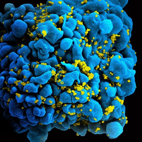

 Scoop.it!