Your new post is loading...

|
Scooped by
Gilbert C FAURE
January 29, 1:27 PM
|
Platelets are cell fragments that plug leaks in blood vessels, preventing bleeding. However, they also form clots that impede blood flow (thrombosis) and increase inflammation.
Activated platelets, which can bind to each other, and hyporesponsive platelets, which do not form clots, are detected in patients with acute severe infections, such as COVID-19 and sepsis. How to uncouple the protective and harmful proinflammatory functions of platelets is a therapeutic dilemma.
In Science, researchers report the formation of platelet-derived integrin-and tetraspanin-enriched tethers (PITTs) in patients with severe infections.
PITTs, which are enriched in specific platelet proteins, remain anchored to the blood vessel wall (endothelium) and promote neutrophil recruitment, inflammation, and tissue damage, whereas the main platelet body shears off and reenters circulation in a hyporesponsive state. PITTs may contribute to blood clotting, immune dysregulation, and bleeding complications that characterize severe infections.
Learn more in a new #SciencePerspective: https://scim.ag/4pYNOyA
|
|
Scooped by
Gilbert C FAURE
January 3, 2022 3:23 AM
|
Blood platelets play a central hemostatic role, (i) as they repair vascular epithelial damage, and (ii) they play immune defense roles, as they have the capacity to produce and secrete various cytokines, chemokines, and related products. Platelets sense and respond to local dangers (infectious or not). Platelets, therefore, mediate inflammation, express and use receptors to bind infectious pathogen moieties and endogenous ligands, among other components. Platelets contribute to effective pathogen clearance. Damage-associated molecular patterns (DAMPs) are danger signals released during inflammatory stress, such as burns, trauma and infection. Each pathogen is recognized by its specific molecular signature or pathogen-associated molecular pattern (PAMP). Recent data demonstrate that platelets have the capacity to sense external danger signals (DAMPs or PAMPs) differentially through a distinct type of pathogen recognition receptor (such as Toll-like receptors). Platelets regulate the innate immune response to pathogens and/or endogenous molecules, presenting several types of “danger” signals using a complete signalosome. Platelets, therefore, use complex tools to mediate a wide range of functions from danger sensing to tissue repair. Moreover, we noted that the secretory capacity of stored platelets over time and the development of stress lesions by platelets upon collection, processing, and storage are considered stress signals. The key message of this review is th

|
Suggested by
LIGHTING
May 24, 2021 3:13 AM
|
In 2019 10 million people developed symptomatic tuberculosis (TB) disease and 1.2 million died. In active TB the inflammatory response causes tissue destruction, which leads to both acute morbidity and mortality. Tissue destruction in TB is driven by host innate immunity and mediated via enzymes, chiefly matrix metalloproteinases (MMPs) which are secreted by leukocytes and stromal cells and degrade the extracellular matrix. Here we review the growing evidence implicating platelets in TB immunopathology. TB patients typically have high platelet counts, which correlate with disease severity, and a hypercoagulable profile. Platelets are present in human TB granulomas and platelet-associated gene transcripts are increased in TB patients versus healthy controls. Platelets most likely drive TB immunopathology through their effect on other immune cells, particularly monocytes, to lead to upregulation of activation markers, increased MMP secretion, and enhanced phagocytosis. Finally, we consider current evidence supporting use of targeted anti-platelet agents in the treatment of TB due to growing interest in developing host-directed therapies to limit tissue damage and improve treatment outcomes. In summary, platelets are implicated in TB disease and contribute to MMP-mediated tissue damage via their cellular interactions with other leukocytes, and are potential targets for novel host-directed therapies.
|
|
Scooped by
Gilbert C FAURE
March 18, 2020 4:37 PM
|
|
|
Scooped by
Gilbert C FAURE
October 12, 2019 5:27 AM
|
Platelets are central players in thrombosis and hemostasis but are increasingly recognized
as key components of the immune system. They shape ensuing immune responses by recruiting
leukocytes, and support the development of adaptive immunity.
|
|
Scooped by
Gilbert C FAURE
June 18, 2019 3:54 AM
|
The role of platelets has been extensively studied in the context of coagulation and vascular integrity. Their hemostatic imbalance can lead to known conditions as atherosclerotic plaques, thrombosis and ischemia. Nevertheless, the knowledge regarding the regulation of different cell types by platelets has been growing exponentially in the past years. Among these biological systems, the innate immune response is remarkably affected by the crosstalk with platelets. This interaction can come from the formation of platelet-leukocyte aggregates, signaling by direct contact between membrane surface molecules or by the stimulation of immune cells by soluble factors and active microparticles secreted by platelets. These ubiquitous blood components are able to sense and react to danger signals, guiding leukocytes to an injury site and providing a scaffold for the formation of extracellular traps for efficient microbial killing and clearance. Using several different mechanisms, platelets have an important task as they regulate the release of different cytokines and chemokines upon sterile or infectious damage, the expression of cell markers and regulation of cell death and survival. Therefore, platelets are more than clotting agents, but critical players within the fine inflammatory equilibrium for the host. In this review, we present pointers to a better understanding about how platelets control and modulate innate immune cells, as well as a summary of the outcome of thi
|
|
Rescooped by
Gilbert C FAURE
from Hematology
May 10, 2019 4:24 AM
|
Megakaryocytes (MKs) are mostly recognized for their role in the generation of platelets. In this issue of Blood , [Campbell et al][1] demonstrate that MKs are also capable of preventing the spread of viral infection, suggesting that they too participate in immunity.[1][2]
![Figure][3]
MKs
|
|
Scooped by
Gilbert C FAURE
July 13, 2018 4:44 AM
|
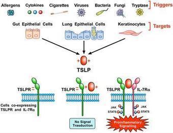
Thymic Stromal Lymphopoietin (TSLP) is a pleiotropic cytokine originally isolated from a murine thymic stromal cell line. TSLP exerts its biological effects by binding to a high affinity heteromeric complex composed of TSLPR chain and IL-7R. TSLP is primarily expressed by activated lung and intestinal epithelial cells, keratinocytes, and fibroblasts. However, dendritic cells, mast cells and presumably other immune cells can also produce TSLP. Different groups of investigators have demonstrated the existence of two variants for TSLP in human tissues: the main isoform expressed in steady state is the short form (sf TSLP), which plays a homeostatic role, whereas the long form (lfTSLP) is upregulated in inflammatory conditions. In addition, there is evidence that in pathological conditions TSLP can be cleaved by several endogenous proteases. Several cellular targets for TSLP have been identified, including immune (dendritic cells, ILC2, T and B cells, NKT and Treg cells, eosinophils, neutrophils, basophils, monocytes, mast cells, and macrophages) and non-immune cells (platelets and sensory neurons). TSLP has been originally implicated in a variety of allergic diseases (e.g., atopic dermatitis, bronchial asthma, eosinophilic esophagitis). Emerging evidence indicates that TSLP is also involved in chronic inflammatory (i.e., COPD, celiac disease) and autoimmune (e.g., psoriasis, rheumatoid arthritis) disorders and several cancers. These emerging observations greatly widen th
|
|
Scooped by
Gilbert C FAURE
July 24, 2017 12:44 PM
|
Platelets participate in many important physiological processes, including hemostasis and immunity. However, despite their broad participation in these evolutionarily critical roles, the anucleate platelet is uniquely mammalian. In contrast with the large nucleated equivalents in lower vertebrates, we find that the design template for the evolutionary specialization of platelets shares remarkable similarities with human-engineered unmanned aerial vehicles in terms of overall autonomy, maneuverability, and expendability. Here, we review evidence illustrating how platelets are uniquely suited for surveillance and the manner in which they consequently provide various types of support to other cell types.
|
|
Suggested by
LIGHTING
December 4, 2016 2:59 PM
|
|
|
Scooped by
Gilbert C FAURE
October 10, 2015 4:32 PM
|
Clinical & Translational Immunology focuses on the general functioning of the immune system in its broadest sense, with a particular emphasis on its cell biology.
|
|
Scooped by
Gilbert C FAURE
April 20, 2015 2:13 PM
|
Mol Immunol. 2015 Apr 14. pii: S0161-5890(15)00348-X. doi: 10.1016/j.molimm.2015.03.244. [Epub ahead of print] REVIEW
|
|
Scooped by
Gilbert C FAURE
February 20, 2015 2:14 PM
|
Small fragments circulating in the blood were formally identified by the end of the 19th century, and it was suggested that they assisted coagulation via interactions with vessel endothelia. Wright...
|
|
|
Scooped by
Gilbert C FAURE
January 28, 2023 1:58 AM
|
|
|
Scooped by
Gilbert C FAURE
June 1, 2021 1:24 PM
|
Viewpoint Free access | 10.1172/JCI151092 Is complement the culprit behind COVID-19 vaccine-related adverse reactions? Dimitrios C. Mastellos,1 Panagiotis Skendros,2 and John D. Lambris3 Published May 4, 2021 - More info View PDF Rare thrombotic reactions to adenovirus-based COVID-19 vaccines Undeniably, a global and coordinated COVID-19 vaccination effort is a prerequisite for taming the spread of emerging SARS-CoV-2 variants and achieving population-wide immunity that can thwart future viral surges. Ongoing vaccination programs have relied on both mRNA and adenoviral-based formulations eliciting potent humoral responses and showing safety in the majority of the population vaccinated so far. However, rare vaccine-related adverse events were recently reported in individuals who received the adenoviral-encoded ChAdOx1 nCov-19 and Ad26.COV2.S vaccines (1–3). Most of these cases developed acute thrombotic complications (cerebral venous thrombosis or disseminated intravascular coagulation) and severe thrombocytopenia following the first dose of the vaccine, without any prior history of heparin exposure or known risk factors for thrombophilia. This rare syndrome, termed vaccine-induced immune thrombotic thrombocytopenia (VITT), clinically resembles the hallmarks of autoimmune heparin-induced thrombocytopenia (autoimmune HIT) and is linked to the consistent presence of circulating autoantibodies against platelet factor 4 (PF4), which can directly activate platelets in the absence of heparin (2). The prothrombotic action of anti-PF4 antibodies likely involves antibody-mediated platelet activation through IgG-FcγR interactions and similar FcR-mediated engagement of immune effector cells such as monocytes/macrophages and neutrophils (Figure 1). Since VITT is a newly described syndrome, its clinical management mostly relies on its similarities with HIT and non–heparin-dependent autoimmune thrombotic thrombocytopenias. While nonheparin anticoagulants and intravenous immunoglobulin (IVIg) have been proposed as first-line treatments for patients with VITT, these approaches are not fail-safe and entail drawbacks that may curtail their effectiveness (e.g., risk of promiscuous FcR activation, acute anaphylaxis, or even thromboembolic events in the case of intravenous immunoglobulin) (4). Several second-line therapies for HIT have also been suggested, including plasmapheresis, especially when nonheparin anticoagulation is contraindicated because of major bleeding events (5). Thus, more insight into the precise pathophysiology of VITT is needed to enable more personalized medicine approaches. Figure 1 A schematic diagram of the plausible mechanisms by which complement may contribute to the VITT-associated prothrombotic response. Vaccine-induced immune thrombotic thrombocytopenia (VITT) has been described in individuals presenting with high titers of platelet-activating anti–platelet factor 4 (PF4) autoantibodies. These antibodies recognize large multiantigenic complexes comprising PF4 and polyanionic structures similar to heparin, and resemble autoimmune heparin-induced thrombocytopenia (HIT). Complement activation can be triggered in multiple ways in patients presenting with VITT and anti-PF4 antibodies. First, the formation of complexes between PF4 and certain vaccine constituents (e.g., adenoviral capsid proteins or DNA) (23) may serve as a scaffold for C3 activation, similarly to what has been observed with PF4-heparin ultra-large complexes. In addition, complement activation can be triggered by the binding of C1q to the anti-PF4 immune complexes that are deposited on the endothelium, monocyte, or platelet surface via binding to polyanionic structures such as glycosaminoglycans (GAGs). Classical pathway activation leads to C3 cleavage, amplification of complement responses via the alternative pathway, downstream generation of proinflammatory C3a and C5a anaphylatoxins, and the formation of the C5b-9 complex. Complement activation fragments mediate a broad range of thromboinflammatory reactions by interacting with complement receptors on platelets, monocytes, and neutrophils. These interactions can induce or enhance FcR-mediated platelet activation, neutrophil-platelet aggregation, and release of TF-loaded neutrophil extracellular traps (NETs) from activated neutrophils. All these processes are fueled by complement activation and can collectively contribute to a prothrombotic environment that may lead to VITT. Moreover, C3-opsonized immune complexes can enhance FcγR-dependent effector responses on platelets further promoting thrombotic responses. Abbreviations: CP, classical pathway; AP, alternative pathway; TF, tissue factor. A proposed role for complement Complement is a host innate immune sentinel comprising over 50 proteins that can be swiftly activated through any of three initiating pathways (classical, lectin, or alternative) that sense microbial invaders or other inflammatory cues, such as immune complexes. In fact, complement activation has been implicated as a perpetrator of immune complex–driven thromboinflammation in autoimmune pathologies such as the antiphospholipid syndrome, a systemic disorder that increases risk for blood clots (6). Prompted by the striking findings in patients who developed VITT after a single dose of adenoviral-associated vaccine, we posit here that the observed thrombotic complications may partly reflect immune complex–triggered complement activation through the classical pathway. PF4-containing immune complexes can be recognized by C1q, a soluble pattern recognition molecule of the classical pathway that binds to the Fc portion of IgG molecules. Anti-PF4 complex–driven classical pathway activation can lead to C3 activation, alternative pathway–mediated amplification of complement responses, and downstream generation of potent proinflammatory mediators and effectors (C3a, C5a and membrane attack complex) that can potentiate thromboinflammation (7). In fact, the thrombogenic capacity of distinct complement activation fragments has been linked to a broad spectrum of immune-related mechanisms that underpin the immunothrombosis of severe COVID-19 (7, 8). Considering that certain allelic combinations of complement gene polymorphisms within the population confer differential susceptibility to deregulated complement activity, it is tempting to speculate that these vaccinated individuals may represent cases where complement deregulation occurs due to genetic alterations that result in ineffective host regulatory control (9). Deregulated complement responses can perpetuate a vicious cycle of inflammatory tissue damage that renders the vascular endothelium and platelet/monocyte compartment more thrombogenic. In this regard, circulating anti-PF4 immune complexes may well serve as a first hit that ignites complement activation, in a way similar to complement-mediated thrombotic microangiopathies (10). The fact that a fraction of these anti-PF4 autoantibodies may recognize antigenic complexes between PF4 and polyanionic structures (such as GAGs or heparan sulfate) raises the possibility that these immune complexes may skew the binding of endogenous complement regulators to host polyanionic surfaces, thereby altering the capacity of these surfaces to withstand autologous complement attack. C3 activation may play a pivotal pathogenic role in this vaccine-induced immunothrombotic phenotype. C3-opsonized anti-PF4 immune complexes can enhance the phagocytic and inflammatory properties of monocytes and neutrophils through the synergistic engagement of FcγR and complement receptors, thereby increasing the thrombogenic milieu in the vasculature (Figure 1). C3 fragment deposition on the endothelium and on platelets/neutrophils can potentiate thromboinflammatory responses by promoting CR3-dependent platelet-neutrophil interactions that favor platelet adhesion and clotting (11). Also, C3aR signaling on platelets can further drive thrombotic responses (12). Of note, it has been shown that PF4–heparin complexes, a hallmark of HIT pathology, can potently activate C3 in the fluid phase and C3 inhibitors such as Cp40 can abrogate C3 activation triggered by HIT-associated PF4–heparin complexes (13). In line with this, the Cp40-based drug candidate AMY-101 has been shown to effectively decrease both neutrophils and neutrophil extracellular trap (NET) formation, two major drivers of HIT-associated thrombosis, in patients with COVID-19 (7, 14–16). Potential treatment approaches In an analogy to HIT, clinical-stage complement C3 inhibitors may represent a novel therapeutic strategy for ameliorating the thrombotic complications elicited in VITT. On one hand, C3 inhibition may quench anti-PF4 antibody production by reducing antigen uptake or processing in the lymphoid tissue. On the other hand, it can broadly suppress multiple paths of immune complex–driven thromboinflammation in VITT (Figure 1). Moreover, C3 inhibition may offer broader therapeutic control of thromboembolic reactions mediated by pathogenic autoantibodies. In this regard, C3 activation, but not C5, was shown to be indispensable for antiphospholipid antibody-induced tissue factor (TF) activation and thrombosis in antiphospholipid syndrome (17). The recently reported resolution of vaccine-induced thrombotic microangiopathy in two patients following combined treatment with anticoagulants and the C5 inhibitor eculizumab further corroborates our hypothesis that therapeutic complement modulation may offer significant clinical benefits to patients with VITT (18). In this regard, C3 inhibitors may exert a broader therapeutic effect than anti-C5 agents by suppressing both terminal pathway activation and thrombogenic pathways operating upstream of C5 (14, 19). Because this newly described immune syndrome is still poorly defined, it would be of great benefit to patients with VITT if the algorithm for VITT clinical management considered diagnostic monitoring of complement biomarkers as a means of validating the use of specific complement inhibitors. As a cautionary note, we should stress that plasma levels of complement proteins reflect both turnover and synthesis. Therefore, the accurate monitoring of complement activity should rely on markers of ongoing activation combined with corroborative functional assays that gauge the impact of complement activation on the tissue or cellular level. mRNA vaccine reactions In addition to these cases of VITT in individuals receiving adenovirus vectored vaccines, the rollout of COVID-19 mRNA vaccines has brought to the spotlight rare adverse events involving a maladaptive immune reaction to certain vaccine constituents. Rare cases of severe allergic reactions (anaphylaxis) were reported following the first dose of the Pfizer–BioNTech or Moderna SARS-CoV-2 mRNA vaccines (20). Such allergic reactions are likely triggered by the PEG or lipid moieties of these mRNA vaccines. They involve complement and mast cell activation in an IgE-independent manner and closely resemble the complement mediated pseudo-allergy reaction (CARPA) that has been described in the case of liposomal carriers (21). Complement activation in this case may be initiated by IgG or IgM directed against the PEGylated lipids of these vaccine formulations. Concluding remarks Taken together, these paradigms of vaccine-related adverse reactions highlight the potential contribution of complement activation to vaccine-related pathology, raise the importance of incorporating complement diagnostic monitoring in the early clinical management of such patients, and endorse complement inhibitors as potential treatment options. In addition to the complement-activating capacity of anti-PF4 antibodies, we cannot rule out the possibility that complement activation in VITT may also be fueled by preformed (natural IgM) or induced IgG antibodies against adenoviral capsid antigens or direct interaction of pattern recognition molecules with capsid elements (22). The relevance of these plausible mechanisms to VITT pathology warrants further investigation. Footnotes Conflict of interest: JDL is the founder of Amyndas Pharmaceuticals, which is developing complement inhibitors for therapeutic purposes. He is the inventor on patents or patent applications that describe the use of complement inhibitors for therapeutic purposes, some of which are developed by Amyndas Pharmaceuticals: US patents 8946145/9371365 (Modified compstatin with peptide backbone and C-terminal modifications), 9630992 (Compstatin analogs with improved pharmacokinetic properties), 9579360 (Methods of treating or preventing periodontitis and diseases associated with periodontitis). JDL is also the inventor of the compstatin technology licensed to Apellis Pharmaceuticals, i.e., 4(1MeW)7W/POT-4/APL-1 and PEGylated derivatives such as APL-2/pegcetacoplan/empaveli and APL-9: US patents 6319897 (Peptides which inhibit complement activation), 7989589 (Compstatin analogs with improved activity), 7888323 (Potent compstatin analogs). Copyright: © 2021, American Society for Clinical Investigation. Reference information: J Clin Invest. 2021;131(11):e151092. https://doi.org/10.1172/JCI151092. References Muir K-L, et al. Thrombotic thrombocytopenia after Ad26.COV2.S vaccination. [published online April 14, 2021]. N Engl J Med. https://doi.org/10.1056/NEJMc2105869. View this article via: PubMed Google Scholar Scully M, et al. Pathologic antibodies to platelet factor 4 after ChAdOx1 nCoV-19 vaccination. [published online April 16, 2021]. N Engl J Med. https://doi.org/10.1056/NEJMoa2105385. View this article via: PubMed Google Scholar Schultz NH, et al. Thrombosis and thrombocytopenia after ChAdOx1 nCoV-19 vaccination. [published online April 9, 2021]. N Engl J Med. https://doi.org/10.1056/NEJMoa2104882. View this article via: PubMed Google Scholar Kapoor M, et al. Thromboembolic risk with IVIg: incidence and risk factors in patients with inflammatory neuropathy. Neurology. 2020;94(6):e635–e638. View this article via: PubMed CrossRef Google Scholar Marchetti M, et al. Heparin-induced thrombocytopenia: a review of new concepts in pathogenesis, diagnosis, and management. J Clin Med. 2021;10(4):683. View this article via: PubMed CrossRef Google Scholar Chaturvedi S, et al. Complement in the pathophysiology of the antiphospholipid syndrome. Front Immunol. 2019;10:449. View this article via: PubMed Google Scholar Skendros P, et al. Complement and tissue factor-enriched neutrophil extracellular traps are key drivers in COVID-19 immunothrombosis. J Clin Invest. 2020;130(11):6151–6157. View this article via: JCI PubMed CrossRef Google Scholar Risitano AM, et al. Complement as a target in COVID-19? Nat Rev Immunol. 2020;20(6):343–344. View this article via: PubMed CrossRef Google Scholar Heurich M, et al. Common polymorphisms in C3, factor B, and factor H collaborate to determine systemic complement activity and disease risk. Proc Natl Acad Sci U S A. 2011;108(21):8761–8766. View this article via: PubMed CrossRef Google Scholar Gavriilaki E, Brodsky RA. Complementopathies and precision medicine. J Clin Invest. 2020;130(5):2152–2163. View this article via: JCI PubMed CrossRef Google Scholar Hamad OA, et al. Contact activation of C3 enables tethering between activated platelets and polymorphonuclear leukocytes via CD11b/CD18. Thromb Haemost. 2015;114(6):1207–1217. View this article via: PubMed Google Scholar Sauter RJ, et al. Functional relevance of the anaphylatoxin receptor C3aR for platelet function and arterial thrombus formation marks an intersection point between innate immunity and thrombosis. Circulation. 2018;138(16):1720–1735. View this article via: PubMed CrossRef Google Scholar Khandelwal S, et al. Novel immunoassay for complement activation by PF4/heparin complexes. Thromb Haemost. 2018;118(8):1484–1487. View this article via: PubMed CrossRef Google Scholar Mastellos DC, et al. Complement C3 vs C5 inhibition in severe COVID-19: early clinical findings reveal differential biological efficacy. Clin Immunol. 2020;220:108598. View this article via: PubMed Google Scholar Gollomp K, et al. Neutrophil accumulation and NET release contribute to thrombosis in HIT. JCI Insight. 2018;3(18):99445. View this article via: JCI Insight PubMed Google Scholar Perdomo J, et al. Neutrophil activation and NETosis are the major drivers of thrombosis in heparin-induced thrombocytopenia. Nat Commun. 2019;10(1):1322. View this article via: PubMed CrossRef Google Scholar Müller-Calleja N, et al. Complement C5 but not C3 is expendable for tissue factor activation by cofactor-independent antiphospholipid antibodies. Blood Adv. 2018;2(9):979–986. View this article via: PubMed CrossRef Google Scholar Tiede A, et al. Prothrombotic immune thrombocytopenia after COVID-19 vaccine. [published online April 30, 2021]. Blood. https://doi.org/10.1182/blood.2021011958. View this article via: PubMed Google Scholar Hillmen P, et al. Pegcetacoplan versus eculizumab in paroxysmal nocturnal hemoglobinuria. N Engl J Med. 2021;384(11):1028–1037. View this article via: PubMed CrossRef Google Scholar Castells MC, Phillips EJ. Maintaining safety with SARS-CoV-2 vaccines. N Engl J Med. 2021;384(7):643–649. View this article via: PubMed CrossRef Google Scholar Klimek L, et al. Allergenic components of the mRNA-1273 vaccine for COVID-19: possible involvement of polyethylene glycol and IgG-mediated complement activation. [published online March 3, 2021]. Allergy. https://doi.org/10.1111/all.14794. View this article via: PubMed Google Scholar Chéneau C, Kremer EJ. Adenovirus-extracellular protein interactions and their impact on innate immune responses by human mononuclear phagocytes. Viruses. 2020;12(12):1351. View this article via: PubMed CrossRef Google Scholar Greinacher A, et al. Towards understanding ChAdOx1 nCov-19 vaccine-induced immune thrombotic thrombocytopenia (VITT) [preprint]. https://doi.org/10.21203/rs.3.rs-440461/v1 Posted on Research Square April 20, 2021. Version history Version 1 (May 4, 2021): In-Press Preview Version 2 (June 1, 2021): Electronic publication
|
|
Scooped by
Gilbert C FAURE
May 13, 2021 12:41 PM
|
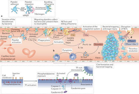
Thrombosis is the most feared complication of cardiovascular diseases and a main cause of death worldwide, making it a major health-care challenge. Platelets and the coagulation cascade are effectively targeted by antithrombotic approaches, which carry an inherent risk of bleeding. Moreover, antithrombotics cannot completely prevent thrombotic events, implicating a therapeutic gap due to a third, not yet adequately addressed mechanism, namely inflammation. In this Review, we discuss how the synergy between inflammation and thrombosis drives thrombotic diseases. We focus on the huge potential of anti-inflammatory strategies to target cardiovascular pathologies. Findings in the past decade have uncovered a sophisticated connection between innate immunity, platelet activation and coagulation, termed immunothrombosis. Immunothrombosis is an important host defence mechanism to limit systemic spreading of pathogens through the bloodstream. However, the aberrant activation of immunothrombosis in cardiovascular diseases causes myocardial infarction, stroke and venous thromboembolism. The clinical relevance of aberrant immunothrombosis, referred to as thromboinflammation, is supported by the increased risk of cardiovascular events in patients with inflammatory diseases but also during infections, including in COVID-19. Clinical trials in the past 4 years have confirmed the anti-ischaemic effects of anti-inflammatory strategies, backing the concept of a prothrombotic function of inflammation. Targeting inflammation to prevent thrombosis leaves haemostasis mainly unaffected, circumventing the risk of bleeding associated with current approaches. Considering the growing number of anti-inflammatory therapies, it is crucial to appreciate their potential in covering therapeutic gaps in cardiovascular diseases. In this Review, Stark and Massberg discuss how the interplay between innate immunity, platelet activation and coagulation, known as immunothrombosis, functions as a host defence mechanism to limit pathogen spreading, yet its aberrant activation, termed thromboinflammation, results in thrombotic complications, highlighting the therapeutic potential of anti-inflammatory strategies in cardiovascular pathologies.
|
|
Scooped by
Gilbert C FAURE
December 12, 2019 4:52 AM
|
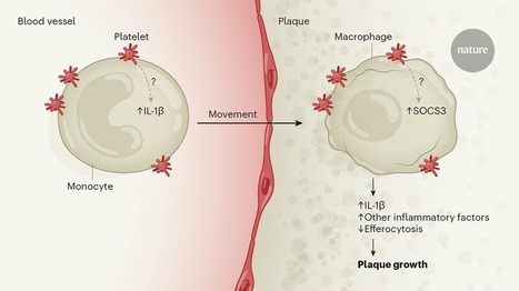
Platelets reprogram macrophages to accelerate plaque formation.
|
|
Scooped by
Gilbert C FAURE
July 27, 2019 12:15 PM
|
Besides mediating hemostatic functions, platelets are increasingly recognized as important players involved in inflammation. Data from experiments in mice and men revealed various points of intersection between thrombosis, hemostasis and inflammation, which are addressed and discussed in this review in detail. One such example is the intrinsic coagulation cascade that is initiated after platelet activation thereby further propagating and re-enforcing wound healing or thrombus formation, which contributes to the pathophysiology of severe diseases. FXII of the intrinsic pathway connects platelet activation with the coagulation cascade during immune reactions. It can activate the contact system thereby either creating an inflammatory state or accelerating inflammation. Recent insights into platelet biology could show that platelets are equipped with complement receptors. Platelets are important for tissue remodeling after injury has been inflicted to the endothelial barrier and to the subendothelial tissue. Thus, platelets are increasingly recognized as more than just cells relevant for bleeding arrest or thrombus formation, and further interesting insights into platelet biology are to be discovered. In the future this understanding will potentially offer novel opportunities for therapeutic intervention in diseases, where platelets are involved.

|
Suggested by
Société Francaise d'Immunologie
June 16, 2019 5:45 AM
|
Summary G‐protein–coupled receptor (GPCR) signaling is essential for the spatiotemporal control of leukocyte dynamics during immune responses. For efficient navigation through mammalian tissues, most leukocyte types express more than one GPCR on their surface and sense a wide range of chemokines and chemoattractants, leading to basic forms of leukocyte movement (chemokinesis, haptokinesis, chemotaxis, haptotaxis, and chemorepulsion). How leukocytes integrate multiple GPCR signals and make directional decisions in lymphoid and inflamed tissues is still subject of intense research. Many of our concepts on GPCR‐controlled leukocyte navigation in the presence of multiple GPCR signals derive from in vitro chemotaxis studies and lower vertebrates. In this review, we refer to these concepts and critically contemplate their relevance for the directional movement of several leukocyte subsets (neutrophils, T cells, and dendritic cells) in the complexity of mouse tissues. We discuss how leukocyte navigation can be regulated at the level of only a single GPCR (surface expression, competitive antagonism, oligomerization, homologous desensitization, and receptor internalization) or multiple GPCRs (synergy, hierarchical and non‐hierarchical competition, sequential signaling, heterologous desensitization, and agonist scavenging). In particular, we will highlight recent advances in understanding GPCR‐controlled leukocyte navigation by intravital microscopy of immune cells in mice. 1 INTRODUCTION Innate and adaptive immune responses depend on the coordination and spatiotemporal control of leukocyte dynamics between and within lymphoid organs and peripheral tissues. A wide spectrum of chemically diverse molecules including small proteins, peptides, lipids, nucleotides, and extracellular matrix fragments can act as chemoattractants, regulating leukocyte motility, migration, chemotaxis, positioning, and cell‐cell interaction. Leukocytes perceive these signals mainly through G‐protein–coupled receptors (GPCRs) that, upon agonist binding, induce intracellular promigratory responses.1, 2 Based on the cell type–specific expression of cell surface GPCR type combinations and intracellular GPCR‐controlling protein networks, each leukocyte subset has evolved specific trafficking paths to fulfill their designated effector functions in the body.3-5 Over the last decades, the analysis of GPCR‐deficient mouse models has advanced our understanding of how specific GPCR signals determine immune cell positioning and compartmentalization in situ. Recent work now also started to shed light on the establishment of chemokine patterns and chemotactic fields that guide leukocytes in mammalian tissues.6, 7 In particular, intravital imaging of control and GPCR‐deficient leukocytes in tissues of live anesthetized mice has brought unprecedented insight into the dynamic migration patterns of immune cell subsets in situ.8, 9 Despite these achievements, it is still largely unclear how leukocytes make directional decisions in lymphoid and inflamed organs where they constantly encounter GPCR signals emanating from multiple sources. The detailed mechanisms of how leukocytes sense, integrate, and prioritize between multiple chemotactic fields in situ and how this influences their physiological trafficking are still largely unknown. Dissecting these molecular events experimentally in mammalian tissues is very challenging and demanding. In particular, we are missing important pieces of information (eg, the exact distribution and concentrations of known chemottractant, GPCR dynamics in leukocytes) and cannot control for unknown chemotactic fields. Due to these current limitations many of the emerging concepts for leukocyte navigation in complex chemoattractant milieus come from studies in controlled in vitro systems2 or lower model organisms.10, 11 However, these concepts from reductionist models may not always be easily translatable to the complex situation in mammalian lymphoid organs and inflamed peripheral tissues. In recent years, several systematic studies in mice have started to disentangle how multiple GPCR signals shape leukocyte migration patterns in the complexity of mammalian tissues. In this review, we will summarize common concepts and mechanisms of GPCR‐controlled leukocyte navigation coming from studies with mouse and human leukocytes. We outline the protein networks that control GPCR signaling and GPCR activity and discuss how these may control leukocyte navigation strategies. In particular, we highlight our current knowledge on leukocyte navigation in complex chemoattractant fields and summarize them for three leukocyte subsets (neutrophils, T cells, and dendritic cells [DC]) with different physiological trafficking paths. For each cell type, we will critically evaluate the relevance of current concepts for leukocyte navigation in mouse tissue. While it is very likely that some navigation strategies are only relevant for certain immune cell types and adapted to specific physiological tissue environments, leukocyte subsets may also share basic strategies for directional decision‐making in lymphoid and inflamed tissues of mammals. 1.1 Concepts and mechanisms of GPCR‐controlled leukocyte navigation Many chemokines and chemoattractants are classical inducers of leukocyte polarity, adhesion, and locomotion. They act through GPCRs on intracellular signaling pathways to establish biochemical asymmetry within the cell, preceding the formation of a polarized cell with an actin‐rich front and a myosin II–rich rear.12, 13 Key molecular pathways involved in establishing functional polarity downstream of GPCRs in leukocytes depend on phosphoinositide 3‐kinases (PI3K) and members of the Rho family of small GTPases.14 Rac isoforms, Cdc42 and RhoA represent major signal hubs that act through several effector molecules to regulate actin polymerization and actomyosin cytoskeleton. The balanced interplay of these components shapes the typical amoeboid migration mode of each leukocyte subset.15, 16 In homogeneous fields of soluble chemokines and chemoattractants, most leukocytes undergo self‐polarization, before increasing their motility, migration speed, and movement in a random and undirected fashion (chemokinesis) (Figure 1A).17 Chemokinesis is considered a migratory pattern that helps leukocytes scan large tissue volumes. We have learnt from intravital microscopy studies in mice that lymphocytes show such continuous undirected (random‐walk) migration in lymphoid organs to enable encounters and interactions with other leukocytes.9 Together with other determining factors (eg, confinement between stromal tissue elements), uniform chemokine signals critically control chemokinesis by increasing the speed of randomly moving lymphocytes in situ.18 Once leukocytes perceive external gradients of soluble chemokines and chemoattractants, they can perform directional movement along the concentration gradients of these diffusing attractants (chemotaxis) (Figure 1B).17 Some leukocyte subsets have also been reported to be repelled from high chemoattractant concentrations (chemorepulsion), leading to their movement against the gradient (fugetaxis).19 However, the underlying molecular mechanisms of this process are still under investigation (Figure 1C).20, 21 Apart from soluble molecular cues, leukocytes also perceive surface‐bound attractants as gradients or as homogeneous fields and undergo movement along these surfaces in a directed (haptotaxis) (Figure 1B) or undirected manner (haptokinesis) (Figure 1A), respectively.16, 22 The aspect of leukocyte navigation along surface‐bound attractants is particularly relevant for chemokines with binding capacities to extracellular glycosaminoglycans (GAGs), which decorate many stromal cell elements in tissues.22, 23 This form of imprinting non‐diffusible chemokine gradients into the tissue has been shown critical for leukocyte trafficking across the blood endothelium and entry into lymphatic vessels.6, 23 The terms “chemotaxis” and “haptotaxis” subsume a spectrum of navigation strategies, which leukocytes can use to follow attractant gradients.24 When perceiving an external attractant gradient, some leukocytes (eg, unstimulated neutrophils in culture dish) undergo a full morphological change from unpolarized shape to the establishment of a front‐back axis, which then leads to directional movement along the gradient. In contrast, other leukocytes already migrate with pre‐established polarity (eg, DCs and neutrophils in 3D), before they steer along the gradient by redirecting their migration path. Alternatively, prepolarized leukocytes may move along a gradient without redirecting their path and start to accelerate the closer they get to the attractant source (eg, neutrophils in response to dying cells). Once at the highest attractant source, leukocytes may also actively decelerate and slow down their migration.24 As evidenced from intravital microscopy of select leukocyte subsets, we know that this spectrum of chemotactic responses also occurs in mouse tissues. While we acknowledge that chemokines and chemoattractants are central for many aspects of these migratory responses, we are missing a detailed understanding about how these signals shape the navigation of leukocytes in situ. Is the cellular movement guided by one or more attractant signal(s)? How do leukocytes navigate in complex gradient fields and sense the right direction? How do leukocytes sense their final destination and know where to stop? Are there inherent differences between leukocyte subsets in their navigation strategies? As many other cell types, leukocytes possess several mechanisms that directly regulate the functionality of GPCRs.13, 25-28 These are mainly controlled by GPCR interactions with three protein families: heterotrimeric (α, β, γ) G‐proteins, G‐protein–coupled receptor kinases (GRKs), and β‐arrestins. Upon stimulation, a GPCR functions as a guanine nucleotide exchange factor that catalyzes the exchange of GTP for GDP on Gα subunits of the heterotrimeric G‐protein. In leukocytes, Gα subunits of the pertussis toxin‐sensitive Gi subfamily play critical roles for the signaling of many chemokine and chemoattractant receptors. The dissociation of Gβγ from Gα‐GTP allows them both to independently activate downstream signaling pathways. Leukocyte chemotactic responses are largely controlled by the released Gβγ, which activates phosphoinositide 3‐kinase (PI3K) and phosphoinositide‐specific phospholipase Cβ (PLC)/inositol‐trisphosphate (IP3)/diacylglycerol effector pathways.29, 30 The termination of GPCR signaling relies on the intrinsic GTPase activity of the Gα subunit that hydrolyzes GTP and reassembles as Gα‐GDP with Gβγ to form an inactive heterotrimer. This Gα GTPase reaction is accelerated by a family of proteins, called “regulators of G‐protein signalling” (RGS proteins) to facilitate the rapid turning off GPCR signaling. Hence, RGS proteins are considered key desensitizers of G‐protein‐signaling pathways.27 In addition, another negative feedback loop evolved to prevent the overstimulation of cells through excessive GPCR stimulation. Following agonist binding and G‐protein activation, GPCRs become rapidly phosphorylated on their cytoplasmic tails and intracellular C‐termini by specific enzymes, usually members of the GRK family.31, 32 The pattern of receptor phosphorylation creates a barcode with high‐affinity binding sites for the recruitment of β‐arrestin proteins.33 As a consequence, the increased binding of β‐arrestin to the receptor sterically hinders the GPCR interaction with G‐proteins. This uncoupling of G‐proteins from the receptor, referred to as “receptor desensitization”, prevents further GPCR activation by repeated agonist stimulation over seconds to minutes.34 β‐arrestins can also control more long‐lasting desensitization of GPCRs over minutes to hours. As they can interact with clathrin‐coated pits, their binding to GPCRs also targets receptors for internalization. Besides, GPCR internalization can also follow other pathways (arrestin independent, clathrin independent) or not occur at all.35 Depending on the intracellular trafficking route, the internalized receptors may be subject to degradation, leading to the downregulation of cell surface GPCRs. Alternatively, targeting internalized GPCR to recycling endosomes and back to the plasma membrane can quickly replenish GPCRs on the cell surface.35 In addition to their important roles in receptor inactivation and internalization, GPCR‐recruited β‐arrestins also act as signaling mediators by directly coupling to numerous signaling molecules.36 At many GPCRs, β‐arrestin‐mediated signaling and classical G‐protein signaling can have distinct biochemical and physiological actions from one another. A relatively new paradigm on GPCR signaling considers the allosteric interaction among the ligand, the GPCR, and the transducers (β‐arrestins and GRKs) and how their contribution influences the bias for G‐protein– or β‐arrestin–mediated signaling.28 Resulting from this paradigm, the concept of biased agonism, ie, that different ligands at the same GPCR are able to activate some signaling pathways while blocking others, is of particular interest for many chemokine receptors with prominent roles in leukocyte navigation.37 In the following, we will point out the most commonly and recently discussed mechanisms, which would allow the control of leukocyte navigation. Given the before‐mentioned regulation mechanisms for GPCR functionality, a “One‐GPCR system” appears already sufficient to control chemokinesis and chemotaxis of leukocytes (Figure 2A‐E). To increase the sensitivity for attractant gradients, leukocytes can upregulate the cell surface expression of GPCRs. Some leukocytes store GPCRs in intracellular vesicles, which can fuse with the plasma membrane under conditions of leukocyte activation.38 While the release of GPCR from vesicular stores occurs within minutes to hours, most leukocytes often respond to external stimuli by changing their cell surface GPCR profile over hours to days. These changes are primarily based on the induction of gene‐transcription networks, which also include GPCR proteins, when leukocytes transition between cell states or undergo differentiation (Figure 2A). Multimerization of GPCRs, including homomerization of the same GPCR type, provides another level of GPCR regulation.25 A recent study highlighted the functional importance of GPCR oligomerization in promoting leukocyte chemotaxis (Figure 2B). By lowering cellular cholesterol, inflammatory stimuli enabled GPCR oligomers that acted as scaffolds for distinct promigratory signaling pathways to foster efficient leukocyte migration.39 However, GPCR multimerization has also been reported to dampen GPCR responsiveness.25 Several other mechanisms can attenuate the sensitivity of leukocytes for chemokines and chemoattractants. One involves the presence of a receptor antagonist that binds to the same GPCR as the gradient‐forming attractant agonist, but exerts blocking function for promigratory signaling (Figure 2C). More cell‐intrinsic mechanisms include the downregulation of GPCR expression by lowering GPCR mRNA stability40 or enhancing GPCR degradation.41 As discussed above, agonist‐induced receptor desensitization also attenuates the signaling responsiveness of GPCRs. Upon homologous desensitization, when GRKs phosphorylate agonist‐specific GPCRs, the further recruitment of β‐arrestins can lead to G‐protein uncoupling and internalization of GPCRs (Figure 2D). This process explains why many leukocyte chemotaxis responses toward single attractants are dose dependent and follow a bell‐shaped curve, with higher concentrations resulting in stasis.42 Homologous desensitization may be particularly relevant when leukocytes sense high attractant concentration in situ, eg, at the center of an attractant source.43 Under such conditions, the majority of cell surface GPCRs is likely to undergo desensitization, internalization, and degradation, which may then result in cellular unresponsiveness to continuous or repeated stimulation through the same attractant. As a strategy to remain responsive to subsequent stimulation in gradients, some leukocytes internalize GPCRs, deploy its ligand cargo into lysosomes, and quickly recycle unloaded GPCRs back to the plasma membrane.44 Thus, by eliminating chemotactic factors from the extracellular environments, classic GPCRs can acquire additional scavenging function. This “consumption” of external attractant may also impact the chemotactic milieu by reducing the strength of attractant cues.44 Instead of only binding one agonist, many “One‐GPCR systems” can bind and respond to two or more chemoattractants and activate downstream signaling (Figure 2E). Sensing these distinct attractants through the same GPCR in a spatial or temporal sequence can provide another navigation strategy. In our body, leukocytes are rarely exposed to only one chemotactic factor. For efficient trafficking between organs and coordinated movement within tissues, most leukocytes express more than one GPCR on their surface and sense a wide range of chemokines and chemoattractants. Directional decision‐making in lymphoid and inflamed organs requires leukocytes to constantly sense, compare, and integrate numerous GPCR signals emanating from multiple sources. To efficiently navigate in a complex milieu of attractant gradients, leukocytes need to respond to combinational signals and prioritize between competing signals. While this aspect of GPCR signal integration has already been of interest decades ago, it has regained momentum with the recent developments in fabricating microfluidic‐based chemotaxis devices. We will now briefly summarize common mechanisms and concepts for leukocyte navigation, which become already relevant in the presence of a second GPCR signaling system (“Two‐GPCR systems”) (Figure 3). As leukocytes often use combinations of more than two‐GPCR systems, these principles could be further expanded. Manifold scenarios of attractant interpretation are conceivable, when leukocytes sense two chemoattractant signals through two different GPCR types (Figure 3). The outcome for leukocyte guidance depends on several determining parameters: (a) the spatial distribution of each signal (uniform, graded), (b) the spatial orientation of both signals toward each other (same or opposing directions), and (c) the ratio of signaling input that leukocytes receive from both signals (equal, unequal). The latter point includes both cell‐intrinsic features and cell‐extrinsic conditions. Unequal signaling input can result from different cell surface expression of the two‐GPCR types or different strength of the downstream signaling pathway. In both cases, one chemoattractant signal dominates over the other. This can also occur in leukocytes with equal signaling input from both GPCR systems under conditions of unequal concentrations of both signals in the extracellular space. Many of these scenarios have already been addressed with select leukocyte subsets and their cell‐specific GPCR combinations in controlled in vitro microenvironments, and examples will be discussed in the next paragraphs about specific leukocyte subsets (neutrophils, T cell, and DCs). As discussed before, homogenous chemoattractant fields can induce leukocyte polarity and chemokinesis. A second‐graded signal “on top” can then redirect prepolarized leukocytes and guide them toward other locations. However, if the uniform attractant field is very potent, it will retain leukocytes and attenuate chemotaxis toward the second‐graded signal (Figure 3A).45-48 Under conditions when both attractant signals are oriented in the same direction and overlap, the two‐GPCR signals have usually additive effects for leukocyte chemotaxis (Figure 3B). In the case of promigratory signals, chemotactic factors act often synergistically to improve chemotaxis along the same direction.49 Physiologically, this might be of particular relevance for tissue sites where overlapping attractant gradients originate from the same local area. A special variant of GPCR signal synergism has initially been observed in migrating human and mouse neutrophil populations in vitro. During their chemotaxis in response to shallow N‐formyl‐peptides (fMLF) gradients, neutrophils released the lipid leukotriene B4 (LTB4) that acted on top of the early chemotactic factor as a signal relay molecule to improve chemotaxis of the whole neutrophil population.50 Under conditions when leukocytes encounter two spatially opposing gradients, the two‐GPCR signals compete for guiding leukocytes into one or the other direction (Figures 3C and 4). Leukocytes exposed to two opposing attractants with equal signaling strength will likely accumulate and find migratory equilibrium in the middle of both gradients (Figure 3C).47, 48, 51, 52 However, an unequal signaling input from both signals favors the movement of leukocytes into one direction (Figure 4A).47 It is known for decades that specific GPCR signals can dominate over other GPCR signals, which enables leukocytes to prioritize some chemoattractant signals over others (Figure 4B).42, 45 This aspect of GPCR crosstalk provided a first conceptual framework for neutrophils traveling in a stepwise fashion from the blood into infected tissues.45 Initially, it was discussed that heterologous desensitization of GPCRs might be an underlying mechanism for prioritizing chemotactic cues. In contrast to homologous desensitization (see above), heterologous GPCR desensitization is agonist independent and results from the phosphorylation of one GPCR by a kinase activated through the signaling cascade of another activated GPCR (Figure 3D). Thus, one‐GPCR signal can act as desensitizing signal for multiple other GPCR types, causing leukocytes to become refractory to several chemotactic stimuli within minutes. This rapid cross‐phosphorylation has been linked to second‐messenger kinases, particularly protein kinase C, which phosphorylates the GPCR at sites different from GRKs.53 For neutrophil navigation in opposing gradients, it was later proposed that the dominance of certain GPCR signals is regulated by events downstream of the receptors. According to this concept, referred to as “signaling hierarchy”, special GPCRs induce intracellular signaling pathways that can override signaling pathways downstream of other GPCR signals42, 54 (see below for details). Leukocyte populations in tissues rarely consist of identical cell clones, but are rather characterized by many cells with slightly heterogeneous expression of GPCR combinations. Thus, single‐cell heterogeneity on the basis of combinatorial GPCR expression may influence the population response in two opposing attractants with equal signaling strength (Figure 4C). In many physiological settings, leukocytes are thought to navigate step‐by‐step through sequential chemotactic fields. Under conditions of two spatially segregated chemotactic signals in sequence (Figure 3E), leukocytes may first move along a first attractant gradient (Figure 1A), and then subsequently sense the gradient of a second, more distant attractant. During the transition from first to second attractant gradient leukocytes then encounter a competitive situation between two gradients (Figure 3C). When the second signal is dominant over the first one, it is plausible that leukocytes are recruited to and then retained at the distant site. Importantly, even without any dominance between the signals, leukocytes can migrate away from a first attractant and travel toward a second, more distant attractant.45 In principle, this also allows leukocytes to leave chemotactic fields of high, saturating concentrations. Moreover, leukocytes remain responsive for additional attractant changes, while moving back and forth in the equilibrium between both gradients. Thus, interpreting two distinct signals in sequence allows efficient navigation patterns that are not possible with only one attractant presented in the same spatial array. We want to point out that the sequential sensing of attractant gradients can also be temporally controlled (Figure 3E). Leukocytes may undergo sensing of a first attractant gradient, while a second attractant arises newly in close vicinity. In the course of an immune response in lymphoid or inflamed tissues, this may result from chemoattractant secretion of nearby activated tissue‐resident cells or recently recruited, blood‐derived leukocytes. As most immune responses are choreographed by a sequence of timely separated cellular events, it is likely that this aspect of sequential sensing has strong influence on leukocyte navigation in situ. In our contemplation on leukocyte navigation by “Two‐GPCR systems”, we have focused until now on GPCRs with signaling function. However, many leukocyte subsets additionally express members of the atypical chemokine receptor (ACKR) family on their surface. Also referred to as scavenging receptors, ACKRs are GPCRs with promiscuous binding affinity for multiple chemokines. As they do not couple to G‐proteins, they are unable to induce intracellular signaling pathways. Instead, ACKRs can actively internalize and target chemokines for lysosomal degradation.55 Functional co‐expression of conventional chemokine receptors and ACKRs in leukocytes has been implicated in the fine‐tuning of leukocyte chemotaxis, with ACKRs acting as rheostats in restricting excessive migration in response to moderate chemokine levels (Figure 3F).56-58 As we have just outlined, leukocytes have evolved several molecular mechanisms that allow an enormous spectrum of GPCR‐controlled navigation strategies. Many of the emerging concepts for leukocyte navigation in complex chemoattractant fields have been developed in controlled in vitro systems, some of which by studying only one select leukocyte subset. Rigorous testing and proving the relevance of these concepts for leukocyte migration in the complexity of mammalian organs and tissues is technically very challenging. However, systematic studies in mice have started to delineate GPCR‐controlled navigation strategies and their influence on physiological trafficking patterns through lymphoid organs and inflamed peripheral tissues. In the following, we will discuss our current knowledge on the role of GPCR signals for the migration patterns of three leukocyte subsets (neutrophils, T cells, and DCs). Each of these immune cell types has evolved their own trafficking paths to fit their effector functions. We will highlight recent studies that conceptually advanced our understanding of GPCR‐controlled leukocyte navigation in mammalian tissues. 2 NEUTROPHILS Neutrophils are pivotal effector cells of our innate immune response and the first line of defense for eliminating bacteria and fungi in tissues.59 Recent studies also implicate regulatory functions of neutrophils in tissue homeostasis and cancer.60 In mammals, they are produced in the bone marrow and are continuously released into the bloodstream to patrol the intravascular compartment under homeostasis. Neutrophils circulate in the blood for ca. 12 hours under the influence of circadian rhythms, before they are mostly cleared into the bone marrow, but also other extramedullary tissues.61 In contrast to the general believe that naive peripheral tissues are free of neutrophils, recent work suggests that low numbers of neutrophils are present in many tissues under steady state.62 Upon inflammation and infection, the release of neutrophils from bone marrow increases their concentration in the blood by several folds.63 Moreover, large numbers of circulating neutrophils then infiltrate inflamed tissues where they sense activated vascular endothelium. In agreement with their critical role as first responders, neutrophils have evolved robust strategies to infiltrate inflamed tissues extremely fast and orient themselves to detect potential sites of microbial invasion with great precision. In many organs, the exit from the vascular compartment involves a sequence of exquisitely coordinated processes: (a) intravascular arrest, migration, and crawling along the activated endothelial cells, and (b) breaching multiple layers of the vascular wall (venular endothelium, venular basement membrane, and pericytes).64 Once neutrophils have traversed this barrier, they are primed for the rapid detection of damaged tissue or microbes in the extravascular space. At sites of cell death in the interstitial space, neutrophils undergo sequential phases of highly coordinated chemotaxis and cell cluster formation, now commonly referred to as neutrophil swarming.65, 66 As true sentinel cells, they sense almost any form of tissue insult, rapidly localize it in any inflamed organ and efficiently move toward the core of the inflammation or infection site. Neutrophils have been shown to express on their cell surface more than 30 GPCR types, which can all bind chemotactic molecules. Accordingly, neutrophils are responsive to a broad array of chemically diverse chemoattractants, including chemokines, lipids, complement factors, formylated peptides, nucleotides, alarmins, and proteolytic (ECM) fragments.67-69 Many of these chemotactic factors are usually present at sites of in vivo inflammation. How the crosstalk between these GPCR signals guides neutrophil recruitment in inflamed tissues has been of huge interest since decades. Early in vitro work with human polymorphonuclear neutrophils (PMN) characterized the strength of chemically distinct chemoattractant classes to desensitize calcium‐ion mobilization, as a read‐out of GPCR downstream signaling, to one another (Figures 2D and 3D).53, 70 Peptide chemoattractants, including N‐formylmethionyl‐leucyl‐phenylalanine (fMLF), the complement factor C5a, and the chemokine CXCL8 (IL‐8), cross‐desensitized one another to different extents (fMLF > C5a > IL‐8). Moreover, peptide chemoattractants, in particular fMLF, cross‐desensitized lipid chemoattractants, including platelet‐activating factor (PAF) and LTB4. In contrast, lipid and purinergic agonists failed to desensitize to the peptide chemoattractants.70 These results were extended to neutrophil chemotaxis in the presence of multiple chemoattractants, showing a hierarchy of resistance to competing signals, with fMLF and C5a inhibiting migration to other chemoattractants (Figure 3D).71, 72 Elegant under‐agarose chemotaxis experiments proposed the concept that neutrophils can navigate through complex chemoattractant fields by migrating in a step‐by‐step manner in response to one agonist source after another. Thus, their ultimate positions are determined combinatorially by the array of GPCRs they express and by the sequence of chemoattractant gradients they encounter (Figure 3E).45 In this concept, pathogen‐derived “end‐target” chemoattractants (N‐formyl peptides, C5a) will dominate over host tissue‐derived “intermediate‐target” chemotactic factors (CXCL8, LTB4) in their ability to guide neutrophil migration (Figure 3A, C, E).45 This dominance of end‐target attractants has been suggested to depend on the activation of the p38 MAPK pathway, which can override the phosphoinositide 3‐kinase (PI3K) pathway activated by intermediate‐target attractants.42, 54 Importantly, neutrophil navigation is also possible in a spatially sequential array of intermediate‐target chemoattractants (Figure 3E).45, 47, 51, 73 Another aspect of crosstalk between end‐target and intermediate‐target chemoattractants has not been considered in these early studies, but is also functionally relevant for neutrophil navigation. Already small amounts of fMLF or C5a can stimulate neutrophils to secrete intermediate‐target attractants, in particular LTB4.50, 74 This paracrine signaling mechanism becomes very relevant in chemotaxing neutrophil populations. Neutrophil‐released LTB4 functions as a signal relay molecule and amplifies in a feed‐forward manner the chemotactic response to subsaturating concentrations of end‐target attractants (Figure 3B).50, 75 Recent developments in microfabricated devices and microfluidic‐based chemotaxis systems now allow a more detailed analysis of neutrophil migration responses in complex chemoattractant milieus, providing deeper insight into neutrophil navigation in opposing gradients (Figure 3C),47, 73, 76-78 synergistic gradients (Figure 3B),49 and geometrically complex environments.49, 77 Moreover, even the analysis of GPCR signaling underlying neutrophil swarming dynamics is now possible due to novel micropatterning techniques.75 As human PMNs, mouse neutrophils express a range of functional GPCRs to sense N‐formyl peptides (through FPR1 and FPR2), C5a (mainly through C5ar1), LTB4 (mainly through LTB4R1/BLT1), and several chemokines including CXCL1, CXCL2, CXCL5 (through CXCR2), CXCL12 (through CXCR4), and several CC‐chemokines (through CCR1).67 Some of these GPCRs are present in secretory vesicles that can rapidly fuse with the plasma membrane upon neutrophil activation, allowing the cells to increase their responsiveness to chemotactic cues (Figure 2A).38 Other reported mechanisms to control GPCR functionality in neutrophils include competitive antagonism or partial agonism (Figure 2C),79 receptor multimerization (Figure 2B), differential agonist responses (Figure 2E),80 and agonist scavenging through simultaneous expression of ACKRs (Figure 3F).56 Homologous desensitization is a well‐known phenomenon for almost all neutrophil‐expressed GPCRs (Figure 2D). While GPCR transducers (G‐protein subunit types, GRKs, and arrestins) and RGS proteins have been linked to the function of some GPCRs in primary mouse neutrophils,32, 81-88 we are still missing a detailed understanding of their exact contributions to neutrophil navigation in complex chemoattractant fields. Clearly, neutrophils are equipped with many GPCR types and mechanisms to dynamically control GPCR activity, allowing them to use various navigation strategies in the presence of multiple chemotactic cues. Moreover, neutrophils have been shown to perform fugetaxis against the direction of a gradient of high concentration chemokine, a process that is mechanistically not well understood (Figure 1C).89 However, there are still many open questions of how these mechanisms contribute to the neutrophil migration patterns observed in situ. Intravital microscopy studies of GPCR‐deficient neutrophils provided novel insight into how GPCR signals choreograph the physiological trafficking paths of neutrophils. In the following, we will point out that GPCR signals are important throughout many steps in the lifecycle of a neutrophil. In particular, we will highlight studies that advanced our concepts of GPCR‐controlled neutrophil navigation in mouse tissues. 2.1 Bone marrow release: inhibiting chemokine‐mediated retention The majority of mature neutrophils in the body reside in the bone marrow, with an estimated ratio of 300:1 between neutrophils in the bone marrow and blood. Neutrophil release into the blood is controlled by the interplay of retention and mobilization signals, provided by two antagonistically operating chemokine systems (Figure 3A,C). During homeostasis, the chemokine CXCL12 is produced in the bone marrow and essential for the retention of mature neutrophils that express the corresponding chemokine receptor CXCR4.90 CXCL12 is expressed at highest levels by “CXCL12‐abundant reticular cells” (CAR cells), but also by other stromal cells of the bone marrow, including osteoblasts.63, 91 The expression levels of CXCL12 in stromal cells follow a circadian rhythm, controlled by the sympathetic nervous system.92 When neutrophils were freshly isolated from the bone marrow, they expressed only low levels of cell surface CXCR4, but contained high intracellular levels of CXCR4.90, 93 After placing these cells in medium lacking CXCL12, they upregulated cell surface CXCR4 within hours.90, 93 These findings suggested receptor desensitization and internalization in response to constitutively high local concentrations of CXCL12 in the bone marrow microenvironment (Figure 2D). Moreover, these data underlined that neutrophils receive constant CXCL12 signals, which position them in the bone marrow, probably by activating integrin‐mediated adhesion to stromal cells.94 While high concentrations of CXCL12 retain neutrophils in the bone marrow via CXCR4 signaling, low concentrations of CXCR2 ligands expressed on endothelial cells and produced by megakaryocytes facilitate neutrophil egress via CXCR2.63 Thus, CXCR2 signals recruit neutrophils out of the retentive CXCL12 field and direct them to the endothelial sinus walls for entry into the blood circulation (Figure 3A). This intricately balanced interplay between retention and mobilization signals can be shifted in both directions. G‐CSF treatment or acute inflammation, which increases blood serum levels of G‐CSF and CXCR2 ligands, shifts the balance toward neutrophil mobilization from the bone marrow.95-98 Several modes of action have been proposed for G‐CSF, either leading to the lowering of CXCL12 levels in the bone marrow or triggering the release of CXCR2 ligands from endothelial cells or megakaryocytes (summarized in63). The same shift toward neutrophil egress can be achieved by conditional gene depletions of CXCR4 or CXCL12 in mice.99, 100 The administration of the CXCR4 antagonist AMD3100 also results in a threefold increase in the number of circulating neutrophils and a corresponding decrease in bone marrow numbers.93, 101 Originally, this drug effect was interpreted as direct antagonism of CXCR4 expressed by neutrophils, thereby disrupting the CXCL12‐CXCR4 axis in the bone marrow (Figure 2C).90 Surprisingly, subsequent studies found that AMD3100 also acted on CXCR4 expressed by bone marrow endothelial cells, and inhibited the translocation of CXCL12 across bone marrow endothelial cells, suggesting a rapid rise of CXCL12 in the blood and reversal of the CXCL12 gradient (Figure 3B).102, 103 These data were questioned by intravital imaging studies that could not observe clear neutrophil egress from the bone marrow upon AMD3100 treatment. Instead, the increase in circulating neutrophils through AMD3100 was explained by the release of marginated neutrophils from the lung microvasculature.104 Moreover, AMD3100 had a blocking effect on the trafficking of aged blood neutrophils, which upregulate CXCR4 expression over time, enabling them to home back to the bone marrow for clearance.104 The exact mode of action of AMD3100 is still controversially discussed.90 Insight into the physiological consequences of shifting the chemokine balance in the opposite direction, toward retention in the bone marrow, comes from an extremely rare congenital human immunodeficiency disease. Warts, hypogammaglobulinemia, infections, and myelokathexis (WHIM) syndrome is an autosomal dominant human disorder caused by gain‐of‐function mutations in the C‐tail of CXCR4.105, 106 WHIM syndrome involves myelokathexis, a condition characterized by the retention of mature neutrophils in the bone marrow and peripheral neutropenia. WHIM‐associated truncations or mutations in the carboxy‐terminal tail of CXCR4 result in the loss of multiple serine residues, which are target sites for GRK‐mediated receptor phosphorylation. Truncated forms of CXCR4 are impaired in agonist‐induced homologous desensitization and internalization, and cause enhanced signaling upon CXCL12 stimulation.106 This leads to stronger anchorage of mature neutrophils in the bone marrow, counteracting CXCR2‐mediated chemotaxis toward vessels and neutrophil egress. However, administration of G‐CSF or ADM3100 to WHIM patients can overcome this imbalance and cause an increase in circulating neutrophil numbers.106 2.2 Getting to the site of inflammation: navigation along multiple chemokine fields Blood‐circulating neutrophils detect sites of tissue injury and infection by interacting with the luminal surface of activated venular endothelium. In response to tissue‐released inflammatory cytokines, endothelial cells upregulate P‐selectin, E‐selectin, and integrin ligands, such as ICAM‐1 and VCAM‐1, to promote interactions between neutrophils and the vessel wall. This induces neutrophils to undergo a multistep adhesion cascade along the endothelial lining before emigrating from the vessel into the inflamed tissue interstitium.107 Chemokines are present on the luminal surface of endothelial cells and cause arrest of rolling neutrophils by triggering the activation of integrins. Many studies showed an involvement of CXCR2 ligands, in particular CXCL1, but also CXCL2 and CXCL5, in this process.108, 109 These chemokines are primarily expressed and released by endothelial cells,110, 111 but also tissue‐resident cells, including pericytes, perivascular macrophages, and mast cells, can act as important chemokine sources.112-115 Due to their high‐affinity binding sites for heparin sulfate (HS) GAG most chemokines become immobilized by HS GAG on the luminal side of the endothelium,116 and defects in HS GAG synthesis have been shown to decrease immobilization of chemokines and neutrophil transmigration into inflamed tissues.117, 118 While the involvement of chemokines for neutrophil transmigration is widely established, there has been ongoing debate whether and how leukocytes sense stable chemokine gradients across the vessel wall on their way into the tissue (Figure 1B). There are ample references to support the common believe that chemotactic gradients between the apical and basolateral part of postcapillary venules promote diapedesis, ie, the outward passage of neutrophils through the vessel wall. Immunofluorescence stainings of HS GAG moieties revealed steep gradient of HS GAG scaffolds between the luminal and abluminal aspect of skin postcapillary venules.119 Under inflammatory conditions, HS GAG distribution became even more polarized by massive depositions of HS and laminin in the basolateral basement membrane of skin vessels.119 Given the GAG‐binding properties of many chemokines, it was proposed that these sharp HS GAG scaffold gradients might favor chemokine gradients across the venular wall. In agreement with this idea, excessive deposition of the GAG‐binding chemokine CXCL1 on the apical part of inflamed blood vessels resulted in decreased neutrophil diapedesis.120 Other supporting data come from experiments in which an extravascular source of CXCR2 ligands was placed in form of a gel on one side of a blood vessel.121 Under these conditions, neutrophil crawling was directed to emigrate preferentially from that side of the vessel. HS GAG on the surface of the endothelium was required to present CXCL2, forming a chemotactic gradient that was followed by neutrophils. This sequestration of endothelial chemokine occurred exclusively in venules, but not other blood vessel types.121 HS GAG, together with the atypical receptor ACKR1, was also discussed to establish gradients by transcytosis of chemokines.118, 122, 123 Transcytosis involves the internalization and transport of tissue‐derived inflammatory chemokines from the basal surface onto the luminal surface of endothelial cells. ACKR1 is specifically expressed by postcapillary venules and was shown to support promigratory chemokine patterns across the vessel wall for optimal leukocyte migration across the vessel wall.123, 124 Unlike other ACKR, ACKR1 does not act as classical scavenger receptor, and was initially discussed to promote the transcytosis of tissue‐derived inflammatory chemokines from perivascular pools to the apical surface of venules.122, 123 Novel insight into the functional role of ACKR1 and the directional cues that guide neutrophils during diapedesis come from recent elegant experiments of Nourshargh and colleagues.125 By studying TNF‐induced neutrophil emigration in the inflamed mouse cremaster muscle, it was shown that neutrophils encounter the two CXCR2 ligands CXCL1 and CXCL2 in a sequential manner during their passage through the vessel wall. In a three‐step process, neutrophils first sense CXCL1 produced by TNF‐stimulated endothelial cells on the luminal surface of venules, promoting crawling along the endothelium. Next, neutrophils encounter CXCL2 that is retained at endothelial cell junctions by ACKR1. Notably, CXCL2 is secreted by neutrophils themselves and then presented by ACKR1, which is enriched in endothelial junctions. This self‐guided migration response of neutrophils was critical for breaching of endothelial junctions and unidirectional transendothelial migration. Lastly, CXCL1 expressed by pericytes governed neutrophil crawling in the subendothelial space.125 While earlier work indicated relatively restricted expression of CXCL1 to vascular and tissue‐resident cells, and CXCL2 with activated neutrophils,126 these novel findings highlight unexpected non‐redundant roles for CXCL1 and CXCL2 to guide neutrophils through venular walls as governed by their distinct cellular sources.125 Having identified this critical spatiotemporal sequence of chemokine‐guided steps across the vessel wall (Figure 3E), it will be of future interest to understand how other tissue‐derived attractant signals may act on top of this and support further guidance into the underlying inflamed interstitium. As an example, neutrophils entering an inflamed peritoneum were suggested to encounter two sequential gradients in a step‐by‐step manner (Figure 3E).114 In a lipopolysaccharide‐induced peritonitis model, initial recruitment of neutrophils out of the blood vessel into the peritoneal tissue was regulated by rapid secretion of CXCL1 and CXCL2 from perivascular mast cells. Further guidance of extravasated neutrophils through the interstitium and into the peritoneal cavity depended on CXCL1 and CXCL2 produced by tissue‐resident interstitial macrophages.114 2.3 Getting to the site of inflammation: interpreting temporal cascades of chemically distinct chemoattractants Most of our discussions until now focused on the role of chemokines for neutrophil guidance in the tissue, but also putative non‐GAG‐binding, soluble chemoattractants including C5a, LTB4, and formylated peptides are sensed by neutrophils and relevant for special types of inflammatory reactions. A mouse model of autoimmune rheumatoid arthritis, the K/BxN serum transfer model, has provided unprecedented insight into how four different chemoattractant receptors, C5aR1, LTB4R1, CCR1, and CXCR2, collaborate to recruit neutrophils into the inflamed tissue.127 In this model, the inflammatory response is driven by immunoglobulin G autoantibodies to the ubiquitous glycolytic enzyme glucose‐6‐phosphate isomerase (GPI) leading to the formation of immune complexes that drive immune cell activation. Neutrophil infiltration into the joint is critical for the clinical signs of arthritis in this model. Several consecutive studies have elucidated a temporal cascade in which neutrophils encounter complement, lipid, and chemokine signals to enter the joint and contribute to disease progression (Figure 3E).74, 128-131 While it has long been known that immune complexes lead to the activation of the alternative complement system in arthritis,132, 133 it has only recently been found that C5aR signaling in circulating neutrophils is the key initiator for a cascade of events that ignite inflammation.131 The deposition of immune complexes in the joint space induces the generation of C5a, which unexpectedly binds to HS GAG on the synovial endothelium. Neutrophils sense C5a on the joint endothelium, induce integrin‐dependent arrest, and facilitate crawling along the vasculature. Subsequently, C5a signals stimulate neutrophils to secrete LTB4, which induces the entry of the first neutrophils into the joint and initiates the onset of disease.74, 128, 129, 131 After disease onset, recruited neutrophils in the joint secrete IL‐1β, which in turn stimulates fibroblast‐like synoviocytes to produce CCR1 ligands at first and later CXCR2 ligands.130 CCR1 ligands are required to recruit the next wave of neutrophils into the joint, and this recruitment of neutrophils is broadly amplified in the last step of the cascade when neutrophils themselves release CXCL2 to potently attract large numbers of neutrophils into the joint.67, 130 Thus, neutrophil entry into the inflamed joint is a prime example where neutrophil recruitment is choreographed by non‐redundant roles of distinct chemoattractants that collaborate sequentially in a temporal cascade (Figure 3E). Moreover, this process highlights the rapid production of complement and LTB4 to initiate a recruitment cascade at the local site of inflammation. By contrast, chemokines act later in this cascade to amplify and prolong the inflammatory response.67 2.4 At the site of inflammation: synergism through lipid‐mediated amplification Another example where complement‐induced LTB4 secretion rapidly shapes neutrophil migration patterns has recently been described.134 In a simplified fungemia mouse model, the fungi Candida albicans sequestered in the pulmonary vaculature and actively recruited neutrophils. Neutrophils showed swarm‐like migration patterns in response to sequestered yeast, similar to previously described swarming responses in other tissues.66 A small number of lung neutrophils responded within minutes and performed intravascular chemotaxis, before larger numbers of neutrophils were recruited and formed cell clusters. The immediate capture of live C. albicans by neutrophils depended on complement‐mediated chemotaxis.134 In addition, complement activation stimulated neutrophils to secrete LTB4, which amplified the directional recruitment and clustering of neutrophils in a synergistic manner (Figure 3B). This was in agreement with a previous study highlighting the importance of LTB4‐mediated signal amplification during neutrophil swarming in interstitial tissue spaces at sites of sterile skin injury and in bacteria‐infected lymph nodes.135 Neutrophil swarming is an essential process of the neutrophil tissue response and has been observed in diverse tissues under conditions of sterile inflammation and infection with various pathogens, including bacteria, fungi, and parasites.65, 66 The swarming response comprises sequential phases of highly coordinated chemotaxis followed by neutrophil accumulation and the formation of substantial neutrophil clusters, which has led to the multistep attraction model of neutrophil swarming.66 While the underlying signals and molecular players for some of these steps are still unclear, we know that neutrophil‐derived LTB4 acted on top of early released, yet unknown, chemoattractants and mediates intercellular signal relay among neutrophils to amplify interstitial recruitment in a feed‐forward manner (Figure 3B). This model was in accordance with earlier in vitro studies identifying LTB4 acted as a signal relay molecule (see above).50 As a consequence, LTB4‐LTB4R1 signaling enhances the radius of neutrophil recruitment from distant tissue sites and maintains the amplified chemotactic response of the neutrophil population. Together with signaling through other GPCRs, such as CXCR2 and FPR2, LTB4R1 signaling also optimizes neutrophil clustering.135 Importantly, LTB4 and CXCR2 signals are also relevant for the swarming response of human neutrophils.75 The formation of focalized dense neutrophil aggregates is considered beneficial for isolating sites of tissue wounding from viable surrounding tissue, containing microbial invaders and concentrating the microbicidal activity of a whole neutrophil population in one swarm center.66 However, neutrophil aggregation can also lead to tissue damage and removal of other cells.135, 136 In the context of the lung microvasculature, intravascular neutrophil swarms that formed in response to C. albicans infection lead to vessel occlusion and pulmonary hemorrhages, which were reverted in the absence of LTB4R1.134 How neutrophil‐derived LTB4 shapes and maintains a promigratory chemotactic field in vivo remains unclear. LTB4 is considered to have a short half‐life and can be rapidly turned into LTB4 metabolites, which act as natural inhibitors of LTB4‐mediated responses. However, it has also been demonstrated that neutrophils release LTB4 in form of exosomes, which may support more prolonged effects of LTB4 gradients.137 Interesting insights into this issue come from studies with diabetic mice receiving skin infections with methicillin‐resistant Staphylococcus aureus (MRSA), which leads to large neutrophil clusters in form of abscesses.138 Diabetic mice produce higher levels of LTB4 in the infected skin compared to non‐diabetic mice. Surprisingly, neutrophils in the skin of these mice failed to assemble into an organized abscess, as excessive LTB4 levels likely disrupted chemoattractant gradients in the skin and impaired directed neutrophil chemotaxis.138 These findings exemplify that the appearance or absence of neutrophil swarms depends on the specific mixture of chemoattractants in a specific inflammatory environment. Although not yet studied in detail, we know that several factors, including tissue damage size, presence of pathogens, induction of cell death, and neutrophil numbers, are important for shaping the exact neutrophil‐swarm phenotype in an inflamed tissue. All these factors can potentially initiate the release of chemotactic fields that may strengthen or attenuate LTB4‐mediated signal amplification in situ.66 2.5 Getting to the site of inflammation: following hierarchies True prioritization of chemoattractant fields has been observed for neutrophils in a model of thermal‐induced liver injury.139 In this model, large numbers of neutrophils are recruited over hours from the liver sinusoids to a focal site of sterile hepatic necrosis. The necrotic area releases several molecules that shape the adjacent hepatic environment and ultimately form two distinct chemotactic fields at different distances from the necrotic focus. During their search for damaged tissue neutrophils sequentially navigate through these two chemotactic fields. First, neutrophils follow an intravascular gradient of GAG‐bound immobilized CXCL2 that supports integrin‐dependent crawling toward the necrotic focus. This CXCL2 haptotactic gradient abruptly halts, approximately 150 μm proximal to the border of the necrotic tissue. Second, neutrophils at this border continue to migrate on top of platelets along non‐perfused vasculature, before entering the necrotic area.139, 140 When neutrophils were lacking the formyl‐peptide receptor FPR1, they migrated along the healthy sinusoids to the highest concentration at the end of the haptotactic CXCL12 gradient and never entered into the platelet‐rich and following necrotic zone. Given the important role of FPR1 signaling during this process, mitochondrial‐derived formyl peptides released from dying hepatocytes were suggested to attract neutrophils into the necrotic area. Of interest, FPR1 signaling has also been shown to regulate neutrophil recruitment into necrotic areas of hepatic ischemia‐reperfusion injury.141 In contrast to formyl peptides released from bacteria, endogenous formyl peptides are produced by mitochondria and become released upon cellular damage.142 Similar to bacterial formyl peptides, also mitochondrial formyl peptides can promote neutrophil chemotaxis in an FPR‐dependent manner.139, 142 Thus, neutrophil navigation in the liver injury model follows a spatial cascade of chemoattractants (Figure 3E) and appears to depend on the prioritization of an end‐target attractant (eg, fMLF) over an intermediate‐target attractant (eg, CXCL2) (Figure 3A). Recent elegant work by Kubes and colleagues revealed that neutrophils play a critical role in fully repairing the hepatic injury. In the course of this process, neutrophils at the border of the injury left the injury site and reentered healthy liver sinusoids.143 However, it remains to be shown if GPCR‐controlled mechanisms are involved in this form of reverse migration into the vasculature. 3 T CELLS T cells and B cells represent the cellular hallmark of the adaptive immune system. These lymphocytes are generally long‐lived cells and are driven by GPCR‐mediated guidance throughout their life cycle. Being released from the bone marrow as a common lymphoid progenitor, T‐cell precursor settle in the thymus where they tread a highly regulated developmental path, guided by GPCRs. Once T cells have been fully matured and selected, they exit the thymus and commence their recirculation pattern between secondary lymphoid organs (SLO). As the T‐cell receptor (TCR) repertoire needs to be highly diverse in order to raise the chance to efficiently detect foreign antigens against all kind of pathogens, the naive T‐cell precursor frequency for a specific antigenic determinant is very low (≈1/105‐1/106). Consequently, during a newly arising infection, antigen‐presenting DC need to interact with millions of T cells in order to activate the few rare naive T cells that optimally react to the foreign antigenic determinant. To facilitate this process T cells need to efficiently migrate between and navigate within SLOs to optimize such DC encounters. Once T cells are activated and primed by a DC carrying and displaying the proper antigen, they differentiate and change their GPCR expression pattern in order to leave the SLO and enter infected tissues via the blood stream. Here, GPCRs pilot them toward infected cells where they execute their specific effector function in order to eliminate or at least control the microbial invasion. In vitro experiments to address how lymphocytes respond to chemokines were primarily based on transwell, under agarose assays or 3D ECM‐like gels, showing that chemokinesis, chemotaxis, and homologous and heterologous desensitization apply to GPCR‐mediated navigation of T cells.144-149 However, these approaches have limitations when trying to address how T cells respond to more complex chemokine fields involving multiple GPCRs. With the development of microfluidic devices150 and more recently, microfluidic mazes, T‐cell exploratory patterns and chemotaxis in response to chemokines could be analyzed in more detail, while providing the investigator with a higher level of control and precision.151 Overall, however, there are only a few studies that have addressed complex GPCR interactions in T cells in vitro. In the following sections we will focus on what we have learned from in vivo experiments regarding the trafficking of T cells. 3.1 Searching for the antigen: exploration and compartmentalization In vivo, chemo‐ and haptokinesis are particularly prevalent in lymphoid organs and underlie the continuous undirected or random‐walk migratory activity of lymphocytes (Figure 1A).9 Functionally, this migratory behavior is required to enable encounters and consecutive interactions between leukocytes. In other words it reflects the search strategy of lymphocytes to detect rare interaction partners in a large and densely populated three‐dimensional space. Given the low precursor frequency of ly
|
|
Scooped by
Gilbert C FAURE
March 12, 2019 5:53 AM
|
Summary Two types of extracellular vesicles (EVs), exosomes and ectosomes, are generated and released by all cells, including immune cells. The two EVs appear different in many properties: size, mechanism and site of assembly, composition of their membranes and luminal cargoes, sites and processes of release. In functional terms, however, these differences are minor. Moreover, their binding to and effects on target cells appear similar, thus the two types are considered distinct only in a few cases, otherwise they are presented together as EVs. The EV physiology of the various immune cells differs as expected from their differential properties. Some properties, however, are common: EV release, taking place already at rest, is greatly increased upon cell stimulation; extracellular navigation occurs adjacent and at distance from the releasing cells; binding to and uptake by target cells are specific. EVs received from other immune or distinct cells govern many functions in target cells. Immune diseases in which EVs play multiple, often opposite (aggression and protection) effects, are numerous; inflammatory diseases; pathologies of various tissues; and brain diseases, such as multiple sclerosis. EVs also have effects on interactive immune and cancer cells. These effects are often distinct, promoting cytotoxicity or proliferation, the latter together with metastasis and angiogenesis. Diagnoses depend on the identification of EV biomarkers; therapies on various mechanisms such as (1) removal of aggression‐inducing EVs; (2) EV manipulations specific for single targets, with insertion of surface peptides or luminal miRNAs; and (3) removal or re‐expression of molecules from target cells. Origin and structure of the two types of extracellular vesicles The presence in the extracellular space of membrane structures, part of which look like small vesicles, has long been known to electron microscopists. For decades, however, these structures were mostly interpreted as fragments of cells and their membranes, released during degeneration, cell death or simple fixation. More than 30 years ago, however, during in‐vitro incubation of erythrocytes, vesicles visible using the light microscope, and thus of considerable size, were released by shedding from small areas of the plasma membrane 1. These vesicles, although variously named, including shedding vesicles and microvesicles, are also called ectosomes (i.e. vesicles released from the surface of the cell) 2, 3 (Fig. 1), a nomenclature employed in this review. Ectosomes are released by all living cells 2-4. For almost 2 decades, these vesicles remained largely unknown. A detailed level of knowledge about them was reached only 10 years ago 3-6. In the meantime, intracellular vacuoles, named multi‐vesicular bodies (MVBs), present in all cells, were recognized as endosome cisternae recycled from the cell surface. Compared to the other endosome cisternae, the MVBs exhibit a distinctive property: the occurrence within their lumen of many small vesicles generated at their membrane by reverse exocytosis. Similar to many other endosome cisternae, a fraction of MVBs undergo surface exocytosis reinforced by cell stimulation. By such exocytosis the MVB lumen, including the intact vesicles, are released to the extracellular space. Upon their release these vesicles assume the name of exosomes 2, 3 (Fig. 1). For more than 20 years, the two families of extracellular vesicles (EVs), large ectosomes and small exosomes (with diameters of 100–600 and 50–150 nm, respectively) 3 have been known to be delimited by membranes that include some molecules typical of their membranes of origin, plasma membranes and endosomal membranes. Together with molecules of their membrane of origin the EVs include many other distinct molecules, concentrated here during the course of their assembly. Within their lumen, both EV types contain largely specific cargoes primarily composed of different proteins, glycans, lipids and nucleotides. The latter include many RNAs, some coding (mRNAs), the others non‐encoding, especially microRNAs (miRNAs) 1, 2, 4, 6-8. In addition, the EVs can contain short DNA sequences (Fig. 1). Further information about these vesicles can be found in recent reviews 2, 3, 5, 7. Extracellular vesicles: functions in general cell biology and immunology The EVs of the two types, initially discovered by studies of their membranes and cargoes, were considered physiological structures competent for exchange of distinct specific molecules from and to various types of cells (see [2, 3, 6]). However, recent studies have clarified that the various proteins and miRNAs, abundant in one type of EV, are also present in the other type, although at lower levels. In other words, ectosomes and exosomes are more similar than previously believed 3, 9. In addition, many EV populations published so far are reported to include only one of the two types, and not the other. In many other studies, however, they include mixtures of the two. For these considerations, the distinct nomenclature of ectosomes and exosomes is employed only rarely in the present review, where both vesicles are presented together as EVs. This does not mean that the EV populations are always homogeneous. In fact, EV heterogeneities are common – dependent, however, not only from their different origin, but also from the functional state, variable in both EV assembly and release. As an example of functional heterogeneity consider the release of EVs, rare when the cells are at rest, increased to a large extent, with an at least partially different composition, when the same cells are appropriately stimulated. Similar results, demonstrated in vivo in mice and humans, have also been reported in single cells grown in vitro, when their fluids are significantly changed 2, 3, 9, 10. Upon their release, navigating EVs can have a different fate. A small fraction of them dissolves, with solution of their cargo components into the extracellular fluids. Most EVs, however, interact with specific types of cells, not only in the proximity, but in some cases also at large distance from their site of release. The binding of EVs to target cells does not take place at random but depends on the expression, at the surface of the cells, of specific receptors, many of which are not yet identified 3, 5 (Fig. 2). Upon such binding, some EVs induce activation of intracellular signalling processes. Upon binding, other EVs undergo reverse exocytic fusion of their membrane, taking place either at the surface or upon EV internalization by endocytosis or phagocytosis. The result of such fusions is the release of EV cargoes to the cytosol of target cells (Fig. 2). The cargo molecules received by target cells do not necessarily remain there. Upon their mixing with local molecules, they can participate in the generation of other EVs, which are released and undergo fusion with other cells by a process defined as recycling of intercellular communications 3, 5, 11 (Fig. 2). Several papers concerning basic EV processes reported so far illustrate many cell types, including immune cells 7-12. In many cases, in fact, the latter cells operate by the described basic and functional mechanisms, analogous to the cells of other types. In view of this analogy, this review is focused primarily on the events of physiological and pathological relevance taking place in cells of the immune system, in particular in B and T lymphocytes, dendritic cells, granulocytes and macrophages. A subsequent section also concerns specific diseases, including cancer, in which immune cells often play key roles. A final section deals with the relevance of EVs in the diagnosis and therapy of those diseases 3, 5-7, 12. The results reported in this review illustrate many recent developments of the EV role in immune cells, presented in general terms. For further details, readers are addressed to articles and reviews listed in the Reference section and in the rest of the literature. Physiological EV effects in immunology Until approximately 2 decades ago, most connections among distinct immune cells, necessary for the acquisition and maintenance of adaptive immunity, were attributed to two distinct mechanisms: direct connection between different cells and secretion of specific molecules released by cells upon appropriate stimulation. A third mechanism recognized now is due to EVs released from, and then fused to, target cells. In this case, the effects are due to the intercellular exchange of specific membrane and cargo molecules 7-12. The functional relevance of such mechanism depends on the age of the participating animals and humans. In particular, the blood concentration of EVs decreases with advancing age due to their increased internalization 13. Together with unique proteins and lipids, the EV cargoes are rich in various miRNAs. When transferred to the cytoplasm of immune target cells these RNAs govern various processes, including expression of immune genes and proteins relevant for development and function 12, 14. The human blood plasma contains many EVs rich in miRNAs of various types. Among these miRNAs, some are targeted from donors to distinct immunological cells; for example, monocytes and lymphocytes 15-17. miRNAs also appear critical for dendritic cells, with ensuing anti‐pathogenic effects and tolerant phenotypes of T lymphocyte target cells 18-20. Binding of EVs to their specific target cells takes place by a type of agonist–receptor interaction, depending on particular proteins present in the two interacting surfaces. Such interactions justify the selective effects of EVs to their target cells. For example, the EVs released by human platelets, targeted to granulocytes and monocytes, do not have such an effect on lymphocytes of all types. Interestingly, this distinction depends on the affinity of the binding, which is approximately 100‐fold higher for granulocytes with respect to lymphocytes 15, 20, 21. Immune cell diseases Considered together, the EVs released from immune cells modulate ample aspects of the system by either enhancing or suppressing its various activities 22, 23. This role of EVs has been confirmed by in‐vitro studies, where the cells employed for their release are often of a single type, and target cells are known. EVs are often important for animals and humans, especially when afflicted by infectious diseases. The EVs from pathogens are relevant for various reasons: on one hand, for the co‐ordinated activity of bacteria; on the other hand, for lesions of host cells, including alterations of immune recognition and cellular responses 24-26. The EVs most effective for intervention in the latter responses are those derived from mesenchymal stromal cells (MSCs) 26. When the investigation of inflammation depends on the analysis of EVs from the blood plasma, the origin of these vesicles includes various types of cells, especially platelets, endothelia and leucocytes. The effects of EVs revealed by these studies include increases of inflammation, with ensuing increases of pathology and various disorders. However, effects induced by miscellaneous EVs have also been of anti‐inflammatory type. It appears, therefore, that the EV cocktails from the blood or other origin can induce apparently opposite effects, programmed towards pro‐ or anti‐inflammatory cell phenotypes 23, 27-32. EVs also operate in tissue pathology. Extracellular vesicles from human liver stem cells reduce injury in an ex‐vivo normo‐thermic hypoxic rat liver perfusion model 33. In the lung, anti‐inflammatory EVs addressed to alveolar macrophages result in the protection of epithelial cells 34. Upon heart infarction, stem cell therapy, induced by myocardial or myocyte progenitor cells, is mediated by factors affecting T cell proliferation and function 35, 36. Among the intermediate mechanisms activated within T cells are the up‐ and down‐regulation of several intracellular pathways. The success of the progenitor cells employed depends on their strong capacity for immunosuppression 34-36. Brain diseases are also modulated by EVs. EVs established between neurones and glial cells induce inflammation and alteration of synapses 37, 38. Among the effects induced by brain injury are neuronal degeneration, microgliosis and astrocytosis, all reduced by treatment with EVs generated by MSC 18, 20, 38. In multiple sclerosis, an autoimmune disease of the central nervous system characterized by localized neuro‐axonal degenerations, the function of regulatory T cells is impaired. EVs generated by restimulation of such cells regulate the disease development 39. Analysis of cerebrospinal fluid in multiple sclerosis has demonstrated an enrichment of EVs in the plasma, which is important for the disease 40, while EVs containing interleukin (IL)‐4 modulate neuro‐inflammation 41. A potential for multiple sclerosis and its therapy is also demonstrated by EVs generated from human MSCs 42. In another disease of the central nervous system, amyotrophic lateral sclerosis, neuronal degeneration is located in multiple areas. Such disease depends, at least in part, on EVs enriched in toxic proteins, released by blood leucocytes from sporadic patients 43. Finally, wound healing remains a challenging clinical problem. In this case, the challenge is to consider the role of leucocyte EVs in the generation of the process and in its therapy 44. Immune cell–cancer cell interactions As emphasized in publications already mentioned, various types of EV play critical roles in the interaction of cancer cells with the immune system (see, for example, [7, 14, 20, 21]). Recent evidence has demonstrated that the EVs of both exosomes and ectosomes originate not only from immune but also from cancer cells. Depending on their origin, these EVs exhibit profound heterogeneities of both their membranes and luminal cargoes, affecting highly important molecules such as growth factors, cytokines, chemokines, various receptors, oncoproteins, oncogenes and multiple miRNAs. EVs from both immune and cancer cells transfer immunologically active molecules that influence physiological and pathological processes. EV heterogeneities participate, therefore, in the generation and control of the intricate complexity of cancer biology. In addition to processes already mentioned in this review, the EVs involved in back‐and‐forth traffic from immune and cancer cells have a role in external processes. Among these processes are the development of angiogenesis and the establishment of distant pro‐metastatic cell niches 45-47 (Fig. 3). A key function played by EVs in the cancer environment is modification of the phenotype and function of cancer cells. The result of these effects can be either the progression or depletion of cancer cells 48, 49. The latter effect is induced by EVs from CD8+ T cells activated by dendritic cells. Upon their infiltration into tumour lesions, these EVs participate in the elimination of non‐cancer cells, such as fibroblasts and MSCs, after which they attack tumour cells. These EVs can thus exhibit direct cytotoxicity against tumour cells 49-52 (Fig. 3). However, EVs derived from immune cells can also reinforce cancer progression 49, 53-55 (Fig. 3). Many other EVs, often concentrated in the hypoxic cancer environment or in the blood plasma, induce effects such as cell proliferation and invasion, which promote cancer progression. In this case, the EVs, mostly of tumour origin, play a role in the targeting of non‐malignant or other cancer cells 56. Upon their generation from many cells of single cancers, targeting of these EVs is addressed not only to nearby cells of origin, but also to others at greater distance. In addition to angiogenesis and metastasis 45-47, targeting occurs during cell migration and invasion 53, 54. A mechanism of these EV effects is immune suppression of lymphocytes, such as CD8+ T, or macrophages 55-59, possibly dependent on the protein 14‐3‐3ζ over‐expressed at the surface of the vesicles 60. Within cancer target cells, the operative effects induced by the EV binding include the activation of specific signalling pathways 61. Diagnosis and therapy Information developed concerning EVs offers opportunities for their use in diagnosis and therapy. Important for these studies is the identification of biomarkers, useful for the strategy of medical action. Such identification, based on the analyses of plasma EVs, has led to the revelation (or confirmation) of their involvement in specific cancers. Among biomarkers, some correspond to whole EVs, known to be released from particular cancer cells 45-47; others are based on EV surface proteins, such as tetraspanins and 14‐3‐3ζ 3, 60 or on miRNAs, such as miR‐301a‐3p and many others, abundant in the lumen of cancer EVs 46, 48. These biomarkers are tools to be employed, together with the classical tools of medicine, in the diagnosis of various cancers and in the evaluation and prediction of their development 20, 51, 57, 62. The relevance of EVs in therapy, already established in the last few years, is still growing, promising important developments for next future. Various approaches have already been developed. MSC‐derived EVs have attracted attention because of their ability to migrate towards inflammatory areas, including tumours and other diseases. Distinct types of EVs, such as those of cancer origin, can exacerbate the cell microenvironment. In this case, a considered therapeutic approach has been EV removal carried out by affinity‐based methods. An additional approach, considered of great interest, is the manipulation of exosomes and/or ectosomes carried out by insertion at their surface of peptide ligands specific for target cell receptors, within the lumen, of appropriate miRNAs and/or chemotherapeutic drugs. A final possibility is another manipulation, making it possible for the EVs to remove molecules from their target cells 6, 62, 63. At present, therapy with EVs takes place not only for cancer but also for other diseases. In infectious diseases, EVs from MSCs are employed as novel therapeutic tools. In fact, they suppress proinflammatory processes, also reducing oxidative stress and fibrosis with concomitant tissue regeneration, allowing endogenous stem and progenitor cells to repair affected tissues. Such approaches, not yet fully standardized, have already been successfully employed in humans 30, 64. EVs from MSCs are also of interest for treatment of brain degenerative diseases. Specifically, these treatments induce protection from both short‐term defects of myelination and long‐term lesions of white matter, accompanied by reduction of various types of alteration: neuronal degeneration, microgliosis and reactive astrocytosis. Thus, EVs from MSCs induce considerable structural and functional improvements in the brain 37, 64. In addition, the same EVs demonstrate therapeutic potential not only in brain diseases, but also in biological processes. Among these is wound‐healing taking place in many tissues, including kidney, heart and liver 37, 64. Cancer therapy governed by EVs has been the most extensively investigated process. A few years ago, significant differences had already been reported between the EVs released by tumour cells, which induce immunoregulation, compared to the EVs from immune cells, which induce immunomodulation 65. At the time, progress was expected particularly for the EV‐based immunotherapies 65-67. Improvements developed recently are focused on the treatment of single or various cancers. A few examples have already been reported in this review 48, 50, 62. Consider now three other examples specific for single cancers. A permissive anti‐tumoral approach is specific for a brain cancer, the glioblastoma multiforme. The therapeutic mechanism is based on the re‐expression, within these cancer cells, of leucine‐rich repeat C4 (LRRC4), a tumour suppressor gene. When transported by EVs, this gene can ultimately inhibit the proliferation of regulatory T cells that infiltrate the tumour 68. Another approach, already promising for in‐vivo therapy of breast cancer, is based on the engineering of MSC cells competent for the release of EVs enriched in the microRNA, miR‐379. The systemic administration of such EVs has demonstrated therapeutic effects in cancer and its metastases 69. Another interesting therapeutic process has been developed for hepatocellular carcinoma cells. In this case, EVs derived from stellate cells (1) could be loaded with an miRNA of choice (miR‐335‐5p); (2) were taken up by hepatocellular carcinoma cells in vitro and, more importantly, also in vivo; and (3) were found to inhibit proliferation and invasion of hepatocellular carcinoma cells in vitro and to induce their tumour shrinkage in vivo. The summing‐up of this system is highly promising for new therapeutic strategies 70. Most of the EV‐governed anti‐cancer processes are recommended not to be used alone, but in combination with classical chemotherapeutic drugs 63, 71. This combination, however, is not always successful. EVs from various origins operate in chemotherapy drug resistance by participating in various processes, such as expression of glutathione S‐transferase or other enzymes; sequestration of cytotoxic drugs; capture of monoclonal antibodies by their binding to membrane proteins; and release to the cancer cell cytoplasm of specific proteins together with various coding and non‐coding RNAs (see, among others, [72, 73]). These effects are at least potentially critical because, together with the cross‐talk of cancer cells with stromal cells, they play substantial roles in the establishment of cancer. Therapy in this case consists of the prevention or elimination of the EV mechanisms of action 62, 74. Conclusions Both types of EV, the exosomes and ectosomes, participate in numerous processes and are therefore of high importance. The 2–3‐decade history of these vesicles, which appears recent when compared to other organelles, is indeed complex and innovative. Their discovery and the initial identification of their functions in a limited number of cells were unexpected in basic cell biology. Rapid release, targeting and fusion of EVs were recognized to involve many, and then all, types of cells. In parallel, the accumulation of knowledge concerning these vesicles revealed that their activity is not limited to physiology, but is also relevant for a growing number of diseases. Currently, mechanisms have been developed for the diagnosis of specific diseases and, even more importantly, for the progress of new, highly promising therapies. It can be concluded that medicine is the main field of current EV research, carried out by basic scientists in collaboration with highly qualified medical specialists of the field. This explains why the number of publications concerning these vesicles has grown so much during the last 10 years: from 200 in 2007 to more than 2000 in 2017. Immunity is one of the fields of great interest for EV research. Experimental studies have revealed the critical role of these vesicles and the mechanisms of their action in processes that, until recently, were believed to depend on different mechanisms. Many results and conclusions along these lines, often focused on specific areas of immunity, have been convincing. Therefore, they have been the target of immune reviews, and have also been included in specific immunology publications. Approaches of this type have become potentially valuable for specialists in specific areas. In the present review, many areas of the immune EV functions have been illustrated. Interest has been directed to mechanistic analogies of the latter functions with respect to those of other cell types. The physiological properties of EVs have been followed by properties specific for a number of diseases, including the interactions of various EVs from and with immune and cancer cells. The presentation has been focused mainly on processes that have emerged during the last few years, including those that have contributed new ideas. Current developments expected for the near future include the molecular characterization of EVs released from various types of immune cells and their interaction with specific target cells. Another group of intensely investigated areas includes therapeutic EVs composed by appropriately addressed membranes together with their specific, drug‐containing cargoes 3, 75-78. In conclusion, it is hoped that this review will be useful to immune scientists interested in the updating of existing information combined with the development of innovative studies, currently under operation and/or approval. In other words, it is hoped that information on EV functions in immune cells will be useful to readers interested in EV activities and their expected development during the next few years. Disclosures The extracellular vesicles, presented here for immune vesicles, have been previously investigated by the author, specifying the general criteria and the interests specific for neural cells. References
Research Highlight
Platelets actively migrate at inflamed sites and can collect and bundle bacteria and promote neutrophil activation.
Via Krishan Maggon
|
|
Scooped by
Gilbert C FAURE
July 7, 2017 11:17 AM
|
Chemokines have been shown to play immune-modulatory functions unrelated to steering cell migration. CXCL4 is a chemokine abundantly produced by activated platelets and immune cells. Increased levels of circulating CXCL4 are associated with immune-mediated conditions, including systemic sclerosis. Considering the central role of dendritic cells (DCs) in immune activation, in this article we addressed the effect of CXCL4 on the phenotype and function of monocyte-derived DCs (moDCs). To this end, we compared innate and adaptive immune responses of moDCs with those that were differentiated in the presence of CXCL4. Already prior to TLR- or Ag-specific stimulation, CXCL4-moDCs displayed a more matured phenotype. We found that CXCL4 exposure can sensitize moDCs for TLR-ligand responsiveness, as illustrated by a dramatic upregulation of CD83, CD86, and MHC class I in response to TLR3 and TLR7/8-agonists. Also, we observed a markedly increased secretion of IL-12 and TNF-α by CXCL4-moDCs exclusively upon stimulation with polyinosinic-polycytidylic acid, R848, and CL075 ligands. Next, we analyzed the effect of CXCL4 in modulating DC-mediated T cell activation. CXCL4-moDCs strongly potentiated proliferation of autologous CD4+ T cells and CD8+ T cells and production of IFN-γ and IL-4, in an Ag-independent manner. Although the internalization of Ag was comparable to that of moDCs, Ag processing by CXCL4-moDCs was impaired. Yet, these cells were more potent at stimulating Ag-specific CD8+ T cell responses. Together our data support that increased levels of circulating CXCL4 may contribute to immune dysregulation through the modulation of DC differentiation.
|
|
Scooped by
Gilbert C FAURE
May 7, 2016 2:31 AM
|
This study identifies a previously unanticipated role for platelets as pathfinders, guiding leukocytes to the sites at which they can exit the microvasculature; this process appears to be critical for an effective immune response.
|
|
Scooped by
Gilbert C FAURE
April 28, 2015 3:16 PM
|
Blood platelets are first aimed at ensuring primary hemostasis.
|
|
Scooped by
Gilbert C FAURE
March 6, 2015 2:00 PM
|
Platelets contribute to processes beyond thrombus formation and may play a so far underestimated role as an immune cell in various circumstances.
|