Your new post is loading...

|
Scooped by
Gilbert C FAURE
September 7, 2019 11:28 AM
|
Clinical MedicineImmunologyPulmonology Free access | 10.1172/JCI128654 The alveolar immune cell landscape is dysregulated in checkpoint inhibitor pneumonitis Karthik Suresh,1 Jarushka Naidoo,2,3 Qiong Zhong,1 Ye Xiong,1 Jennifer Mammen,4 Marcia Villegas de Flores,5 Laura Cappelli,5 Aanika Balaji,2 Tsvi Palmer,1 Patrick M. Forde,2,3 Valsamo Anagnostou,2,3 David S. Ettinger,2 Kristen A. Marrone,2,3 Ronan J. Kelly,2,3 Christine L. Hann,2,3 Benjamin Levy,2,3 Josephine L. Feliciano,2,3 Cheng-Ting Lin,6 David Feller-Kopman,1 Andrew D. Lerner,1 Hans Lee,1 Majid Shafiq,1 Lonny Yarmus,1 Evan J. Lipson,3,4 Mark Soloski,5 Julie R. Brahmer,2,3 Sonye K. Danoff,1 and Franco D’Alessio1 First published July 16, 2019 - More info Abstract BACKGROUND. Checkpoint inhibitor pneumonitis (CIP) is a highly morbid complication of immune checkpoint immunotherapy (ICI), one which precludes the continuation of ICI. Yet, the mechanistic underpinnings of CIP are unknown. METHODS. To better understand the mechanism of lung injury in CIP, we prospectively collected bronchoalveolar lavage (BAL) samples in ICI-treated patients with (n = 12) and without CIP (n = 6), prior to initiating first-line therapy for CIP (high-dose corticosteroids). We analyzed BAL immune cell populations using a combination of traditional multicolor flow cytometry gating, unsupervised clustering analysis, and BAL supernatant cytokine measurements. RESULTS. We found increased BAL lymphocytosis, predominantly CD4+ T cells, in patients with CIP. Specifically, we observed increased numbers of BAL central memory T cells, evidence of type I polarization, and decreased expression of cytotoxic T lymphocyte–associated protein 4 and programmed cell death protein 1 in BAL Tregs, suggesting both activation of proinflammatory subsets and an attenuated suppressive phenotype. CIP BAL myeloid immune populations displayed enhanced expression of IL-1β and decreased expression of counterregulatory interleukin-1 receptor antagonist. We observed increased levels of T-cell chemoattractants in the BAL supernatant, consistent with our proinflammatory, lymphocytic cellular landscape. CONCLUSION. We observe several immune cell subpopulations that are dysregulated in CIP, which may represent possible targets that could lead to therapeutics for this morbid immune-related adverse event. FUNDING. NIH, Department of Defense, and the Bloomberg~Kimmel Institute for Cancer Immunotherapy. Graphical Abstract Introduction With recent clinical trials demonstrating clear efficacy for immunotherapy in patients with locally advanced and advanced-stage non–small-cell lung cancer (NSCLC) as well as other tumors, the use of immune checkpoint inhibitors (ICIs) for the treatment of NSCLC has rapidly increased (1–3), becoming the standard of care. ICIs, however, are associated with a constellation of toxicities termed immune-related adverse events (irAEs). These toxicities include arthritis, colitis, endocrinopathies, and lung injury; the last is termed checkpoint inhibitor pneumonitis (CIP) (4, 5). Clinically, patients with CIP present with acute to subacute onset of dyspnea, hypoxemia, and pulmonary infiltrates similar to that seen in patients with lung injury from acute respiratory distress syndrome (6). Although CIP can result in high morbidity, it was previously thought to be an uncommon complication of ICI therapy, with an incidence of around 3% to 5% (7, 8) based on clinical trial data. Recent evidence from our group and others suggests, however, that the occurrence of CIP may be higher in real-world settings (9, 10). For instance, using a multidisciplinary, standardized approach (11), we recently observed an incidence of 19% in a cohort of 205 patients with NSCLC treated with ICI (12). In addition, we also observed an association between CIP development and increased mortality rates in patients with NSCLC treated with ICI (13). Despite the rising incidence of CIP and its association with increased mortality, the current paradigms for diagnosis and treatment of CIP are largely based on anecdotal evidence, primarily because fundamental knowledge of CIP pathobiology is lacking (14). CIP is diagnosed by the presence of compatible symptoms (shortness of breath, hypoxia, cough), new radiographic infiltrates, which can be either unilateral or bilateral (15, 16), typically with ground glass and consolidative components (Figure 1), and the exclusion of infectious etiologies (with sputum cultures or bronchoalveolar lavage [BAL]). There are currently no diagnostic biomarkers for CIP, so the diagnosis remains largely one of exclusion. Once diagnosed, clinical severity is used to determine CIP grade (Supplemental Table 1; supplemental material available online with this article; https://doi.org/10.1172/JCI128654DS1). For CIP grade 2 (i.e., symptomatic patients with compatible radiographic infiltrates) and higher, ICI therapy is immediately discontinued, and empiric high-dose steroids are initiated. More targeted, disease-specific therapy is not instituted as first-line treatment for CIP in part because there are currently no available data on the mechanism of lung injury in CIP. Due to the lack of diagnostic and therapeutic options, patients diagnosed with CIP are typically also not eligible for further ICI; this is particularly disadvantageous in individuals with ongoing tumor response. Figure 1 Radiographic presentation of CIP. Representative computed tomography images of an ICI-treated NSCLC patient (A) prior to development of CIP, (B) at the time of CIP diagnosis, and (C) after 3 weeks of steroid treatment. *Denotes area of preexisting post-radiotherapy changes that were stable before initiation of ICI. As part of a multidisciplinary immune-related toxicity (irTox) team (11) engaged in diagnosis, management, and study of irAEs following ICI therapy, we prospectively collected BAL fluid (BALF) specimens from patients treated with ICI who have no evidence of CIP as well as those with suspected CIP. Clinical, laboratory, and radiographic data of patients suspected of having CIP were subsequently reviewed by the irTox team, and a determination was made as to whether the presenting symptoms were due to CIP or another etiology. Using these specimens, we performed multiparametric flow cytometric analysis on BALF samples to better understand the landscape of immune dysregulation in CIP. In part due to the lack of available data on the biology of lung injury in CIP, we utilized unbiased clustering analytic techniques to examine our flow cytometric results. Such approaches have the advantage of detecting changes in small cell populations that may otherwise be excluded with manual gating. Importantly, the control group comprised patients who also received ICI but did not exhibit any clinical evidence of CIP at the time of bronchoscopy. Results BAL lymphocytosis is a hallmark for CIP. Study design as well as baseline clinical characteristics for the patients enrolled in this observational study are shown in Figure 2 and Table 1, respectively. Clinical grade, management, and outcomes data for the 12 patients with CIP are presented in Supplemental Table 2. We first manually counted BAL cell differentials in a subset of control and CIP samples. We found a relative increase in lymphocytes with a concomitant decrease in monocytes in CIP (Supplemental Figure 2) compared with patients without CIP. Notably, BAL neutrophils were not abundant in patients with CIP. To further characterize subsets of BAL immune cells, we performed multiparametric flow cytometric analysis using optimized T cell and monocyte panels (Supplemental Table 3). We initially analyzed these data using traditional gating methods, and similar to our manual cell differentials, found an increase in the percentage of T lymphocytes in patients with CIP (Figure 3). Specifically, we found an increase in CD3+CD4+ cells (Figure 3A; P = 0.04) and a possible association with increased CD3+CD8+ cells (Figure 3B; P = 0.073). We also noted a decrease in monocytes, specifically CD3–CD19–CD14+ cells (Figure 3C; P = 0.04). We did not observe any differences in the percentage of Tregs (CD3+CD4+CD127loCD25+Foxp3+) among patients who were CIP+ and CIP– (data not shown). Figure 2 Study design and participating patients. Consort diagram showing study enrollment and adjudication of patients into control and CIP groups. *Pertinent clinical, radiographic, laboratory and microbiologic (including BAL culture when available) data were reviewed by the immune-related toxicity (irTox) team before and (in cases of suspected CIP) after bronchoscopy. At both time points, patients with suspected CIP with an alternative etiology for symptoms were excluded from the CIP group (n = 2). ICI, immune checkpoint inhibitor; VATS, video-assisted thoracic surgery; CIP, checkpoint inhibitor pneumonitis. Figure 3 BAL lymphocytosis in patients with CIP. Scatter plots showing number of (A) CD4+, (B) CD8+, (C) CD14+, and (D) CD16+ T cells (A and B) and monocytes (C and D), respectively, in control and CIP samples. n = 6 (CIP–), 12 (CIP+). Comparisons between groups performed using Mann-Whitney test. Table 1 Baseline characteristics Unsupervised clustering reveals differential T cell subpopulations in CIP. To understand immune cell subpopulations in our samples in more granular detail, we next turned to unsupervised clustering analysis. The total numbers of cells per condition used for our unsupervised analyses are shown in Supplemental Table 4. We represent the results of our clustering analysis using star charts. As shown in Supplemental Figure 1, groups of cells that share similar cytokine profiles are identified as a node and represented by a circle. The diameter of the circle reflects the number of cells present within that subpopulation. The cell surface or intracellular mean fluorescence intensity (MFI) for each fluorophore is expressed as a wedge within the circle; the radius of the wedge segment represents the expression level of that particular marker. For instance, in Supplemental Figure 1B, a node with very high PD-1, CD45RA, and CD127 expression is shown. Topologically, nodes are arranged by similarity to each other in a cluster map (Supplemental Figure 1C). Cell subsets occupy distinct areas within a map; for instance, in the T cell cluster map, as expected, CD4+ and CD8+ cells are clustered together in opposite ends because they are very distinct from each other (Supplemental Figure 1, D and E). In control, unstimulated T cells, we observed clustering around 2 cell populations: CD4+ cells with high PD-1 expression (Figure 4A) and CD8+ cells with moderate PD-1 expression (Figure 4A). Unstimulated CIP samples exhibited increased CD8+ cell populations compared with unstimulated controls (Figure 4B) as well as a local shift in CD4+ Treg populations (Figure 4B), as discussed in more detail in the information to follow. Figure 4 T cell populations in CIP. Unsupervised clustering of T cells in BALF samples of patients without (control, n = 6) and with CIP (n = 12). Cluster maps showing distribution of T cell subpopulations in (A) unstimulated controls and (B) unstimulated CIP. Within each cluster map, larger cell populations distinct to that particular condition are highlighted (square boxes). To better understand the specific T cell subsets that were up/downregulated in patients with CIP, we examined the differential cluster map of T cell subsets, which highlights only clusters where the magnitude of difference between groups was greater than 95%. As shown in Figure 5, in CIP, we observed a significant increase in CD4+CD45RA+CD25– cells that also expressed CD62L. Because this cytokine profile resembled that of central memory T cells (Tcms), a non-Treg (i.e., conventional) T cell subpopulation characterized by high CD62L and low CD45RA expression, we performed manual gating for Treg and non-Treg subpopulations (Supplemental Figure 3) and observed a significantly higher percentage of Tcm in CIP samples (P = 0.01). As mentioned earlier, we observed a shift in CD4+FoxP3+ cells between unstimulated control and CIP cluster maps. Closer examination of these clusters revealed that while clusters of PD-1loCTLA-4lo Tregs were similarly expressed in both CIP and controls, a subpopulation of Tregs with high PD-1 and CTLA-4 expression was only seen in controls, and these effector molecules were downregulated in alveolar Tregs in CIP (Figure 5). Compared with controls, multiple CD8+TNF-αhi subpopulations were upregulated at baseline in CIP (Figure 5). Ex vivo stimulation of CIP samples polarized T cells toward a type 1 phenotype with increased TNF-α and IFN-γ production across multiple cell subsets with varying degrees of CD8 expression; these cell populations were not increased in control cells following stimulation (Supplemental Figure 4). Figure 5 Abnormal T cell subsets in CIP. Differential cluster map (center) shows clusters where the number of cells within the cluster were increased by 95% in controls (red, n = 6) or CIP (cyan, n = 12). Cytokine profile (inset) and scatter plot of relevant cytokines showing MFI in the selected clusters (red) compared with MFI across all clusters (black) in (counterclockwise): (i) CD4+FoxP3loCD25–CD62LhiCD45RAlo cluster increased in CIP; (ii) PD-1hiCTLA-4hi clusters of Tregs increased in controls, scatter plot showing PD-1/CTLA-4 MFI in selected clusters; (iii) similar (i.e., <95% difference) expression of PD-1loCTLA-4lo Treg clusters in CIP and controls, scatter plot showing PD-1/CTLA-4 MFI in selected clusters; (iv) a CD3+CD4lo CD8–TNF-αhi population increased in CIP, scatter plot showing CD4/TNF-α MFI in selected clusters; (v) CD8+TNF-αhiPD-1hi clusters increased in CIP, scatter plot showing CD8/TNF-α MFI in selected clusters; and (vi) a second set of CD8+TNF-αhi clusters increased in CIP. In summary, these findings suggest multiple dysregulated T cell subsets in patients with CIP. At baseline, we observe in CIP: (a) increased Tcms, (b) loss of PD-1hi/CTLA-4hi CD4+ Tregs and (c) upregulation of proinflammatory (i.e., TNF-αhi, IFN-γhi) CD8+ cells. With stimulation, we observe an increase in numbers of CD8+ TNF-αhi subsets and the amount of TNF-α expression in stimulated CIP samples compared with controls. Upregulation of IL-1βhi monocytes in CIP and IL-1RA–expressing B cells in controls. Next, we sought to examine population differences in non–T (i.e., CD3–) cells. Similar to our T cell analyses, we represented the results in cluster maps where the distinct cell populations (e.g., CD14+ monocytes, CD16+ monocytes, B cells) occupy various regions within the map (Supplemental Figure 5). We observed clear differences between unstimulated control and CIP samples (Figure 6). As shown in Figure 6, A and B, and in closer detail in Figure 7, two reciprocal populations were upregulated in controls and CIP, respectively. In controls, we observed a large increase in several clusters corresponding to IL-1RA–expressing CD86+ B cells (CD19+). While this cluster was downregulated in CIP, a different cluster of IL-1βhiTNF-αhiCD-11bhi myeloid cells (CD19–, CD14int/CD16int) was significantly upregulated in CIP. Similar to our T cell analysis, we confirmed the presence of a TNF-αhiIL-1βhiCD11bhi population in CIP samples with manual gating (Supplemental Figure 6). Unlike T cells, we did not observe significant differences in cluster profiles between unstimulated and stimulated cells either in the control or CIP condition (Supplemental Figure 7). Figure 6 Monocyte populations in CIP. Unsupervised clustering of non–T cells (singlet, live, CD3–) in BALF samples of patients without (control, n = 6) and with CIP (n = 12). Cluster maps showing distribution of myeloid subpopulations in (A) unstimulated controls and (B) unstimulated CIP. Figure 7 Abnormal monocyte subsets in CIP. Differential cluster map of myeloid cells showing clusters that are increased by at least 95% between unstimulated controls (n = 6) and unstimulated CIP (n = 12) samples. Cytokine profiles and scatter plot of relevant cytokines showing MFI in the selected clusters (red) compared with MFI across all clusters (black) showing: (i) population of IL-1RAhi B cells (CD19+) increased in controls and (ii) large population of related clusters of IL-10hiIL-1βhi myeloid cells (CD14loCD16loCD19–) increased in CIP. We also compared the subpopulations identified previously as being significantly different in controls or CIP to the results of a meta-clustering analysis, to determine whether the subpopulations selected to be differentially upregulated in our prior analyses were also identified as distinct populations using an autogating strategy. As shown in Supplemental Figure 8, meta-clustering identified the clusters previously examined in our T cell and monocyte/B cell cluster maps (Figure 4 and Figure 6) as distinct subpopulations. Upregulation of lymphocyte chemoattractants in the BALF of patients with CIP. To determine whether BALF cytokines were promoting the cellular phenotypes observed in our flow cytometry data, we measured key cytokines in the cell-free BAL supernatant (Figure 8, A–C, and Supplemental Table 5). Surprisingly, despite observing an increased number of IL-1βhi cells in our flow analysis, we observed decreased levels of IL-1β in CIP BAL supernatants. We observed no differences in TNF-α levels, but discovered increased levels of the type 1 skewing cytokine IL-12p40. We also measured levels of cytokines involved in the recruitment of inflammatory cells to the alveolus. We observed lower levels of IL-8, the classical neutrophil chemoattractant, in CIP. Although no differences were seen in levels of monocyte chemoattractant proteins 1 or 4, we observed lower levels of macrophage inflammatory protein-3α (MIP-3α), a significant increase in levels of the IFN-γ–induced protein 10 (IP-10, or CXCL-10) and a trend toward increased levels of T cell chemoattractant protein TARC (also known as CCL17; P = 0.06). Figure 8 BALF cytokine analysis. (A) Heatmap showing expression of various cytokines in control and CIP BAL supernatant samples. Cytokines are scaled, centered, and hierarchically clustered (using the Euclidean distance). (B) Box-and-whisker plots showing median, minimum, and maximum with individual data point overlay (dots) for select cytokines involved in alveolar inflammation and immune cell skewing (B) or inflammatory cell recruitment/chemotaxis (C). *Denotes significant difference from control BALF samples (Mann-Whitney, P < 0.05). Discussion In this study, we describe multiple baseline and functional abnormalities in both lymphoid and myeloid alveolar cell types in patients who developed CIP. These abnormalities involve both upregulation of proinflammatory subsets and downregulation of the counterregulatory antiinflammatory process in both T cells and myeloid cells (Figure 9). In healthy adults, the BAL is composed primarily of macrophages (>85%) and lymphocytes (10%) (17). These percentages are similar to the pattern seen in our control samples (i.e., patients who received ICI but did not have CIP at the time of bronchoscopy), suggesting that ICI therapy alone does not appear to significantly alter the alveolar immune cell pattern. In contrast, we observed lymphocytosis of greater than 20% in most of our CIP+ BAL samples. BAL lymphocytosis has been reported in other conditions such as sarcoidosis, hypersensitivity pneumonitis, cryptogenic organizing pneumonia, nonspecific interstitial pneumonia, and radiation pneumonitis. Our finding of lymphocytosis in the BALF of patients with CIP argues for the use of BAL cell count differentials and flow cytometry for CD4+/CD8+ cells as part of the clinical evaluation scheme during BAL in patients with suspected CIP. As no biomarker currently exists for this disease, this discovery represents a translational application of our current findings. Figure 9 Summary of dysregulated immune cell phenotypes in CIP. Our unbiased clustering approach identified several subpopulations of T cells that are likely to be playing key roles in the pathobiology of CIP. First, CD4+ central memory subsets (Tcms, CD4+CD45RA–CD62L+) were increased in CIP. Tcms have been shown to be more resistant to steroid-induced apoptosis than other conventional T cells, such as effector memory T cells. Moreover, CD62L+ cells play an important role in adhesion to inflammatory sites and can perpetuate injury (18). Increased Tcm in CIP might explain why some patients fail high-dose steroid therapy. We recently reported steroid-refractory disease in up to 40% of patients with CIP in our cohort (10); from a lung injury standpoint, this feature of CIP is unique compared with other lymphocytic pneumonitides, which generally tend to be steroid responsive. The incidence of CIP is significantly higher in patients with underlying NSCLC than other cancers, and we have shown (5) that within patients with NSCLC, tumor histology further stratifies CIP incidence and risk. These findings, coupled with our current data, suggest that Tcm could be responding to tumor-specific antigens. T cell receptor sequencing of the T cell subsets in CIP samples will be useful in this regard. Second, a subpopulation of CD4+ cells skewed toward a type I phenotype with high IFN-γ and TNF-α production is upregulated in CIP. Type I lymphocytes have been linked to several lung diseases including sarcoidosis, hypersensitivity pneumonitis, and lung allograft rejection (19–21). Thus, the combination of “sticky” lung CD4+ T cells (i.e., CD62L+ CD4+ cells) and type I skewing may be synergistically contributing to lung injury seen in patients with CIP. Third, we observed decreased CTLA-4 and PD-1 expression within Treg (i.e., FoxP3+) populations, suggesting an attenuated Treg suppressive phenotype. One explanation for our findings is that, in CIP, loss of Treg suppression may be promoting exuberant Th1 T cell responses. We have shown that alveolar Tregs play a pivotal role orchestrating resolution of lung inflammation and are present in humans with lung injury (22), while others have shown that PD-1+ Tregs are more suppressive to control CD8+ T cells (23). In addition to PD-1, the lack of CTLA-4 may further impair Treg ability to control conventional T cell (such as Tcm) and macrophage proinflammatory responses (24). Overall, our findings suggest highly activated alveolar T cells with loss of a regulatory, antiinflammatory Treg suppressive phenotype contributing to unchecked immune dysregulation seen in CIP. Interestingly, while we observed decreased numbers of CD14+ monocytes based on traditional gating methods, our clustering data show additional dramatic shifts in myeloid populations between controls and patients with CIP, such as a significant increase in CD11bhiIL-1βhi, myeloid cells with varying degrees of CD14/CD16 expression. This is accompanied by a loss of IL-1RA+CD19+ cells in patients with CIP, reflected in the cluster maps as a relative upregulation of these cells in controls. These findings suggest that an imbalance in IL-1 signaling, along with overexuberant TNF-α signaling may be contributing to the pathobiology of lung injury in patients with CIP. The concomitant presence of increased Tcms, as discussed earlier, may also serve to augment T cell and monocyte inflammation. Our BALF cytokine results also point toward a proinflammatory, chemoattractant cytokine milieu. Interestingly, we observed a decrease in soluble IL-1β, while an increase in IL-1β–expressing monocyte subsets was observed in flow cytometry. The dynamics of IL-1β production and release is complex, however, and thought to be related to the strength of the inflammatory stimulus (25). Thus, one possibility is that, in CIP, the underlying source of inflammation promotes IL-1β translation and endosomal storage, but not membrane release. Another possibility is that soluble IL-1β release occurs earlier in injury and is decreased by the time our samples are obtained (generally 2 to 3 days at a minimum, after symptom onset). This lack of time resolution in our BALF data may also explain why TNF-α levels were not significantly different. Another explanation is that, although our controls did not have CIP, they underwent bronchoscopy prior to tumor sampling/resection; this bias may be skewing our control IL-1β results. Despite these findings, our IL-12p40 and CXCL-10 (IP-10) data further implicate CD4+ cells in the pathobiology of CIP. IL-12 is a known orchestrator of tissue inflammation and type I polarization. IL-12p40 can form heterodimers with IL-12p70 and IL-23 (26); however, neither of these cytokines was elevated in the BALF of subjects with CIP (Supplemental Table 5). Thus, we postulate that the increased IL-12p40 observed in CIP constitutes the monomeric form. This secreted form has been reported to be 10- to 20-fold in excess compared with IL-12p70 in stimulated human peripheral blood cells (27) and has been known to be elevated in patients with asthma during airway inflammation (28). Additionally, IP-10 is known to guide Tcm lymphocytes (a T cell subset seen to be upregulated in our flow cytometry data) to their destination within lymph nodes (29). Therapeutically, antibody-mediated blockade of IL-12p40 and CXCL10 has been used to treat inflammatory diseases (30, 31). Our chemotactic cytokine data collectively reflect a lack of neutrophil chemoattraction to the lung (decreased IL-8). Similarly, MIP-3α, which is decreased in patients with CIP BALF, has been previously observed in the context of airway infections (32), is thought to have antimicrobial properties. This observation, along with our IL-8 data and lack of significant neutrophil predominance in our BAL cell differentials (Supplemental Figure 2) further supports the notion that CIP may not be a bacterial infection–triggered phenomenon. Lastly, our finding of increased CCL17 levels correlates with our flow cytometric finding of increased CD11bhi populations of myeloid cells; CD11b+ cells have been previously identified as a key source of the CCL17-honing chemokine in the lung (33). Our findings suggest several targets for therapeutic consideration in patients with steroid-refractory CIP. We note upregulation of several TNF-αhi subsets (lymphoid and myeloid) at baseline in CIP; this finding provides some tissue-specific rationale for the use of infliximab for steroid-refractory CIP, although our BAL cytokine data suggest that timing of TNF-α inhibition may need to be further explored. Importantly, our data also identify several potentially novel populations upregulated in CIP (such as CD62Lhi Tcms and IL-1β–expressing monocytes) that could be targeted using existing therapies. Anti-CD62L antibodies or small-molecule inhibitors have been used to attenuate models of lung injury (34, 35), although these inhibitors are not currently approved for any clinical indication. Biological agents against IL-1β (e.g., anakinra or canakinumab) are currently either in trials or in use, and thus, further validation of these results could provide the rationale for testing these therapies either as first-line adjuncts or as salvage therapies for high-grade CIP. It is known that transient expression of IL-1β can induce lung inflammation, increase TNF-α, and contribute to progressive tissue fibrosis (36); hence, targeting IL-1β could represent an attractive target in treating CIP. CCL-17 (TARC) the ligand for CCR4 is usually considered a selective chemoattractant for type 2 cells, although it has been shown to be elevated in sarcoidosis, a classical type I–mediated lung disease (37). Blocking TARC or its receptor CCR4 could decrease T cell infiltration into the inflamed CIP lungs. Alternatively, transiently enhancing Treg suppressive function could lead to multiple beneficial effects, such as improving control of exuberant type I responses and limiting proliferation, abrogating macrophage proinflammatory responses and ultimately orchestrating lung repair (22, 38). For instance, we have previously shown that a short-course administration of the DNA methyltransferase inhibitor decitabine can potently augment endogenous Tregs and mediate resolution of lung inflammation and promote lung repair (39). Analysis of CIP rates in ongoing trials utilizing ICI/DNA methyltransferase inhibitor combinations could provide further insight into a potential beneficial effect for these agents from a CIP standpoint. Although our data provide insight into potential pathobiologic mechanisms in CIP, CIP is unique in comparison to other irAEs regarding incidence (across cancer types) (40) and relationship to overall survival (OS). CIP is much more common in lung cancers compared with other cancers, and although other irAEs have been associated with improved OS, we did not observe a similar association with CIP (13). Thus, we do not believe that our results are necessarily generalizable to other irAEs. There are several limitations to this study. First, due to the logistical challenges associated with identifying and promptly performing lavage in patients with suspected CIP before antibiotic or steroid administration, our sample sizes are low and thus preclude adjustment for clinical comorbidities (such as chronic obstructive pulmonary disease) that may confound our results. Second, while only patients with a negative infectious work-up were included in the CIP cohort, it is possible that BAL cultures did not identify a focus of infection in patients thought to have CIP. Third, although BAL of CIP infiltrates were performed in areas not previously affected by tumor, it is possible that presence of malignancy in the nearby airways could have influenced our results. In conclusion, our data provide several hypothesis-generating insights into the dysregulated alveolar immune dysregulation in patients with CIP. In the absence of a preclinical model for CIP, our findings provide the first rigorous report to our knowledge of immunological mechanisms underlying CIP. In addition to validation in larger clinical cohorts, these data could inform the design of preclinical and translational studies aimed at further understanding the mechanistic basis of CIP, so that targeted therapies can be developed for this morbid complication of immunotherapy. Methods Study population. Patients were enrolled in this prospective observational study if they were (a) diagnosed with NSCLC and (b) treated with ICIs. Patients who received neoadjuvant ICI underwent bronchoscopy with the collection of BALF prior to surgery. Otherwise, BALF was collected whenever patients underwent bronchoscopy. If CIP was suspected, the BALF sampled was categorized as “CIP” if (a) the sample was obtained before initiating steroids and antibiotics and (b) a clinical diagnosis of CIP was adjudicated by the multidisciplinary irTox team (information to follow). After adjudication, patients with CIP were treated with high-dose steroids (1 mg/kg prednisone). Second-line agents (infliximab, i.v. immunoglobulin, or mycophenolate mofetil) were added at the discretion of the treating team if no improvement was noted after 72 hours, as described previously (12). CIP diagnosis. CIP was defined as (a) shortness of breath, decreased exercise tolerance, exertional desaturation, and/or cough along with (b) the presence of new radiographic infiltrates and (c) lack of evidence of infection (negative cultures on BAL, negative respiratory viral swab) or alternate etiologies (diffuse alveolar hemorrhage, heart failure). Radiographic assessment was performed based on response evaluation criteria in solid tumors (RECIST); cases where the new infiltrates were deemed to represent tumor progression were excluded from both control and CIP groups. A diagnosis of CIP was adjudicated following review and discussion of the pertinent microbiologic and radiographic (11, 12) data by the primary oncologist, a second oncologist (JN), 2 pulmonologists (KS, SD), and a radiologist (CTL), with additional input from other members of the immune-related toxicity team (11) (such as radiation oncology or infectious disease), as needed. Patients in whom clinical equipoise regarding infection was present (e.g., clinical presence of fever, purulent sputum, sick contacts, elevated bands on complete blood count differential) were not adjudicated as CIP even if the BAL cultures were negative. BAL. In control patients, the middle lobe was lavaged. In patients with CIP, an area with new infiltrates not previously known to be associated with tumor was lavaged. The volume of instilled and returned saline was abstracted from the BAL procedure note. BAL specimens were processed with ammonium chloride–potassium lysis solution. Cells were then counted following trypan blue staining to exclude dead cells. Manual cell differentials. BALF cells were stained with Diff-Quik (Thermo Fisher) and equal numbers of total cells (n = 500) were counted per specimen by 2 investigators blinded to the sample group classification as previously described (22). BAL cytokine measurements. BAL supernatant was collected following centrifugation of the cellular components and stored at –80° until further processing. Cytokine measurements were performed using the Mesoscale Discovery platform. Values were normalized to total volume of BAL fluid returned, as noted during bronchoscopy. Flow cytometry. After thawing samples at 37°C, cells were stained for flow cytometry. Approximately 1 × 10⁶ cells per sample were stained with violet LIVE/DEAD (Invitrogen). Cells were incubated with human IgG (Rockland Immunochemicals) to block Fc receptors. Cells were then surface stained with BD Biosciences–Pharmingen antibodies: BV510-conjugated anti-CD3 (UCHT1), BUV395-conjugated anti-CD4 (RPA-T4), allophycocyanin-Cy7–conjugated anti-CD25 (M-A251), BUV737-conjugated anti-CD8 (SK1), PE-CF594–conjugated anti-CD62L (DREG-56), BV421-conjugated anti-CD127 (HIL-7R-M21), BV650-conjugated anti-CD45RA (HI100), PE-Cy7–conjugated anti-CD14 (MoP9), BV711-conjugated anti–PD-1 (EH12), BB700-conjugated anti-CD19 (SJ25C1), PE-Cy7–conjugated anti-CD80 (L307), APC-Cy7–conjugated anti–HLD-DR (G46-6), BV650-conjugated anti-CD11b (M1/70), BV711-conjugated anti-C86 (2331-FUN-1), APC-R700–conjugated anti-CD274 (MIH1), BV786-conjugated anti-CD206 (19.2), PE-conjugated anti–IL-1RA (AS-17), and BUV395-conjugated anti-CD16 (3G8), and intracellularly stained with allophycocyanin-conjugated anti-Foxp3 (PCH101; eBioscience). The following intracellular antibodies from BD Biosciences were also used: Alexa-488–conjugated anti–IL-4 (8D4-8), PE-conujgated anti–IL-10 (JES3-9D7), APC-R700–conjugated anti–IL-17A (N49-653), BV605-conjugated anti–IFN-γ (B27), BV750-conjugated anti–TNF-α (Mab11), BV786-conjugated anti–CTLA-4 (BNI3), PE-CF594–conjugated anti–TGF-β (TW4-9E7), BV510-conjugated anti–IL-8 (G265-8), and BV421-conjugated anti–IL-1β (H1b-98; BioLegend). A UV-excitable LIVE/DEAD discrimination assay (Invitrogen) was applied. Cells were then analyzed on a FACSAria (BD Biosciences). Data were analyzed using either FlowJo (TreeStar, Inc.) for traditional gating analyses or R/Bioconductor for unsupervised clustering, as detailed in the information to follow. BAL cell ex vivo stimulation. Cells were resuspended in a 96-well U-bottom plate using Iscove’s modified Dulbecco medium (Thermo Fisher) (10% heat-inactivated fetal bovine serum, 1% sodium pyruvate, 1% HEPES, 2 mM GlutaMax, 100 U/mL penicillin/streptomycin, and 50 μM β-mercaptoethanol). For lymphocyte stimulation, cells were stimulated with PMA (40 ng/mL) and ionomycin (500 ng/mL) for a total for 4 hours, and GolgiStop and GolgiPlug (BD Biosciences) were added the last 3 hours. For myeloid stimulation, cells were stimulated with LPS (1 μg/mL) and IFN-γ (100 ng/mL) for a total of 4 hours, with GolgiStop and GolgiPlug added for the last 3 hours. BAL biomarker measurements using Vplex immunoassays. BAL supernatants were used to measure C-reactive protein, eotaxin, eotaxin-3, FGF (basic), GM-CSF, ICAM-1, IFN-γ, IL-1α, IL-1β, IL-2, IL-4, IL-5, IL-6, IL-7, IL-8, IL-8 (HA), IL-10, IL-12 (p40), IL-12 (p70), IL-13, IL-15, IL-16, IL-17A, IL-21, IL-22, IL-23, IL-27, IL-31, IP-10, MCP-1, MCP-4, MDC, MIP-1α, MIP-1β, MIP-3α, PlGF, SAA, TARC, Tie-2, TNF-α, TNF-β, VCAM-1, VEGF-A, VEGF-C, VEGF-D, and VEGFR-1/Flt-1 using Vplex immunoassays (Meso-Scale Discovery), according to the manufacturer’s instructions. Samples were run in duplicate. All BAL supernatants were diluted equivalently, the cytokine results were normalized for the amount of BALF recovered, and the results are thus expressed as micrograms per milliliter of recovered BALF, as per the guidelines for measurement of acellular BALF components (17, 41). Statistics. Unsupervised clustering analysis was conducted using the FlowSOM and flowCore packages in R/Bioconductor (42). Briefly, scaled, transformed MFIs for each cell are used as the coordinates for a data point in n-dimensional space, where n is the number of fluorophores. A self-organizing map of nodes in this space was to maximize similarity within each node. The distances between nodes reflects the degree of similarity between groups of cells. Importantly, MFI is treated as a continuous variable, thus allowing visualization and analysis of cell subsets where a surface marker expression may be intermediate. Further, because this method of analyzing flow cytometry data incorporates the MFIs for each fluorophore for each cell, it allows for greater resolution of differences in cytokine expression in a multiparametric flow cytometric data set. A graphical abstract of the algorithm is provided in Supplemental Figure 1A. All MFI values are compensated, scaled, and transformed as previously described (43, 44). For both T cells (i.e., singlet, live, CD3+ cells) and monocytes/B cells (i.e., singlet, live, CD3–), the cluster map was first constructed on a concatenated data set composed of unstimulated control, stimulated control, unstimulated CIP, and stimulated CIP samples. This concatenated set represents the sum total of biological replicates (n = 18; 6 controls and 12 CIP cases). Next, group comparisons between controls and CIP as well as unstimulated and stimulated samples were made by generating group-specific cluster maps as well as a differential cluster map, where a node was considered to be upregulated if there was a greater than 95% difference between groups. As the initial conditions for the clustering algorithm is randomly chosen, the map shape can differ slightly with each run; each analysis was rerun 5 times to ensure that the same clusters were upregulated across multiple runs (map stability). Lastly, a meta-clustering analysis was performed. This represents an auto-gating strategy where the algorithm attempts to classify cell populations across clusters based on their cytokine profile. The code used to generate the clustering analysis and high-resolution copies of cluster maps are provided in the supplemental data. Scaled, centered values were used to generate the heatmap for BALF cytokine data (gplots package, R). Cytokine profiles were hierarchically clustered (using complete linkage clustering and Euclidean distance). Individual cytokine comparisons were plotted and compared using GraphPad Prism. Two-tailed nonparametric tests (Mann-Whitney) were used to compare mean differences between control and CIP cytokine values. A P value less than 0.05 was accepted as significant. Study approval. IRB and ethical approval as well as consent was obtained for all participants in this study. All human work was approved by the IRB at the Johns Hopkins Hospital. Author contributions KS was responsible for flow cytometric and multiplex experimental design, data processing and statistical analysis, manuscript writing and review, and figure preparation. JN was responsible for study design, IRB and clinical database design/administration, clinical data analysis, and manuscript writing and review. KS, FD, JN, JRB, SKD, M Shafiq, PMF, JM, MVF, LC, M Soloski, and VA were responsible for study design. KS, FD, QZ, YX, TP, AB, JM, MVF, LC, VA, DSE, KAM, RJK, CLH, BL, JLF, CTL, DFK, ADL, HL, M Shafiq, LY, M Soloski, and EJL were responsible for conducting experiments/adjudication of patient data/data acquisition. QZ, YX, TP, KS, FD, and AB were responsible for data analysis. KS, FD, JN, and SKD were responsible for writing the manuscript. All authors reviewed and edited the manuscript. Supplemental material Acknowledgments We wish to thank Naresh Punjabi, Larissa Shimoda, and Mahendra Damarla for their comments on design and interpretation of unsupervised clustering analyses, the Bayview Immunomics Core for their technical expertise with BALF cytokine measurement, and the Bloomberg~Kimmel Institute for Cancer Immunotherapy for research and administrative support. This research supported in part by the National Institutes of Health (NIH HL132055, HL131812), Department of Defense (DoDW81XWH-16-1-0510) and the Bloomberg~Kimmel Institute for Cancer Immunotherapy. Footnotes Conflict of interest: The authors have declared that no conflict of interest exists. Copyright: © 2019, American Society for Clinical Investigation. Reference information: J Clin Invest. https://doi.org/10.1172/JCI128654. References Lipson EJ, Forde PM, Hammers HJ, Emens LA, Taube JM, Topalian SL. Antagonists of PD-1 and PD-L1 in cancer treatment. Semin Oncol. 2015;42(4):587–600. View this article via: PubMed CrossRef Google Scholar Brahmer J, et al. Nivolumab versus docetaxel in advanced squamous-cell non-small-cell lung cancer. N Engl J Med. 2015;373(2):123–135. View this article via: PubMed CrossRef Google Scholar Hellmann MD, et al. Nivolumab plus ipilimumab as first-line treatment for advanced non-small-cell lung cancer (CheckMate 012): results of an open-label, phase 1, multicohort study. Lancet Oncol. 2017;18(1):31–41. View this article via: PubMed CrossRef Google Scholar Balaji A, Verde F, Suresh K, Naidoo J. Pneumonitis from anti-PD-1/ PD-L1 therapy. Oncology (Williston Park, NY). 2017;31(10):739–746, 754. Suresh K, Naidoo J, Lin CT, Danoff S. Immune checkpoint immunotherapy for non-small cell lung cancer: benefits and pulmonary toxicities. Chest. 2018;154(6):1416–1423. View this article via: PubMed CrossRef Google Scholar ARDS Definition Task Force , et al. Acute respiratory distress syndrome: the Berlin Definition. JAMA. 2012;307(23):2526–2533. View this article via: PubMed Google Scholar Khunger M, et al. Incidence of pneumonitis with use of programmed death 1 and programmed death-ligand 1 inhibitors in non-small cell lung cancer: a systematic review and meta-analysis of trials. Chest. 2017;152(2):271–281. View this article via: PubMed CrossRef Google Scholar Forde PM, Chaft JE, Pardoll DM. Neoadjuvant PD-1 blockade in resectable lung cancer. N Engl J Med. 2018;379(9):e14. View this article via: CrossRef Google Scholar Cho JY, et al. Characteristics, incidence, and risk factors of immune checkpoint inhibitor-related pneumonitis in patients with non-small cell lung cancer. Lung Cancer. 2018;125:150–156. View this article via: PubMed CrossRef Google Scholar Suresh K, et al. Pneumonitis in non-small cell lung cancer patients receiving immune checkpoint immunotherapy: incidence and risk factors. J Thorac Oncol. 2018;13(12):1930–1939. View this article via: PubMed CrossRef Google Scholar Naidoo J, et al. A multidisciplinary toxicity team for cancer immunotherapy-related adverse events. J Natl Compr Canc Netw. 2019;17(6):712–720. View this article via: PubMed CrossRef Google Scholar Suresh K, et al. Pneumonitis in non-small cell lung cancer patients receiving immune checkpoint immunotherapy: incidence and risk factors. J Thorac Oncol. 2018;13(12):1930–1939. View this article via: PubMed CrossRef Google Scholar Suresh K, et al. Impact of checkpoint inhibitor pneumonitis on survival in NSCLC patients receiving immune checkpoint immunotherapy. J Thorac Oncol. 2019;14(3):494–502. View this article via: PubMed CrossRef Google Scholar Puzanov I, et al. Managing toxicities associated with immune checkpoint inhibitors: consensus recommendations from the Society for Immunotherapy of Cancer (SITC) Toxicity Management Working Group. J Immunother Cancer. 2017;5(1):95. View this article via: PubMed CrossRef Google Scholar Naidoo J, et al. Pneumonitis in patients treated with anti-programmed death-1/programmed death ligand 1 therapy. J Clin Oncol. 2017;35(7):709–717. View this article via: PubMed CrossRef Google Scholar Tirumani SH, et al. Radiographic profiling of immune-related adverse events in advanced melanoma patients treated with ipilimumab. Cancer Immunol Res. 2015;3(10):1185–1192. View this article via: PubMed CrossRef Google Scholar Meyer KC, et al. An official American Thoracic Society clinical practice guideline: the clinical utility of bronchoalveolar lavage cellular analysis in interstitial lung disease. Am J Respir Crit Care Med. 2012;185(9):1004–1014. View this article via: PubMed CrossRef Google Scholar Lucas M, et al. Ex vivo phenotype and frequency of influenza virus-specific CD4 memory T cells. J Virol. 2004;78(13):7284–7287. View this article via: PubMed CrossRef Google Scholar Agostini C, et al. Involvement of the IP-10 chemokine in sarcoid granulomatous reactions. J Immunol. 1998;161(11):6413–6420. View this article via: PubMed Google Scholar Prasse A, et al. Th1 cytokine pattern in sarcoidosis is expressed by bronchoalveolar CD4+ and CD8+ T cells. Clin Exp Immunol. 2000;122(2):241–248. View this article via: PubMed CrossRef Google Scholar Yamasaki H, Ando M, Brazer W, Center DM, Cruikshank WW. Polarized type 1 cytokine profile in bronchoalveolar lavage T cells of patients with hypersensitivity pneumonitis. J Immunol. 1999;163(6):3516–3523. View this article via: PubMed Google Scholar D’Alessio FR, et al. Resolution of experimental lung injury by monocyte-derived inducible nitric oxide synthase. J Immunol. 2012;189(5):2234–2245. View this article via: PubMed CrossRef Google Scholar Gianchecchi E, Fierabracci A. Inhibitory receptors and pathways of lymphocytes in the role of PD-1 in Treg development and their involvement in autoimmunity onset and cancer progression. Front Immunol. 2018;9:2374. View this article via: PubMed Google Scholar Rowshanravan B, Halliday N, Sansom DM. CTLA-4: a moving target in immunotherapy. Blood. 2018;131(1):58–67. View this article via: PubMed Google Scholar Lopez-Castejon G, Brough D. Understanding the mechanism of IL-1β secretion. Cytokine Growth Factor Rev. 2011;22(4):189–195. View this article via: PubMed CrossRef Google Scholar Cooper AM, Khader SA. IL-12p40: an inherently agonistic cytokine. Trends Immunol. 2007;28(1):33–38. View this article via: PubMed CrossRef Google Scholar D’Andrea A, et al. Production of natural killer cell stimulatory factor (interleukin 12) by peripheral blood mononuclear cells. J Exp Med. 1992;176(5):1387–1398. View this article via: PubMed CrossRef Google Scholar Walter MJ, Kajiwara N, Karanja P, Castro M, Holtzman MJ. Interleukin 12 p40 production by barrier epithelial cells during airway inflammation. J Exp Med. 2001;193(3):339–351. View this article via: PubMed CrossRef Google Scholar Sung JH, et al. Chemokine guidance of central memory T cells is critical for antiviral recall responses in lymph nodes. Cell. 2012;150(6):1249–1263. View this article via: PubMed CrossRef Google Scholar Mannon PJ, et al. Anti-interleukin-12 antibody for active Crohn’s disease. N Engl J Med. 2004;351(20):2069–2079. View this article via: PubMed CrossRef Google Scholar Mayer L, et al. Anti-IP-10 antibody (BMS-936557) for ulcerative colitis: a phase II randomised study. Gut. 2014;63(3):442–450. View this article via: PubMed CrossRef Google Scholar Starner TD, Barker CK, Jia HP, Kang Y, McCray PB. CCL20 is an inducible product of human airway epithelia with innate immune properties. Am J Respir Cell Mol Biol. 2003;29(5):627–633. View this article via: PubMed CrossRef Google Scholar Medoff BD, et al. CD11b+ myeloid cells are the key mediators of Th2 cell homing into the airway in allergic inflammation. J Immunol. 2009;182(1):623–635. View this article via: PubMed CrossRef Google Scholar Ridings PC, et al. A dual-binding antibody to E- and L-selectin attenuates sepsis-induced lung injury. Am J Respir Crit Care Med. 1995;152(1):247–253. View this article via: PubMed CrossRef Google Scholar Seekamp A, Regel G, Rother K, Jutila M. The effect of anti-L-selectin (EL-246) on remote lung injury after infrarenal ischemia/reperfusion. Shock. 1997;7(6):447–454. View this article via: PubMed CrossRef Google Scholar Kolb M, Margetts PJ, Anthony DC, Pitossi F, Gauldie J. Transient expression of IL-1beta induces acute lung injury and chronic repair leading to pulmonary fibrosis. J Clin Invest. 2001;107(12):1529–1536. View this article via: JCI PubMed CrossRef Google Scholar Heffner DK. Treatments for pulmonary sarcoidosis. Respir Med. 2008;102(11):1674. View this article via: PubMed CrossRef Google Scholar Mock JR, et al. Foxp3+ regulatory T cells promote lung epithelial proliferation. Mucosal Immunol. 2014;7(6):1440–1451. View this article via: PubMed CrossRef Google Scholar Singer BD, et al. Regulatory T cell DNA methyltransferase inhibition accelerates resolution of lung inflammation. Am J Respir Cell Mol Biol. 2015;52(5):641–652. View this article via: PubMed CrossRef Google Scholar Puzanov I, et al. Managing toxicities associated with immune checkpoint inhibitors: consensus recommendations from the Society for Immunotherapy of Cancer (SITC) Toxicity Management Working Group. J Immunother Cancer. 2017;5(1):95. View this article via: PubMed CrossRef Google Scholar Haslam PL, Baughman RP. Report of ERS Task Force: guidelines for measurement of acellular components and standardization of BAL. Eur Respir J. 1999;14(2):245–248. View this article via: PubMed Google Scholar R Core Team. R: A language and environment for statistical computing. R Foundation for Statistical Computing, Vienna, Austria. http://www.R-project.org/ Accessed July 11, 2019. Hahne F, et al. flowCore: a Bioconductor package for high throughput flow cytometry. BMC Bioinformatics. 2009;10:106. View this article via: PubMed Google Scholar Van Gassen S, et al. FlowSOM: using self-organizing maps for visualization and interpretation of cytometry data. Cytometry. 2015;87(7):636–645. View this article via: PubMed Google Scholar Version history Version 1 (July 16, 2019): In-Press Preview Version 2 (September 4, 2019): Electronic publication
|
|
Scooped by
Gilbert C FAURE
August 13, 2019 1:59 PM
|
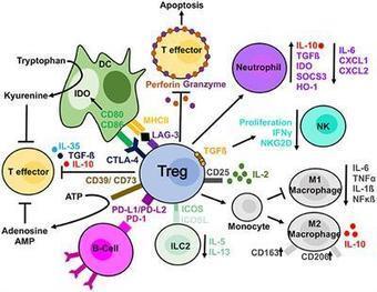
The inflammation is the protective response of the body against various harmful stimuli, however, the aberrant and inappropriate activation tends to become harmful. The acute inflammatory response tends to resolved once the offending agent is subside but this acute response becomes chronic in nature when the body is unable to successfully neutralized the noxious stimuli. This chronic inflammatory microenvironment is associated with the release of various pro-inflammatory and oncogenic mediators such as nitric oxide (NO), cytokines (IL-1β, IL-2, IL-6 and TNF-α), growth factor and chemokines. These mediators make the inflammatory microenvironment more vulnerable towards tumorigenesis. The pro-inflammatory mediators released during the chronic inflammation tends to induce several molecular signaling cascades such as NF-κB, MAPKinase, Nrf2, Pi3K, JAK/STAT, Wnt/B-catenin, and CREB. The immune system and its components have a pleiotropic effect on inflammation and cancer progression. Immune components such as T cells, NK cells, macrophages, and neutrophils either inhibit or enhance tumor initiation depending upon the type of tumor and immune cells involved. Tumor-associated macrophages (TAMs) and tumor-associated neutrophils (TANs) are pro-tumorigenic cells highly prevalent in inflammation-mediated tumors. Similarly, presence of T regulatory (Treg) cells in an inflammatory and tumor setting suppresses the immune system, thus paving the way for oncogenesis. However, Treg cell
|
|
Scooped by
Gilbert C FAURE
August 5, 2019 11:26 AM
|
T regulatory (Treg) cells were discovered more than 20 years ago and have remained a topic of intense investigation by immunologists. The initial doubts about their existence were dissipated by the discovery in 2003 of the lineage specific transcription factor Foxp3. In this article, I will discuss some of the questions that I believe still need to be answered before we will be able to fully apply Treg therapy to the clinic. The major issue that remains to be resolved is how they mediate their suppressive functions. In order to correct defective suppression in autoimmune disease (assuming it is a causative factor) or to augment suppression in graft versus host disease (GVHD) or during organ transplantation, we still need to fully understand the biochemical nature of suppressor mechanisms. Similarly, in cancer, it is now widely accepted that reversal of Treg suppression would be highly desirable, yet which of the many purported pathways of suppression are operative in different tumors in different anatomic sites. Many of the concepts we have developed are based on in vitro studies, and it remains unclear if these concepts can readily be applied to Treg function in vivo. Our lack of a specific cell surface marker that readily allows us to identify and target Treg in vivo, particularly in man, remains a major stumbling block. Lastly, I will review in some detail controversies regarding the origin of Treg, thymus versus periphery, and attempts to reverse Treg suppression b
|
|
Scooped by
Gilbert C FAURE
June 15, 2019 12:38 PM
|
Il1rl1 (also known as ST2) is a member of the IL-1 superfamily and its only known ligand is IL-33. ST2 exists in two forms as splice variants: a soluble form (sST2), which acts as a decoy receptor, sequesters free IL-33, and does not signal, and a membrane bound form (ST2), which activates the MyD88/NF-κB signaling pathway to enhance mast cell, Th2, regulatory T cell (Treg), and innate lymphoid cell type 2 (ILC2) functions. sST2 levels are increased in patients with active inflammatory bowel disease, acute cardiac and small bowel transplant allograft rejection, colon and gastric cancers, gut mucosal damage during viral infection, pulmonary disease, heart disease, and graft-versus-host disease (GVHD). Recently, sST2 has been shown to be secreted by intestinal pro-inflammatory T cells during gut inflammation; on the contrary, protective ST2 expressing Tregs are decreased, implicating that ST2/IL-33 signaling may play an important role in intestinal disease. This review will focus on what is known on its signaling during various inflammatory disease states and highlight potential avenues to intervene in ST2/IL-33 signaling as treatment options.

|
Suggested by
Société Francaise d'Immunologie
June 7, 2019 2:00 AM
|
Abstract The transcription factor Helios is expressed in a large percentage of Foxp3+ regulatory T (Treg) cells and is required for the maintenance of their suppressive phenotype, as mice with a selective deficiency of Helios in Treg cells spontaneously develop autoimmunity. However, mice with a deficiency of Helios in all T cells do not exhibit autoimmunity, despite the defect in the suppressor function of their Treg cell population, suggesting that Helios also functions in non-Treg cells. Although Helios is expressed in a small subset of CD4+Foxp3− and CD8+ T cells and its expression is upregulated upon T cell activation, its function in non-Treg cells remains unknown. To examine the function of Helios in CD4+Foxp3− T cells, we transferred Helios-sufficient or -deficient naive CD4+Foxp3− TCR transgenic T cells to normal recipients and examined their capacity to respond to their cognate Ag. Surprisingly, Helios-deficient CD4+ T cells expanded and differentiated into Th1 or Th2 cytokine-producing effectors in a manner similar to wild-type TCR transgenic CD4+ T cells. However, the primed Helios-deficient cells failed to expand upon secondary challenge with Ag. The tolerant state of the Helios-deficient memory T cells was not cell-intrinsic but was due to a small population of Helios-deficient naive T cells that had differentiated into Ag-specific peripheral Treg cells that suppressed the recall response in an Ag-specific manner. These findings demonstrate that Helios plays a role in the determination of CD4+ T cell fate.
|
|
Suggested by
Société Francaise d'Immunologie
February 17, 2019 1:39 PM
|
An inflammatory environment threatens the stability of Foxp3+ Treg cells. Garg et al. show that by expressing the transcriptional regulator Blimp1, Treg cells resist the IL-6-driven loss of Foxp3 in inflamed tissues.
|
|
Scooped by
Gilbert C FAURE
December 11, 2018 1:33 PM
|
Regulatory T cell localization to inflammatory sites is key to their homeostatic function.
Kishore and colleagues demonstrate that Treg cell migration requires the activation
of glycolysis by the enzyme glucokinase induced via a Treg cell-selective PI3K-mTORC2
pathway.
|
|
Scooped by
Gilbert C FAURE
October 28, 2018 3:48 PM
|
Regulatory T (Treg) cells prevent autoimmunity by limiting immune responses and inflammation in the secondary lymphoid organs and nonlymphoid tissues. While unique subsets of Treg cells have been described in some nonlymphoid tissues, their relationship to Treg cells in secondary lymphoid organs and circulation remains unclear. Furthermore, it is possible that Treg cells from similar tissue types share largely similar properties. We have identified a short-lived effector Treg cell subset that expresses the α2 integrin, CD49b, and exhibits a unique tissue distribution, being abundant in peripheral blood, vasculature, skin, and skin-draining lymph nodes, but uncommon in the intestines and in viscera-draining lymph nodes. CD49b+ Treg cells, which display superior functionality revealed by in vitro and in vivo assays, appear to develop after multiple rounds of cell division and TCR-dependent activation. Accordingly, single-cell RNA-seq analysis placed these cells at the apex of the Treg developmental trajectory. These results shed light on the identity and development of a functionally potent subset of mature effector Treg cells that recirculate through and survey peripheral tissues.

|
Suggested by
LIGHTING
August 4, 2018 11:46 AM
|
|
|
Scooped by
Gilbert C FAURE
July 13, 2018 4:44 AM
|
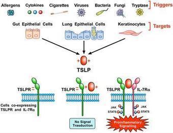
Thymic Stromal Lymphopoietin (TSLP) is a pleiotropic cytokine originally isolated from a murine thymic stromal cell line. TSLP exerts its biological effects by binding to a high affinity heteromeric complex composed of TSLPR chain and IL-7R. TSLP is primarily expressed by activated lung and intestinal epithelial cells, keratinocytes, and fibroblasts. However, dendritic cells, mast cells and presumably other immune cells can also produce TSLP. Different groups of investigators have demonstrated the existence of two variants for TSLP in human tissues: the main isoform expressed in steady state is the short form (sf TSLP), which plays a homeostatic role, whereas the long form (lfTSLP) is upregulated in inflammatory conditions. In addition, there is evidence that in pathological conditions TSLP can be cleaved by several endogenous proteases. Several cellular targets for TSLP have been identified, including immune (dendritic cells, ILC2, T and B cells, NKT and Treg cells, eosinophils, neutrophils, basophils, monocytes, mast cells, and macrophages) and non-immune cells (platelets and sensory neurons). TSLP has been originally implicated in a variety of allergic diseases (e.g., atopic dermatitis, bronchial asthma, eosinophilic esophagitis). Emerging evidence indicates that TSLP is also involved in chronic inflammatory (i.e., COPD, celiac disease) and autoimmune (e.g., psoriasis, rheumatoid arthritis) disorders and several cancers. These emerging observations greatly widen th
|
|
Scooped by
Gilbert C FAURE
June 15, 2018 8:23 AM
|
Scientists now report that Treg cells, a type of regulatory lymphocyte, may be protecting babies in the womb from getting infected with the HIV virus when the mother is infected.
|
|
Scooped by
Gilbert C FAURE
June 1, 2018 2:47 PM
|
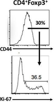
CD4+Foxp3+ Regulatory T-cells (Tregs) are a unique subset of helper T-cells, which regulate immune response and establish peripheral tolerance. Tregs not only maintain the tone and tenor of an immune response by dominant tolerance, but in recent years have also been identified as key players in resolving tissue inflammation and as mediators of tissue healing. Apart from being diverse in their origin (thymic and peripheral) and location (lymphoid and tissue resident), Tregs are also phenotypically heterogeneous as per the orientation of ongoing immune response. In this review, we discuss the recent advances in the field of Treg biology in general, and non-lymphoid and tissue resident Tregs in particular. We elaborate upon well-known visceral adipose tissue, colon, skin and tumor infiltrating Tregs and newly identified tissue Treg populations as in lungs, skeletal muscle, placenta and other tissues. Our attempt is to differentiate Tregs based on distinctive properties of their location, origin, ligand specificity, chemotaxis and specific suppressive mechanisms. Despite ever expanding roles in maintaining systemic homeostasis, Tregs are employed by large varieties of tumors to dampen anti-tumor immunity. Thus, a comprehensive understanding of Treg biology in the context of inflammation can be instrumental in effectively managing tissue transplantation, autoimmunity and anti-tumor immune responses.
|
|
Scooped by
Gilbert C FAURE
December 31, 2017 1:05 PM
|
Skin dendritic cells (DCs) are divided into several subsets with distinctive functions. This study shows a previously unappreciated role of dermal CD11b-type Langerin− DCs in maintaining immunological self-tolerance after UVB exposure. After UVB exposure, dermal CD11b-type Langerin− DCs upregulated surface CD86 expression, induced proliferation of Foxp3+ regulatory T (Treg) cells without exogenous Ags, and upregulated a set of genes associated with immunological tolerance. This Treg-expansion activity was significantly hampered by CD80/CD86 blockade in vivo. These results indicate that CD11b-type Langerin− DCs from the UVB-exposed skin are specialized to expand Treg cells in the skin, which suppress autoimmunity.
|
|
|
Scooped by
Gilbert C FAURE
August 14, 2019 8:29 AM
|
Abstract Regulatory T cells (Tregs) are specialized in immune suppression and play a dominant role in peripheral immune tolerance. Treg cell lineage development and function maintenance is determined by the Foxp3 transcriptional factor, whose activity is fine-tuned by its post-translational modifications (PTMs) and interaction partners. In this review, we summarize current studies in the crystal structures, the PTMs and interaction partners of Foxp3 protein, and discuss how these insights may provide a roadmap for new approaches to modulate Treg suppression, and new therapies to enhance immune tolerance in autoimmune diseases. This article is protected by copyright. All rights reserved.
|
|
Scooped by
Gilbert C FAURE
August 9, 2019 1:23 PM
|
Immunotherapy with immune checkpoint blockers (ICBs) has revolutionized cancer treatment. Unfortunately, ICB therapy usually benefits <15% of patients and causes immune-related adverse events in a substantial number of patients. Another immunotherapy—engineered chimeric antigen receptor (CAR) T cells that specifically target tumor-associated antigens—has transformed the treatment of multiple hematological cancers and exhibited therapeutic potential in solid tumors. Emerging data show that the function of blood vessels associated with tumors is critical in the response to these immunotherapies (1, 2). The vasculature in tumors is abnormal, which impairs blood flow and limits the delivery of oxygen, nutrients, and therapeutics, including antibodies and immune cells (3) (see the figure). The resulting hypoxia (low oxygen concentration) and low pH can in turn induce the production of immunosuppressive molecules, such as transforming growth factor–β (TGFβ), vascular endothelial growth factor (VEGF), and adenosine in the tumor microenvironment (TME). Some of these molecules lower the expression of adhesion molecules on tumor vascular endothelium, the lining of vessel walls that normally supports immune cell adhesion and transmigration into tissues. Reducing the expression of adhesion molecules therefore interferes with the ability of immune cells to adhere to and migrate across the vessel wall, preventing their entry into the tumor. In addition, pro-angiogenic molecules produced by cancer, stromal, and immune cells up-regulate Fas ligand on endothelial cells and thereby induce the apoptosis of infiltrating immune cells. VEGF can hinder maturation of dendritic cells (DCs), which are needed to present tumor-associated antigens and activate effector immune cells. Furthermore, hypoxia directly up-regulates the expression of immune checkpoint molecules [e.g., programmed cell death protein ligand 1 (PD-L1)], which suppress effector T cell activation, on various cells in the TME, including myeloid-derived suppressor cells (MDSCs) and DCs. Hypoxia also promotes the recruitment of immunosuppressive regulatory T lymphocytes (Treg cells) and reprograms tumor-associated macrophages (TAMs) from an antitumor to a protumor phenotype. Notably, the immunosuppressive molecules generated in the TME can enter the circulation and cause systemic immunosuppression. Given the consequences of malfunctioning blood vessels, multiple therapeutic strategies to improve the function of the tumor vasculature have been developed (4). Because excess VEGF contributes to vascular abnormalities, it was proposed that the judicious use of anti-VEGF agents could normalize tumor vessels and thereby improve oxygenation and drug delivery. This was supported by experiments showing that low-dose anti–VEGF receptor 2 (VEGFR2) treatment enhances trafficking of immune cells to tumors, reprograms TAMs to an antitumor phenotype, and improves outcomes of vaccine therapy in mouse breast cancer models (5). A number of preclinical studies have now shown the benefit of combining anti-VEGF agents with various immunotherapies, including ICBs and adoptively transferred engineered CAR T cells (3). Anti-VEGF–induced vascular normalization is transient, which limits efficacy. Thus, a number of strategies are emerging to extend the window of normalization. One such strategy is to target angiopoietin-2 (ANG-2), which begins to increase toward the end of the normalization window. Importantly, dual blockade of VEGF and ANG-2 prolonged the duration of normalization and survival in a number of mouse tumor models, compared with inhibiting either molecule alone. Relevant for immunotherapies, VEGF and ANG-2 dual targeting reprogrammed TAMs to an antitumor phenotype (6) and enhanced tumor response to ICBs (7). Furthermore, circulating ANG-2 concentrations are initially elevated or increase in melanoma patients with an unfavorable response to ICBs. Moreover, blood ANG-2 concentrations inversely correlate with response to ICBs with or without anti-VEGF therapy in melanoma patients (3), providing additional rationale for VEGF and ANG-2 dual inhibition. A number of other agents that target endothelial cells and/or pericytes, which support the structure and function of blood vessels, have been shown to transiently normalize tumor vessels. These targets include the receptor tyrosine kinase TIE-2, Notch signaling, endothelial cell metabolism, and oxygen sensors (4). Other strategies that can induce vessel normalization—either directly or indirectly—include restoring perivascular nitric oxide gradients, some chemotherapeutics such as eribulin, and aerobic exercise. Notably, two recent studies showed that ICBs can normalize tumor vessels in breast cancer models in mice (8, 9), which is intriguing because it implies that T cells can directly affect vascular function. These studies showed that the increased vessel perfusion by ICB is positively correlated with its efficacy (9). Future efforts will determine whether these agents can be combined with particular ICBs in specific cancer types. Physical forces generated by cells and the extracellular matrix (ECM) in tumors can also impair the function of blood and lymphatic vessels through compression (4). This is particularly the case in desmoplastic tumors, which are characterized by excessive fibrosis and activated stroma [e.g., pancreatic ductal adenocarcinoma (PDAC) and triple-negative breast cancer] and which generally have abundant carcinoma-associated fibroblasts (CAFs) and ECM. Killing cells with cytotoxic agents or depleting specific ECM components by using enzyme-based drugs can reopen these vessels. However, as cancer cells become resistant to cytotoxic agents and begin to proliferate, vessels collapse again. Similarly, enzymes that target a single ECM component (e.g., hyaluronan), become ineffective when other ECM components (e.g., collagen I) begin to contribute to compressive forces. Agents that reprogram CAFs so that they stop producing multiple ECM components can overcome this compression. For example, angiotensin system inhibitors (ASIs) are a class of molecules commonly used to treat hypertension that also reprogram CAFs and have been shown to improve the function of tumor vessels as well as the delivery and efficacy of various therapeutics (10). Moreover, these widely used drugs can activate both innate and adaptive antitumor immune responses in mouse models and patients (10). On the basis of a successful phase II clinical trial, this approach is now being tested in a randomized clinical trial in patients with locally advanced PDAC (NCT03563248). Because ASIs can cause hypotension, new formulations are needed to lower the systemic exposure of these drugs. Recently, valsartan—an ASI linked to pH-sensitive polymers—was shown to reprogram CAFs in the acidic tumor environment to alleviate antitumor immunosuppression and improve T lymphocyte activity without adverse hypotension. This approach improved the response to ICBs in mice bearing primary and metastatic breast tumors (2). Other strategies are also emerging for targeting CAF-mediated desmoplasia and vessel compression. For example, blocking C-X-C motif chemokine 12 (CXCL12)-C-X-C chemokine receptor 4 (CXCR4) signaling in mouse models of primary and metastatic breast cancer reduces fibrosis, vessel compression, and hypoxia while alleviating immunosuppression, which enhances the efficacy of ICBs (1). Similarly, the activity of tumor-associated immunosuppressive cells and the highly desmoplastic stroma in PDACs are fueled by cancer cell focal adhesion kinase (FAK) activity. Selective FAK inhibitors normalized the fibrotic TME, improved T cell infiltration of the tumor, and increased survival of tumor-bearing mice (11). Endothelial cells present in tertiary lymphoid organs (organized structures consisting of lymphocytes and stromal cells that form outside of the spleen or lymph nodes) within tumors can also regulate antitumor immunity. By combining VEGFR2 and PD-L1 antibodies, it is possible to induce the formation of specialized vessels called high endothelial venules (HEVs) in some tumor models, which improved survival (12). HEVs facilitate the entry of immune cells into lymph nodes and might serve a similar function in tumors. Moreover, depletion of Treg cells can induce the formation of HEVs outside of lymph nodes and the spleen, where they are normally found. When HEVs form in tumors, they can support lymphocyte infiltration and subsequent tumor cell killing by cytotoxic T lymphocytes (13). Lymphatic vessels are the routes from tissue to lymph nodes and back to the blood circulation. Therefore, lymphatic endothelial cells (LECs) are among the first cells to contact antigens that drain from tissue. LECs can control DC maturation and migration as well as directly present antigens on major histocompatibility complex class I (MHC I) and MHC II molecules to T cells (14). They also produce cytokines that modulate the immune response. Numerous lines of evidence demonstrate that LECs help limit and resolve effector T lymphocyte responses, modulate leukocyte function, and maintain peripheral tolerance, which prevents autoimmune responses (14). In cancer, LECs also suppress immune cell antitumor responses (14). Paradoxically, tumors that contain LECs (even in the absence of functional lymphatic vessels) show better responses to ICBs. Tumors with more intratumoral LECs have more T cells, but they become quiescent. These dysfunctional yet antigen-educated T cells can be reawakened by ICBs to produce antitumor immunity. Immune-related toxicities are a major problem with ICB and CAR–T cell immunotherapies. However, discontinuing or reducing the dose of these agents can reduce or even abrogate these toxicities. Improving the function of tumor blood vessels can improve the delivery of antibodies and cells and thus may lower the required immunotherapy dose. Such approaches hold promise for reducing toxicities while improving efficacy. However, one of the biggest challenges in realizing this goal is the lack of validated biomarkers for guiding the dose and schedule of drugs that directly or indirectly target blood vessels. This is further complicated by the recently described “paradoxical effects of obesity.” Obesity is known to fuel tumor progression, desmoplasia, hypoxia, immunosuppression, and resistance to various therapies, including chemotherapies and anti-VEGF therapies (3). Yet in mouse models of melanoma and lung cancer, and in various human cancers, obesity is associated with improved response to ICBs (15). Given the epidemic of obesity, a better understanding of the mechanisms underlying these paradoxical effects are likely to yield strategies to improve immunotherapy further. Combining strategies that improve function of tumor vessels with ICBs holds promise. For example, three recent phase III trials have shown the benefit of combining PD1 or PD-L1 antibodies with anti-VEGF or anti-VEGFR agents, leading to U.S. Food and Drug Administration approvals for the combination in lung and kidney cancers (NCT02684006, NCT02853331, and NCT02366143). However, the relative contribution to patient survival from immunomodulatory versus nonimmunomodulatory effects of VEGF blockade with ICBs in these trials is not known. The ongoing trial that aims to test the role of adding losartan—a safe, inexpensive, and widely prescribed antihypertensive ASI shown to decompress tumor blood vessels—to chemo-radiation and PD1 antibody therapy will reveal the potential of this approach in improving the treatment outcome in patients with PDAC (NCT03563248). http://www.sciencemag.org/about/science-licenses-journal-article-reuse This is an article distributed under the terms of the Science Journals Default License. References and Notes ↵ I. X. Chen et al., Proc. Natl. Acad. Sci. U.S.A. 116, 4558 (2019). ↵ V. P. Chauhan et al., Proc. Natl. Acad. Sci. U.S.A. 116, 10674 (2019). ↵ D. Fukumura et al., Nat. Rev. Clin. Oncol. 15, 325 (2018).OpenUrlCrossRefPubMed ↵ J. D. Martin et al., Annu. Rev. Physiol. 81, 505 (2019).OpenUrl ↵ Y. Huang et al., Proc. Natl. Acad. Sci. U.S.A. 109, 17561 (2012). ↵ J. Kloepper et al., Proc. Natl. Acad. Sci. U.S.A. 113, 4476 (2016). ↵ M. Schmittnaegel et al., Sci. Transl. Med. 9, eaak9670 (2017). ↵ L. Tian et al., Nature 544, 250 (2017).OpenUrlCrossRef ↵ X. Zheng et al., J. Clin. Invest. 128, 2104 (2018).OpenUrl ↵ M. Pinter, R. K. Jain, Sci. Transl. Med. 9, eaan5616 (2017).OpenUrl ↵ H. Jiang et al., Nat. Med. 22, 851 (2016).OpenUrlCrossRefPubMed ↵ E. Allen et al., Sci. Transl. Med. 9, eaak9679 (2017). ↵ C. F. Sautès-Fridman et al., Nat. Rev. Cancer 19, 307 (2019).OpenUrl ↵ K. Maisel et al., Adv. Drug Deliv. Rev. 114, 43 (2017).OpenUrl ↵ Z. Wang et al., Nat. Med. 25, 141 (2019).OpenUrl Acknowledgments: L.L.M. and R.K.J. thank Z. Amoozgar, D. G. Duda, D. Fukumura, Y. Huang, and T. Padera for helpful comments. R.K.J. received honorarium from Amgen; consultant fees from Chugai, Enlight, Merck, Ophthotech, Pfizer, SPARC, and SynDevRx; owns equity in Enlight, Ophthotech, and SynDevRx; and serves on the Boards of Trustees of Tekla Healthcare Investors, Tekla Life Sciences Investors, Tekla Healthcare Opportunities Fund, and Tekla World Healthcare Fund. The Massachusetts General Hospital has applied for patents related to concepts discussed in this article. L.L.M. owns equity in Bayer AG.
|
|
Suggested by
Société Francaise d'Immunologie
July 9, 2019 1:33 PM
|
KEY POINTS Skint8 shares sequence and structural similarity to existing B7 family members. Skint8-Ig fusion protein inhibits T cell functions in vitro. Skint8-Ig alleviates experimental autoimmune encephalomyelitis in mice. Abstract Immune responses are tightly controlled by T cell costimulatory and coinhibitory molecules. In this study, we identify Skint8 as a new member of the T cell coinhibitory group, whose extracellular domains share significant homology with existing B7 family members. Skint8 mRNA is expressed in resting and activated B cells, monocytes, and CD4 T cells. The Skint8 putative receptor is expressed on activated CD4 and CD8 T cells, B cells, monocytes and dendritic cells. Recombinant Skint8–IgG Fc fusion protein inhibits T cell proliferation, activation, and cytokine production in vitro. In vivo administration of Skint8–IgG Fc reduces T cell activation and alleviates experimental autoimmune encephalomyelitis in mice. The findings broaden our understanding of the regulation of immune responses and may have implications for treating immune-related diseases. Footnotes This work was supported by grants from the National Institutes of Health (1R01AI123131) and the Connecticut Regenerative Medicine Research Fund (16-RMB-UCONN-02). The online version of this article contains supplemental material. Abbreviations used in this article: BTN butyrophilin BTNL BTN-like DC dendritic cell EAE experimental autoimmune encephalomyelitis qRT-PCR quantitative RT-PCR Skint selection and upkeep of intraepithelial T cells Skint8-Ig Skint8–IgG Fc Treg regulatory T cell. Received May 3, 2018. Accepted May 17, 2019. Copyright © 2019 by The American Association of Immunologists, Inc.
|
|
Scooped by
Gilbert C FAURE
June 15, 2019 12:37 PM
|
Regulatory T cells (Tregs) are important for the induction and maintenance of peripheral tolerance therefore, they are key in preventing excessive immune responses and autoimmunity. In the last decades, several reports have been focussed on understanding the biology of Tregs and their mechanisms of action. Preclinical studies have demonstrated the ability of Tregs to delay/prevent graft rejection and to control autoimmune responses following adoptive transfer in vivo. Due to these promising results, Tregs have been extensively studied as a potential new tool for the prevention of graft rejection and/or the treatment of autoimmune diseases. Currently, solid organ transplantation remains the treatment of choice for end-stage organ failure. However, chronic rejection and the ensuing side effects of immunosuppressant represent the main limiting factors for organ acceptance and patient survival. Autoimmune disorders are chronic diseases caused by the breakdown of tolerance against self-antigens. This is triggered either by a numerical or functional Treg defect, or by the resistance of effector T cells to suppression. In this scenario, patients receiving high doses of immunosuppressant are left susceptible to life-threatening opportunistic infections and have increased risk of malignancies. In the last 10 years, a few phase I clinical trials aiming to investigate safety and feasibility of Treg-based therapy have been completed and published, while an increasing numbers o
|
|
Suggested by
Société Francaise d'Immunologie
April 26, 2019 2:50 PM
|
Summary In response to infection, naive CD4+ T‐cells proliferate and differentiate into several possible effector subsets, including conventional T helper effector cells (TH1, TH2, TH17), T regulatory cells (Treg) and T follicular helper cells (TFH). Once infection is cleared, a small population of long‐lived memory cells remains that mediate immune defenses against reinfection. Memory T lymphocytes have classically been categorized into central memory cell (TCM) and effector memory cell (TEM) subsets, both of which circulate between blood, secondary lymphoid organs and in some cases non‐lymphoid tissues. A third subset of memory cells, referred to as tissue‐resident memory cells (TRM), resides in tissues without recirculation, serving as ‘first line’ of defense at barrier sites, such as skin, lung and intestinal mucosa, and augmenting innate immunity in the earliest phases of reinfection and recruiting circulating CD4+ and CD8+ T‐cells. The presence of multiple CD4+ T helper subsets has complicated studies of CD4+ memory T‐cell differentiation, and the mediators required to support their function. In this review, we summarize recent investigations into the origins of CD4+ memory T‐cell populations and discuss studies addressing CD4+ TRM differentiation in barrier tissues. Introduction CD4+ T lymphocytes are a key element of adaptive immunity, acting to direct and enhance functions of innate cells, B‐cells and CD8+ T‐cells in response to diverse pathogens. Following infection, naive CD4+ T‐cells proliferate and have the potential to differentiate into at least seven functionally distinct T helper (TH) subsets with unique effector functions within the circulation, secondary lymphoid organs (SLO) and infected tissues.1 Depending on the type of immunological threat, early host–pathogen interactions result in an infection milieu that directs differentiation of naive CD4+ T‐cells to acquire the helper functions to ensure that each class of pathogen is countered with the appropriate immune response.2 Once infection has cleared, the majority of the effector cells die via apoptosis during a contraction phase, while a small proportion persists and differentiates into long‐lived memory cells. This memory population enables a rapid and robust secondary response against recurring pathogens and, thus, is pivotal in conferring lasting cellular immunity particularly against pathogens where neutralizing antibodies alone are insufficient at providing long‐term protection. While significant advances have been made in understanding the generation and maintenance of memory CD8+ T‐cells and B‐cells, the molecular mechanisms underlying the generation of memory CD4+ T‐cells remain relatively elusive. Two major obstacles have contributed to this knowledge deficit. First, CD4+ T‐cells are inherently less proliferative and the CD4+ memory T‐cell population appears to decline following antigen clearance, while the CD8+ memory T‐cell population, if established, is typically stable3-5 and thus, fewer cells are available for study. Second, the existence of functionally distinct effector TH cell subsets hinders the ability to characterize a common CD4+ memory T‐cell precursor. Further, TH effector and memory T‐cells also exhibit significant plasticity and can interconvert between lineages, both in vivo and in vitro, adding an additional layer of complexity to identifying memory precursor cells in CD4+ T memory studies.6-11 Memory T‐cells have been conventionally divided into central memory (TCM) cells, which circulate between the blood and SLO, and effector memory (TEM) cells, which can migrate from the blood into non‐lymphoid tissues 1, 11. Over the past decade, evidence of a novel subset of memory cells called tissue‐resident memory T‐cells (TRM) has emerged. TRM are seeded in non‐lymphoid tissues, particularly at barrier sites like the skin and intestinal mucosa.12-15 TRM have limited recirculation out of tissues and serve as sentinels at sites of potential reinfection, where they coordinate the initial response to pathogens and provide a substantial boost to tissue immunity via direct antigen recognition and recruitment of circulating immune cells.13, 16 The majority of studies on TRM have focused on CD8+ tissue‐resident lymphocyte differentiation, survival and function, while less is known about their CD4+ counterparts, which are also known to mediate antiviral responses.17, 18 The relationship between circulating and tissue‐resident CD4+ memory T‐cell origins and TH effectors subsets is currently unknown. A better understanding of the precursors of CD4+ TRM and the molecular mechanisms mediating their differentiation will allow us to harness the protective capacity of this memory population and modulate their activity in the context of infection or inflammatory diseases. Despite the challenges in studying memory CD4+ T‐cells, efforts in recent years focusing on different aspects of memory development have begun to elucidate a more comprehensive picture for the generation and maintenance of memory CD4+ T‐cells. In this review, we summarize recent studies addressing the identity of memory CD4+ T‐cell populations and their precursors in both the periphery and non‐lymphoid tissues. CD4+ T‐cell memory in secondary lymphoid organs Despite clear differences between memory CD4+ and CD8+ T‐cell populations, including the range of effector cell heterogeneity,3 the models for memory CD8+ T‐cell formation have served as a useful framework for investigation of memory CD4+ T‐cells. During the primary response of antigen‐specific cytotoxic CD8+ T‐cells (CTL), two effector CD8+ T‐cell populations can be identified based on surface expression of killer cell lectin‐like receptor subfamily G member 1 (KLRG1) and interleukin‐7 receptor‐α (CD127).19 The KLRG1hiCD127lo population, termed terminal effector cells (TE), is predominantly lost during the contraction phase, while the KLRG1loCD127hi subset contains memory‐precursor cells (MP), which can differentiate into long‐lived memory CD8+ T‐cells.19 CD4+ T‐cells also express KLRG120 and CD127.21 However, the roles of these molecules in memory CD4+ populations are not well established nor are clear strategies for distinguishing shorter‐lived effector and precursors of memory TH populations. Evidence for long‐lived CD4+ memory T‐cells capable of responding to pathogen re‐challenge has been documented in studies of adoptive transfer of T‐cell receptor (TCR) transgenic T‐cells22-25 and endogenous immune responses.23 However, the diversity of functional TH phenotypes has made identification of distinct CD4+ TE and MP effector populations challenging. Additionally, it is unclear whether all CD4+ TH effector T‐cells possess the same potential to differentiate into long‐lived memory cells. A separate MP may exist for each subset or there may be a unique effector subset with an inherent memory programme that can give rise to memory populations with the potential to generate TH subsets with all or some effector functions [TH1, TH2, TH17, T follicular helper cells (TFH), T regulatory cells (Treg)] in a secondary infection. An elegant study by Tubo et al.26 addressed this issue by following the differentiation of individual CD4+ T‐cells responding to infection. Utilizing over 80 distinct TCR clones that can specifically respond to Listeria monocytogenes (LM) infection, they demonstrated that all microbe‐specific naive CD4+ T‐cells have the potential to give rise to memory cells following acute infection.26 Different individual naive CD4+ T‐cells generated antigen‐specific effector populations with varying frequencies of TH1 and TFH effector cells. Notably, the relative frequencies of these subsets were preserved into the memory phase, suggesting that both TH1 and TFH effector populations contain precursors of memory cells that retain their effector TH characteristics (Fig. 1a). These data favour the idea that some CD4+ memory cells are relatively lineage‐committed; however, a range of expansion potential and plasticity among progeny was also observed, suggesting that not all CD4+ memory precursor cells may be equivalent. TH1 and TFH CD4+ memory T‐cells In efforts to address these questions, several groups have used lymphocytic choriomeningitis virus (LCMV) to characterize the response of adoptively transferred SMARTA (SM) cells, which have transgenic expression of an MHC Class II‐restricted T‐cell antigen receptor (TCR) specific for LCMV glycoprotein amino acids 66–77.24, 25, 27, 28 Meanwhile, other investigators have studied the endogenous polyclonal response by utilizing the peptide‐loaded major histocompatibility complex class II (pMHCII) tetramer‐based approach to identify antigen‐specific CD4+ T‐cells.22, 23, 25 During acute infection with LCMV‐Armstrong, antigen‐specific CD4+ T‐cells differentiate into two main helper subtypes in the spleen and lymph nodes: TH1 and TFH. TH1 cells express the transcriptional regulator T‐bet and are known for secreting their signature effector molecule, interferon gamma (IFNɣ), while TFH cells express Bcl6 and their hallmark surface molecule C‐X‐C chemokine receptor type 5 (CXCR5), which allows for homing to germinal centres to support B‐cell responses. To explore the origins of TH1 and TFH memory cells, investigators utilized fluorescence‐activated cell sorting to isolate TH1 and TFH effector and memory cells based on known markers, and studied their characteristics in the context of reinfection.21, 24-26 Marshall et al.24 found that within the primary effector populations from the spleen at day 8, two CD4+ T‐cell subsets that resembled the CD8+ TE and MP T‐cells were observed. The TE‐like population was marked by high expression of both P‐selectin glycoprotein ligand‐1 (PSGL‐1) and lymphocyte antigen 6 complex (Ly6C), while the MP‐like effector cells were PSGL‐1hiLy6Clo. In contrast to the PSGL‐1hiLy6Chi cells, the PSGL‐1hiLy6Clo MP‐like population exhibited greater longevity in uninfected hosts, increased proliferation following antigen re‐challenge, and similar gene‐expression profiles with day 60 PSGL‐1hi memory CD4+ T‐cells.24 These results led the authors to propose that differential expression of Ly6C can distinguish TE from MP cells within the TH1 subset. At day 8, PSGL‐1loLy6Clo effector cells showed high expression of known TFH markers (ICOS, CXCR5, PD‐1). This PSGL‐1loLy6Clo subset was found along with PSGL‐1hiLy6Chi and PSGL‐1hiLy6Clo TH1 cells within the memory cells at day 150 after infection, suggesting that MP of both TH1 and TFH phenotypes may persist long term.24 Interestingly, while the PSGL‐1hiLy6Clo MP population was thought to be primarily TH1 cells, it was later shown by Choi et al.25 that the PSGL‐1hiLy6Clo MP population actually contains both CXCR5− TH1 and CXCR5+ TFH cells at comparable frequencies. These results highlight the complexity and heterogeneity within CD4+ memory T‐cells and the need for further studies to fully understand the nature of the CD4+ memory T‐cell pool. To investigate the potential of TFH memory cells for re‐differentiation upon reinfection, Hale et al.27 utilized expression of CXCR5 and Ly6C to distinguish between TH1 (CXCR5−Ly6Chi) and TFH (CXCR5+Ly6Clo & CXCR5+Ly6Cint) memory populations following acute infection with LCMV‐Armstrong, then transferred each of the three subsets into naive hosts for reinfection. TH1 memory cells mostly maintained high Ly6C expression with few effector cells gaining CXCR5 expression, while TFH memory cells were able to give rise to both CXCR5−Ly6Chi TH1 cells and CXCR5+Ly6Clo/int TFH cells. This multi‐potency of TFH memory cells during re‐challenge has also been observed in acute bacterial infection with LM29 as well as in viral influenza infection.28 In a concurrent study, Pepper et al. addressed CD4+ memory T‐cell differentiation using LM infection and the expression of CXCR5 and CC chemokine receptor 7 (CCR7), a marker used in previous studies to identify TCM. During acute infection, antigen‐specific effector cells segregated into a CXCR5− population favouring the TH1 phenotype and a CXCR5+ population.30 A fraction of the CXCR5− TH1 population, which the authors termed TH1 effector memory cells, survived to a memory time point and, upon re‐challenge, produced TH1 effector cells. The CXCR5+ effector population included cells with high expression of the lineage‐defining factor Bcl6, were localized to follicles and were termed TFH, while cells with lower Bcl6 levels showed co‐expression of CCR7 and were termed TCM. It is worth noting that the TFH subset resembled what some studies term germinal centre TFH cells (GC TFH); GC TFH can lose expression of Bcl6 after infection, suggesting that, depending on the time point, the CXCR5+ population can include cells that did not enter the GC as well as those that were transiently in the GC. While both TFH and TCM in this study expressed CXCR5, TCM were not seen in the follicles and, upon re‐challenge, produced both TH1 effector cells and CXCR5+ cells that likely include TFH and GC TFH.30 Notably, Choi et al.25 found that precursors of TFH or the CXCR5+ populations show greater potential to develop into memory cells compared with TH1 precursors and share gene‐expression signatures with memory CD8+ T‐cells. These results suggest that both TH1 and TFH effector T‐cells can give rise to memory cells, and CXCR5+ TFH‐derived memory cells have greater plasticity in generating secondary effector phenotypes. Corroborative reports affirming the increased plasticity of TFH memory relative to TH1 memory upon re‐challenge suggest that TFH memory populations may retain a greater cellular ‘stem‐ness’ and are capable of providing a more comprehensive and robust secondary response during re‐infection. Two possible models can explain the multi‐potency demonstrated by CXCR5+ memory cells (Fig. 1b). One possible explanation is that TFH memory cells are inherently more plastic compared with other TH memory cells and, therefore, retain the ability to differentiate into alternative helper lineages upon reinfection. A second possibility is that the CXCR5+ memory population actually contains distinct subsets that are programmed or biased towards a specific TH lineage upon secondary challenge. In this case, CXCR5+CCR7+ could distinguish memory cells with the greater potential for re‐expansion, while CXCR5+CCR7− cells may be long‐lived TFH/GC TFH cells that have downregulated Bcl6 and PD‐1, and are more similar to long‐lived effector subsets. Based on the data currently available, neither hypothesis can be eliminated and further characterization of TFH memory cells, perhaps using single‐cell approaches, is needed to determine whether the multi‐potency of TFH memory is the result of cellular plasticity or population heterogeneity, or both. In line with this idea, a recent study by Ciucci et al.31 utilized single‐cell RNA sequencing to investigate the heterogeneity of antigen‐specific CD4+ effector T‐cells in response to acute LCMV infection. Visualization with t‐distributed stochastic neighbour embedding (t‐SNE) of day 7 effector T‐cells yielded multiple transcriptionally distinct clusters showcasing the heterogeneity exhibited by TH1 and TFH effector cells. At 30 days post‐infection, single cell analysis also showed multiple distinct transcriptional clusters with shared TFH features, supporting the idea that memory CXCR5+ TFH multi‐potency may be the result of population heterogeneity. TH2 CD4+ memory T‐cells TH2 memory cells have been best characterized in the context of allergic inflammatory disorders,32 though some studies have highlighted this population's role in defense against parasitic worm infection. As mentioned previously, antigen‐experienced CD4+ TH cells contract more rapidly after pathogen clearance compared with CD8+ T‐cells,4 which is why early investigations into TH2 memory relied on adoptive cell transfers of in vitro polarized TH2 effectors.33 This system involved activating CD4+ T‐cells in vitro with antigen and antigen‐presenting cells (APCs) followed by culturing in TH polarizing conditions33 and subsequent adoptive transfer. Interestingly, in vitro generated TH1 and TH2 cells retained their expression of lineage‐defining transcription factors (TFs), T‐bet and GATA3, respectively, for months after transfer into naive hosts.34 However, upon viral infection with LCMV, in vitro‐derived TH2 memory cells were able to adapt a TH1 phenotype and persist as a ‘hybrid’ memory cell with combined TH1 and TH2 characteristics.34 Utilizing a similar in vitro polarization system, Endo et al.35 identified an interleukin‐5 (IL‐5)‐producing subset of TH2 memory cells in the spleen that is primarily responsible for asthmatic symptoms, such as eosinophilic infiltration into the airway, airway hyper‐responsiveness and mucus hyper‐production in a murine model of TH2‐driven allergic airway inflammation. These studies provided early evidence of the potential existence of TH2 memory populations, but data demonstrating direct in vivo generation were lacking until recently. A study by Hondowicz et al.36 provided key insights into TH2 memory studying the endogenous allergen‐specific CD4+ T‐cells induced in response to house dust mite (HDM) inoculation. Using pMHCII tetramers to follow antigen‐specific CD4+ T‐cells, the authors showed an expansion of allergen‐specific CD4+ TH2 cells in SLOs and the lung following intranasal HDM administration. Notably, this allergen system induces both antigen‐specific TH2 and TFH cells, analogous to the TH1 and TFH response against LCMV‐Armstrong. The allergen‐specific memory pool in the SLOs consisted of CXCR5+ and CXCR5− cells that also expressed CCR7+, consistent with the earlier observations that memory T‐cells retain characteristics of TH effector phenotypes. The authors further explored characteristics of allergen‐specific TH2 resident in the lung, which will be discussed in the lung CD4+ TRM section below. TH17 CD4+ memory T‐cells Though not as extensively characterized as other helper subsets, memory TH17 cells have been documented in both humans and mice, primarily in the context of autoimmunity.37 Early memory experiments using LM infection showed that TH17 cells existed only transiently following intranasal infection.23 However, it is worth noting that LM may not be an optimal infection for TH17 studies as it is an intracellular pathogen38 and most efficiently induces TH1 cells. Muranski et al.39 reported on long‐lived memory TH17 cells but, similar to early TH2 studies, these cells required in vitro polarization prior to transfer into host mice. In a recent study of dry eye disease, Chen et al.40 utilized a pre‐clinical murine model of autoimmune ocular disease, where mice were subjected to 14 days of environmental desiccating stress followed by rest in normal conditions for 14 days, and found disease‐specific pathogenic memory TH17 cells in both the inflamed site and draining lymph nodes. Two cytokines associated with CD4+ memory T lymphocytes, IL‐7 and IL‐1541 were shown to be crucial in the maintenance of these pathogenic TH17 cells. Neutralization of these cytokines with topical application of anti‐IL‐7 or anti‐IL‐15 antibody decreased the number of TH17 cells in both the conjunctivae and lymph nodes, offering a potential therapy for autoimmune disorders. One crucial caveat to note is that these ‘memory’ TH17 cells were studied under the chronic inflammatory environment of autoimmunity, perhaps under prolonged or recurrent exposure to antigen; therefore, this population's identity as true resting memory cells remains uncertain. Uncovering the origin and identity of resting memory or MP cells within a particular helper T‐cell lineage will lay the foundation for future molecular studies into how each memory TH lineage is uniquely regulated. However, in the next section, we will review two biological requirements crucial for memory formation that appear to be conserved across all TH subsets. Memory differentiation cues: TCR signalling and IL‐2 A recent comprehensive review of studies aimed at resolving the signals required for CD4+ memory T‐cell formation42 discussed the instructive signals both during the ‘early priming’ phase of initial antigen recognition and activation as well as at ‘late‐acting checkpoints’ prior to contraction that play a role in the effector‐to‐memory transition. Much like the signals important for CD8+ memory T‐cell generation, strengths of TCR and co‐stimulatory signalling also have profound effects on memory TH development.42, 43 Recent results from Snook et al.44 demonstrated that TCR signalling has a direct impact on TH memory formation. Utilizing a panel of TCRs specific for the same viral antigen, the authors showed substantial variability in TCR signal strength, expression of IL‐2‐receptor alpha (CD25) and activation of downstream TFs across the CD4+ memory T‐cell population.44 TCR clones with stronger TCR signalling appear to differentiate towards a more TE state and become largely depleted by memory time points, while clones with comparatively lower signalling were memory‐like and able to persist after antigen clearance. Interestingly, it seems that stronger TCR signalling was associated with higher expression of TH1 markers, while weaker TCR signals correlated with higher expression of TFH markers,44 suggesting that there may be a connection between lineage differentiation and memory potential for CD4+ helper T‐cells. Utilizing influenza A virus (IAV) as an infection model, McKinstry et al.45 showed that IL‐2 is crucial at a late checkpoint for effector helper T‐cells to survive the contraction phase, allowing for the transition into resting memory cells. To circumvent defects in initial T‐cell priming caused by IL‐2 deficiency, the authors first activated CD4+ T‐cells in vitro with exogenous IL‐2 and then transferred these cells into naive mice for infection. Following IAV challenge, both in vitro primed wild‐type and IL‐2‐deficient donors showed similar cell numbers at the peak of infection and production of IFNɣ; however, the IL‐2‐deficient population quickly declined and was undetectable by day 28 of infection. Exogenous administration of IL‐2 during days 5–7 of infection successfully restored memory cell numbers for IL‐2‐deficient CD4+ T‐cells, demonstrating the importance of IL‐2 for CD4+ memory T‐cell generation in this context. Furthermore, a recent study by DiToro et al.46 with LM infection showed that as early as 20 hr after antigen exposure in vivo, IL‐2 production in CD4+ TH effector cells strongly correlated with TH fate differentiation during infection, again supporting a link between lineage specification and memory formation. To further highlight the importance of IL‐2 in TH memory, Shakya et al.47 identified a role for TF Oct1 and its coactivator OCA‐B in poising the Il2 locus for robust expression in memory CD4+ T‐cells, unveiling an important mechanism by which memory CD4+ T‐cells control IL‐2 production. However, these studies regarding TCR signalling and IL‐2 in CD4+ memory T‐cells were done without investigation of specific TH lineages. Therefore, further investigations into the required transcriptional and epigenetic regulation for generation and maintenance of memory TH subsets are needed. Additionally, while targeting peripheral memory cells in vaccination strategies can provide systemic protection, in some cases a localized strategy in which tissue‐resident cells at barrier surfaces are activated as front‐line defense against recurrent infections may be more effective, thus how these signals pertain to TRM will be informative.48 Tissue‐resident CD4+ memory T‐cells Much like circulating CD4+ memory T‐cells, studies of tissue‐resident lymphocytes have predominantly focused on CD8+ TRM due to the heterogeneity of CD4+ memory T‐cells and the existing gaps in knowledge regarding mechanisms governing memory CD4+ T‐cell formation. Nevertheless, recent studies have highlighted a prominent population of long‐lived CD4+ T‐cells within many non‐lymphoid tissues, including the lungs,36, 49-57 small intestine (SI),12, 18, 58-60 skin,15, 61-64 and female reproductive tract (FRT)18, 65, 66 (Fig. 2). Similar to their CD8+ T‐cell counterparts, CD4+ TRM have been shown to facilitate rapid immune defense upon re‐exposure to antigen and can supplant innate immunity in recognizing and responding to recurrent infections.67 However, much remains to be explored about the phenotype, function and maintenance of CD4+ TRM during infections. Additionally, differences exist between CD4+ and CD8+ TRM in tissue localization, surface marker expression and cytokine cues driving TRM formation; these outstanding questions in the field must be addressed to better define the identity and differentiation of CD4+ TRM. Classically, tissue‐resident memory T lymphocytes have been defined using parabiosis experiments in which a naive mouse and an immune mouse, previously exposed to antigen, are surgically joined to create a shared circulatory system.56, 68 Thus, all circulating cells will normalize between both partners while the non‐circulating tissue‐resident cells remain lodged in the tissues of the immune mouse. Alternative methods have been developed and validated to assess whether cells remain in tissues, including intravenous injection of a fluorescently labelled antibody to mark cells in the circulating system.69 Any cells positive for the label are considered ‘circulating’, while unlabelled cells are assumed to have limited access to circulation and are therefore ‘tissue‐resident’.56 To determine the protective functions of tissue‐resident lymphocytes in secondary infection, immune mice are treated with FTY‐720, an agonist of sphingosine‐1‐phosphate receptor 1 (S1PR1), which causes decreased surface expression of S1PR1 and therefore prevents egress of circulating memory cells from lymph nodes.70 When these mice are re‐challenged with the original pathogen, any immune response at the local site of infection will be mediated only by cells resident to that tissue.71 Lung CD4+ TRM cells The lungs contain a population of CD4+ TRM that have been shown to play a critical role in recruiting CD8+ T‐cells and enhancing secondary immune responses against bacterial, viral and worm infections.51, 55, 56, 72 In an influenza infection model, antigen‐specific memory CD4+ T‐cells migrate to the lungs and are retained in the tissue without recirculation, as demonstrated by parabiosis experiments.56 This subset of CD4+ T‐cells shows a distinct phenotype from circulating populations; specifically, high expression of CD69, a membrane‐bound type II C‐lectin receptor and known marker of tissue retention.73, 74 Functionally, these lung‐resident CD4+ T‐cells provide protection from influenza virus when transferred to naive mice. Similar to its role in directing long‐term memory fate as discussed above, IL‐2 also supports the formation of antigen‐specific lung CD4+ TRM with a transcriptional signature distinct from that of circulating CD4+ T‐cell populations but similar to that of CD8+ TRM.51, 75 Interestingly, Strutt et al.51 also identified an IL‐2‐independent population of influenza‐specific lung CD4+ TRM following infection, suggesting that IL‐2 may not be the only cytokine regulating lung TRM development and maintenance. In fact, IL‐15 was shown to be essential for these IL‐2‐independent cells, acting as an ‘alarm’ at local sites of infection to promote both CD8+ T‐cell responses and induce long‐lived CD4+ TRM.42, 51 Using fluorescently labelled antibody injection, Hondowicz et al.49 showed that LCMV‐specific CD4+ T‐cells migrated to the lungs as a Tbethi TH1 subset with CD69 expression similar to influenza experiments. Again, the establishment of LCMV‐specific lung TH1 TRM cells required IL‐2 signalling; however, for long‐term maintenance and survival, additional interactions with B‐cells in tissues were necessary.49 Several groups have shown that allergens in an asthma model also elicit CD4+ TRM responses in the lung. After exposure to HDM, allergen‐specific CD4+ TRM in the lungs were shown to be resident by parabiosis experiments.36 These cells expressed high levels of CD69 and required IL‐2 signalling for their migration to the lungs, similar to the studies above with viral pathogens. Interestingly, the TF Bcl6, the fate‐determining factor for TFH cells, prevented HDM‐specific CD4+ TRM from entering the lungs, and loss of Bcl6 actually increased the TRM population in the tissue. These results suggest a possible antagonistic relationship between TRM and memory TFH cells, and highlight a role for TFs in directing the development of tissue‐resident populations. In another study using the HDM model, lung CD4+ TRM clustered around the airways at rest and rapidly reactivated upon secondary exposure within their clusters to produce IL‐4, IL‐5 and IL‐17 and recruit dendritic cells.50 Additional studies highlight a role for IL‐7, along with IL‐2, in the maintenance of allergen‐specific CD4+ TRM cells in the lung parenchyma and airways. However, these IL‐7‐dependent CD4+ TRM did not express CD69,52 suggesting either that CD69 may not be a conclusive marker for CD4+ TRM or that CD4+ TRM are highly heterogeneous, and further studies must focus on elucidating the different subsets. Additionally, in both viral infection and allergen‐induced asthma models, it remains unclear whether the same cells can become resident in both the parenchyma and airways, and if similar survival signals sustain such subsets. Skin CD4+ TRM cells A second well‐studied tissue for CD4+ TRM cells is the skin particularly in a herpes simplex virus (HSV) infection model. Initial studies by Gebhardt et al.15 using intravital microscopy demonstrated that HSV‐specific gDT‐II CD4+ T‐cells showed a migration pattern distinct from that of CD8+ T‐cells in the skin following infection, homing to the dermis as opposed to the preference of CD8+ T‐cells for the epidermis. Additionally, dermal TH cells were significantly more motile and had lower expression of CD103 compared with CD8+ T‐cells. Thus, it appears that CD4+ TRM can migrate between the skin and circulation much more easily than CD8+ TRM and may not be a fully resident population. In another study of skin HSV response, Collins et al. examined skin from naive mice and found resting CD4+ T‐cells preferentially clustered around hair follicles. Upon infection, circulating CD4+ T‐cells were rapidly recruited to the skin, specifically to the perifollicular regions; the majority of recruited cells were lost by day 30, but a small percentage survived in tissue long after initial antigen exposure.64 These skin CD4+ TRM, as demonstrated by parabiosis experiments, also aggregated in APC‐associated clusters, consistent with experiments done in the FRT,66, 76 as discussed below. Additional studies of skin CD4+ TRM following Leishmania major (L. major) infection showed evidence of a L. major‐specific resident population in the dermis with increased expression of IFN genes and chemokine signalling pathways.61 These CD4+ TRM cells were able to control parasite growth following a secondary challenge, presumably through the aforementioned IFN response and recruitment of circulating immune cells. Through FTY‐720 treatment experiments, resident CD4+ T‐cells were found to be the main contributors to protection against L. major, recruiting inflammatory monocytes for production of reactive oxygen species to kill the parasites.62 These results highlight another function of CD4+ TRM in enhancing secondary responses and protection against recurring pathogens. CD4+TRM cells in other mucosal tissues Through parabiosis experiments, CD4+ TRM have been shown to be active in the FRT in response to HSV‐2 infection.66, 77 Intravaginal HSV‐specific CD69+ CD4+ TRM localized to memory lymphocyte clusters containing CD8+ T‐cells, macrophages and other APCs, and were maintained in these structures by chemokines secreted by macrophages. Upon a secondary re‐challenge with lethal HSV‐2, resident TH cells alone were sufficient for protection, largely mediated by CD4+ TRM production of IFNγ to inhibit viral replication. In this context, FRT‐resident CD4+ T‐cells were more directly involved in clearing the infection as opposed to their classic role in alarming and recruiting circulating populations. The SI contains a potential subset of tissue‐resident CD4+ T‐cells, though no parabiosis experiment has been performed to definitively characterize this population as resident. In general, intestinal helper T‐cells can be found as both intraepithelial lymphocytes (IELs) and lamina propria lymphocytes (LPLs) with a greater proportion of cells in the LP. The majority of studies of gut‐resident CD4+ T‐cell subsets have focused on endogenous polyclonal IELs that originate from the circulating population.59, 78, 79 Upon entering the SI, these CD4+ IELs downregulate the TF ThPOK responsible for thymic CD4+ T‐cell fate and upregulate Runx3, the fate‐determining factor for differentiating CD8+ thymocytes. This switch in programming towards a more cytolytic function appears to be important in the CD4+ T‐cell response to endogenous gut microbiota.78 Nevertheless, this study characterizes polyclonal CD4+ T‐cells with variable TCR specificity and affinity, and further investigations are necessary to determine whether similar transcriptional changes are seen in antigen‐specific responses. In an LM infection, antigen‐specific CD4+ T‐cells preferentially migrated into the LP (though a small population was seen in the IEL) to form a long‐lived antigen‐specific memory population that was predominantly TH1, expressed high levels of CD69 and, unlike lung‐resident CD4+ T‐cells, was independent of IL‐15 signalling.60 Interestingly, this gut ‘TRM’ population had low Ly6C expression relative to other lymphocytes in circulation, similar to the MP population of circulating TH1 cells discussed in the TH1 and TFH CD4+ memory T‐cells above. This suggests that circulating and resident memory cells may share a common precursor. As discussed in the studies above, CD4+ TRM differ greatly between infection models and tissues, although some general principles can be drawn (Fig. 2). As is the case with CD8+ TRM, the majority of CD4+ TRM express high levels of CD69, but CD103 expression varies within and between tissues.15, 56, 60, 66 Across multiple tissues – lung, skin, FRT – CD4+ TRM form clusters with other resident immune subsets including CD8+ T‐cells, macrophages and APCs.52, 64, 66 These clusters position CD4+ TRM in close proximity to the cells they need to activate in case of a recurrent infection. This may be an optimized way for helper TRM cells to perform their ‘sense‐and‐alarm’ function,68 initiating the innate response to recruit circulating cells and activating CD8+ T‐cells to fight off pathogen. Additionally, cytokines appear to be critical in the recruitment, formation and maintenance of CD4+ TRM in many tissues.50-52, 60 However, which cytokines are important for which tissues remain unknown, although IL‐2 may play a key role for all TRM given its known effects in circulating memory populations.80 It is also unclear which CD4+ TH effector subset, if any, gives rise to TRM, or whether the tissue‐resident precursor is distinct from the peripheral population completely, residing in the tissue until the right signals are detected to induce differentiation. Conclusion The diversity and plasticity of effector CD4+ T‐cells create a heterogeneous memory pool, making the study of helper T‐cell memory differentiation complex. While there are some promising markers to differentiate between memory TH1 and TFH memory subsets,24, 27 it is still unclear whether both helper memory populations originate from their respective effector cells or whether the ‘stem‐like’ properties of TFH cells make them the primary precursor.25 Likewise, in other infection systems that elicit TH2 or TH17 effector cells, we do not know how these effector populations contribute to the final pool of memory cells. Adding further to the complexity, CD4+ TRM cells are highly variable across tissues, and different cues drive their establishment and retention. It is unclear whether the precursor for CD4+ TRM cells originates from the same memory‐precursor population that yields circulating memory cells or if it is found even earlier, before the effector versus memory decision. TFH cells may also contribute to the CD4+ TRM pool, although currently no evidence exists for a TFH‐like subset in tissues. Lastly, it is clear that surface markers do not accurately identify subpopulations in effector and memory pools, and further work requires examining TFs and regulators that may direct the memory programme. One possible approach to parse the heterogeneity of CD4+ memory T‐cells is through bulk and single‐cell epigenetic and transcriptional profiling of cells in the circulation and tissues over the course of an infection to identify whether an early memory precursor exists or whether memory potential is programmed as effector cells contract and die.75, 81, 82 Paired with adoptive transfers of putative subsets, it will be possible to identify key factors at each step in the effector‐to‐memory transition, and in the formation and survival of the tissue‐resident subset. Acknowledgements This review was supported by the American Heart Association Predoctoral Fellowship to QPN, and US National Institutes of Health R01 AI072117 and U19 AI109976 to AWG. The authors thank Drs Kyla D. Omilusik and J. Ty Crowl for critical review of the manuscript. Disclosures None declared. References
|
|
Scooped by
Gilbert C FAURE
January 8, 2019 5:18 AM
|
Regulatory T cells (Tregs) can impair anti-tumor immune responses and are associated with poor prognosis in multiple cancer types. Tregs in human tumors span diverse transcriptional states distinct from those of peripheral Tregs, but their contribution to tumor development remains unknown. Here, we used single cell RNA-Seq to longitudinally profile conventional CD4+ T cells (Tconv) and Tregs in a genetic mouse model of lung adenocarcinoma. Tissue-infiltrating and peripheral CD4+ T cells differed, highlighting divergent pathways of activation during tumorigenesis. Longitudinal shifts in Treg heterogeneity suggested increased terminal differentiation and stabilization of an effector phenotype over time. In particular, effector Tregs had enhanced expression of the interleukin 33 receptor ST2. Treg-specific deletion of ST2 reduced effector Tregs, increased infiltration of CD8+ T cells into tumors, and decreased tumor burden. Our study shows that ST2 plays a critical role in Treg-mediated immunosuppression in cancer, highlighting new potential paths for therapeutic intervention.
Visualizing TGF-β1 regulation by GARP
Regulatory T cells (Tregs) can suppress immune responses through a variety of mechanisms. One such mechanism involves the activation of a surface-bound latent form of the cytokine transforming growth factor–β1 (TGF-β1). Within the cell, newly synthesized pro-TGF-β1 homodimers form disulfide bonds with the transmembrane protein GARP, which acts to chaperone and orient the cytokine for activation at the cell surface. Liénart et al. reveal how GARP interacts with TGF-β1, using a crystal structure in which the complex was stabilized using a Fab fragment from a monoclonal antibody (MHG-8) that binds to the complex. In so doing, they also demonstrate how MHG-8 prevents membrane-associated TGF-β1 release. These structural and mechanistic insights may inform treatments of diseases with altered TGF-β1 functionality and dysfunctional Treg activity, including cancer immunotherapy.
Via Krishan Maggon
|
|
Scooped by
Gilbert C FAURE
September 13, 2018 3:06 AM
|
IV. THE ORIGIN OF DARWINIAN IMMUNITY IN VERTEBRATES Above, we argued that the origin of Darwinian immunity constitutes a major transition in evolution. We now speculate on how it might have happened in the lineage of vertebrates. We propose that the transition occurred only once, before the split between jawed and jawless vertebrates, and explain why we believe that the transition was limited by a difficult evolutionary innovation, rather than the presence or absence of selection pressure for Darwinian immunity. We offer a hypothesis on the nature of the limiting innovation, and outline possible routes of stepwise evolution once the bottleneck had been passed. (1) A single origin There are good reasons to believe that the Darwinian immune systems of jawless and jawed vertebrates can be traced back to a common root, and thus that the major transition occurred only once, in a common ancestor of the two lineages. Lampreys have three distinct classes of lymphocytes that provide cellular and humoral immunity, resembling both major lineages of T cells and B cells of jawed vertebrates, respectively (Guo et al., 2009; Hirano et al., 2013). The similarities between not only functions, but also gene expression profiles suggest that the three kinds of lymphocytes are homologous between the two groups and pre‐date the divergence of jawed and jawless vertebrates (Flajnik, 2014; Kasahara & Sutoh, 2014). In addition, jawless fish have thymus‐like lympho‐epithelial structures (‘thymoids’) that are thought to serve as the sites of lymphocyte development (Bajoghli et al., 2011), and express the lamprey orthologue of the gene encoding forkhead box N1 (Foxn1) transcription factor, a marker of the thymopoietic microenvironment in jawed vertebrates. Finally, receptor diversity in jawless fish is generated by the action of enzymes that are closely related to the gnathostome activation‐induced cytosine deaminase (AID) (Rogozin et al., 2007), which is active in the diversification of B‐cell receptors. The apparent homology of multiple components of clonal selection‐based immunity between jawed and jawless vertebrates strongly suggests that the roots of the system originated in the common ancestor of all vertebrates. (2) Chance or necessity The apparently unique origin of Darwinian immunity can be explained in two possible ways. Either, the transition involved a difficult (i.e. low‐probability) event that occurred only once, ‘by chance’, in a common ancestor of all vertebrates [classifying this transition as ‘variation‐limited’ (Számadó & Szathmáry, 2006)]; or, the selective forces that favour the emergence of Darwinian immunity appeared first (and only) in vertebrates and have then driven, ‘by necessity’, the stepwise evolution of the system [in the frame of a ‘selection‐limited’ transition (Számadó & Szathmáry, 2006)]. Historically, the discovery of the intricate molecular mechanisms of V(D)J recombination (the only mechanism of somatic receptor diversity then known) led researchers to favour the first alternative, assuming a once‐only low‐probability event for the origin of this system. Marchalonis & Schluter (1990, p. 16) termed this event ‘a “Big Bang” because sophisticated rearranging systems consisting of multiple elements appear in a fully functional form without foreshadowing in the antecedent species’. This notion was further strengthened by the recognition that the molecular machinery of V(D)J recombination likely arose by the integration of recombination‐activating genes (RAGs) into the vertebrate genome by horizontal gene transfer (Bernstein et al., 1996; Fugmann, 2010). However, subsequent discoveries have challenged the key role of V(D)J recombination in the origin of Darwinian immunity. First, the discovery of RAG1/2 in sea urchins (Fugmann et al., 2006) suggested that the original horizontal gene transfer event must have preceded the origin of vertebrates. Second, we now know that RAG‐mediated V(D)J recombination [enhanced with non‐templated nucleotide addition diversity (Kallenbach et al., 1992)] is far from being the only mechanism that can generate somatic receptor diversity. Jawless fish generate receptor diversity by RAG‐independent gene conversion (Nagawa et al., 2007), subsets of immunoglobulin (Ig) genes in some jawed vertebrates (sharks, birds, rabbits, sheep) rely heavily on gene conversion and hypermutation to generate antibody diversity (Flajnik & Kasahara, 2010), and invertebrate systems of shotgun immunity generate somatic diversity by gene conversion, alternative splicing, RNA editing, post‐translational modifications, and possibly even somatic recombination (Ghosh et al., 2011). Mechanisms of somatic diversity have thus evolved multiple times independently, and are unlikely to be a limiting ‘bottleneck’ in the evolution of Darwinian immunity. Although it cannot be ruled out that the evolution of V(D)J recombination in particular might have been triggered by a second (intragenomic) transposition event that inserted the RAG transposon into a variable innate immune receptor gene (Koonin & Krupovic, 2015), this can no longer be regarded as ‘the Big Bang’ of adaptive immunity, but rather as one of several ‘smaller bangs’ (Bartl et al., 2003; Flajnik, 2014). Kasahara (1997, 1998) argued that the triggering event of the ‘Big Bang’ might have been the one or two rounds of whole‐genome duplication (WGD) that occurred close to the origin of vertebrates (Smith et al., 2013; Smith & Keinath, 2015). This event duplicated many genes related to immunity, and it ‘might have provided unique opportunities to create many accessory and effector molecules of the adaptive immune system’ (Kasahara et al., 1997, p. 92). We will return to this idea in Section IV.4, proposing possible scenarios as to how the WGD event might have triggered the origin of Darwinian immunity. In turn, several studies have argued against the key role of a single triggering event. Klein & Nikolaidis (2005, p. 174) [along the lines of an earlier argument by Bartl et al. (2003)] favour gradual evolution that ‘consisted initially of changes unrelated to immune response that were selected to serve other functions’ and that, by chance, attained a combination that integrated the elements into a new function giving rise to adaptive immunity. Litman, Rast & Fugmann (2010) also emphasized co‐option and redirection of pre‐existing systems as the main source of innovation, at the same time perceiving ‘no reason to assume that vertebrates require a complex immune system any more than do complex invertebrates’ (Litman et al., 2010, p. 552). However, if the origin of Darwinian immunity is not dependent on a ‘difficult’ (i.e. low‐probability) transition, then vertebrates must have some specific traits that favour Darwinian immunity in this group, but are absent from others. (3) Selective scenarios: not exclusive to vertebrates Long lifespan (Klein, 1989; Lee, 2006) and slow reproduction (Flajnik, 1998; Lee, 2006; Flajnik & Kasahara, 2010), high metabolic intensity (Rolff, 2007; Sandmeier & Tracy, 2014), efficient closed circulation (van Niekerk, Davis & Engelbrecht, 2015), low population density (Klein, 1989) and large (Klein, 1989; Flajnik & Kasahara, 2010) or morphologically complex (Boehm, 2012) bodies have been invoked as factors favouring (Darwinian) adaptive immunity. However, these traits are not exclusive to vertebrates, and, in fact, the last common chordate ancestor (and therefore also the ancestral vertebrate) was probably a lancelet‐(amphioxus‐)like creature (Lowe et al., 2015): small, not particularly long‐lived, and rather inconspicuous. The most extensive phylogenetic analysis so far estimated that the lineages of jawed vertebrates and jawless fish diverged about 650 million years ago (Blair & Hedges, 2005). While molecular clock estimates might be sensitive to assumptions on the tempo and mode of evolution, fossil evidence of two distinct types of jawless fish dated to around 520 million years ago (Shu et al., 1999) confirms that the split must have occurred before or shortly after the Cambrian Explosion: Darwinian immunity must therefore have provided a selective advantage already in the Precambrian or early Cambrian world of small body sizes and simple body plans. Many extant invertebrates very likely surpass the last common ancestor of vertebrates in both size and life expectancy, and yet (to our current knowledge) lack Darwinian immunity. Cephalopods can have large bodies and long lifespan, but Darwinian immunity (clonal selection acting on heritable somatic receptor diversity) has not been found in the group (Castellanos‐Martínez & Gestal, 2013). It must nonetheless be noted that the species investigated so far have been octopuses that have short lifespans; studies of immunity in Nautilus species that can live for several decades (Saunders, 1984) are much awaited. Some further ancestral traits of vertebrates might also have facilitated or favoured the evolution of Darwinian immunity. A closed circulatory system, which seems to be an ancestral chordate character (Stach, 2008), may well be a prerequisite of effective immune surveillance by lymphocytes; however, cephalopods also have a closed circulation. Filter feeding seems to be an ancestral trait for deuterostomes (Gans & Northcutt, 1983; Yu & Holland, 2009; Lowe et al., 2015), and is present in echinoderms (sea urchins, sea cucumbers), tunicates (sea squirts), and also cephalochordates (amphioxus), which are thought to most closely resemble the common ancestor of vertebrates (Gans & Northcutt, 1983; Yu & Holland, 2009). The evolution of this lifestyle probably generated selection pressure for improved immunity (to fight pathogens, and to avoid unnecessary or harmful responses to the myriad harmless microorganisms in the filtrate). Echinoderms (Hibino et al., 2006), amphioxus (Huang et al., 2008) and, independently, also mussels (Gerdol & Venier, 2015) and sponges (Degnan, 2015), the most ancient group of filter‐feeding organisms, took the path of expanding their repertoire of innate pattern‐recognition receptors. While expanded innate receptors indeed imply selection pressure for improved immunity, the defining traits of Darwinian immunity have not been found in any of these groups to date. It has also been noted that vertebrates harbour more complex microbiomes than invertebrates, which tend to have either relatively simple microbial communities or rely on microbial partners that are shielded from immunity within the cells or in compartments enclosed in physical barriers (McFall‐Ngai, 2007). Managing a complex microbiome has been invoked as a selection pressure that may have driven the evolution of Darwinian immunity specifically in vertebrates (Pancer & Cooper, 2006; Weaver & Hatton, 2009; Lee & Mazmanian, 2010; Boehm, 2012). However, this explanation only leads one step back, to another question: why would vertebrates be special in terms of needing a complex microbiome? We find it more plausible that Darwinian immunity evolved for another reason (a rare event that opened up a difficult evolutionary path), and could then enable the acquisition of a more complex microbiome – which then might have provided an evolutionary edge to vertebrates. A further hypothesis has been proposed by Pancer & Cooper (2006, p. 512), who posited that novel selection pressure might have arisen at the origin of vertebrates because a large arsenal of innate receptors ‘presented serious autoimmunity problems at a time of rapid developmental and morphologic innovation’, and rapid changes in the endosymbiotic communities might also have occurred. As a consequence, the complexity of the innate immune system might have been reduced, creating increased selection pressure for the evolution of an alternative system. However, innate receptors, even those belonging to complex families, tend to target classes of molecules that are not present in the host, and the complexity of the vertebrate body plan increased not so much by expanding the set of molecular building blocks, but rather by regulatory and organizational complexity (Heimberg et al., 2008; Lowe et al., 2011). Such an evolutionary trajectory would not have raised the risk of autoimmunity by innate recognition. It is also unclear why the evolution of vertebrate characteristics would have generated a selection pressure for rapid shifts in the microbiome, sufficiently strong to compensate for drastically reduced (innate) immune defence against pathogens. The discoverer of clonal selection, Burnet himself entertained the idea that it might have been the increased developmental flexibility of vertebrates that created the selection pressure for adaptive immunity (Burnet, 1968). He argued that flexible development resulted in an increased risk of cancer, and the threat from the ‘modified self’ of tumours called for a mechanism that was itself variable and adaptable. However, Darwinian immunity requires reliable mechanisms of immune tolerance to be able to target patterns that are similar to those found in the host self. As we will explain in later sections, it is likely to have started targeting motifs that showed relatively small similarity to host motifs, and could expand to riskier targets only as gradual evolution improved the specificity of targeting and the capacity for antigen‐specific tolerance. Distinguishing tumours from normal self is likely to be the most challenging task for Darwinian immunity that could only be added at advanced stages of its evolution – it cannot have been the initial trigger. Finally, we note that extant vertebrates encompass huge diversity in terms of lifestyles, body size (from shrews to the blue whale) and lifespan (from weeks to >100 years), and while some species have lost or simplified elements of adaptive immunity, the presence of clonal selection‐based Darwinian immunity seems ubiquitous across this dizzying diversity of size and form. (Moreover, the species with reduced adaptive immunity do not seem to follow any discernible pattern of size or lifestyle: these examples may simply reflect stochastic loss in some lineages). Considering that most components of vertebrate Darwinian immunity appear to be scalable in terms of diversity, and a higher diversity of innate immune recognition would probably be quite straightforward to re‐evolve [indeed Atlantic cod (Gadus morhua) have lost MHC class II and have expanded their innate Toll‐like receptor (TLR) repertoire (Star et al., 2011)], the ubiquitous maintenance of Darwinian immunity in vertebrates suggests that this type of adaptive immune defence provides benefits across a very wide range of life‐history parameters. It is hard to see how this wide range also would not cover the lifestyles of a large number of invertebrate species. To conclude, while a number of life‐history traits likely exerted selection pressure on the ancestral vertebrate to develop sophisticated immunity, and some features of the vertebrate body plan might have acted as necessary pre‐adaptations, none of these selection pressures and physical traits seem to be exclusive to this group, and Darwinian immunity would likely be beneficial for many invertebrates as well. We therefore argue that a key piece of the puzzle is still missing: there must have been a difficult evolutionary innovation that emerged, as far as we know, only in vertebrates. (4) Immunological Big Bang 2.0 What had to be invented for the transition from the invertebrate immunity of an amphioxus‐like ancestor to Darwinian vertebrate immunity? The necessary components for the somatic generation of receptor diversity were all in place: amphioxus has RAG1 (Huang et al., 2014; Zhang et al., 2014) and proto‐MHC (Abi‐Rached et al., 2002); sea urchins have RAG1/2 (Fugmann et al., 2006); and the presence of orthologous ancestral genes in both jawed and jawless vertebrates indicates that the vertebrate ancestor had both BCR/TCR and VLR precursors (Flajnik & Kasahara, 2010). In addition, lymphocyte‐like cells have been found in amphioxus (Huang et al., 2007), along with homologues of several genes that are active in immune signalling in the Darwinian immunity of vertebrates (Yu et al., 2005), and recently discovered innate lymphoid cells in mammals perform many functions associated with T cells without expressing T‐cell receptors (Walker, Barlow & McKenzie, 2013). These cells can be induced by microbial products, and NK cells that bear germline‐encoded antigen receptors (specific, e.g. for conserved structures of viruses) establish immune memory by the survival of an amplified cell population (O'Sullivan et al., 2015), possibly constituting a system of proto‐Darwinian immunity. Similar lymphocyte‐like cells bearing germline‐encoded receptors might have existed in the ancestral vertebrate [innate lymphoid cells might be present in jawless fish, as well (Eberl, Di Santo & Vivier, 2015)], and might already have possessed both the genetic circuitry required for pathogen‐induced proliferation and antimicrobial effector mechanisms. In addition to these pre‐existing components, clonal selection‐based Darwinian immunity requires two key properties (Du Pasquier, 2006). First, as recognized very early by Burnet (1970), monoallelic (or at most oligoallelic) expression of the somatically generated, clonally heritable antigen receptors is needed to allow for specific amplification (clonal selection) of an appropriate response. Stable expression and clonal heritability are required to maintain targeting specificity over time and across cell divisions; monoallelic expression is necessary to prevent the simultaneous presence of useful and useless or harmful receptors on the same cell, which would greatly abrogate the efficiency of clonal selection. Second, antigen‐specific immune tolerance is needed to avoid autoimmunity when a somatically generated receptor responds to a molecular pattern of the host (‘self’). We propose that the evolution of antigen‐specific immune tolerance is a difficult (low‐probability) transition that requires major innovations in gene regulation, and therefore imposes a critical bottleneck in the evolution of Darwinian immunity. We argue that in the evolution of vertebrates this transition was made possible by an abrupt increase in regulatory complexity [precipitated by a WGD event and a series of segmental genome duplications (Smith et al., 2013; Smith & Keinath, 2015)] before the divergence of jawless and jawed vertebrates, and once this difficult transition had been achieved, pre‐existing mechanisms of somatic receptor diversity could quickly be co‐opted for clonal selection. We term this concept the ‘Immunological Big Bang 2.0’, and below provide further arguments in its support. Of the two components of the transition, monoallelic expression of receptor genes does not seem to be particularly difficult to evolve. In addition to the antigen receptors of lymphocytes in jawed and jawless vertebrates (Pancer et al., 2004), monoallelic expression occurs in many mammalian genes not associated with immunity (Nag et al., 2013), while inhibitory receptors on mammalian NK cells (Cichocki, Miller & Anderson, 2011) are characterized by the stochastic expression of a subset of receptor genes from a larger germline‐encoded repertoire, and the 185/333 immune‐response genes expressed in sea urchin coelomocytes (a type of immune cell) display near‐monoallelic expression from a set of germline‐encoded alleles (Majeske et al., 2014). However, amplifying lymphocytes with ‘random’ (i.e. somatically generated) receptors carries the risk of autoimmunity – which brings us to the necessity of antigen‐specific tolerance for clonal selection‐based Darwinian immunity. Whereas an autoreactive response without amplification inflicts damage analogous to a fixed dose of a toxic substance, an amplifiable response is analogous to an infectious agent that can multiply and do great harm even at a very low initial dose. As soon as clonal amplification extends to immune recognition motifs that can potentially target self patterns, protective mechanisms are needed to neutralize effector cells based on their self‐reactive targeting specificity. Two main mechanisms operate in jawed vertebrates: clonal deletion (‘negative selection’) removes autoreactive cells during the maturation of lymphocytes (Palmer, 2003) to enable ‘recessive tolerance’ (tolerance by the absence of autoreactivity); by contrast, regulatory T cells (Tregs) enable ‘dominant tolerance’ by actively downregulating autoreactive immune responses in the targeted tissues (Coutinho et al., 2001; Sakaguchi, 2004). Both mechanisms are based on intricate gene regulation mechanisms that are likely to be difficult to evolve, and the (near) simultaneous appearance of both systems is highly unlikely. We propose that the ‘Big Bang’ of vertebrate immunity might have been triggered by the evolution of Treg‐mediated dominant tolerance, facilitated by the greatly increased potential for regulatory complexity following the WGD event that gave rise to vertebrates. Below we explain why dominant, rather than recessive tolerance might have been the key innovation, and show that its main genomic components probably originated at or near the WGD event. We argue that reliable immune tolerance can be achieved by Treg‐mediated dominant tolerance, but not by negative selection alone. Both mechanisms are necessarily imperfect (and must have been even less efficient in the beginning), but there is an important difference in the way the two mechanisms can ‘fail’. Imperfect negative selection is imperfect in terms of coverage: some auto‐reactive clones escape selection; imperfect dominant tolerance is imperfect in terms of degree: all autoimmune reactions are affected, but the degree of control is limited. In the former case, a single escaped clone could wreak havoc without additional control by Tregs in the peripheries, because repeated rounds of clonal expansion would induce exponential growth of the autoimmune reaction. By contrast, imperfect dominant tolerance can afford mistakes, because a self‐reactive clone activated by a stochastic glitch in tolerance could still be brought under control later: negative selection has one chance to act, dominant tolerance has many. To suffice alone, negative selection should be perfect; dominant tolerance just needs to be ‘good enough’ to have a statistically high chance of bringing self‐reactive clones under control before they can do too much damage. We therefore argue (in agreement with Janeway, 2001) that the evolution of regulatory T cells (dominant tolerance) was probably necessary for the emergence of Darwinian immunity. Once dominant tolerance jumpstarted the evolution of Darwinian immunity, the evolution of mechanisms for negative selection against major self‐antigens could provide an economical advantage, removing highly autoreactive cells before they had their first chance to expand. If dominant immune tolerance was a necessary innovation to achieve Darwinian immunity, it certainly cannot have been an easy one. Foxp3 acts as a central switch: it forms complexes with hundreds of genes (Rudra et al., 2012), and affects the expression of more than 2000 genes in mouse T cells (Xie et al., 2015). The task is indeed not trivial. Foxp3+ Tregs often have to respond in the opposite reaction compared with conventional (non‐regulatory) effector T cells: TCR signalling (with co‐stimulation) induces effector functions in conventional T cells, but repressor functions acting on neighbouring T cells in Tregs. In addition, regulatory activity must strike a delicate balance between too little regulation resulting in runaway autoimmunity, and too much, which could downregulate useful responses against pathogens (self antigens are also presented in the vicinity of pathogen invasion). To achieve this complex functionality, Foxp3 acts not only as a repressor of activation‐associated genes, but also upregulates a large number of genes (Zheng et al., 2007), and is likely to operate a bistable autoregulatory loop to maintain a stable identity of regulatory cell clones (Rubtsov et al., 2010). The complexity and difficulty of the task supports the notion that Treg‐mediated tolerance might indeed constitute the major bottleneck towards Darwinian immunity that, in vertebrates, could only be passed by a rare burst of regulatory complexity. Phylogenetic evidence is compatible with the origin of Treg‐mediated dominant tolerance in the vertebrate common ancestor. Foxp3, the key regulatory gene for the development of regulatory T cells (Hori, Nomura & Sakaguchi, 2003), belongs to the ancient eukaryotic family of Forkhead box (Fox) transcription factors. Remarkably, the Foxp class of the family has a single orthologue in invertebrates (including sea urchin), but four members in most vertebrates (Andersen, Nissen & Betz, 2012), which is consistent with the origin of the class at the WGD event [followed by segmental duplication involving Foxp loci (Smith & Keinath, 2015)]. The analysis of the sea lamprey (Petromyzon marinus) genome identified homologues of Foxp1, 2 and 4, but did not find Foxp3 (Smith et al., 2013). However, Foxp3 is most closely related to Foxp4, and both were created by the last gene duplication in the family (Santos et al., 2011). The Foxp4 ortholog identified in lamprey might therefore be homologous to the common ancestor of Foxp3 and Foxp4 [a situation with known precedents among duplicated transcription factors (Kasahara & Sutoh, 2014)], and might perform the regulatory role of Foxp3 in jawless fish. While Foxp3 is at the top of the regulatory cascade of dominant tolerance, the evolution of this complex regulatory function likely required the involvement of a whole suite of regulatory genes – which may have depended on the sudden availability of duplicated genes in the ancestral vertebrate. Of note, the transcription factors Helios and GATA‐3, which are key interacting partners of Foxp3 in the orchestration of the regulatory phenotype (Rudra et al., 2012; Kim et al., 2015), both belong to gene families that were duplicated in the WGD event (Gillis et al., 2009; John, Yoong & Ward, 2009). Another member of the Foxp class, Foxp1 is involved in the regulation of B‐ and T‐cell development and homeostasis (Hu et al., 2006; Feng et al., 2010), and further classes of duplicated regulatory genes might also have contributed to the expanding genetic circuitry of immune cell fates (Rothenberg & Pant, 2004; John et al., 2009). In addition to duplicated transcription factors, the increased regulatory complexity of vertebrates arose partly from a massive increase in microRNAs (miRNAs) in the stem lineage of vertebrates (preceding the split between jawless and jawed vertebrates), both due to genome duplication and to the acquisition of new miRNA families (Heimberg et al., 2008, 2010). miRNAs play multiple complex roles in the development and control of vertebrate adaptive immunity (Xiao & Rajewsky, 2009; Mehta & Baltimore, 2016), including mechanisms of both central and peripheral tolerance (reviewed in Simpson & Ansel, 2015). In particular, the selective disruption of miRNAs in Tregs results in autoimmune pathology closely resembling that caused by deficiency in Foxp3 (Zhou et al., 2008), while the selective knockout of miRNAs in thymic epithelial cells compromises promiscuous gene expression (Ucar et al., 2013) that is crucial for the thymic induction of tolerance against peripheral self‐antigens. By contrast, V(D)J recombination does not seem to require miRNA control (Xiao & Rajewsky, 2009). Compatible with our scenario, the operation of specific immunological tolerance depends on regulatory complexity acquired at the origin of vertebrates, but the generation of receptor diversity does not. Thus many components of the genetic circuitry (transcription factors, miRNAs) seem to have appeared in the series of genomic duplications that occurred at the root of the vertebrate lineage. Since duplicated genes tend to get inactivated then lost unless they acquire new functions, the integration of a large number of elements, duplicated within a short time frame, is consistent with a rapid, ‘Big Bang’ like episode of evolution. Conversely, the construction of the highly complex genetic circuitry of dominant tolerance might have depended on the simultaneous presence of a large number of recently duplicated elements. The analysis of the gene regulatory networks (Martinez‐Sanchez et al., 2015) might eventually elucidate how the duplicated regulatory elements might have triggered the evolution of a Treg cell phenotype. We have thus argued that dominant immune tolerance might be a necessary condition for Darwinian immunity, that the regulatory circuitry required for this function might be very difficult to evolve, and that in vertebrates the origin of the involved genetic machinery apparently goes back to the rare burst of genomic innovation that gave rise to the lineage. Thus, while Burnet (1968) believed that the greater flexibility of development in vertebrates created the selection pressure for adaptive (Darwinian) immunity, we suggest that it created not the need, but the opportunity. However, we note that the presence of Tregs in jawless fish still needs to be demonstrated, and while it is plausible to assume a crucial role of dominant tolerance in Darwinian immunity, the evidence is not unequivocal. We argued that both somatic receptor diversity and clonal selection might have had pre‐existing components, and it was the linking of the two that required a difficult evolutionary innovation: specific (and probably dominant) immune tolerance. However, while the existence of multiple mechanisms of somatic diversity has clearly been demonstrated, clonal selection (amplification) has not been described in any invertebrate to date. It is possible that the machinery for clonal amplification by itself is difficult to evolve, and we cannot exclude that it was this step that imposed a bottleneck for the evolution of Darwinian immunity (L. Du Pasquier, personal communication) that could only be passed by the increased regulatory complexity of vertebrates. We also note that the ‘burst of regulatory complexity’ at the root of the lineage is not quite straightforward to explain. WGDs have occurred rarely, but still multiple times in animals, and much more frequently in plants (Otto & Whitton, 2000). However, while some of these events have given rise to successful new clades and/or duplicated regulatory factors, the origin of vertebrates appears to be unique with respect to the number of regulatory elements retained, and the abrupt increase in regulatory complexity and developmental flexibility that accompanied it. It remains to be elucidated what additional factors (selection pressures, pre‐adaptations, low‐probability genomic events) might have contributed to the rare constellation of conditions that allowed for the rapid increase in regulatory complexity that very likely laid the foundations for the evolutionary success of vertebrates, and opened the trajectory towards Darwinian immunity. To summarize, the lack of a selective scenario specific to vertebrates argues very strongly for a ‘Big Bang’‐type origin of Darwinian immunity, limited by a difficult evolutionary innovation; the abrupt increase in regulatory complexity at the origin of vertebrates was very likely a prerequisite (and possible trigger) to passing this bottleneck; and antigen‐specific dominant immune tolerance is a plausible (but not the sole) candidate for the limiting evolutionary innovation. (5) The chicken and egg problem of Darwinian immunity Beyond the initial bottleneck for Darwinian immunity, an apparent chicken and egg problem arises. Clonal amplification of immune responses with stochastic (somatically diversified) targeting is unsafe without specific (dominant) tolerance; however, specific tolerance might not make much sense without stochastic immune targeting. We argued previously that the emergence of dominant tolerance might have been the key to the evolution of Darwinian immunity in vertebrates – but what drove it in the first place? If specific tolerance evolved against the backdrop of innate or shotgun immunity that did not allow for the clonal amplification of somatically diversified immune responses, what was then the initial selective advantage? There are two ways to resolve this apparent paradox. First, some limited form of clonal amplification, involving immune responses with a limited scope of diversified targeting, might be beneficial even without specific tolerance, if the benefits of improved defence outweigh the costs (including some limited auto‐immunity). Below we shall discuss possible incremental stages in the evolution of randomized immune targeting: it is not impossible that the very first steps could be taken without dominant immune tolerance. In this scenario, a slightly enhanced form of proto‐Darwinian immunity (with restricted somatic diversification, and amplification limited in both space and time) might have preceded the emergence of dominant immune tolerance, and mitigating the low‐level auto‐immunity associated with the former might have provided an immediate evolutionary benefit. If this is true, this level of proto‐Darwinian immunity should eventually be found in extant invertebrates [the near‐monoclonally expressed 185/333 immune‐response genes of sea urchins (Majeske et al., 2014) might constitute a candidate system]. Note that while this scenario somewhat blurs the line between shotgun immunity and Darwinian immunity, a large gap still remains, and bridging that gap very likely required the evolution of specific immune tolerance. Alternatively, Treg‐mediated dominant tolerance might have evolved first to afford specific tolerance to beneficial symbiotic bacteria (Weaver & Hatton, 2009). Innate and shotgun immunity tend to target broad classes of conserved microbial patterns and cannot discriminate and selectively spare potentially beneficial species. By providing this function (downregulating innate mechanisms with narrow targeting), specific tolerance, even in early rudimentary forms, might have provided an immediate benefit even in the absence of somatically diversified immune effector targeting. Remarkably, the gut of extant vertebrates (mice), which holds the largest diversity and biomass of the microbiome, is enriched in Tregs that are reactive to commensal microbes and are essential for the maintenance of immune tolerance against these (Chai, Zhou & Hsieh, 2014; Sefik et al., 2015). Improved microbiome management might afford a huge metabolic benefit (McFall‐Ngai, 2007), and is thought to have been a major driver of immune evolution from the earliest animals (Bosch, 2014). In this scenario, Tregs might even have been the first cell type to evolve somatically diversified targeting, which could then be co‐opted for effector targeting, as the broadening scope of specific tolerance allowed it. Because the generation of regulatory cells depends on an education period when they encounter antigens under non‐inflammatory conditions, the scope of specific tolerance under this scenario could easily be extended to cover self‐antigens. (6) Stepwise evolution of Darwinian immunity after the ‘Big Bang’ After the emergence of an early form of specific immune tolerance, the subsequent evolution of vertebrate Darwinian immunity could proceed in small incremental steps, increasing the potential of somatic receptor diversification and clonal amplification to match and drive further the improving capacity of specific tolerance. We consider in turn how the scope and potential of somatic receptor diversification, clonal amplification and specific immune memory, and specific tolerance might have evolved through a series of gradual improvements. Receptor targeting might have evolved in terms of broadening epitope coverage, shifting from germline‐encoded receptors to increasing somatic diversification, and towards higher specificity. Mechanisms of receptor diversification very likely existed even before the immunological ‘Big Bang’ (Loker et al., 2004), either to generate shotgun immunity [e.g. by somatic hypermutation (Du Pasquier et al., 1998; Lee et al., 2002)] or expressed in the germline to generate variation rapidly across generations. LRR‐ and RAG/Ig‐based systems of gene assembly might also have had their origin at this stage, e.g. the sea urchin homologues of Rag1/2 are expressed in coelomocytes that perform immune functions (Fugmann et al., 2006), and RAG transposition still appears to play a role in generating germline‐encoded receptor diversity across generations in sharks (Lee et al., 2000; Hsu et al., 2006). Such pre‐existing mechanisms of receptor diversity could then be conveniently co‐opted for clonally selected lymphocytes once specific tolerance had appeared. Initially, the germline‐encoded receptors must have targeted safe molecular patterns that were reliably associated with potential pathogens but were absent from the host species (Ohno, 1990), and the scope of somatic diversification in the frame of shotgun immunity must have been optimized (limited) to keep the repertoire within these safe boundaries. Then, after the ‘Big Bang’, the gradual improvements of specific tolerance allowed these safe boundaries to expand, and the mechanisms of somatic receptor diversity had multiple ways to take advantage of this opportunity and expand accordingly (Fig. 4). First, germline‐encoded receptor genes (or their modular components) might have expanded by gene duplication and divergence to allow the targeting of novel domains in the ‘epitope space’ of possible targeting motifs. Second, the extent of somatic diversification (the possible distance from the germline‐encoded target specificities) might also have increased gradually, e.g. by increasing the rate of hypermutation or by expanding the genomic regions affected. The first mechanism could create new foci of epitope targeting, while the second could increase the action radius of existing foci in epitope space. Both would allow immune targeting to expand gradually into domains of epitope space that used to carry a high risk of autoimmunity, but were becoming safe due to improving specific tolerance. Mechanisms based on gene assembly also offer multiple ‘scalable’ solutions for both aspects of expanding epitope coverage. The number of genomic segments is freely scalable, and the set and probability of possible combinations can also be regulated. For example, Ig genes of cartilaginous fish are still characterized by the (probably) ancestral cluster organization of V(D)J miniloci, which involves very limited numbers of gene segments within each locus, and rearrangements are allowed only within the miniloci (Hsu et al., 2006) (Fig. 5A). This genomic arrangement constrains the possible foci of epitope targeting. By contrast, most Ig genes in tetrapods feature translocon organization, in which multiple gene segments are allowed to recombine, generating much greater combinatorial diversity (Fig. 5B). Remarkably, teleost fish have both cluster and translocon organization in different Ig genes or in different species, underlining the flexibility of gradual evolution towards increasing (or decreasing) combinatorial diversity (Hsu et al., 2006). Furthermore, even with translocon organization, ‘random’ somatic recombination does not necessarily imply that all possible combinations (specificities) are produced with the same probability. The generation of V(D)J recombinants can be skewed (Jackson et al., 2013; Elhanati et al., 2014), and some lymphocyte subsets [e.g. several types of unconventional T cells (Godfrey et al., 2015)] are characterized by a highly focused receptor repertoire with limited gene combinations and diversity. The degrees of freedom in combinatorial diversity might have evolved gradually with improving specific tolerance, and if some parts of ‘receptor space’ were more likely to be useful, regulatory mechanisms could apparently evolve to ensure the skewed production of these predictably useful specificities. There is also no reason why additional mechanisms of somatic receptor diversity could not be fine‐tuned towards generating broader or more constrained diversity. For example, some vertebrates first generate a limited repertoire relying on somatic recombination only, and switch on the expression of terminal deoxynucleotidyl transferase (TdT; responsible for nucleotide addition diversity) only at later stages of ontogeny (Schwager et al., 1991; Bogue et al., 1992). Knock‐out mice lacking TdT display reduced lymphocyte receptor diversity (Gilfillan, Benoist & Mathis, 1995), but are also less prone to autoimmune disease (Conde et al., 1998). The extent of TdT‐mediated junctional diversity could probably be flexibly tuned during evolution to match the evolving capabilities of tolerance mechanisms, and the same is likely to be true for the additional mechanisms of hypermutation and gene conversion. In jawed vertebrates, MHC restriction of adaptive immune responses offers a further scalable solution for the coverage of somatic receptor diversity. Most peptide antigens are able to elicit an immune response only when presented on the surface of a cell bound to an MHC molecule. MHC presentation requires the successive steps of proteasomal cleavage (for class I MHC only), translocation into the lumen of the endoplasmic reticulum (where MHC molecules are loaded), and binding to an MHC molecule. Each of these steps are selective (Hoof et al., 2012), and the degree of selectivity can be fine‐tuned by the substrate specificity of cleavage and translocation, and the number and binding specificity of MHC alleles. The analysis of the highly conserved genome of the elephant shark (Callorhinchus milii) (Venkatesh et al., 2014) suggests that MHC alleles were originally in genetic linkage with the genes of the antigen receptors that could bind to them. Such an arrangement might have facilitated the control of the set of peptides involved in MHC presentation, and might also have allowed somatic diversity to get started without thymic positive selection of lymphocytes (because coupled MHC–TCR pairs could be selected for binding over generations). Tissue‐specific restriction offers a further solution to restricting autoimmune collateral damage when specific tolerance is not (yet) efficient. Of note, unconventional T cells tend to recognize antigens in the context of non‐polymorphic antigen‐presenting molecules, some of which are expressed in a tissue‐specific manner (Godfrey et al., 2015). Clonal amplification and specific memory might also have evolved in incremental steps, contributing to the stepwise co‐evolution of the effector and regulatory arms of Darwinian immunity. Clonal amplification can be safe even without specific tolerance for effector cells bearing germline‐encoded receptors that are selected for safe targeting across generations (Boehm, 2006), and the genetic circuitry for inducible expansion might have evolved prior to the origins of Darwinian immunity for such cell types (as in NK cells). Then clonal amplification might have been co‐opted for cell types using a limited repertoire of somatically diversified receptors [focused on patterns typically associated with pathogens, similar to some classes of unconventional T cells (Godfrey et al., 2015) in extant organisms], and finally also for cells with the broadest diversity of targeting. The evolving genetic circuitry of programmed cell expansion and contraction also incorporated transcription factors that were created in the ancient vertebrate genome duplication event (Rothenberg & Pant, 2004). In addition to the breadth of targeting involved in clonal amplification, the extent and durability of the amplification could also evolve in gradual steps. In particular, if an immune reaction is short‐lived and no memory cells survive, then collateral damage is limited to the time span of the primary immune reaction (launched against an invading pathogen), and this one‐time cost might be outweighed by the benefit of efficient defence against the pathogen. That immune effector cells cross‐reactive to self would be induced against potentially dangerous non‐self antigens, but not to self tissues in the first place, could be ensured by the dependence of clonal amplification on danger signals from the very beginning of Darwinian immunity. The use of danger signals was probably easy to evolve: the new effector mechanisms simply needed to be built on top of the original (innate) decision cascades, co‐opting pre‐existing inducers of innate immunity as ‘danger signals’ for evolving Darwinian immunity. This way, self‐reactive cells inflicted only limited collateral damage during acute immune responses, and starting from such a situation, any (initially imperfect) measure of specific tolerance would have been useful and favoured by selection. The scalability of clonal amplification and specific immune memory can still be observed in the immune systems of extant vertebrates. For example, in sharks ‘the memory response is clearly inferior to that of the higher vertebrates’ (Flajnik & Kasahara, 2010, p. 50), and repeated challenge with an antigen cannot boost the antibody response beyond the peak of the initial response (Dooley & Flajnik, 2005). Even mammals have several lymphocyte subsets that display limited receptor diversity, tend to target conserved microbial structures, and are able to launch very rapid responses, but generate limited immune memory (Baumgarth, Tung & Herzenberg, 2005; Godfrey et al., 2015). This combination of characteristics may reconstitute (or preserve) the early stages of the evolution of Darwinian immunity, in that restricted somatic diversity and limited memory allow for safe responses without strict check‐points (that delay the response of highly diverse classes of lymphocytes) and advanced mechanisms of immune tolerance. The efficiency of tolerance mechanisms is also likely to have evolved in a stepwise manner. Genome duplication created surplus copies of regulatory factors, but wiring these into a genetic circuitry for regulatory T cells must have taken considerable evolutionary time, and each improvement in regulatory function could further potentiate the evolution of the effector components of Darwinian immunity. Of note, the deletion of Foxp3 in zebrafish results in only a moderate inflammatory phenotype (in contrast to the fatal autoimmune disease observed in Foxp3‐deficient mice) (Sugimoto et al., 2017), which is compatible with the view that the capacity of both effector and regulatory immune mechanisms has improved gradually during the evolution of vertebrates. The action of Treg cells could then also be complemented by the evolution of negative selection, improving not only the reliability, but also the cost efficiency of immune tolerance, by neutralizing autoreactive cells before they had their first chance to expand. In principle, some simple form of negative selection might even have preceded Treg‐mediated dominant tolerance in the frame of proto‐Darwinian immunity with restricted germline‐encoded receptor diversity. In a possible extant analogy, mammalian NK cells go through a period of ‘education’ early in their development, during which they are able to tune their responsiveness according to the level of inhibitory and stimulatory ligands in their environment (Orr & Lanier, 2010). However, we note that although NK cells are traditionally regarded as components of innate immunity, they are still embedded in the higher regulatory complexity of vertebrates, and it is unclear whether such fine‐tuned regulation had been possible before the ‘Big Bang’ of the WGD event. We cannot rule out that the ‘Big Bang’ of increasing regulatory complexity opened the way simultaneously to both Treg‐mediated dominant tolerance and recessive tolerance by negative selection; remarkably, Foxn1 transcription factor, a marker of the thymopoietic microenvironment, also originated at the WGD event (Singh, Arora & Isambert, 2015). Then, at least in jawed vertebrates, the evolution of the intricate mechanism of promiscuous gene expression in dedicated cells of the thymus (Derbinski et al., 2001) could extend the education of thymocytes (and thereby improve the efficiency of tolerance) to self antigens that are normally restricted to specific tissues. The gene of the transcription factor Aire, the central orchestrator of promiscuous gene expression in the thymus, has been found in the elephant shark (Venkatesh et al., 2014), an ancestral jawed vertebrate, but not yet in lamprey (Smith et al., 2013). Recent studies in mice indicate that promiscuous gene expression promotes the generation of Treg cells involved in dominant tolerance to tissue‐specific antigens (Aschenbrenner et al., 2007; Yang et al., 2015); in a recurring theme of immune evolution, new components of immunity tend to evolve interdependencies with pre‐existing components. As the increasing capacity for somatic receptor variability and clonal amplification allowed for increasing repertoire diversity, targeting could also evolve towards higher specificity. This allowed the targeting of variable (not evolutionarily conserved) patterns of potential pathogens, and facilitated the differential recognition of not only self and non‐self (Borghans, Noest & De Boer, 1999), but also of distinct pathogens that can be controlled by different effector mechanisms (Borghans & De Boer, 2002). The evolutionary scenario (including pre‐‘Big Bang’ pre‐adaptations and selection pressures) for the evolution of Darwinian immunity in vertebrates is shown in Fig. 6. (7) Darwinian immunity as a key driver of vertebrate evolution Finally, we argue that the origin and evolution of Darwinian immunity might have played a crucial role at several stages in the evolution of vertebrates. There are no known vertebrates without Darwinian immunity. Thus, either the innovation was necessary for the subsequent evolution of the vertebrate body plan, or the evolutionary advantage was so large that all other forms without it were outcompeted and went extinct without descendants. The latter possibility becomes highly unlikely once considerable adaptive radiation has occurred, so the emergence of the fundamental framework of vertebrate Darwinian immunity must have happened either shortly after the adaptive radiation of early vertebrates, or even before it, possibly contributing to the evolutionary success of vertebrates. The situation is somewhat analogous to the origin of eukaryotes and mitochondria. All extant eukaryotes either possess mitochondria or are derived from ancestors that had them. While it is unclear whether it was the acquisition of mitochondria that triggered the burst of evolutionary innovations that led to the last common eukaryotic ancestor (Poole & Gribaldo, 2014), the symbiogenetic event conferred sufficient selective advantage to drive all other protoeukaryotic lineages to extinction. Darwinian immunity evolved along with a whole package of evolutionary innovations triggered by the WGD event. While the exact contribution of Darwinian immunity to the evolutionary success of vertebrates cannot be directly estimated, the apparent extinction of several intermediate stages of its evolutionary trajectory argues that it must have been a major driver of vertebrate evolution. As argued in previous sections, the selective advantage provided by Darwinian immunity might have included improved cost efficiency of defence against pathogens and/or improved microbiome management. In addition, the pattern of two alternative implementations of Darwinian immunity in jawless and jawed vertebrates is far from straightforward to explain, and may have further implications for the evolution of vertebrates. Assuming that the evolution of Darwinian immunity was indeed initiated by the establishment of a framework for specific immune tolerance in the common ancestor of all vertebrates, two alternative scenarios can explain the extant pattern of two unrelated implementations of receptor diversity. In the first scenario, one of the two systems (VLR in jawless fish; TCR/BCR in jawed vertebrates) evolved first in the common ancestor of both lineages, but was then replaced by the other system in one of the lineages. It has been speculated that VLR might have evolved first, because all the required genes seem to have been present in the last common vertebrate ancestor, while the horizontal gene transfer that inserted RAG genes into an ancestral TCR/BCR‐like gene locus occurred after the split, in the jawed vertebrate lineage (Kato et al., 2012; Kasahara & Sutoh, 2014). Alternatively, the two systems might have arisen independently, each in the common ancestor of one of the lineages, over the background of some form of shotgun and/or proto‐Darwinian immunity. The first scenario (replacement) would imply that the more recent of the two systems had, already in its early rudimentary form, a selective advantage over the more ancient system, which at that time had already undergone some period of adaptive evolution. If VLR is indeed more ancient (Kato et al., 2012; Kasahara & Sutoh, 2014), then the BCR/TCR system must be more efficient, and it is tempting to speculate that it might have contributed to the much greater evolutionary success of jawed versus jawless vertebrates. Under the replacement scenario (irrespective of which system appeared first), the evolution of the second, more powerful system might have been helped by the presence of the tolerance mechanisms that co‐evolved with the first system of somatic diversity. In turn, the alternative scenario of independent origins of both systems from shotgun immunity would imply that two vertebrate lineages that acquired Darwinian immunity remained successful to this day, while all ancestral lineages without it have (apparently) been lost. Finally, we note that while at the moment, Darwinian immunity is practically synonymous with vertebrate adaptive immunity, independently evolved systems of Darwinian immunity might yet be found in invertebrates. The lessons from vertebrates suggest that higher developmental complexity and, in particular, extensive genome duplications might be prerequisites for the emergence of Darwinian immunity, while filter‐feeding and/or reliance on symbiotic microorganisms might give rise to particularly strong selection pressure for improved immunity: invertebrate groups displaying combinations of these traits should be investigated with particular scrutiny. If the last decade has taught us anything, it was that the diversity and ingenuity of invertebrate immune systems is far greater than previously thought: we are certain that the explosive growth of comparative immunology will not fail to deliver further surprises. We list some of the outstanding questions below. (8) Outstanding questions of Darwinian immunity What conditions (life‐history traits) favour Darwinian immunity over other types of adaptive and innate immunity? Has Darwinian immunity evolved in any invertebrate taxa? Is the monoallelic expression of variable immune receptors in sea urchins associated with clonal selection? Is there clonal selection (based on clonally stable receptor identity) in NK cells? Has Darwinian immunity been lost completely in any vertebrate? How exactly did the genomic duplication(s) at the origin of vertebrates facilitate the emergence of specific immune tolerance? What drove the exceptional increase in regulatory complexity, in contrast to other genome duplication events? How do species that appear to have no homologues of Foxp3 [some birds (Andersen et al., 2012); possibly sea lamprey] operate dominant immune tolerance? Do they have a divergent form of the gene (Denyer et al., 2016), or an alternative mechanism has taken over its function? Is the LRR‐based somatic receptor diversity of jawless vertebrates the ancestral vertebrate condition, or did both LRR‐based and Ig/RAG‐based somatic diversity evolve after the split of jawless and jawed vertebrates? Is MHC restriction a fortuitous ‘complication’ in jawed vertebrates, or is this function necessary (inevitable) beyond some level of complexity or potency of Darwinian immunity? In the latter case, are jawless fish below this level, or do they have an analogous system to perform this function?
|
|
Scooped by
Gilbert C FAURE
July 19, 2018 2:41 PM
|
Regulatory T cells (Tregs) are crucial for suppressing autoimmunity and inflammation mediated by conventional T cells. To be useful, some Tregs should have overlapping specificity with relevant self-reactive or pathogen-specific clones. Whether matching recognition between Tregs and non-Tregs might arise through stochastic or deterministic mechanisms has not been addressed. We tested the hypothesis that some Tregs that arise in the thymus or that are induced during Ag-driven expansion of conventional CD4+ T cells might be clonally related to non-Tregs by virtue of asymmetric Foxp3 induction during cell division. We isolated mouse CD4+ thymocytes dividing in vivo, wherein sibling cells exhibited discordant expression of Foxp3 and CD25. Under in vitro conditions that stimulate induced Tregs from conventional mouse CD4+ T cells, we found a requirement for cell cycle progression to achieve Foxp3 induction. Moreover, a substantial fraction of sibling cell pairs arising from induced Treg stimulation also contained discordant expression of Foxp3. Division-linked yet asymmetric induction of Treg fate offers potential mechanisms to anticipate peripheral self-reactivity during thymic selection as well as produce precise, de novo counterregulation during CD4+ T cell–mediated immune responses.
|
|
Scooped by
Gilbert C FAURE
July 7, 2018 12:40 AM
|
Interleukin-2 (IL-2), the first cytokine that was molecularly cloned, was shown to be a T cell growth factor essential for the proliferation of T cells and the generation of effector and memory cells. On the basis of this activity, the earliest therapeutic application of IL-2 was to boost immune responses in cancer patients. Therefore, it was a surprise that genetic deletion of the cytokine or its receptor led not only to the expected immune deficiency but also to systemic autoimmunity and lymphoproliferation. Subsequent studies established that IL-2 is essential for the maintenance of Foxp3+ regulatory T cells (Treg cells), and in its absence, there is a profound deficiency of Treg cells and resulting autoimmunity. We now know that IL-2 promotes the generation, survival, and functional activity of Treg cells and thus has dual and opposing functions: maintaining Treg cells to control immune responses and stimulating conventional T cells to promote immune responses. It is well documented that certain IL-2 conformations result in selective targeting of Treg cells by increasing reliance on CD25 binding while compromising CD122 binding. Recent therapeutic strategies have emerged to use IL-2, monoclonal antibodies to IL-2, or IL-2 variants to boost Treg cell numbers and function to treat autoimmune diseases while dealing with the continuing challenges to minimize the generation of effector and memory cells, natural killer cells, and other innate lymphoid populations.
|
|
Scooped by
Gilbert C FAURE
June 12, 2018 11:32 AM
|
The cytokine IL-2 is critical for promoting the development, homeostasis, and function of regulatory T (Treg) cells. The cellular sources of IL-2 that promote these processes remain unclear. T cells, B cells, and dendritic cells (DCs) are known to make IL-2 in peripheral tissues. We found that T cells and DCs in the thymus also make IL-2. To identify cellular sources of IL-2 in Treg cell development and homeostasis, we used Il2FL/FL mice to selectively delete Il2 in T cells, B cells, and DCs. Because IL-15 can partially substitute for IL-2 in Treg cell development, we carried out the majority of these studies on an Il15−/− background. Deletion of Il2 in B cells, DCs, or both these subsets had no effect on Treg cell development, either in wild-type (WT) or Il15−/− mice. Deletion of Il2 in T cells had minimal effects in WT mice but virtually eliminated developing Treg cells in Il15−/− mice. In the spleen and most peripheral lymphoid organs, deletion of Il2 in B cells, DCs, or both subsets had no effect on Treg cell homeostasis. In contrast, deletion of Il2 in T cells led to a significant decrease in Treg cells in either WT or Il15−/− mice. The one exception was the mesenteric lymph nodes where significantly fewer Treg cells were observed when Il2 was deleted in both T cells and DCs. Thus, T cells are the sole source of IL-2 needed for Treg cell development, but DCs can contribute to Treg cell homeostasis in select organs.
|
|
Scooped by
Gilbert C FAURE
February 3, 2018 1:02 PM
|
FOXP3‐expressing CD4+ T regulatory (Treg) cells are instrumental for the maintenance of self‐tolerance. They are also involved in the prevention of allergy, allograft rejection, foetal rejectio
|