The field of cardiac lymphatic research has expanded considerably over the past decade. Clinical studies have uncovered lymphatic remodelling in a wide range of cardiovascular diseases, and experimental research has demonstrated that these structural alterations often lead to dysfunction of lymphatic transport. Given the vital physiological role of lymphatics, insufficient lymphatic drainage can affect several aspects of cardiac pathophysiology, including myocardial fluid balance, the immune microenvironment, collagen turnover and lipid handling. In this Review, current knowledge on cardiac lymphatics is summarized, including the structural and molecular specializations underlying their diverse homeostatic functions, and how these features can be altered in cardiovascular diseases. The latest research on the effects of inflammation on lymphatics is presented, together with the mechanisms by which lymphatics modulate immunity. The regulation of cardiac lymphangiogenesis is discussed, including accumulating evidence of immune cell–lymphatic crosstalk in the heart, the role of metabolic and biomechanical stimulation of lymphangiogenesis, and examples of experimental approaches to therapeutic lymphangiogenesis and their current limitations. Finally, areas for future research are highlighted, including the translation of lymphatic imaging and lymphangiogenic therapies to the clinic for patients with cardiovascular disease. In this Review, Brakenhielm summarizes current knowledge on cardiac lymphatics, including structural and molecular features of the lymphatic system, the regulation of cardiac lymphangiogenesis, the lymphatic-mediated modulation of immunity and inflammation, and the alterations in cardiovascular diseases, and discusses potential approaches for therapeutic lymphangiogenesis as well as areas for future research.
✅ It’s open access—free to read and share! Let’s spread the science! 🚀
Natural killer (NK) play a key role in controlling tumor dissemination by mediating cytotoxicity towards cancer cells without the need of education. These cells are pivotal in eliminating circulating tumor cells (CTCs) from the bloodstream, thus limiting cancer spread and metastasis. However, aggressive CTCs can evade NK cell surveillance, facilitating tumor growth at distant sites. In this review, we first discuss the biology of NK cells, focusing on their functions within the tumor microenvironment (TME), the lymphatic system, and circulation. We then examine the immune evasion mechanisms employed by cancer cells to inhibit NK cell activity, including the upregulation of inhibitory receptors. Finally, we explore the clinical implications of monitoring circulating biomarkers, such as NK cells and CTCs, for therapeutic decision-making and emphasize the need to enhance NK cell-based therapies by overcoming immune escape mechanisms.
👏👏👏 Doryan Masmoudi, Martin Villalba, Catherine Alix-Panabières CHU de Montpellier, Recherche et innovation – CHU de Montpellier, Renan Targhetta, Samir JABER Liquid Biopsy LCCRH Lab - Laboratoire Cellules Circulantes Rares Humaines 🩸 European Liquid Biopsy Society (ELBS) PANCAID, GUIDE.MRD OncoDaily Mauro Castelli
Lymph nodes are secondary lymphoid organs in which immune responses of the adaptive immune system are initiated and regulated. Distributed throughout the body and embedded in the lymphatic system, local lymph nodes are continuously informed about the state of the organs owing to a constant drainage of lymph. The tissue-derived lymph carries products of cell metabolism, proteins, carbohydrates, lipids, pathogens and circulating immune cells. Notably, there is a growing body of evidence that individual lymph nodes differ from each other in their capacity to generate immune responses. Here, we review the structure and function of the lymphatic system and then focus on the factors that lead to functional heterogeneity among different lymph nodes. We will discuss how lymph node heterogeneity impacts on cellular and humoral immune responses and the implications for vaccination, tumour development and tumour control by immunotherapy. This Review from Wolfgang Kastenmüller and colleagues highlights the heterogeneity that exists among lymph nodes at different anatomical locations. The authors consider the factors that contribute to lymph node heterogeneity and explain the relevance of this for the immune response, particularly in the contexts of vaccination and cancer.
Introducing the ultimate medical accuracy in digital 3D modeling: our human lymphatic system model. With over 119 individual meshes, each correctly named and organized on layers, this model covers all lymphatic vessels and nodes from head to toe.
Perfect for animation studios, app developers, illustrators, and universities, this model features non-overlapping and non-intersecting geometry, ensuring a clean and accurate representation of the human lymphatic system. With 99.5% quads and minimal triangles, the model boasts a top-quality topology, and 99% of overlapping and intersecting geometry has been eliminated for optimal precision. Elevate your project’s medical accuracy with our digital 3D lymphatic system model.
The influx and efflux of cells and antigens to and from the draining lymph nodes largely take place through the subcapsular, cortical and medullary sinus systems. Recent analyses in mice and humans have revealed unexpected diversity in the lymphatic endothelial cells, which form the distinct regions of the sinuses. As a semipermeable barrier, the lymphatic endothelial cells regulate the sorting of lymph-borne antigens to the lymph node parenchyma and can themselves serve as antigen-presenting cells. The leukocytes entering the lymph node via the sinus system and the lymphocytes egressing from the parenchyma migrate through the lymphatic endothelial cell layer. The sinus lymphatic endothelial cells also orchestrate the organogenesis of lymph nodes, and they undergo bidirectional signalling with other sinus-resident cells, such as subcapsular sinus macrophages, to generate a unique lymphatic niche. In this Review, we consider the structural and functional basis of how the lymph node sinus system coordinates immune responses under physiological conditions, and in inflammation and cancer. Recent single-cell studies have revealed a previously unappreciated heterogeneity among endothelial cells that line the lymphatic sinuses of the lymph nodes. In this Review, the authors describe these various lymphatic endothelial cell types and how they support the trafficking of cells and antigens through lymph nodes.
Immunotherapy with immune checkpoint blockers (ICBs) has revolutionized cancer treatment. Unfortunately, ICB therapy usually benefits <15% of patients and causes immune-related adverse events in a substantial number of patients. Another immunotherapy—engineered chimeric antigen receptor (CAR) T cells that specifically target tumor-associated antigens—has transformed the treatment of multiple hematological cancers and exhibited therapeutic potential in solid tumors. Emerging data show that the function of blood vessels associated with tumors is critical in the response to these immunotherapies (1, 2). The vasculature in tumors is abnormal, which impairs blood flow and limits the delivery of oxygen, nutrients, and therapeutics, including antibodies and immune cells (3) (see the figure). The resulting hypoxia (low oxygen concentration) and low pH can in turn induce the production of immunosuppressive molecules, such as transforming growth factor–β (TGFβ), vascular endothelial growth factor (VEGF), and adenosine in the tumor microenvironment (TME). Some of these molecules lower the expression of adhesion molecules on tumor vascular endothelium, the lining of vessel walls that normally supports immune cell adhesion and transmigration into tissues. Reducing the expression of adhesion molecules therefore interferes with the ability of immune cells to adhere to and migrate across the vessel wall, preventing their entry into the tumor. In addition, pro-angiogenic molecules produced by cancer, stromal, and immune cells up-regulate Fas ligand on endothelial cells and thereby induce the apoptosis of infiltrating immune cells. VEGF can hinder maturation of dendritic cells (DCs), which are needed to present tumor-associated antigens and activate effector immune cells. Furthermore, hypoxia directly up-regulates the expression of immune checkpoint molecules [e.g., programmed cell death protein ligand 1 (PD-L1)], which suppress effector T cell activation, on various cells in the TME, including myeloid-derived suppressor cells (MDSCs) and DCs. Hypoxia also promotes the recruitment of immunosuppressive regulatory T lymphocytes (Treg cells) and reprograms tumor-associated macrophages (TAMs) from an antitumor to a protumor phenotype. Notably, the immunosuppressive molecules generated in the TME can enter the circulation and cause systemic immunosuppression. Given the consequences of malfunctioning blood vessels, multiple therapeutic strategies to improve the function of the tumor vasculature have been developed (4). Because excess VEGF contributes to vascular abnormalities, it was proposed that the judicious use of anti-VEGF agents could normalize tumor vessels and thereby improve oxygenation and drug delivery. This was supported by experiments showing that low-dose anti–VEGF receptor 2 (VEGFR2) treatment enhances trafficking of immune cells to tumors, reprograms TAMs to an antitumor phenotype, and improves outcomes of vaccine therapy in mouse breast cancer models (5). A number of preclinical studies have now shown the benefit of combining anti-VEGF agents with various immunotherapies, including ICBs and adoptively transferred engineered CAR T cells (3). Anti-VEGF–induced vascular normalization is transient, which limits efficacy. Thus, a number of strategies are emerging to extend the window of normalization. One such strategy is to target angiopoietin-2 (ANG-2), which begins to increase toward the end of the normalization window. Importantly, dual blockade of VEGF and ANG-2 prolonged the duration of normalization and survival in a number of mouse tumor models, compared with inhibiting either molecule alone. Relevant for immunotherapies, VEGF and ANG-2 dual targeting reprogrammed TAMs to an antitumor phenotype (6) and enhanced tumor response to ICBs (7). Furthermore, circulating ANG-2 concentrations are initially elevated or increase in melanoma patients with an unfavorable response to ICBs. Moreover, blood ANG-2 concentrations inversely correlate with response to ICBs with or without anti-VEGF therapy in melanoma patients (3), providing additional rationale for VEGF and ANG-2 dual inhibition. A number of other agents that target endothelial cells and/or pericytes, which support the structure and function of blood vessels, have been shown to transiently normalize tumor vessels. These targets include the receptor tyrosine kinase TIE-2, Notch signaling, endothelial cell metabolism, and oxygen sensors (4). Other strategies that can induce vessel normalization—either directly or indirectly—include restoring perivascular nitric oxide gradients, some chemotherapeutics such as eribulin, and aerobic exercise. Notably, two recent studies showed that ICBs can normalize tumor vessels in breast cancer models in mice (8, 9), which is intriguing because it implies that T cells can directly affect vascular function. These studies showed that the increased vessel perfusion by ICB is positively correlated with its efficacy (9). Future efforts will determine whether these agents can be combined with particular ICBs in specific cancer types. Physical forces generated by cells and the extracellular matrix (ECM) in tumors can also impair the function of blood and lymphatic vessels through compression (4). This is particularly the case in desmoplastic tumors, which are characterized by excessive fibrosis and activated stroma [e.g., pancreatic ductal adenocarcinoma (PDAC) and triple-negative breast cancer] and which generally have abundant carcinoma-associated fibroblasts (CAFs) and ECM. Killing cells with cytotoxic agents or depleting specific ECM components by using enzyme-based drugs can reopen these vessels. However, as cancer cells become resistant to cytotoxic agents and begin to proliferate, vessels collapse again. Similarly, enzymes that target a single ECM component (e.g., hyaluronan), become ineffective when other ECM components (e.g., collagen I) begin to contribute to compressive forces. Agents that reprogram CAFs so that they stop producing multiple ECM components can overcome this compression. For example, angiotensin system inhibitors (ASIs) are a class of molecules commonly used to treat hypertension that also reprogram CAFs and have been shown to improve the function of tumor vessels as well as the delivery and efficacy of various therapeutics (10). Moreover, these widely used drugs can activate both innate and adaptive antitumor immune responses in mouse models and patients (10). On the basis of a successful phase II clinical trial, this approach is now being tested in a randomized clinical trial in patients with locally advanced PDAC (NCT03563248). Because ASIs can cause hypotension, new formulations are needed to lower the systemic exposure of these drugs. Recently, valsartan—an ASI linked to pH-sensitive polymers—was shown to reprogram CAFs in the acidic tumor environment to alleviate antitumor immunosuppression and improve T lymphocyte activity without adverse hypotension. This approach improved the response to ICBs in mice bearing primary and metastatic breast tumors (2). Other strategies are also emerging for targeting CAF-mediated desmoplasia and vessel compression. For example, blocking C-X-C motif chemokine 12 (CXCL12)-C-X-C chemokine receptor 4 (CXCR4) signaling in mouse models of primary and metastatic breast cancer reduces fibrosis, vessel compression, and hypoxia while alleviating immunosuppression, which enhances the efficacy of ICBs (1). Similarly, the activity of tumor-associated immunosuppressive cells and the highly desmoplastic stroma in PDACs are fueled by cancer cell focal adhesion kinase (FAK) activity. Selective FAK inhibitors normalized the fibrotic TME, improved T cell infiltration of the tumor, and increased survival of tumor-bearing mice (11). Endothelial cells present in tertiary lymphoid organs (organized structures consisting of lymphocytes and stromal cells that form outside of the spleen or lymph nodes) within tumors can also regulate antitumor immunity. By combining VEGFR2 and PD-L1 antibodies, it is possible to induce the formation of specialized vessels called high endothelial venules (HEVs) in some tumor models, which improved survival (12). HEVs facilitate the entry of immune cells into lymph nodes and might serve a similar function in tumors. Moreover, depletion of Treg cells can induce the formation of HEVs outside of lymph nodes and the spleen, where they are normally found. When HEVs form in tumors, they can support lymphocyte infiltration and subsequent tumor cell killing by cytotoxic T lymphocytes (13). Lymphatic vessels are the routes from tissue to lymph nodes and back to the blood circulation. Therefore, lymphatic endothelial cells (LECs) are among the first cells to contact antigens that drain from tissue. LECs can control DC maturation and migration as well as directly present antigens on major histocompatibility complex class I (MHC I) and MHC II molecules to T cells (14). They also produce cytokines that modulate the immune response. Numerous lines of evidence demonstrate that LECs help limit and resolve effector T lymphocyte responses, modulate leukocyte function, and maintain peripheral tolerance, which prevents autoimmune responses (14). In cancer, LECs also suppress immune cell antitumor responses (14). Paradoxically, tumors that contain LECs (even in the absence of functional lymphatic vessels) show better responses to ICBs. Tumors with more intratumoral LECs have more T cells, but they become quiescent. These dysfunctional yet antigen-educated T cells can be reawakened by ICBs to produce antitumor immunity. Immune-related toxicities are a major problem with ICB and CAR–T cell immunotherapies. However, discontinuing or reducing the dose of these agents can reduce or even abrogate these toxicities. Improving the function of tumor blood vessels can improve the delivery of antibodies and cells and thus may lower the required immunotherapy dose. Such approaches hold promise for reducing toxicities while improving efficacy. However, one of the biggest challenges in realizing this goal is the lack of validated biomarkers for guiding the dose and schedule of drugs that directly or indirectly target blood vessels. This is further complicated by the recently described “paradoxical effects of obesity.” Obesity is known to fuel tumor progression, desmoplasia, hypoxia, immunosuppression, and resistance to various therapies, including chemotherapies and anti-VEGF therapies (3). Yet in mouse models of melanoma and lung cancer, and in various human cancers, obesity is associated with improved response to ICBs (15). Given the epidemic of obesity, a better understanding of the mechanisms underlying these paradoxical effects are likely to yield strategies to improve immunotherapy further. Combining strategies that improve function of tumor vessels with ICBs holds promise. For example, three recent phase III trials have shown the benefit of combining PD1 or PD-L1 antibodies with anti-VEGF or anti-VEGFR agents, leading to U.S. Food and Drug Administration approvals for the combination in lung and kidney cancers (NCT02684006, NCT02853331, and NCT02366143). However, the relative contribution to patient survival from immunomodulatory versus nonimmunomodulatory effects of VEGF blockade with ICBs in these trials is not known. The ongoing trial that aims to test the role of adding losartan—a safe, inexpensive, and widely prescribed antihypertensive ASI shown to decompress tumor blood vessels—to chemo-radiation and PD1 antibody therapy will reveal the potential of this approach in improving the treatment outcome in patients with PDAC (NCT03563248). http://www.sciencemag.org/about/science-licenses-journal-article-reuse This is an article distributed under the terms of the Science Journals Default License. References and Notes ↵ I. X. Chen et al., Proc. Natl. Acad. Sci. U.S.A. 116, 4558 (2019). ↵ V. P. Chauhan et al., Proc. Natl. Acad. Sci. U.S.A. 116, 10674 (2019). ↵ D. Fukumura et al., Nat. Rev. Clin. Oncol. 15, 325 (2018).OpenUrlCrossRefPubMed ↵ J. D. Martin et al., Annu. Rev. Physiol. 81, 505 (2019).OpenUrl ↵ Y. Huang et al., Proc. Natl. Acad. Sci. U.S.A. 109, 17561 (2012). ↵ J. Kloepper et al., Proc. Natl. Acad. Sci. U.S.A. 113, 4476 (2016). ↵ M. Schmittnaegel et al., Sci. Transl. Med. 9, eaak9670 (2017). ↵ L. Tian et al., Nature 544, 250 (2017).OpenUrlCrossRef ↵ X. Zheng et al., J. Clin. Invest. 128, 2104 (2018).OpenUrl ↵ M. Pinter, R. K. Jain, Sci. Transl. Med. 9, eaan5616 (2017).OpenUrl ↵ H. Jiang et al., Nat. Med. 22, 851 (2016).OpenUrlCrossRefPubMed ↵ E. Allen et al., Sci. Transl. Med. 9, eaak9679 (2017). ↵ C. F. Sautès-Fridman et al., Nat. Rev. Cancer 19, 307 (2019).OpenUrl ↵ K. Maisel et al., Adv. Drug Deliv. Rev. 114, 43 (2017).OpenUrl ↵ Z. Wang et al., Nat. Med. 25, 141 (2019).OpenUrl Acknowledgments: L.L.M. and R.K.J. thank Z. Amoozgar, D. G. Duda, D. Fukumura, Y. Huang, and T. Padera for helpful comments. R.K.J. received honorarium from Amgen; consultant fees from Chugai, Enlight, Merck, Ophthotech, Pfizer, SPARC, and SynDevRx; owns equity in Enlight, Ophthotech, and SynDevRx; and serves on the Boards of Trustees of Tekla Healthcare Investors, Tekla Life Sciences Investors, Tekla Healthcare Opportunities Fund, and Tekla World Healthcare Fund. The Massachusetts General Hospital has applied for patents related to concepts discussed in this article. L.L.M. owns equity in Bayer AG.
Acquista Online La Prescrizione Di Perdita Di Peso Crediamo che i farmaci a volte possano essere molto urgenti da assumere. Se hai urgente bisogno di farmaci, possiamo anche fornirti una consegna espressa,
Summary Lymphoid organs guarantee productive immune cell interactions through the establishment of distinct microenvironmental niches that are built by fibroblastic reticular cells (FRC). These specialized immune‐interacting fibroblasts coordinate the migration and positioning of lymphoid and myeloid cells in lymphoid organs and provide essential survival and differentiation factors during homeostasis and immune activation. In this review, we will outline the current knowledge on FRC functions in secondary lymphoid organs such as lymph nodes, spleen and Peyer's patches and will discuss how FRCs contribute to the regulation of immune processes in fat‐associated lymphoid clusters. Moreover, recent evidence indicates that FRC critically impact immune regulatory processes, for example, through cytokine deprivation during immune activation or through fostering the induction of regulatory T cells. Finally, we highlight how different FRC subsets integrate innate immunological signals and molecular cues from immune cells to fulfill their function as nexus between innate and adaptive immune responses. 1 FIBROBLASTIC RETICULAR CELLS UNDERPIN THE STRUCTURE OF LYMPHOID ORGANS The activation of adaptive immune responses depends on the interaction of professional antigen‐presenting cells (APC) with T and B cells in specialized compartments of lymphoid organs. Secondary lymphoid organs (SLO) are strategically positioned at routes of pathogen invasion and thereby increase the likelihood of lymphocytes to encounter their cognate antigens at a particular location and during a certain time window. Lymph nodes are found at convergence points of afferent lymph vessels and surveil extracellular fluids from separate areas of peripheral tissue. Other classical SLO such as Peyer′s patches are located right below the area of the intestinal surface they surveil, while the splenic white pulp provides specialized niches for the development of immune responses against blood‐borne pathogens.1 Nonclassical SLO such as fat‐associated lymphoid clusters (FALC) serve as surveillance hubs of the body cavities by sampling the fluids that are secreted by mesothelial cells.2 Transient lymphoid aggregates that display T‐ and B‐cell zone segregation and that appear in inflamed tissues are referred to as tertiary lymphoid structures (TLS, also known as tertiary lymphoid organs).3 Fluid flow in lymphoid organs is sustained by endothelial stromal cells with blood endothelial cells facilitating the delivery of oxygen and nutrients via blood vessels and lymphatic endothelial cells granting drainage of extracellular liquids via lymphatic vessels. The structural integrity of all lymphoid organs is determined by fibroblastic stromal cells that build, for example, the capsule of lymph nodes or the spleen. In addition, specialized immune‐interacting fibroblasts, generally termed fibroblastic reticular cells (FRC), form the scaffold structures that underpin the distinct microenvironments required for efficient immune cell interactions (Figure 1). FRC crucially contribute to the functioning of the immune system through the secretion of homeostatic chemokines to coordinate the interaction between APC and lymphocytes and provide growth and survival factors that nurture both innate and adaptive lymphocytes.4, 5 Moreover, FRC are equipped with a large range of pattern recognition receptors6-8 that play a crucial role in the control innate immune reactions. The detection of pathogen‐associated molecular signatures by germline‐encoded receptors is a key determinant for the immune system to distinguish harmful molecules of foreign origin from inoccuous self‐molecules.9, 10 Sensing of microbial products by pattern recognition receptors expressed by APC such as dendritic cells and macrophages has been considered as a key step for the activation of the adaptive immune system.9, 11 However, recent studies have revealed that FRC in classical and nonclassical SLO actively shape adaptive immune responses through the integration of innate immunological signals.8, 12 In this review, we will highlight the ability of FRC to generate tissue‐specific microenvironmental niches that orchestrate complex immunological processes and discuss recent work that has revealed the role of FRC as an important nexus of innate and adaptive immune responses. 2 DIVERSITY OF FIBROBLASTIC RETICULAR CELLS IN LYMPHOID ORGANS The most commonly used distinction of endothelial and fibroblastic stromal cells of SLO relies on the expression of the endothelial marker CD31 (platelet endothelial cell adhesion molecule‐1, PECAM‐1) and the fibroblast marker podoplanin (PDPN).13 While PDPN expression captures the majority of FRC in lymph nodes, the expression of this marker in the spleen is restricted mainly to FRC in the T‐cell zone.14 Moreover, PDPN is a surface molecule that is expressed by fibroblasts in nonlymphoid tissues and in tumors15 rendering this marker unsuitable to specifically track lymphoid tissue FRC. In contrast, the promoter activity of the FRC signature genes Ccl19 and Cxcl13 is well‐suited to capture the complexity of the immune‐interacting fibroblasts in SLO.16, 17 Indeed, the Ccl19‐Cre model facilitates targeting of FRC in all relevant microenvironments in lymph nodes,16, 18, 19 in Peyer's patches12 and in the white pulp of the spleen.20 Likewise, the Cxcl13‐Cre/tdTomato transgene targets the majority of FRC in all SLO.17 The combination of such advanced transgenic mouse models with single‐cell RNA‐seq‐based analyses of lymph node7, 21 and splenic white pulp22 FRC will enable a series of novel studies to further explore the functional complexity of FRC in lymphoid organs. 2.1 The many shapes of FRC in classical secondary lymphoid organs While the differentiation trajectories of splenic white pulp FRC from perivascular progenitors have been delineated recently using Ccl19 promoter‐based cell fate mapping22 and lineage tracing,20 the origin of lymph node FRC has not yet been fully elucidated. Nevertheless, the aggregation of Ccl19‐Cre+ and Cxcl13‐Cre+ cells in the vicinity of blood vessels of the lymph node anlage16, 17 strongly suggests that lymph node FRC originate from myofibroblastic progenitors in the perivascular space. It appears that these precursor cells are able to generate the various FRC subsets that underpin the major compartments of the lymph node (Figure 1A). The lymph fluid from peripheral tissues that arrives to the lymph node via the afferent lymph vessels is drained through the subcapsular sinus which is lined by lymphatic endothelial cells and, beneath the subcapsular sinus floor, by several layers of marginal reticular cells (MRC).23 These cells can be distinguished from other FRC subsets in lymph nodes by the expression of mucosal vascular addressin cell adhesion molecule 1 (MAdCAM‐1), receptor activator of nuclear factor kappa‐Β ligand (RANKL, TNFSF11), and CXCL13.23 In the spleen, MRC support immune responsiveness by supporting the capture and delivery of antigens from the marginal zone to B‐cell follicles.24 The expression of RANKL by Ccl19‐Cre+ lymph node MRC has been shown to be critical for the integrity of lymphatic endothelial cells in the subcapsular sinus25 indicating that MRC actively participate in shaping the cellular environment of the lymph node. Likewise, splenic white pulp MRC impact the phenotype of their interacting immune cell partners. For example, Notch 2‐driven differentiation of marginal zone B cells and of Esam+ dendritic cells requires the expression of the Notch ligand Delta‐like (DL)1 in splenic Ccl19‐Cre+ cells,26 while RANK has been shown to maintain myeloid cell populations in the marginal zone to secure initiation of antiviral immune responses.27 As an extension from the MRC layer unfolds the conduit system through the lymph node parenchyma as a fibrillary network formed by FRC.28 Conduits both in lymph nodes and in the splenic white pulp are ensheathed by FRC and consist of a collagen‐rich core that is surrounded by a microfibrillar zone and a basement membrane.29, 30 The conduit system conveys low‐molecular‐weight substances such as chemokines and antigens from the lymph node subcapsular sinus through the T‐cell area toward high endothelial venules.30 A recent report revealed that IgM transiently gains access to the luminal side of the lymph node conduit system to facilitate rapid export of early‐response IgM antibodies out of the lymph node parenchyma.31 In sum, MRC of lymph nodes and the splenic white pulp shape the border region of lymphoid tissues through the dedicated interaction with immune cells and other stromal cells and by maintaining communication channels for rapid distribution of immunologically relevant information. It will be important to fully exploit existing and novel reticular cell targeting approaches (such as the CollagenVI‐Cre model32) to characterize the function of MRC in Peyer's patches. T‐cell zone reticular cells (TRC), ie, the FRC that co‐localize predominantly with T cells and dendritic cells, express PDPN and the signature chemokines CCL19 and CCL21.12-14, 22 It is important to reiterate that PDPN expression in lymph nodes can be found in almost all FRC subsets and that high PDPN expression is associated with maturation into fully immunocompetent FRC,16 while PDPN expression both in Peyer's patches and in the splenic white pulp is mainly found in TRC.12, 22 Both T cells and dendritic cells are in close contact with TRC and move along their projections within the T‐cell area.33 The mobility of dendritic cells is boosted by the ligation of the C‐type lectin‐like receptor 2 (CLEC‐2) with PDPN on TRC.34 Moreover, lymph node TRC are thought to function as the major source of the cytokine IL‐7 to promote T‐cell homeostasis.13 However, since IL‐7 can also be produced by lymphatic endothelial cells within lymph nodes35 and in the afferent lymphatics,36 the specific function of TRC‐derived IL‐7 for T‐cell sustenance and activation remains to be demonstrated. B‐cell follicles harbor distinct FRC subsets that produce CXCL13, the B cell‐attracting chemokine that binds to CXCR5.37 As mentioned above, CXCL13‐expressing MRC extend into the B cell follicle, while the most prominent reticular cell subset that underpins the B‐cell area has been named follicular dendritic cell (FDC).38 Due to their dendritic morphology, FDC have been assumed to be related to conventional dendritic cells, ie, to originate from bone marrow progenitors. Only recently, the progenitor cell of FDC has been revealed as perivascular myofibroblast that is characterized by the expression of platelet‐derived growth factor receptor β (Pdgfrb, CD140b).39 FDC in the B cell follicle can be identified by the expression the complement receptors CD21 and CD35 and can be found both in primary and secondary follicles.40 The main functions of FDC are the retention and presentation of particulate antigen on their surface to B cells using a broad range of FCγ‐receptors (ie, CD16, CD32)41 and the promotion of affinity maturation and somatic hypermutation of B cells in the germinal center reaction.42 FDC and probably other FRC subsets43 in the B‐cell follicle control the survival of B cells via B cell activation factor (BAFF) or transmembrane activator and CAML interactor (TACI).44 It has been suggested that the dark zone of the germinal center harbors a subset of CXCL12‐expressing reticular cells (CRC) that express PDPN and can be lineage‐traced using the Ccl19‐Cre and CD21‐Cre model.18 However, a unique phenotype of CRC could not be revealed using single‐cell RNA‐seq analysis7 indicating that the phenotypical distinction of FRC/FDC subsets underpinning the germinal center is still unclear. A main fraction of FRC express markers that are indicative for their perivascular location such as Itga7 (integrin α7), Pdgfrb (CD140b), and Acta2 (αSMA) in lymph nodes6, 7 and Ly6a (Sca‐1), Pdgfra (CD140a), and Vcam1 (CD106) in the spleen.22 It is likely that the perivascular reticular cell (PRC) fraction harbors the adult progenitor of all FRC subsets.22, 39 Other regions of the lymph node such as the deep cortical area appear to harbor a subset of FRC that is characterized by the expression of CCL21a, CXCL12, and LepR.19 This area of the lymph node is occupied by T cells, dendritic cells, and B cells suggesting that FRC acquire distinct phenotypical properties when they interact with multiple cell types. Indeed, FRC attain yet other properties when they co‐localize in medullary cords with macrophages, NK cells, and plasma cells.19 In this location, medullary reticular cells (medRC) express high levels of CXCL12, IL‐6, and BAFF and facilitate thereby the formation of dedicated niches for plasma cells.45 Single‐cell RNA‐seq analysis has confirmed the existence of at least two FRC subsets that localize in the medullary region indicating that medRC also promote the maintenance of NK cells in this region.7 Clearly, further studies are required to unveil the molecular properties and function of FRC subsets not only in the lymph node B‐cell niches but also in the different microenvironments of classical SLO. 2.2 Limited FRC heterogeneity in nonclassical SLO and TLS While the formation of classical SLO, ie, lymph nodes, splenic white pulp and Peyer's patches, is fully dependent on the presence of the lymphotoxin‐β receptor,46 the generation of nonclassical SLO (eg, FALC) or TLS (eg, inducible bronchus‐associated lymphoid tissue [BALT]) is largely independent of this pathway.2 For example, the formation of FALC requires the activation of stromal cells via the production of inflammatory cytokines such as the tumor necrosis factor (TNF), which are induced through the presence of microbiota in the intestine.47 Interestingly, the highly activated milieu of the intestinal lamina propria does not provide sufficient cytokine‐mediated stimulation to override the dependence of cryptopatch and isolated follicle formation on lymphotoxin‐β receptor signaling,48 indicating that the pathways employed in the generation of nonclassical SLOs are organ‐dependent. Likewise, TLS, which are locally inducible leukocytic aggregates that form in chronically inflamed nonlymphoid tissues,49 can form in different organs in a context‐dependent manner through triggering of inflammatory circuits involving IL‐17, IL‐6, IL‐1β, and/or IL‐22.50-53 In terms of structural organization and FRC content, both nonclassical SLO (Figure 1B) and TLS (Figure 1C) exhibit a reduced complexity when compared to the classical SLO. We will focus our review here on FALC and inducible BALT as examples of nonclassical SLOs and TLS, respectively, to highlight the few knowns and many unknowns of FRC biology in these compartments. FALC are located beneath the mesothelium and are surrounded by adipose tissues. A clear structural segregation of lymphocytes is not recognizable with a dense cluster of B cells being intermingled with CD4+ T cells and CD11b+ myeloid cells.54, 55 The main B cell population within FALC are B1 B cells that patrol body cavities and are the source of natural, low‐affinity immunoglobulin M (IgM) antibodies that bind to pathogenic bacteria.56 FALC also contain innate lymphoid cells (ILC), particularly type 2 ILCs and NKT cells.47, 55 The production of CCL19 and CCL21 by FALC FRC most likely mediates the attraction and retention of naive T cells.54 PDPN‐expressing FRC that underpin FALC structures are highlighted by the Ccl19‐Cre transgene, express PDPN and occupy perivascular niches with a surface marker profile that resembles splenic PRC (ie, PDGFRα+VCAM‐1+ICAM‐1+).8 Although these cells do not display the general phenotype of MRC or FDC, FALC FRC have been shown to produce the B cell‐attractant CXCL13.57 Hence, it appears that the somewhat random mixture of T and B cells in FALC is due to a high versatility of FALC FRC which permits attraction and interaction with B cells, T cells, and myeloid cells. Clearly, FALC FRC—and probably as well the FRC underpinning intestinal isolated lymphoid follicles—can steer both innate and adaptive immune responses without forming distinct microenvironments such as germinal centers. The formation of TLS is frequently associated with chronic inflammatory and autoimmune diseases.3, 58 Importantly, in the context of cancer, the presence of TLS correlates with improved survival in a growing list of human cancers including breast cancer,59 lung cancer,60 oral squamous cell61 and Merkel cell carcinomas,62 and melanoma.63 Hence, it is tempting to speculate that TLS serve as inducible and transient outposts of the immune system to locally cope with ongoing immunological threats. Moreover, it appears that the coordination of immune cell interaction within these structures relies on organizational principles that are comparable to those in the classical SLO (Figure 1C). During tumor formation, TLS undergo a maturation process that has been suggested to start with the segregation of T‐cell and B‐cell areas in the perivascular space and progresses by the appearance of germinal centers.64, 65 The subsequent development of germinal centers in tumor TLS is accompanied by the appearance of CXCL13‐producing, CD21+ FDC networks both in human colorectal cancer65 and in squamous cell carcinoma of the lung.64 The presence of chemokine‐secreting FRC that underpin TLS has been demonstrated in a variety of models of chronic organ inflammation.52, 53, 66 The formation of inducible BALT in the lung has revealed that the activation of the innate immune system via lipopolysaccharide instillation can drive the formation of local TLS in an IL‐17‐dependent fashion,50 while this cytokine is not necessary to induce BALT formation following intranasal infection with a propagation‐deficient virus.67 Nevertheless, viral infection appears to be sufficient to induce highly organized BALT structures that contain B‐cell follicles underpinned by a network of CXCL13‐expressing FDC as well as CXCL12‐producing, yet undefined, reticular stromal cells.66 Furthermore, the bacterial infection with Pseudomonas aeruginosa triggers BALT formation in a TLR‐dependent manner leading to the emergence of CXCL12+ reticular cells that is dependent on γδ T cell‐derived IL‐17, while FDC fail to develop under these conditions.66 Overall, the emerging view is that remodeling and maturation of immune‐stimulating FRC is one of the initiating events in the establishment of an immune‐competent niche capable of recruiting and retaining disease‐relevant lymphocytes in TLS.3 Hence, targeted modulation of FRC differentiation processes within TLS may lead to treatment modalities that either attenuate TLS formation during chronic inflammatory diseases or foster the development of such immune‐activating structures in malignant diseases. 3 IMMUNE CELL‐FRC INTERACTIONS The main function of secondary lymphoid organs is to preempt68 and to deal with1 the encounter of microbial agents and tumor cells. Pathogens and other antigenic material arrive at the antigen‐sampling zone of SLO, eg, the subcapsular sinus region of lymph nodes, where dedicated macrophage/dendritic cell populations take up and transfer antigen to B cells.69, 70 Consequently, disruption of the subcapsular sinus structure results in defective immune responses during secondary infection in mice.71 The cellular infrastructure of the murine splenic marginal zone functions in the same manner and efficiently retains infectious agents.72 Interestingly, it appears that the function of antigen capture and innate immunological sensing in the marginal zone of murine spleens is assigned to CD169‐positive macrophages and/or dendritic cells,69, 72 while in the human spleen MAdCAM1‐positive MRC operate as coordinators of immune cell interaction and drivers of subsequent immune activation.24 Antigens are further dispersed in the lymph node through the lymphatic sinuses that pervade the lymph node parenchyma, and are taken up by distinct dendritic cell subsets for the delivery to CD8+ or CD4+ T cells.73 The interaction of dendritic cells and T cells depends on the infrastructure provided by TRC74 and is regulated by TRC‐derived factors such as CCL2175 or lysophosphatidic acid.76 The efficient interaction of T and B cells at the T‐B border during viral infection depends on the presence of BAFF‐producing FRC.44 Other FRC subsets contribute during the subsequent steps of B‐cell activation in lymph nodes including antigen presentation in the germinal center,42 regulating B‐cell migration during the germinal center reaction,18, 42 and establishing plasma cell competent microenvironments in the medulla.45 FRC can perform these multiple functions because they are able to integrate a variety of signals through sensing of innate immunological stimuli and the differentiation of cellular signals in their immediate environment (Figure 2A). In the following sections, we will summarize how FRC detect and process pathogen‐derived innate immunological signals and illustrate the molecular pathways employed by FRC to regulate adaptive immune responses in lymph nodes, Peyer's patches and FALC. 3.1 Innate immunological sensing by FRC FRC can directly recognize pathogens and their immune‐activating signals using various pattern recognition receptors and particular sets of immune‐activating molecules.6, 7, 77 The activation of in vitro cultivated FRC from SLO12 or FALC8 with various TLR ligands including lipopolysaccharide, poly(I:C), and zymosan leads to upregulation of adhesion molecules ICAM‐1 and VCAM‐1 and the secretion of inflammatory mediators such as CCL2, IL‐6, and TNF. In vivo, lymph node FRC react rapidly to systemic application of lipopolysaccharide with the activation of antigen presentation and type 1 interferon (IFN) pathways and alterations in the generation of extracellular matrix proteins including matrix metalloproteinase‐9, periostin, collagen type VI, and laminin α2.6 Direct ligation of TLR4 on FDC by lipopolysaccharide leads to increased expression of adhesion molecules and promotes the production of antigen‐specific antibodies when FDC are co‐cultured with B cells in vitro.78 Moreover, the activation of FDC by oxidized phospholipids, which function as endogenous TLR4 ligands, can foster the germinal center reaction by promoting higher rates of class‐switch recombination and somatic hypermutation in B cells.79 The formyl peptide receptor 2 that binds microbial products derived from Escherichia coli or Listeria, interacts with the endogenous ligand LL‐37 to enhance CXCL13 and BAFF production by FDC and thereby promotes B‐cell proliferation.80 Viruses can directly infect FRC as demonstrated for the lymphocytic choriomeningitis virus in mice14, 81 and human viruses including Chikungunya virus82 or Ebola virus.83 Complex immune cell interactions are triggered when intracellular viral RNA is recognized by FRC via TLR712 (Figure 2B). The transcriptomic analysis of lymph node FRCs after subcutaneous infection with herpes simplex virus‐1 (HSV‐1) revealed a pronounced activation of type‐I IFN pathway in FRC.77 Virus‐induced inflammation results in FRC proliferation and the induction of a substantial remodeling of the FRC landscape.35, 77 However, excessive activation of TLR7 ligands, eg, through internalization of ribonucleotide proteins complexes via CD21, can result in type 1 IFN production by FDC which supports the long‐lasting maintenance of the germinal center response and sustained production of antinuclear antibodies with perpetuation of a lupus‐like disease in mice.84 Hence, FRC activation through innate immunological pathways requires dedicated control mechanisms to avoid immunopathological sequela. Innate immunological recognition circuits are integrated intracellularly via particular molecular switches such as the myeloid differentiation primary response 88 (MYD88) protein85 and can be amplified by cellular receptors such as the type‐I IFN receptor (IFNAR).86 During infection with the murine cytomegalovirus, blockade of the type‐I IFN pathway leads to a change in viral tropism with a shift from subcapsular macrophages to MRC as the main target cells. The elevated infection rate of MRC leads to the activation and recruitment of NK cells, which efficiently reduce the viral burden in the subcapsular sinus but consequently destroy the reticular cell network of lymph nodes leading to systemic distribution of the virus.87 Overall, it appears that IFNAR signaling in the stromal cell compartment is important to contain viral replication in a broad range of experimental models. However, whether and to which extent IFNAR signaling in FRC directly contributes to the control of a viral infection has not been determined yet. Further FRC innate activation signals can be derived from immune cells that populate their particular microenvironmental niches. For example, in murine FALC, both FRC and hematopoietic cells attracted by FRC can serve as source for TNF8 (Figure 2C). Likewise, human tonsillar FRC respond in vitro to TNF exposure with increased expression of adhesion molecules and enhanced production of inflammatory cytokines.88 Further consequences of innate immunological sensing by FRC include the production of T cell‐activating factors such as IL‐33, which is produced by splenic PDPN+ FRC during vaccination with a recombinant viral vector.89 Single‐cell RNA‐seq analysis has revealed a population of FRC in naive lymph nodes that express high levels of Cxcl9.7 It appears that CXCL9 is mainly provided by stromal cells while CXCL10 is mainly express by murine myeloid cells DC following immunization of mice with dendritic cells that are pulsed with ovalbumin protein and activated with lipopolysaccharide and poly(I:C).90 In sum, FRC actively participate in the earliest phases of developing immune responses through their function as recipients of innate immunological signals and as coordinators of subsequent immune reactions through autocrine and paracrine signal amplification. 3.2 FRC control adaptive immune responses in lymph nodes and Peyer's patches Molecules of the TNF receptor superfamily are not only crucial for the formation of lymphoid organs2, 46, 68, 91 but also profoundly impact the maturation of myofibroblastic progenitors into fully immunocompetent FRC. For example, genetic ablation of lymphotoxin‐β receptor expression on Ccl19‐Cre+ FRC leads to defective FRC maturation with reduced expression of key molecules such as Ccl19, Ccl21, and Il7, that precipitates high susceptibility to viral infection due to impaired activation of T cells.16 While the lymphotoxin‐β receptor appears to affect the maturation of all FRC subsets in lymph nodes, other molecules from the TNF receptor superfamily appear to be required for subset specification. For example, FDC differentiation depends on TNF produced by B cells,92 while CD30 contributes to proper B‐ and T‐cell zone segregation that is associated with reduced PDPN expression on yet undefined FRC subsets.93 The intricate regulatory circuits of FRC‐immune cell interaction that found the basis of protective immune responses is highlighted in a recent study on intestinal Listeria monocytogenes infection in mice.94 Listeria infection induces intestinal epithelial cell proliferation and depletion of goblet cells, while CX3CR1+ myeloid cells in Peyer's patches produce IL‐23 and thereby activate IL‐17‐secreting γδ T cells. The resulting IL‐11 production by PDPN‐expressing cells in Peyer's patches facilitates the activation of enterocytes and limits intestinal villus invasion by Listeria.94 However, such protective FRC‐immune cell interactions may precipitate immunopathological consequences such as a decreased thickness of the mucus barrier that eventually fosters intestinal inflammation.94 Thus, FRC can function both as activating and regulating cellular entities during immune responses. The ability of FRC to negatively impact T‐cell responses has been first noted by the Turley group who demonstrated that FRC in lymph nodes can present peripheral tissue antigens to T cells and thereby attenuate self‐reactivity.95 It has been proposed that the generation of nitric oxide by lymph nodes FRC regulates the expansion and activity of T cells.96, 97 However, since nitric oxide can be produced by other lymph node stromal cells such as lymphatic endothelial cells,96 it remains to be determined, for example, through FRC‐specific ablation of the inducible nitric oxide synthase gene, to which extent FRC regulate T‐cell activity through this mechanism. It is also possible that FRC regulate global immune responsiveness by impacting regulatory T‐cell differentiation. It is generally assumed that dendritic cells are the main cell population that control the differentiation pathway of CD4+ T cells toward the FoxP3+ regulatory T‐cell phenotype.98, 99 However, a recent study suggests that FRC in mesenteric lymph nodes can modulate resident dendritic cells via a bone morphogenic protein‐2‐dependent pathway to foster the induction of regulatory T cells.21 Further studies are warranted to elaborate such direct and indirect pathways of FRC‐dependent immune regulation. Innate lymphoid cells (ILC) regulate immune responsiveness by bridging innate and adaptive immunity. In contrast to T and B cells, ILC lack rearranged antigen receptors and their development and activation is therefore mainly steered via soluble factors and their receptors. Since ILC are particularly abundant at mucosal sites, they are considered as the main cell population that maintains tissue integrity and homeostasis via innate immune mechanisms.100 ILC accumulation at mucosal surfaces relies on the provision of survival factors such as IL‐7 and IL‐15 that can be produced by FRC13, 101 However, ILC also reside in SLO where they are involved in the regulation of adaptive immunity.102, 103 Recently, it has been shown that group 1 ILC are localized in the T cell zones of Peyer's patches and that Ccl19‐Cre+ FRC generate an essential niche for these cells through the provision of IL‐15.12 However, IL‐15 production by PDPN+ FRC in Peyer's patches is rapidly abrogated under excessive inflammatory conditions such as infection with a cytopathic virus. Importantly, the swift cessation of IL‐15 production by FRC in Peyer's patches and mesenteric lymph nodes is dependent on MyD88 signaling which prevents an overshooting activation of NK1.1+ ILC and immunopathological overstimulation of IFN‐γ‐producing Th1 cells (Figure 2B). As a consequence, unrestrained Peyer's patch FRC lacking MyD88 expression permit the rapid clearance of a cytopathic viral infection through boosted ILC1 and Th1 responses which is accompanied by impaired intestinal integrity, bacterial dysbiosis, and chronical intestinal inflammation.12 This study shows that FRC in lymph nodes and Peyer's patches can act as immune rheostats through the regulation of group 1 ILC activity. It will be important to further elaborate the mechanisms that grant FRC in lymphoid organs control over innate and adaptive immune responses. 3.3 FRC‐dependent immune responses in FALC In case of a breach of pathogenic microorganisms into one of the body cavities, protective immunity needs to be mounted swiftly to prevent harm to the internal organs. The adipose tissue underlying the mesothelial surface of the pericardial, pleural and peritoneal cavities harbors variable numbers of FALC.104 In the omentum, a mesothelium‐covered tissue flap that connects stomach, pancreas, colon, and spleen, FALC have been shown to collect fluids and particles from the peritoneal cavity.105 The uptake of inflammation‐inducing substances from the peritoneal cavity such as lipopolysaccharide106 or the lipid‐based antigen zymosan47 induces an increase of omental FALC size and numbers. It has been shown that opsonization of bacterial antigens by natural, low‐affinity IgM antibodies generated by peritoneal B1 B cells can promote the uptake and elimination of bacteria by myeloid cells.107 Moreover, the presence of viral particles in the peritoneal cavity increases the cellularity and size of FALC with concomitant attraction of macrophages from the peritoneal cavity to these structures.108 Other immune cells in the peritoneal cavity such as B‐2 B cells109 or neutrophils110 reach FALC via the blood vasculature. Although FALC lack the compartmentalization observed in classical SLO, their structural foundations support the rapid generation of both T‐ and B‐cell immune responses. For example, intraperitoneal application of antigens such as ovalbumin leads to the initial activation of antigen‐specific CD4+ and CD8+ T cells in omental FALC.54 Similarly, T‐dependent and T‐independent B‐cell responses are generated first in FALC following intraperitoneal antigen application.47 The role of FRC in the initiation of adaptive immune responses in FALC has only recently been clarified. It appears that CXCL13‐expressing FRC in FALC not only support the attraction and retention of B cells,54, 57 but that Ccl19‐Cre+ FALC FRC can directly sense the presence of microbial products via TLR4 and initiate a MyD88‐dependent immune‐amplifying cascade8 (Figure 2C). Following exposure to TLR2 and TLR4 ligands FALC FRC secrete inflammatory cytokines and chemokines including CCL2 to attract CCR2+ inflammatory monocytes from the circulation.8 Inflammatory monocytes establish a crucial crosstalk with FALC FRC that leads to the rapid growth and remodeling of FALC. TNF functions as the main factor that regulates the reciprocal communication between FRC and the myeloid cells and eventually facilitates the generation of humoral immunity within FALC8, 47 (Figure 2C). Interestingly, although FALC lack FDC, the microenvironmental remodeling provided by FRC is sufficient to promote germinal center‐like B‐cell responses with class‐switch recombination and a discrete somatic hypermutation.47, 54 Currently, it remains unknown whether CXCL13+ and CCL19+ FRC represent two distinct cell types or whether a variable expression of the two chemokines represents different functional states. It will be possible to clarify this question by using appropriate cell fate mapping models to track the lineage commitments of FRC subsets within nonclassical SLO. Beyond their prominent function in the initiation and coordination of innate and adaptive immune responses in FALC, FRC present in visceral adipose tissues may also contribute to the maintenance of immune homeostasis and the regulation of the immune‐suppressive environment of adipose tissues under steady‐state conditions. Adipose tissues, including the omental fat favor the accumulation of IL‐10‐producing B cells111 and regulatory T cells.112 Regulatory T cells in adipose tissues are characterized by high expression of the IL‐33 receptor ST2,112 with IL‐33 being one of the main tissue factors that impacts the development and maintenance of regulatory T cells in this compartment.113 Since IL‐33 is mainly produced by FRC‐like cells in FALC,114 it is tempting to speculate that this circuit not only controls B1 B‐cell activation and local IgM production during lung infection and inflammation,114 but that FALC FRC equilibrate immune‐activating and ‐suppressive circuits in these tissues. The regulation of physiological functions by FRC‐derived IL‐33 in the adipose tissue may even extend to other body functions such a thermogenesis.115 Further studies will be required to better understand the molecular processes underlying FRC‐dependent immune activation in FALC. Moreover, it will be important to dissect the relation of FALC FRC to other PDPN expression fibroblasts in visceral adipose tissues and to elaborate the mechanisms employed by FRC that contribute to the maintenance of tissue homeostasis. 4 CONCLUDING REMARKS The translation of innate immune signals into activating or regulating processes that steer adaptive immune responses has been regarded as one of the main roles of professional APC such as dendritic cells. FRC as dedicated immune‐interacting fibroblasts have now entered the stage to be recognized as cells that crucially contribute to the decision‐making within the immune system. The many functions of FRC are accomplished through the generation of specific microenvironmental niches for various types of immune cells. Distinct subsets of FRC form these niches to support immune cell migration, survival, and differentiation. Interestingly, it seems that particular genetic programs are imprinted in the immune‐interacting fibroblasts depending on their anatomical location. The main differentiation switches of FRC differentiation have been identified with the lymphotoxin‐β receptor representing a necessary “signal 1” for FRC maturation in classical SLO.16 However, further research needs to dissect the intercellular communication pathways between FRCs and the various immune cells that impact the differentiation trajectories of FRC in lymphoid and nonlymphoid organs. Indeed, it appears that the intestinal lamina propria of patients suffering from inflammatory bowel disease harbors at least one mesenchymal stromal cell population that closely resembles lymphoid organ FRC and is highlighted by the expression of IL‐33 and Lysyl oxidases.116 Thus, understanding the mechanisms that govern FRC differentiation in SLO and TLS will, for example, open novel avenues to target drugable FRC differentiation pathways in TLS that form during human chronic inflammatory diseases such as rheumatoid arthritis117, 118 or Sjören's syndrome.119 A major challenge that should to be addressed in the future is the lineage relationship of FRC with other mesenchymal cell types such as adipocytes, chondrocytes, or regular tissue fibroblasts. The definition of mesenchymal cell types needs to be coupled to their development origin and their function within a tissue. Hence, a combination of single‐cell transcriptome analysis and faithful in vivo lineage tracing needs to be employed120 to obtain consistent definitions of the cell types that are currently covered by the broad terms “stromal cells” or “mesenchymal cells”. ACKNOWLEDGEMENTS We thank Dr. Natalia Pikor for critical reading of the manuscript. This study received financial support from the Swiss National Science Foundation (grants 166500 and 177208 to BL). The funders had no role in study design, data collection and analysis, decision to publish, or preparation of the manuscript. CONFLICT OF INTEREST The authors declare no conflict of interest. REFERENCES
The lymphatic system comprises a network of lymphoid tissues and vessels that drains the extracellular compartment of most tissues. During tumor development, lymphatic endothelial cells (LECs) substantially expand in response to VEGFR-3 engagement by VEGF-C produced in the tumor microenvironment, a process known as tumor-associated lymphangiogenesis. Lymphatic drainage from the tumor to the draining lymph nodes consequently increases, powering interstitial flow in the tumor stroma. The ability of a tumor to induce and activate lymphatic growth has been positively correlated with metastasis. Much effort has been made to identify genes responsible for tumor-associated lymphangiogenesis. Inhibition of lymphangiogenesis with soluble VEGFR-3 or with specific monoclonal antibodies decreases tumor spread to LNs in rodent models. Importantly, tumor-associated lymphatics do not only operate as tumor cell transporters but also play critical roles in anti-tumor immunity. Therefore, metastatic as well as primary tumor progression can be affected by manipulating tumor-associated lymphatic remodeling or function. Here, we review and discuss our current knowledge on the contribution of LECs immersed in the tumor microenvironment as immunoregulators, as well as a possible functional specialization of LECs subsets depending on the organ microenvironment.
Commerford et al. characterize the morphological and molecular changes that occur in lymphatic endothelial cells (LECs) in tumor-draining lymph nodes (LNs) in two distinct tumor models. They show an upregulation of integrin αIIb in LN LECs and find that integrin αIIb mediates LEC adhesion to fibrinogen.
The mammalian lymphatic system consists of strategically located lymph nodes (LNs) embedded into a lymphatic vascular network. Mechanisms underlying development of this highly organized system are not fully understood. Using high-resolution imaging, we show that lymphoid tissue inducer (LTi) cells initially transmigrate from veins at LN development sites using gaps in venous mural coverage. This process is independent of lymphatic vasculature, but lymphatic vessels are indispensable for the transport of LTi cells that egress from blood capillaries elsewhere and serve as an essential LN expansion reservoir. At later stages, lymphatic collecting vessels ensure efficient LTi cell transport and formation of the LN capsule and subcapsular sinus. Perinodal lymphatics also promote local interstitial flow, which cooperates with lymphotoxin-β signaling to amplify stromal CXCL13 production and thereby promote LTi cell retention. Our data unify previous models of LN development by showing that lymphatics intervene at multiple points to assist LN expansion and identify a new role for mechanical forces in LN development.
Cellular cross-talk mediated by lymphotoxin αβ–lymphotoxin β receptor (LTβR) signaling plays a critical role in lymph node (LN) development. Although the major role of LTβR signaling has long been considered to occur in mesenchymal lymphoid tissue organizer cells, a recent study using a VE-cadherincreLtbrfl/fl mouse model suggested that endothelial LTβR signaling contributes to the formation of LNs. However, the detailed roles of LTβR in different endothelial cells (ECs) in LN development remain unknown. Using various cre transgenic mouse models ( Tekcre , a strain targeting ECs, and Lyve1cre , mainly targeting lymphatic ECs), we observed that specific LTβR ablation in Tekcre+ or Lyve1cre+ cells is not required for LN formation. Moreover, double- cre –mediated LTβR depletion does not interrupt LN formation. Nevertheless, TekcreLtbrfl/fl mice exhibit reduced lymphoid tissue inducer cell accumulation at the LN anlagen and impaired LN maturation. Interestingly, a subset of ECs ( VE-cadherin + Tekcre-low/neg ECs) was found to be enriched in transcripts related to hematopoietic cell recruitment and transendothelial migration, resembling LN high ECs in adult animals. Furthermore, endothelial Tek was observed to negatively regulate hematopoietic cell transmigration. Taken together, our data suggest that although Tekcre+ endothelial LTβR is required for the accumulation of hematopoietic cells and full LN maturation, LTβR in VE-cadherin+Tekcre-low/neg ECs in embryos might represent a critical portal-determining factor for LN formation.
This recent PNAS paper is making headlines. Why? Because it focuses on something extremely familiar - Tattoos!
Researchers explored how tattoo ink interacts with the immune and lymphatic systems and clearly demonstrated that its effects do not occur only at the tattoo's site. Instead, it can have a significant, long-term impact on the draining lymph nodes, which affect immune responses to events like vaccines and potentially many other situations as well.
Particularly relevant in a world where nearly 1 in 3 people is likely to have at least a tattoo. And a great example of how science can talk so directly to the general public when it feels familiar.
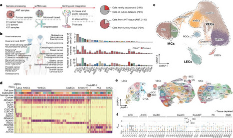
Tumours can obtain nutrients and oxygen required to progress and metastasize through the blood supply1. Inducing angiogenesis involves the sprouting of established vessel beds and their maturation into an organized network2,3. Here we generate a comprehensive atlas of tumour vasculature at single-cell resolution, encompassing approximately 200,000 cells from 372 donors representing 31 cancer types. Trajectory inference suggested that tumour angiogenesis was initiated from venous endothelial cells and extended towards arterial endothelial cells. As neovascularization elongates (through angiogenic stages SI, SII and SIII), APLN+ tip cells at the SI stage (APLN+ TipSI) advanced to TipSIII cells with increased Notch signalling. Meanwhile, stalk cells, following tip cells, transitioned from high chemokine expression to elevated TEK (also known as Tie2) expression. Moreover, APLN+ TipSI cells not only were associated with disease progression and poor prognosis but also hold promise for predicting response to anti-VEGF therapy. Lymphatic endothelial cells demonstrated two distinct differentiation lineages: one responsible for lymphangiogenesis and the other involved in antigen presentation. In pericytes, endoplasmic reticulum stress was associated with the proangiogenic BASP1+ matrix-producing pericytes. Furthermore, intercellular communication analysis showed that neovascular endothelial cells could shape an immunosuppressive microenvironment conducive to angiogenesis. This study depicts the complexity of tumour vasculature and has potential clinical significance for anti-angiogenic therapy. An atlas of tumour vasculature shows that tumour angiogenesis is initiated from venous endothelial cells and extended towards arterial endothelial cells.
Lymphatic vessels are immunological highways that connect lymph nodes to peripheral tissues. Ataide et al. found that tissue-derived UTCs also continuously migrate via this route. This migration pattern leads to characteristic innate and adaptive immune responses that differ between lymph nodes, mirrors the drained tissue, and is based on the immediate effector function of UTCs.
Single-cell RNA sequencing provides a comprehensive map of lymph-node–resident lymphatic endothelial cells, identifying new specific markers and functions of cell subpopulations, including the scavenging of low-density lipoprotein and mediation of rapid lymphocyte egress from lymph nodes.
The lymphatic vasculature is essential to maintain fluid homeostasis and immune surveillance, including in the brain where lymphatic vessels were only recently identified. Here, Jacob et al. provide an anatomical map of lymphatic vessels in the vertebral column, where they find these contribute to...
Summary G‐protein–coupled receptor (GPCR) signaling is essential for the spatiotemporal control of leukocyte dynamics during immune responses. For efficient navigation through mammalian tissues, most leukocyte types express more than one GPCR on their surface and sense a wide range of chemokines and chemoattractants, leading to basic forms of leukocyte movement (chemokinesis, haptokinesis, chemotaxis, haptotaxis, and chemorepulsion). How leukocytes integrate multiple GPCR signals and make directional decisions in lymphoid and inflamed tissues is still subject of intense research. Many of our concepts on GPCR‐controlled leukocyte navigation in the presence of multiple GPCR signals derive from in vitro chemotaxis studies and lower vertebrates. In this review, we refer to these concepts and critically contemplate their relevance for the directional movement of several leukocyte subsets (neutrophils, T cells, and dendritic cells) in the complexity of mouse tissues. We discuss how leukocyte navigation can be regulated at the level of only a single GPCR (surface expression, competitive antagonism, oligomerization, homologous desensitization, and receptor internalization) or multiple GPCRs (synergy, hierarchical and non‐hierarchical competition, sequential signaling, heterologous desensitization, and agonist scavenging). In particular, we will highlight recent advances in understanding GPCR‐controlled leukocyte navigation by intravital microscopy of immune cells in mice. 1 INTRODUCTION Innate and adaptive immune responses depend on the coordination and spatiotemporal control of leukocyte dynamics between and within lymphoid organs and peripheral tissues. A wide spectrum of chemically diverse molecules including small proteins, peptides, lipids, nucleotides, and extracellular matrix fragments can act as chemoattractants, regulating leukocyte motility, migration, chemotaxis, positioning, and cell‐cell interaction. Leukocytes perceive these signals mainly through G‐protein–coupled receptors (GPCRs) that, upon agonist binding, induce intracellular promigratory responses.1, 2 Based on the cell type–specific expression of cell surface GPCR type combinations and intracellular GPCR‐controlling protein networks, each leukocyte subset has evolved specific trafficking paths to fulfill their designated effector functions in the body.3-5 Over the last decades, the analysis of GPCR‐deficient mouse models has advanced our understanding of how specific GPCR signals determine immune cell positioning and compartmentalization in situ. Recent work now also started to shed light on the establishment of chemokine patterns and chemotactic fields that guide leukocytes in mammalian tissues.6, 7 In particular, intravital imaging of control and GPCR‐deficient leukocytes in tissues of live anesthetized mice has brought unprecedented insight into the dynamic migration patterns of immune cell subsets in situ.8, 9 Despite these achievements, it is still largely unclear how leukocytes make directional decisions in lymphoid and inflamed organs where they constantly encounter GPCR signals emanating from multiple sources. The detailed mechanisms of how leukocytes sense, integrate, and prioritize between multiple chemotactic fields in situ and how this influences their physiological trafficking are still largely unknown. Dissecting these molecular events experimentally in mammalian tissues is very challenging and demanding. In particular, we are missing important pieces of information (eg, the exact distribution and concentrations of known chemottractant, GPCR dynamics in leukocytes) and cannot control for unknown chemotactic fields. Due to these current limitations many of the emerging concepts for leukocyte navigation in complex chemoattractant milieus come from studies in controlled in vitro systems2 or lower model organisms.10, 11 However, these concepts from reductionist models may not always be easily translatable to the complex situation in mammalian lymphoid organs and inflamed peripheral tissues. In recent years, several systematic studies in mice have started to disentangle how multiple GPCR signals shape leukocyte migration patterns in the complexity of mammalian tissues. In this review, we will summarize common concepts and mechanisms of GPCR‐controlled leukocyte navigation coming from studies with mouse and human leukocytes. We outline the protein networks that control GPCR signaling and GPCR activity and discuss how these may control leukocyte navigation strategies. In particular, we highlight our current knowledge on leukocyte navigation in complex chemoattractant fields and summarize them for three leukocyte subsets (neutrophils, T cells, and dendritic cells [DC]) with different physiological trafficking paths. For each cell type, we will critically evaluate the relevance of current concepts for leukocyte navigation in mouse tissue. While it is very likely that some navigation strategies are only relevant for certain immune cell types and adapted to specific physiological tissue environments, leukocyte subsets may also share basic strategies for directional decision‐making in lymphoid and inflamed tissues of mammals. 1.1 Concepts and mechanisms of GPCR‐controlled leukocyte navigation Many chemokines and chemoattractants are classical inducers of leukocyte polarity, adhesion, and locomotion. They act through GPCRs on intracellular signaling pathways to establish biochemical asymmetry within the cell, preceding the formation of a polarized cell with an actin‐rich front and a myosin II–rich rear.12, 13 Key molecular pathways involved in establishing functional polarity downstream of GPCRs in leukocytes depend on phosphoinositide 3‐kinases (PI3K) and members of the Rho family of small GTPases.14 Rac isoforms, Cdc42 and RhoA represent major signal hubs that act through several effector molecules to regulate actin polymerization and actomyosin cytoskeleton. The balanced interplay of these components shapes the typical amoeboid migration mode of each leukocyte subset.15, 16 In homogeneous fields of soluble chemokines and chemoattractants, most leukocytes undergo self‐polarization, before increasing their motility, migration speed, and movement in a random and undirected fashion (chemokinesis) (Figure 1A).17 Chemokinesis is considered a migratory pattern that helps leukocytes scan large tissue volumes. We have learnt from intravital microscopy studies in mice that lymphocytes show such continuous undirected (random‐walk) migration in lymphoid organs to enable encounters and interactions with other leukocytes.9 Together with other determining factors (eg, confinement between stromal tissue elements), uniform chemokine signals critically control chemokinesis by increasing the speed of randomly moving lymphocytes in situ.18 Once leukocytes perceive external gradients of soluble chemokines and chemoattractants, they can perform directional movement along the concentration gradients of these diffusing attractants (chemotaxis) (Figure 1B).17 Some leukocyte subsets have also been reported to be repelled from high chemoattractant concentrations (chemorepulsion), leading to their movement against the gradient (fugetaxis).19 However, the underlying molecular mechanisms of this process are still under investigation (Figure 1C).20, 21 Apart from soluble molecular cues, leukocytes also perceive surface‐bound attractants as gradients or as homogeneous fields and undergo movement along these surfaces in a directed (haptotaxis) (Figure 1B) or undirected manner (haptokinesis) (Figure 1A), respectively.16, 22 The aspect of leukocyte navigation along surface‐bound attractants is particularly relevant for chemokines with binding capacities to extracellular glycosaminoglycans (GAGs), which decorate many stromal cell elements in tissues.22, 23 This form of imprinting non‐diffusible chemokine gradients into the tissue has been shown critical for leukocyte trafficking across the blood endothelium and entry into lymphatic vessels.6, 23 The terms “chemotaxis” and “haptotaxis” subsume a spectrum of navigation strategies, which leukocytes can use to follow attractant gradients.24 When perceiving an external attractant gradient, some leukocytes (eg, unstimulated neutrophils in culture dish) undergo a full morphological change from unpolarized shape to the establishment of a front‐back axis, which then leads to directional movement along the gradient. In contrast, other leukocytes already migrate with pre‐established polarity (eg, DCs and neutrophils in 3D), before they steer along the gradient by redirecting their migration path. Alternatively, prepolarized leukocytes may move along a gradient without redirecting their path and start to accelerate the closer they get to the attractant source (eg, neutrophils in response to dying cells). Once at the highest attractant source, leukocytes may also actively decelerate and slow down their migration.24 As evidenced from intravital microscopy of select leukocyte subsets, we know that this spectrum of chemotactic responses also occurs in mouse tissues. While we acknowledge that chemokines and chemoattractants are central for many aspects of these migratory responses, we are missing a detailed understanding about how these signals shape the navigation of leukocytes in situ. Is the cellular movement guided by one or more attractant signal(s)? How do leukocytes navigate in complex gradient fields and sense the right direction? How do leukocytes sense their final destination and know where to stop? Are there inherent differences between leukocyte subsets in their navigation strategies? As many other cell types, leukocytes possess several mechanisms that directly regulate the functionality of GPCRs.13, 25-28 These are mainly controlled by GPCR interactions with three protein families: heterotrimeric (α, β, γ) G‐proteins, G‐protein–coupled receptor kinases (GRKs), and β‐arrestins. Upon stimulation, a GPCR functions as a guanine nucleotide exchange factor that catalyzes the exchange of GTP for GDP on Gα subunits of the heterotrimeric G‐protein. In leukocytes, Gα subunits of the pertussis toxin‐sensitive Gi subfamily play critical roles for the signaling of many chemokine and chemoattractant receptors. The dissociation of Gβγ from Gα‐GTP allows them both to independently activate downstream signaling pathways. Leukocyte chemotactic responses are largely controlled by the released Gβγ, which activates phosphoinositide 3‐kinase (PI3K) and phosphoinositide‐specific phospholipase Cβ (PLC)/inositol‐trisphosphate (IP3)/diacylglycerol effector pathways.29, 30 The termination of GPCR signaling relies on the intrinsic GTPase activity of the Gα subunit that hydrolyzes GTP and reassembles as Gα‐GDP with Gβγ to form an inactive heterotrimer. This Gα GTPase reaction is accelerated by a family of proteins, called “regulators of G‐protein signalling” (RGS proteins) to facilitate the rapid turning off GPCR signaling. Hence, RGS proteins are considered key desensitizers of G‐protein‐signaling pathways.27 In addition, another negative feedback loop evolved to prevent the overstimulation of cells through excessive GPCR stimulation. Following agonist binding and G‐protein activation, GPCRs become rapidly phosphorylated on their cytoplasmic tails and intracellular C‐termini by specific enzymes, usually members of the GRK family.31, 32 The pattern of receptor phosphorylation creates a barcode with high‐affinity binding sites for the recruitment of β‐arrestin proteins.33 As a consequence, the increased binding of β‐arrestin to the receptor sterically hinders the GPCR interaction with G‐proteins. This uncoupling of G‐proteins from the receptor, referred to as “receptor desensitization”, prevents further GPCR activation by repeated agonist stimulation over seconds to minutes.34 β‐arrestins can also control more long‐lasting desensitization of GPCRs over minutes to hours. As they can interact with clathrin‐coated pits, their binding to GPCRs also targets receptors for internalization. Besides, GPCR internalization can also follow other pathways (arrestin independent, clathrin independent) or not occur at all.35 Depending on the intracellular trafficking route, the internalized receptors may be subject to degradation, leading to the downregulation of cell surface GPCRs. Alternatively, targeting internalized GPCR to recycling endosomes and back to the plasma membrane can quickly replenish GPCRs on the cell surface.35 In addition to their important roles in receptor inactivation and internalization, GPCR‐recruited β‐arrestins also act as signaling mediators by directly coupling to numerous signaling molecules.36 At many GPCRs, β‐arrestin‐mediated signaling and classical G‐protein signaling can have distinct biochemical and physiological actions from one another. A relatively new paradigm on GPCR signaling considers the allosteric interaction among the ligand, the GPCR, and the transducers (β‐arrestins and GRKs) and how their contribution influences the bias for G‐protein– or β‐arrestin–mediated signaling.28 Resulting from this paradigm, the concept of biased agonism, ie, that different ligands at the same GPCR are able to activate some signaling pathways while blocking others, is of particular interest for many chemokine receptors with prominent roles in leukocyte navigation.37 In the following, we will point out the most commonly and recently discussed mechanisms, which would allow the control of leukocyte navigation. Given the before‐mentioned regulation mechanisms for GPCR functionality, a “One‐GPCR system” appears already sufficient to control chemokinesis and chemotaxis of leukocytes (Figure 2A‐E). To increase the sensitivity for attractant gradients, leukocytes can upregulate the cell surface expression of GPCRs. Some leukocytes store GPCRs in intracellular vesicles, which can fuse with the plasma membrane under conditions of leukocyte activation.38 While the release of GPCR from vesicular stores occurs within minutes to hours, most leukocytes often respond to external stimuli by changing their cell surface GPCR profile over hours to days. These changes are primarily based on the induction of gene‐transcription networks, which also include GPCR proteins, when leukocytes transition between cell states or undergo differentiation (Figure 2A). Multimerization of GPCRs, including homomerization of the same GPCR type, provides another level of GPCR regulation.25 A recent study highlighted the functional importance of GPCR oligomerization in promoting leukocyte chemotaxis (Figure 2B). By lowering cellular cholesterol, inflammatory stimuli enabled GPCR oligomers that acted as scaffolds for distinct promigratory signaling pathways to foster efficient leukocyte migration.39 However, GPCR multimerization has also been reported to dampen GPCR responsiveness.25 Several other mechanisms can attenuate the sensitivity of leukocytes for chemokines and chemoattractants. One involves the presence of a receptor antagonist that binds to the same GPCR as the gradient‐forming attractant agonist, but exerts blocking function for promigratory signaling (Figure 2C). More cell‐intrinsic mechanisms include the downregulation of GPCR expression by lowering GPCR mRNA stability40 or enhancing GPCR degradation.41 As discussed above, agonist‐induced receptor desensitization also attenuates the signaling responsiveness of GPCRs. Upon homologous desensitization, when GRKs phosphorylate agonist‐specific GPCRs, the further recruitment of β‐arrestins can lead to G‐protein uncoupling and internalization of GPCRs (Figure 2D). This process explains why many leukocyte chemotaxis responses toward single attractants are dose dependent and follow a bell‐shaped curve, with higher concentrations resulting in stasis.42 Homologous desensitization may be particularly relevant when leukocytes sense high attractant concentration in situ, eg, at the center of an attractant source.43 Under such conditions, the majority of cell surface GPCRs is likely to undergo desensitization, internalization, and degradation, which may then result in cellular unresponsiveness to continuous or repeated stimulation through the same attractant. As a strategy to remain responsive to subsequent stimulation in gradients, some leukocytes internalize GPCRs, deploy its ligand cargo into lysosomes, and quickly recycle unloaded GPCRs back to the plasma membrane.44 Thus, by eliminating chemotactic factors from the extracellular environments, classic GPCRs can acquire additional scavenging function. This “consumption” of external attractant may also impact the chemotactic milieu by reducing the strength of attractant cues.44 Instead of only binding one agonist, many “One‐GPCR systems” can bind and respond to two or more chemoattractants and activate downstream signaling (Figure 2E). Sensing these distinct attractants through the same GPCR in a spatial or temporal sequence can provide another navigation strategy. In our body, leukocytes are rarely exposed to only one chemotactic factor. For efficient trafficking between organs and coordinated movement within tissues, most leukocytes express more than one GPCR on their surface and sense a wide range of chemokines and chemoattractants. Directional decision‐making in lymphoid and inflamed organs requires leukocytes to constantly sense, compare, and integrate numerous GPCR signals emanating from multiple sources. To efficiently navigate in a complex milieu of attractant gradients, leukocytes need to respond to combinational signals and prioritize between competing signals. While this aspect of GPCR signal integration has already been of interest decades ago, it has regained momentum with the recent developments in fabricating microfluidic‐based chemotaxis devices. We will now briefly summarize common mechanisms and concepts for leukocyte navigation, which become already relevant in the presence of a second GPCR signaling system (“Two‐GPCR systems”) (Figure 3). As leukocytes often use combinations of more than two‐GPCR systems, these principles could be further expanded. Manifold scenarios of attractant interpretation are conceivable, when leukocytes sense two chemoattractant signals through two different GPCR types (Figure 3). The outcome for leukocyte guidance depends on several determining parameters: (a) the spatial distribution of each signal (uniform, graded), (b) the spatial orientation of both signals toward each other (same or opposing directions), and (c) the ratio of signaling input that leukocytes receive from both signals (equal, unequal). The latter point includes both cell‐intrinsic features and cell‐extrinsic conditions. Unequal signaling input can result from different cell surface expression of the two‐GPCR types or different strength of the downstream signaling pathway. In both cases, one chemoattractant signal dominates over the other. This can also occur in leukocytes with equal signaling input from both GPCR systems under conditions of unequal concentrations of both signals in the extracellular space. Many of these scenarios have already been addressed with select leukocyte subsets and their cell‐specific GPCR combinations in controlled in vitro microenvironments, and examples will be discussed in the next paragraphs about specific leukocyte subsets (neutrophils, T cell, and DCs). As discussed before, homogenous chemoattractant fields can induce leukocyte polarity and chemokinesis. A second‐graded signal “on top” can then redirect prepolarized leukocytes and guide them toward other locations. However, if the uniform attractant field is very potent, it will retain leukocytes and attenuate chemotaxis toward the second‐graded signal (Figure 3A).45-48 Under conditions when both attractant signals are oriented in the same direction and overlap, the two‐GPCR signals have usually additive effects for leukocyte chemotaxis (Figure 3B). In the case of promigratory signals, chemotactic factors act often synergistically to improve chemotaxis along the same direction.49 Physiologically, this might be of particular relevance for tissue sites where overlapping attractant gradients originate from the same local area. A special variant of GPCR signal synergism has initially been observed in migrating human and mouse neutrophil populations in vitro. During their chemotaxis in response to shallow N‐formyl‐peptides (fMLF) gradients, neutrophils released the lipid leukotriene B4 (LTB4) that acted on top of the early chemotactic factor as a signal relay molecule to improve chemotaxis of the whole neutrophil population.50 Under conditions when leukocytes encounter two spatially opposing gradients, the two‐GPCR signals compete for guiding leukocytes into one or the other direction (Figures 3C and 4). Leukocytes exposed to two opposing attractants with equal signaling strength will likely accumulate and find migratory equilibrium in the middle of both gradients (Figure 3C).47, 48, 51, 52 However, an unequal signaling input from both signals favors the movement of leukocytes into one direction (Figure 4A).47 It is known for decades that specific GPCR signals can dominate over other GPCR signals, which enables leukocytes to prioritize some chemoattractant signals over others (Figure 4B).42, 45 This aspect of GPCR crosstalk provided a first conceptual framework for neutrophils traveling in a stepwise fashion from the blood into infected tissues.45 Initially, it was discussed that heterologous desensitization of GPCRs might be an underlying mechanism for prioritizing chemotactic cues. In contrast to homologous desensitization (see above), heterologous GPCR desensitization is agonist independent and results from the phosphorylation of one GPCR by a kinase activated through the signaling cascade of another activated GPCR (Figure 3D). Thus, one‐GPCR signal can act as desensitizing signal for multiple other GPCR types, causing leukocytes to become refractory to several chemotactic stimuli within minutes. This rapid cross‐phosphorylation has been linked to second‐messenger kinases, particularly protein kinase C, which phosphorylates the GPCR at sites different from GRKs.53 For neutrophil navigation in opposing gradients, it was later proposed that the dominance of certain GPCR signals is regulated by events downstream of the receptors. According to this concept, referred to as “signaling hierarchy”, special GPCRs induce intracellular signaling pathways that can override signaling pathways downstream of other GPCR signals42, 54 (see below for details). Leukocyte populations in tissues rarely consist of identical cell clones, but are rather characterized by many cells with slightly heterogeneous expression of GPCR combinations. Thus, single‐cell heterogeneity on the basis of combinatorial GPCR expression may influence the population response in two opposing attractants with equal signaling strength (Figure 4C). In many physiological settings, leukocytes are thought to navigate step‐by‐step through sequential chemotactic fields. Under conditions of two spatially segregated chemotactic signals in sequence (Figure 3E), leukocytes may first move along a first attractant gradient (Figure 1A), and then subsequently sense the gradient of a second, more distant attractant. During the transition from first to second attractant gradient leukocytes then encounter a competitive situation between two gradients (Figure 3C). When the second signal is dominant over the first one, it is plausible that leukocytes are recruited to and then retained at the distant site. Importantly, even without any dominance between the signals, leukocytes can migrate away from a first attractant and travel toward a second, more distant attractant.45 In principle, this also allows leukocytes to leave chemotactic fields of high, saturating concentrations. Moreover, leukocytes remain responsive for additional attractant changes, while moving back and forth in the equilibrium between both gradients. Thus, interpreting two distinct signals in sequence allows efficient navigation patterns that are not possible with only one attractant presented in the same spatial array. We want to point out that the sequential sensing of attractant gradients can also be temporally controlled (Figure 3E). Leukocytes may undergo sensing of a first attractant gradient, while a second attractant arises newly in close vicinity. In the course of an immune response in lymphoid or inflamed tissues, this may result from chemoattractant secretion of nearby activated tissue‐resident cells or recently recruited, blood‐derived leukocytes. As most immune responses are choreographed by a sequence of timely separated cellular events, it is likely that this aspect of sequential sensing has strong influence on leukocyte navigation in situ. In our contemplation on leukocyte navigation by “Two‐GPCR systems”, we have focused until now on GPCRs with signaling function. However, many leukocyte subsets additionally express members of the atypical chemokine receptor (ACKR) family on their surface. Also referred to as scavenging receptors, ACKRs are GPCRs with promiscuous binding affinity for multiple chemokines. As they do not couple to G‐proteins, they are unable to induce intracellular signaling pathways. Instead, ACKRs can actively internalize and target chemokines for lysosomal degradation.55 Functional co‐expression of conventional chemokine receptors and ACKRs in leukocytes has been implicated in the fine‐tuning of leukocyte chemotaxis, with ACKRs acting as rheostats in restricting excessive migration in response to moderate chemokine levels (Figure 3F).56-58 As we have just outlined, leukocytes have evolved several molecular mechanisms that allow an enormous spectrum of GPCR‐controlled navigation strategies. Many of the emerging concepts for leukocyte navigation in complex chemoattractant fields have been developed in controlled in vitro systems, some of which by studying only one select leukocyte subset. Rigorous testing and proving the relevance of these concepts for leukocyte migration in the complexity of mammalian organs and tissues is technically very challenging. However, systematic studies in mice have started to delineate GPCR‐controlled navigation strategies and their influence on physiological trafficking patterns through lymphoid organs and inflamed peripheral tissues. In the following, we will discuss our current knowledge on the role of GPCR signals for the migration patterns of three leukocyte subsets (neutrophils, T cells, and DCs). Each of these immune cell types has evolved their own trafficking paths to fit their effector functions. We will highlight recent studies that conceptually advanced our understanding of GPCR‐controlled leukocyte navigation in mammalian tissues. 2 NEUTROPHILS Neutrophils are pivotal effector cells of our innate immune response and the first line of defense for eliminating bacteria and fungi in tissues.59 Recent studies also implicate regulatory functions of neutrophils in tissue homeostasis and cancer.60 In mammals, they are produced in the bone marrow and are continuously released into the bloodstream to patrol the intravascular compartment under homeostasis. Neutrophils circulate in the blood for ca. 12 hours under the influence of circadian rhythms, before they are mostly cleared into the bone marrow, but also other extramedullary tissues.61 In contrast to the general believe that naive peripheral tissues are free of neutrophils, recent work suggests that low numbers of neutrophils are present in many tissues under steady state.62 Upon inflammation and infection, the release of neutrophils from bone marrow increases their concentration in the blood by several folds.63 Moreover, large numbers of circulating neutrophils then infiltrate inflamed tissues where they sense activated vascular endothelium. In agreement with their critical role as first responders, neutrophils have evolved robust strategies to infiltrate inflamed tissues extremely fast and orient themselves to detect potential sites of microbial invasion with great precision. In many organs, the exit from the vascular compartment involves a sequence of exquisitely coordinated processes: (a) intravascular arrest, migration, and crawling along the activated endothelial cells, and (b) breaching multiple layers of the vascular wall (venular endothelium, venular basement membrane, and pericytes).64 Once neutrophils have traversed this barrier, they are primed for the rapid detection of damaged tissue or microbes in the extravascular space. At sites of cell death in the interstitial space, neutrophils undergo sequential phases of highly coordinated chemotaxis and cell cluster formation, now commonly referred to as neutrophil swarming.65, 66 As true sentinel cells, they sense almost any form of tissue insult, rapidly localize it in any inflamed organ and efficiently move toward the core of the inflammation or infection site. Neutrophils have been shown to express on their cell surface more than 30 GPCR types, which can all bind chemotactic molecules. Accordingly, neutrophils are responsive to a broad array of chemically diverse chemoattractants, including chemokines, lipids, complement factors, formylated peptides, nucleotides, alarmins, and proteolytic (ECM) fragments.67-69 Many of these chemotactic factors are usually present at sites of in vivo inflammation. How the crosstalk between these GPCR signals guides neutrophil recruitment in inflamed tissues has been of huge interest since decades. Early in vitro work with human polymorphonuclear neutrophils (PMN) characterized the strength of chemically distinct chemoattractant classes to desensitize calcium‐ion mobilization, as a read‐out of GPCR downstream signaling, to one another (Figures 2D and 3D).53, 70 Peptide chemoattractants, including N‐formylmethionyl‐leucyl‐phenylalanine (fMLF), the complement factor C5a, and the chemokine CXCL8 (IL‐8), cross‐desensitized one another to different extents (fMLF > C5a > IL‐8). Moreover, peptide chemoattractants, in particular fMLF, cross‐desensitized lipid chemoattractants, including platelet‐activating factor (PAF) and LTB4. In contrast, lipid and purinergic agonists failed to desensitize to the peptide chemoattractants.70 These results were extended to neutrophil chemotaxis in the presence of multiple chemoattractants, showing a hierarchy of resistance to competing signals, with fMLF and C5a inhibiting migration to other chemoattractants (Figure 3D).71, 72 Elegant under‐agarose chemotaxis experiments proposed the concept that neutrophils can navigate through complex chemoattractant fields by migrating in a step‐by‐step manner in response to one agonist source after another. Thus, their ultimate positions are determined combinatorially by the array of GPCRs they express and by the sequence of chemoattractant gradients they encounter (Figure 3E).45 In this concept, pathogen‐derived “end‐target” chemoattractants (N‐formyl peptides, C5a) will dominate over host tissue‐derived “intermediate‐target” chemotactic factors (CXCL8, LTB4) in their ability to guide neutrophil migration (Figure 3A, C, E).45 This dominance of end‐target attractants has been suggested to depend on the activation of the p38 MAPK pathway, which can override the phosphoinositide 3‐kinase (PI3K) pathway activated by intermediate‐target attractants.42, 54 Importantly, neutrophil navigation is also possible in a spatially sequential array of intermediate‐target chemoattractants (Figure 3E).45, 47, 51, 73 Another aspect of crosstalk between end‐target and intermediate‐target chemoattractants has not been considered in these early studies, but is also functionally relevant for neutrophil navigation. Already small amounts of fMLF or C5a can stimulate neutrophils to secrete intermediate‐target attractants, in particular LTB4.50, 74 This paracrine signaling mechanism becomes very relevant in chemotaxing neutrophil populations. Neutrophil‐released LTB4 functions as a signal relay molecule and amplifies in a feed‐forward manner the chemotactic response to subsaturating concentrations of end‐target attractants (Figure 3B).50, 75 Recent developments in microfabricated devices and microfluidic‐based chemotaxis systems now allow a more detailed analysis of neutrophil migration responses in complex chemoattractant milieus, providing deeper insight into neutrophil navigation in opposing gradients (Figure 3C),47, 73, 76-78 synergistic gradients (Figure 3B),49 and geometrically complex environments.49, 77 Moreover, even the analysis of GPCR signaling underlying neutrophil swarming dynamics is now possible due to novel micropatterning techniques.75 As human PMNs, mouse neutrophils express a range of functional GPCRs to sense N‐formyl peptides (through FPR1 and FPR2), C5a (mainly through C5ar1), LTB4 (mainly through LTB4R1/BLT1), and several chemokines including CXCL1, CXCL2, CXCL5 (through CXCR2), CXCL12 (through CXCR4), and several CC‐chemokines (through CCR1).67 Some of these GPCRs are present in secretory vesicles that can rapidly fuse with the plasma membrane upon neutrophil activation, allowing the cells to increase their responsiveness to chemotactic cues (Figure 2A).38 Other reported mechanisms to control GPCR functionality in neutrophils include competitive antagonism or partial agonism (Figure 2C),79 receptor multimerization (Figure 2B), differential agonist responses (Figure 2E),80 and agonist scavenging through simultaneous expression of ACKRs (Figure 3F).56 Homologous desensitization is a well‐known phenomenon for almost all neutrophil‐expressed GPCRs (Figure 2D). While GPCR transducers (G‐protein subunit types, GRKs, and arrestins) and RGS proteins have been linked to the function of some GPCRs in primary mouse neutrophils,32, 81-88 we are still missing a detailed understanding of their exact contributions to neutrophil navigation in complex chemoattractant fields. Clearly, neutrophils are equipped with many GPCR types and mechanisms to dynamically control GPCR activity, allowing them to use various navigation strategies in the presence of multiple chemotactic cues. Moreover, neutrophils have been shown to perform fugetaxis against the direction of a gradient of high concentration chemokine, a process that is mechanistically not well understood (Figure 1C).89 However, there are still many open questions of how these mechanisms contribute to the neutrophil migration patterns observed in situ. Intravital microscopy studies of GPCR‐deficient neutrophils provided novel insight into how GPCR signals choreograph the physiological trafficking paths of neutrophils. In the following, we will point out that GPCR signals are important throughout many steps in the lifecycle of a neutrophil. In particular, we will highlight studies that advanced our concepts of GPCR‐controlled neutrophil navigation in mouse tissues. 2.1 Bone marrow release: inhibiting chemokine‐mediated retention The majority of mature neutrophils in the body reside in the bone marrow, with an estimated ratio of 300:1 between neutrophils in the bone marrow and blood. Neutrophil release into the blood is controlled by the interplay of retention and mobilization signals, provided by two antagonistically operating chemokine systems (Figure 3A,C). During homeostasis, the chemokine CXCL12 is produced in the bone marrow and essential for the retention of mature neutrophils that express the corresponding chemokine receptor CXCR4.90 CXCL12 is expressed at highest levels by “CXCL12‐abundant reticular cells” (CAR cells), but also by other stromal cells of the bone marrow, including osteoblasts.63, 91 The expression levels of CXCL12 in stromal cells follow a circadian rhythm, controlled by the sympathetic nervous system.92 When neutrophils were freshly isolated from the bone marrow, they expressed only low levels of cell surface CXCR4, but contained high intracellular levels of CXCR4.90, 93 After placing these cells in medium lacking CXCL12, they upregulated cell surface CXCR4 within hours.90, 93 These findings suggested receptor desensitization and internalization in response to constitutively high local concentrations of CXCL12 in the bone marrow microenvironment (Figure 2D). Moreover, these data underlined that neutrophils receive constant CXCL12 signals, which position them in the bone marrow, probably by activating integrin‐mediated adhesion to stromal cells.94 While high concentrations of CXCL12 retain neutrophils in the bone marrow via CXCR4 signaling, low concentrations of CXCR2 ligands expressed on endothelial cells and produced by megakaryocytes facilitate neutrophil egress via CXCR2.63 Thus, CXCR2 signals recruit neutrophils out of the retentive CXCL12 field and direct them to the endothelial sinus walls for entry into the blood circulation (Figure 3A). This intricately balanced interplay between retention and mobilization signals can be shifted in both directions. G‐CSF treatment or acute inflammation, which increases blood serum levels of G‐CSF and CXCR2 ligands, shifts the balance toward neutrophil mobilization from the bone marrow.95-98 Several modes of action have been proposed for G‐CSF, either leading to the lowering of CXCL12 levels in the bone marrow or triggering the release of CXCR2 ligands from endothelial cells or megakaryocytes (summarized in63). The same shift toward neutrophil egress can be achieved by conditional gene depletions of CXCR4 or CXCL12 in mice.99, 100 The administration of the CXCR4 antagonist AMD3100 also results in a threefold increase in the number of circulating neutrophils and a corresponding decrease in bone marrow numbers.93, 101 Originally, this drug effect was interpreted as direct antagonism of CXCR4 expressed by neutrophils, thereby disrupting the CXCL12‐CXCR4 axis in the bone marrow (Figure 2C).90 Surprisingly, subsequent studies found that AMD3100 also acted on CXCR4 expressed by bone marrow endothelial cells, and inhibited the translocation of CXCL12 across bone marrow endothelial cells, suggesting a rapid rise of CXCL12 in the blood and reversal of the CXCL12 gradient (Figure 3B).102, 103 These data were questioned by intravital imaging studies that could not observe clear neutrophil egress from the bone marrow upon AMD3100 treatment. Instead, the increase in circulating neutrophils through AMD3100 was explained by the release of marginated neutrophils from the lung microvasculature.104 Moreover, AMD3100 had a blocking effect on the trafficking of aged blood neutrophils, which upregulate CXCR4 expression over time, enabling them to home back to the bone marrow for clearance.104 The exact mode of action of AMD3100 is still controversially discussed.90 Insight into the physiological consequences of shifting the chemokine balance in the opposite direction, toward retention in the bone marrow, comes from an extremely rare congenital human immunodeficiency disease. Warts, hypogammaglobulinemia, infections, and myelokathexis (WHIM) syndrome is an autosomal dominant human disorder caused by gain‐of‐function mutations in the C‐tail of CXCR4.105, 106 WHIM syndrome involves myelokathexis, a condition characterized by the retention of mature neutrophils in the bone marrow and peripheral neutropenia. WHIM‐associated truncations or mutations in the carboxy‐terminal tail of CXCR4 result in the loss of multiple serine residues, which are target sites for GRK‐mediated receptor phosphorylation. Truncated forms of CXCR4 are impaired in agonist‐induced homologous desensitization and internalization, and cause enhanced signaling upon CXCL12 stimulation.106 This leads to stronger anchorage of mature neutrophils in the bone marrow, counteracting CXCR2‐mediated chemotaxis toward vessels and neutrophil egress. However, administration of G‐CSF or ADM3100 to WHIM patients can overcome this imbalance and cause an increase in circulating neutrophil numbers.106 2.2 Getting to the site of inflammation: navigation along multiple chemokine fields Blood‐circulating neutrophils detect sites of tissue injury and infection by interacting with the luminal surface of activated venular endothelium. In response to tissue‐released inflammatory cytokines, endothelial cells upregulate P‐selectin, E‐selectin, and integrin ligands, such as ICAM‐1 and VCAM‐1, to promote interactions between neutrophils and the vessel wall. This induces neutrophils to undergo a multistep adhesion cascade along the endothelial lining before emigrating from the vessel into the inflamed tissue interstitium.107 Chemokines are present on the luminal surface of endothelial cells and cause arrest of rolling neutrophils by triggering the activation of integrins. Many studies showed an involvement of CXCR2 ligands, in particular CXCL1, but also CXCL2 and CXCL5, in this process.108, 109 These chemokines are primarily expressed and released by endothelial cells,110, 111 but also tissue‐resident cells, including pericytes, perivascular macrophages, and mast cells, can act as important chemokine sources.112-115 Due to their high‐affinity binding sites for heparin sulfate (HS) GAG most chemokines become immobilized by HS GAG on the luminal side of the endothelium,116 and defects in HS GAG synthesis have been shown to decrease immobilization of chemokines and neutrophil transmigration into inflamed tissues.117, 118 While the involvement of chemokines for neutrophil transmigration is widely established, there has been ongoing debate whether and how leukocytes sense stable chemokine gradients across the vessel wall on their way into the tissue (Figure 1B). There are ample references to support the common believe that chemotactic gradients between the apical and basolateral part of postcapillary venules promote diapedesis, ie, the outward passage of neutrophils through the vessel wall. Immunofluorescence stainings of HS GAG moieties revealed steep gradient of HS GAG scaffolds between the luminal and abluminal aspect of skin postcapillary venules.119 Under inflammatory conditions, HS GAG distribution became even more polarized by massive depositions of HS and laminin in the basolateral basement membrane of skin vessels.119 Given the GAG‐binding properties of many chemokines, it was proposed that these sharp HS GAG scaffold gradients might favor chemokine gradients across the venular wall. In agreement with this idea, excessive deposition of the GAG‐binding chemokine CXCL1 on the apical part of inflamed blood vessels resulted in decreased neutrophil diapedesis.120 Other supporting data come from experiments in which an extravascular source of CXCR2 ligands was placed in form of a gel on one side of a blood vessel.121 Under these conditions, neutrophil crawling was directed to emigrate preferentially from that side of the vessel. HS GAG on the surface of the endothelium was required to present CXCL2, forming a chemotactic gradient that was followed by neutrophils. This sequestration of endothelial chemokine occurred exclusively in venules, but not other blood vessel types.121 HS GAG, together with the atypical receptor ACKR1, was also discussed to establish gradients by transcytosis of chemokines.118, 122, 123 Transcytosis involves the internalization and transport of tissue‐derived inflammatory chemokines from the basal surface onto the luminal surface of endothelial cells. ACKR1 is specifically expressed by postcapillary venules and was shown to support promigratory chemokine patterns across the vessel wall for optimal leukocyte migration across the vessel wall.123, 124 Unlike other ACKR, ACKR1 does not act as classical scavenger receptor, and was initially discussed to promote the transcytosis of tissue‐derived inflammatory chemokines from perivascular pools to the apical surface of venules.122, 123 Novel insight into the functional role of ACKR1 and the directional cues that guide neutrophils during diapedesis come from recent elegant experiments of Nourshargh and colleagues.125 By studying TNF‐induced neutrophil emigration in the inflamed mouse cremaster muscle, it was shown that neutrophils encounter the two CXCR2 ligands CXCL1 and CXCL2 in a sequential manner during their passage through the vessel wall. In a three‐step process, neutrophils first sense CXCL1 produced by TNF‐stimulated endothelial cells on the luminal surface of venules, promoting crawling along the endothelium. Next, neutrophils encounter CXCL2 that is retained at endothelial cell junctions by ACKR1. Notably, CXCL2 is secreted by neutrophils themselves and then presented by ACKR1, which is enriched in endothelial junctions. This self‐guided migration response of neutrophils was critical for breaching of endothelial junctions and unidirectional transendothelial migration. Lastly, CXCL1 expressed by pericytes governed neutrophil crawling in the subendothelial space.125 While earlier work indicated relatively restricted expression of CXCL1 to vascular and tissue‐resident cells, and CXCL2 with activated neutrophils,126 these novel findings highlight unexpected non‐redundant roles for CXCL1 and CXCL2 to guide neutrophils through venular walls as governed by their distinct cellular sources.125 Having identified this critical spatiotemporal sequence of chemokine‐guided steps across the vessel wall (Figure 3E), it will be of future interest to understand how other tissue‐derived attractant signals may act on top of this and support further guidance into the underlying inflamed interstitium. As an example, neutrophils entering an inflamed peritoneum were suggested to encounter two sequential gradients in a step‐by‐step manner (Figure 3E).114 In a lipopolysaccharide‐induced peritonitis model, initial recruitment of neutrophils out of the blood vessel into the peritoneal tissue was regulated by rapid secretion of CXCL1 and CXCL2 from perivascular mast cells. Further guidance of extravasated neutrophils through the interstitium and into the peritoneal cavity depended on CXCL1 and CXCL2 produced by tissue‐resident interstitial macrophages.114 2.3 Getting to the site of inflammation: interpreting temporal cascades of chemically distinct chemoattractants Most of our discussions until now focused on the role of chemokines for neutrophil guidance in the tissue, but also putative non‐GAG‐binding, soluble chemoattractants including C5a, LTB4, and formylated peptides are sensed by neutrophils and relevant for special types of inflammatory reactions. A mouse model of autoimmune rheumatoid arthritis, the K/BxN serum transfer model, has provided unprecedented insight into how four different chemoattractant receptors, C5aR1, LTB4R1, CCR1, and CXCR2, collaborate to recruit neutrophils into the inflamed tissue.127 In this model, the inflammatory response is driven by immunoglobulin G autoantibodies to the ubiquitous glycolytic enzyme glucose‐6‐phosphate isomerase (GPI) leading to the formation of immune complexes that drive immune cell activation. Neutrophil infiltration into the joint is critical for the clinical signs of arthritis in this model. Several consecutive studies have elucidated a temporal cascade in which neutrophils encounter complement, lipid, and chemokine signals to enter the joint and contribute to disease progression (Figure 3E).74, 128-131 While it has long been known that immune complexes lead to the activation of the alternative complement system in arthritis,132, 133 it has only recently been found that C5aR signaling in circulating neutrophils is the key initiator for a cascade of events that ignite inflammation.131 The deposition of immune complexes in the joint space induces the generation of C5a, which unexpectedly binds to HS GAG on the synovial endothelium. Neutrophils sense C5a on the joint endothelium, induce integrin‐dependent arrest, and facilitate crawling along the vasculature. Subsequently, C5a signals stimulate neutrophils to secrete LTB4, which induces the entry of the first neutrophils into the joint and initiates the onset of disease.74, 128, 129, 131 After disease onset, recruited neutrophils in the joint secrete IL‐1β, which in turn stimulates fibroblast‐like synoviocytes to produce CCR1 ligands at first and later CXCR2 ligands.130 CCR1 ligands are required to recruit the next wave of neutrophils into the joint, and this recruitment of neutrophils is broadly amplified in the last step of the cascade when neutrophils themselves release CXCL2 to potently attract large numbers of neutrophils into the joint.67, 130 Thus, neutrophil entry into the inflamed joint is a prime example where neutrophil recruitment is choreographed by non‐redundant roles of distinct chemoattractants that collaborate sequentially in a temporal cascade (Figure 3E). Moreover, this process highlights the rapid production of complement and LTB4 to initiate a recruitment cascade at the local site of inflammation. By contrast, chemokines act later in this cascade to amplify and prolong the inflammatory response.67 2.4 At the site of inflammation: synergism through lipid‐mediated amplification Another example where complement‐induced LTB4 secretion rapidly shapes neutrophil migration patterns has recently been described.134 In a simplified fungemia mouse model, the fungi Candida albicans sequestered in the pulmonary vaculature and actively recruited neutrophils. Neutrophils showed swarm‐like migration patterns in response to sequestered yeast, similar to previously described swarming responses in other tissues.66 A small number of lung neutrophils responded within minutes and performed intravascular chemotaxis, before larger numbers of neutrophils were recruited and formed cell clusters. The immediate capture of live C. albicans by neutrophils depended on complement‐mediated chemotaxis.134 In addition, complement activation stimulated neutrophils to secrete LTB4, which amplified the directional recruitment and clustering of neutrophils in a synergistic manner (Figure 3B). This was in agreement with a previous study highlighting the importance of LTB4‐mediated signal amplification during neutrophil swarming in interstitial tissue spaces at sites of sterile skin injury and in bacteria‐infected lymph nodes.135 Neutrophil swarming is an essential process of the neutrophil tissue response and has been observed in diverse tissues under conditions of sterile inflammation and infection with various pathogens, including bacteria, fungi, and parasites.65, 66 The swarming response comprises sequential phases of highly coordinated chemotaxis followed by neutrophil accumulation and the formation of substantial neutrophil clusters, which has led to the multistep attraction model of neutrophil swarming.66 While the underlying signals and molecular players for some of these steps are still unclear, we know that neutrophil‐derived LTB4 acted on top of early released, yet unknown, chemoattractants and mediates intercellular signal relay among neutrophils to amplify interstitial recruitment in a feed‐forward manner (Figure 3B). This model was in accordance with earlier in vitro studies identifying LTB4 acted as a signal relay molecule (see above).50 As a consequence, LTB4‐LTB4R1 signaling enhances the radius of neutrophil recruitment from distant tissue sites and maintains the amplified chemotactic response of the neutrophil population. Together with signaling through other GPCRs, such as CXCR2 and FPR2, LTB4R1 signaling also optimizes neutrophil clustering.135 Importantly, LTB4 and CXCR2 signals are also relevant for the swarming response of human neutrophils.75 The formation of focalized dense neutrophil aggregates is considered beneficial for isolating sites of tissue wounding from viable surrounding tissue, containing microbial invaders and concentrating the microbicidal activity of a whole neutrophil population in one swarm center.66 However, neutrophil aggregation can also lead to tissue damage and removal of other cells.135, 136 In the context of the lung microvasculature, intravascular neutrophil swarms that formed in response to C. albicans infection lead to vessel occlusion and pulmonary hemorrhages, which were reverted in the absence of LTB4R1.134 How neutrophil‐derived LTB4 shapes and maintains a promigratory chemotactic field in vivo remains unclear. LTB4 is considered to have a short half‐life and can be rapidly turned into LTB4 metabolites, which act as natural inhibitors of LTB4‐mediated responses. However, it has also been demonstrated that neutrophils release LTB4 in form of exosomes, which may support more prolonged effects of LTB4 gradients.137 Interesting insights into this issue come from studies with diabetic mice receiving skin infections with methicillin‐resistant Staphylococcus aureus (MRSA), which leads to large neutrophil clusters in form of abscesses.138 Diabetic mice produce higher levels of LTB4 in the infected skin compared to non‐diabetic mice. Surprisingly, neutrophils in the skin of these mice failed to assemble into an organized abscess, as excessive LTB4 levels likely disrupted chemoattractant gradients in the skin and impaired directed neutrophil chemotaxis.138 These findings exemplify that the appearance or absence of neutrophil swarms depends on the specific mixture of chemoattractants in a specific inflammatory environment. Although not yet studied in detail, we know that several factors, including tissue damage size, presence of pathogens, induction of cell death, and neutrophil numbers, are important for shaping the exact neutrophil‐swarm phenotype in an inflamed tissue. All these factors can potentially initiate the release of chemotactic fields that may strengthen or attenuate LTB4‐mediated signal amplification in situ.66 2.5 Getting to the site of inflammation: following hierarchies True prioritization of chemoattractant fields has been observed for neutrophils in a model of thermal‐induced liver injury.139 In this model, large numbers of neutrophils are recruited over hours from the liver sinusoids to a focal site of sterile hepatic necrosis. The necrotic area releases several molecules that shape the adjacent hepatic environment and ultimately form two distinct chemotactic fields at different distances from the necrotic focus. During their search for damaged tissue neutrophils sequentially navigate through these two chemotactic fields. First, neutrophils follow an intravascular gradient of GAG‐bound immobilized CXCL2 that supports integrin‐dependent crawling toward the necrotic focus. This CXCL2 haptotactic gradient abruptly halts, approximately 150 μm proximal to the border of the necrotic tissue. Second, neutrophils at this border continue to migrate on top of platelets along non‐perfused vasculature, before entering the necrotic area.139, 140 When neutrophils were lacking the formyl‐peptide receptor FPR1, they migrated along the healthy sinusoids to the highest concentration at the end of the haptotactic CXCL12 gradient and never entered into the platelet‐rich and following necrotic zone. Given the important role of FPR1 signaling during this process, mitochondrial‐derived formyl peptides released from dying hepatocytes were suggested to attract neutrophils into the necrotic area. Of interest, FPR1 signaling has also been shown to regulate neutrophil recruitment into necrotic areas of hepatic ischemia‐reperfusion injury.141 In contrast to formyl peptides released from bacteria, endogenous formyl peptides are produced by mitochondria and become released upon cellular damage.142 Similar to bacterial formyl peptides, also mitochondrial formyl peptides can promote neutrophil chemotaxis in an FPR‐dependent manner.139, 142 Thus, neutrophil navigation in the liver injury model follows a spatial cascade of chemoattractants (Figure 3E) and appears to depend on the prioritization of an end‐target attractant (eg, fMLF) over an intermediate‐target attractant (eg, CXCL2) (Figure 3A). Recent elegant work by Kubes and colleagues revealed that neutrophils play a critical role in fully repairing the hepatic injury. In the course of this process, neutrophils at the border of the injury left the injury site and reentered healthy liver sinusoids.143 However, it remains to be shown if GPCR‐controlled mechanisms are involved in this form of reverse migration into the vasculature. 3 T CELLS T cells and B cells represent the cellular hallmark of the adaptive immune system. These lymphocytes are generally long‐lived cells and are driven by GPCR‐mediated guidance throughout their life cycle. Being released from the bone marrow as a common lymphoid progenitor, T‐cell precursor settle in the thymus where they tread a highly regulated developmental path, guided by GPCRs. Once T cells have been fully matured and selected, they exit the thymus and commence their recirculation pattern between secondary lymphoid organs (SLO). As the T‐cell receptor (TCR) repertoire needs to be highly diverse in order to raise the chance to efficiently detect foreign antigens against all kind of pathogens, the naive T‐cell precursor frequency for a specific antigenic determinant is very low (≈1/105‐1/106). Consequently, during a newly arising infection, antigen‐presenting DC need to interact with millions of T cells in order to activate the few rare naive T cells that optimally react to the foreign antigenic determinant. To facilitate this process T cells need to efficiently migrate between and navigate within SLOs to optimize such DC encounters. Once T cells are activated and primed by a DC carrying and displaying the proper antigen, they differentiate and change their GPCR expression pattern in order to leave the SLO and enter infected tissues via the blood stream. Here, GPCRs pilot them toward infected cells where they execute their specific effector function in order to eliminate or at least control the microbial invasion. In vitro experiments to address how lymphocytes respond to chemokines were primarily based on transwell, under agarose assays or 3D ECM‐like gels, showing that chemokinesis, chemotaxis, and homologous and heterologous desensitization apply to GPCR‐mediated navigation of T cells.144-149 However, these approaches have limitations when trying to address how T cells respond to more complex chemokine fields involving multiple GPCRs. With the development of microfluidic devices150 and more recently, microfluidic mazes, T‐cell exploratory patterns and chemotaxis in response to chemokines could be analyzed in more detail, while providing the investigator with a higher level of control and precision.151 Overall, however, there are only a few studies that have addressed complex GPCR interactions in T cells in vitro. In the following sections we will focus on what we have learned from in vivo experiments regarding the trafficking of T cells. 3.1 Searching for the antigen: exploration and compartmentalization In vivo, chemo‐ and haptokinesis are particularly prevalent in lymphoid organs and underlie the continuous undirected or random‐walk migratory activity of lymphocytes (Figure 1A).9 Functionally, this migratory behavior is required to enable encounters and consecutive interactions between leukocytes. In other words it reflects the search strategy of lymphocytes to detect rare interaction partners in a large and densely populated three‐dimensional space. Given the low precursor frequency of ly
The lymphatics fulfil a vital physiological function as the conduits through which leucocytes traffic between the tissues and draining lymph nodes for the initiation and modulation of immune responses. However, until recently many of the molecular mechanisms controlling such migration have been unclear. As a result of careful research, it is now apparent that the process is regulated at multiple stages from initial leucocyte entry and intraluminal crawling in peripheral tissue lymphatics, through to leucocyte exit in draining lymph nodes where the migrating cells either participate in immune responses or return to the circulation via efferent lymph. Furthermore, it is increasingly evident that most if not all leucocyte populations migrate in lymph and that such migration is not only important for immune modulation, but also for the timely repair and resolution of tissue inflammation. In this article, we review the latest research findings in these areas, arising from new insights into the distinctive ultrastructure of lymphatic capillaries and lymph node sinuses. Accordingly, we highlight the emerging importance of the leucocyte glycocalyx and its novel interactions with the endothelial receptor LYVE-1, the intricacies of endothelial chemokine secretion and sequestration that direct leucocyte trafficking and the significance of the process for normal immune function and pathology.
Lymphatic endothelial cells (LECs) form the structure of the lymphatic vessels and the sinuses of the lymph nodes, positioning them to be key players in many different aspects of the immune response. Following an inflammatory stimulus, LECs produce chemokines that recruit immune cells to the lymph nodes. The recruitment of immune cells aids in the coordination of both LEC and lymph node expansion and contraction. More recent data has demonstrated that to coordinate LEC division and death, cell surface molecules, such as PD-L1 and interferon receptors, are required. During homeostasis, LECs use PD-L1 to maintain peripheral tolerance by presenting specific peripheral tissue antigens in order to eliminate tissue specific responses. LECs also have the capacity to acquire, present and exchange foreign antigens following viral infection or immunization. Here we will review how lymph node LECs require immune cells to expand and contract in response to an immune stimulus, the factors involved and how direct LEC-immune cell interactions are important for programming immunity.
Lane et al. have shown that interferon-γ (IFNγ)-mediated activation of the lymphatic vasculature, as a non-haematopoietic component of the tumour stroma, serves to limit local CD8+ T cell accumulation in melanoma in mice as a mechanism of immune suppression.
In this issue, Bovay et al. (<https://doi.org/10.1084/jem.20180217>;) invoke a compelling model of interplay between the venous and lymphatic vasculature in regulating the developmental genesis and early expansion of LNs.
To get content containing either thought or leadership enter:
To get content containing both thought and leadership enter:
To get content containing the expression thought leadership enter:
You can enter several keywords and you can refine them whenever you want. Our suggestion engine uses more signals but entering a few keywords here will rapidly give you great content to curate.


 Your new post is loading...