Your new post is loading...

|
Scooped by
I2BC Paris-Saclay
October 8, 3:47 AM
|
Tiny proteins, major impact: a new link between bacterial signaling and sporulation
Researchers at I2BC have uncovered a family of small proteins that promotes spore formation in Clostridioides difficile, revealing how the bacterial messenger c-di-GMP controls this key process for the pathogen’s persistence and transmission. Clostridioides difficile is a major intestinal pathogen responsible for severe antibiotic-associated infections. Its ability to form highly resistant spores is central to its success: spores can survive harsh conditions, facilitate transmission and contribute to the persistence of the pathogen. Sporulation is therefore a crucial step in the C. difficile life cycle, yet the mechanisms that determine when the bacterium enters this developmental program remain incompletely understood.
One important regulator of bacterial behavior is c-di-GMP, a signaling molecule that allows bacteria to adapt to changing environments. In C. difficile, c-di-GMP controls processes such as motility, adhesion and biofilm formation. High levels of c-di-GMP also inhibit sporulation, but the molecular mechanisms connecting this signal to spore formation have remained elusive. A distinctive feature of C. difficile is that many responses to c-di-GMP are mediated by riboswitches, regulatory RNA elements that sense this molecule and control gene expression.
Researchers of the I2BC show that the missing connection between c-di-GMP signaling and sporulation involves a previously uncharacterized family of very small, membrane-associated proteins. Several genes encoding these proteins are controlled by c-di-GMP-responsive riboswitches. Increasing the production of one family member strongly promotes the expression of sporulation genes and spore formation. Conversely, progressively deleting members of the family increasingly impairs sporulation, revealing that these proteins act collectively and can partially compensate for one another. High c-di-GMP levels repress their production, providing a molecular explanation for its inhibitory effect on sporulation. These findings uncover a new regulatory pathway connecting bacterial signaling to spore formation and reveal how tiny proteins can collectively control a key developmental decision in this important human pathogen. More information : https://www.nature.com/articles/s41467-026-78452-6 Contact : Johann Peltier johann.peltier@i2bc.paris-saclay.fr https://www.i2bc.paris-saclay.fr/regulatory-rnas-in-clostridia/
|
|
Scooped by
I2BC Paris-Saclay
October 7, 2:58 AM
|
Presence of polymorphism reshapes the crossover distribution in Arabidopsis thaliana
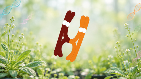
Meiotic crossovers are relocated to polymorphic regions across the Arabidopsis genome through an MSH2-dependent mismatch-repair pathway. Meiotic crossovers reshuffle parental alleles and determine which traits are inherited together. Madec and colleagues demonstrate that, in Arabidopsis thaliana, sequence polymorphism between homologous chromosomes is itself a powerful determinant of where crossovers form. Making use of unique genetic material, the authors managed to compare the same genomic regions when they were polymorphic or non-polymorphic. Polymorphic regions recombined more frequently across the genome and could turn normally cold chromosome-end regions into crossover rich regions, at the expense of other regions of the chromosome. This redistribution depends on the mismatch-repair protein MSH2: without MSH2, crossovers are no longer preferentially directed toward polymorphic regions. The work identifies mismatch repair as a pro-crossover pathway in Arabidopsis and shows how local sequence divergence can extensively reorganize the genome-wide crossover landscape. More information: https://doi.org/10.1038/s41467-026-76213-z Contact: Chloé Girard chloe.girard@i2bc.paris-saclay.fr
|
|
Scooped by
I2BC Paris-Saclay
September 30, 10:00 AM
|
Targeted genome editing of the non-model cyanobacterium Cyanothece PCC 7425 via CRISPR/Cas12a
Unlocking Genetic Engineering in the CaCO3 / SrCO3 granule forming Cyanothece PCC 7425 Cyanobacterial biodiversity represents an outstanding reservoir of biological diversity with considerable potential for addressing challenges in diverse fields, ranging from human nutrition and health to sustainable production of energy and environmental bioremediation. Among non-model cyanobacteria, several species, including Gloeomargarita lithophora and Cyanothece PCC 7425 are distinguished by their remarkable ability to form intracellular CaCO3 granules within the cytoplasm (iACC). In addition to accumulating high levels of calcium (Ca), these cyanobacteria can concentrate other alkaline earth elements, including strontium (Sr), barium (Ba), and radium (Ra). This unique capacity makes them promising candidates for a range of biotechnological applications, notably CO2 capture and sequestration, and bioremediation of nuclear effluents. However, the absence of efficient genetic tools for iACC forming cyanobacteria has been a major obstacle to investigating the molecular mechanisms underlying iACC formation. To address this limitation, the Biology and Biotechnology of Cyanobacteria team at I2BC, CEA Saclay, in collaboration with Biomin team from IMPMC Sorbonne University has established a genetic toolbox for Cyanothece PCC 7425 enabling both gene overexpression and CRISPR-Cas-mediated gene inactivation 1. These advances provide, for the first time, a powerful experimental framework to dissect the molecular basis of iACC formation and pave the way toward a deeper understanding of this unique biomineralization process. The team, using CRISPR-Cas12a mediated homologous recombination, generated deletion mutants of genes acting in metabolism and/or response to stresses. Importantly, full chromosome segregation was rapidly achieved making selection of null-mutants very effective in this cyanobacterium. With these tools in hand, the team now aims to elucidate the molecular basis of iACC mechanisms and the formation of CaCO3 / SrCO3 granules. More information : https://link.springer.com/article/10.1007/s00253-026-13959-y Contact : Soufian Ouchan soufian.ouchane@i2bc.paris-saclay.fr https://www.i2bc.paris-saclay.fr/equipe-biology-and-biotechnology-of-cyanobacteria/
|
|
Scooped by
I2BC Paris-Saclay
September 22, 8:11 AM
|
JIP3 and JIP4 proteins: much more than simple adapters for vesicle transport.
The JIP3 and JIP4 proteins (JIP3/4) do not merely serve as adapters linking the molecular motors kinesin-1 and dynein to their vesicular cargoes for bidirectional transport along microtubules. They also act as key regulators of this transport, capable of activating these motors by relieving their autoinhibited state. In a recent study published in the Journal of Biological Chemistry, scientists from I2BC (CNRS/CEA-Jacob/Université Paris-Saclay, Gif-sur-Yvette) characterized the interaction between JIP3/4 and KIF5B in vitro. To do this, they employed molecular biology techniques alongside a biophysical approach known as MicroScale Thermophoresis (PIM platform, I2BC). The study identified the minimal regions of these proteins required for their interaction: the N-terminal N-RH1 domain of JIP3/4 and the CC4 region of KIF5B. An unexpected discovery emerged from the study: JIP3 and JIP4 exhibit a 40-fold difference in affinity for KIF5B. To further investigate these mechanisms, the researchers conducted extensive site-directed mutagenesis on the N-RH1 region of JIP3/4, targeting accessible charged residues and sequence differences between JIP3 and JIP4. Their results reveal the existence of multiple negatively charged, low-affinity binding sites and the involvement of the unstructured N-terminal domain; together, these elements define an extensive interaction surface with KIF5B on the JIP3 N-RH1 domain.
This work sheds new light on how JIP3/4 might relieve kinesin-1 auto-inhibition. Indeed, in its auto-inhibited form, kinesin-1 is stabilized by the CC4 region. By interacting extensively with the latter, JIP3/4 destabilize this conformation, thereby enabling activation of the molecular motor. More information : https://www.jbc.org/article/S0021-9258(26)02227-1/fulltext Contact : Paola Llinas paola.llinas@i2bc.paris-saclay.fr and Julie Ménétrey julie.menetrey@i2bc.paris-saclay.fr
|
|
Scooped by
I2BC Paris-Saclay
September 14, 5:09 AM
|
Sensing forces: How plants convert mechanical cues into biological responses
Plants adapt their growth to the mechanical stresses of their environment, such as wind, for example. However, the mechanisms by which a mechanical stimulus is translated into a biological response are only just beginning to be understood. Plants encounter mechanical constraints from environmental forces and morphogenesis. Sensing these constraints induces downstream responses that are crucial for plant development and its plasticity in a changing environment. Here, we first provide an overview of the mechanical constraints acting on plants at multiple scales, ranging from organelles to organs. We then focus on the coupling between mechanical stimuli and biological signaling, referred to as mechanotransduction. We highlight distinct modes of coupling, including the sensing of lipid compaction and cell wall status by receptors and channels, as well as the mechanically induced reorganization of cellular components such as the cytoskeleton and plasma membrane. Finally, we discuss the diversity of downstream responses triggered by mechanotransduction and their temporal dynamics.
We propose that the diversity of mechanotransduction modalities allows plants to decode mechanical signatures characterized by specific stimulus intensities, durations, and frequencies, thereby eliciting adaptive cellular and developmental responses. More information: 10.1016/j.devcel.2026.05.003 Contact: Sébastien Thomine sébastien.thomine@i2bc.paris-saclay.fr
|
|
Scooped by
I2BC Paris-Saclay
September 4, 4:22 AM
|
Different Photons, Same Chemistry: Identical Mechanism and Efficiency of Fatty Acid Photodecarboxylase under Blue and UV Excitation.
Blue or violet, FAP plays by the same rules: both wavelengths trigger the same photocycle with identical efficiency, intermediates, and products, overturning the proposed model of violet-enhanced activity. Fatty acid photodecarboxylase (FAP) is a promising photoenzyme for light‑driven hydrocarbon synthesis and a versatile chassis for photobiocatalysis. A recent study proposed that UV/violet illumination enhances FAP activity relative to blue light by accessing a distinct, more efficient photochemical regime. Here, steady-state and time‑resolved fluorescence, transient absorption spectroscopy under single‑turnover conditions, and product quantification under continuous irradiation show that the quantum yield of decarboxylation, the identities and kinetics of reaction intermediates, and the overall chemical yield are identical for blue and UV/violet excitation. These findings establish that FAP operates through a single, wavelength‑independent photocycle, overturning the proposed “violet‑enhanced” reactivity model. More information : https://doi.org/10.1021/acscatal.6c04891 Contact : pavel.muller@i2bc.paris-saclay.fr
|
|
Scooped by
I2BC Paris-Saclay
August 3, 9:14 AM
|
Training session on TEM in cell biology - 13-16 October 2026
|
|
Scooped by
I2BC Paris-Saclay
July 20, 9:20 AM
|
Study on protein–bicelle interactions using switchSENSE technology with the PIM facility at I2BC
Using bicelles as membrane mimetics and switchSENSE technology available at the PIM facility, we uncover new insights into the dynamic mechanisms governing weak protein–membrane interactions, revealing how proteins recognize and interact with membrane environments. To investigate the association of human dystrophin with biomimetic phospholipid bicelles, we employed switchSENSE®, a fluorescence-based biosensing technology that relies on dynamic DNA nanolevers operating in a microfluidic environment while requiring only minimal sample amounts. We demonstrate that this technology enables real-time kinetic analysis of weak and transient interactions, providing a significant advantage over conventional biophysical methods, which often struggle to detect low-affinity protein–lipid binding events. Focusing on two functionally relevant fragments from the central rod domain of dystrophin (R1-3 and R11-15), we quantified their interactions with zwitterionic and anionic bicelles using two complementary experimental configurations: either the proteins were immobilized and the bicelles served as analytes, or vice versa. Both approaches yielded consistent micromolar affinities, in agreement with our previous microscale thermophoresis data, while additionally providing association and dissociation rate constants. This study represents the first kinetic characterization of dystrophin–bicelle interactions. Our results reveal that the observed low affinity is largely driven by the rapid dissociation of the resulting complexes. Notably, the R11-15 fragment displayed a faster association rate and a slower dissociation rate than R1-3, indicating a more stable and long-lasting interaction with lipid assemblies. Beyond providing new mechanistic insights into the reversible membrane association of dystrophin—a process thought to contribute to maintaining sarcolemmal integrity during cycles of muscle contraction and stretching—our work highlights switchSENSE® as a versatile platform for quantifying weak protein–lipid interactions. By delivering kinetic resolution for interactions that lie at the detection limits of conventional techniques, switchSENSE® offers a powerful approach for investigating the dynamic interplay between structural proteins and lipid assemblies. For more information: Article: https://link.springer.com/article/10.1007/s00249-026-01829-4 Contact: sophie.combet@i2bc.paris-saclay.fr ; paloma.fernandez-varela@polytechnique.edu Facility web site: https://www.i2bc.paris-saclay.fr/structural-biology/pim/
|
|
Scooped by
I2BC Paris-Saclay
June 16, 4:34 AM
|
Reassessing Carotenoid Photophysics: Shedding Light on Dark States.
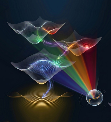
The dark excited states of carotenoids have been debated for decades. Researchers at I2BC now use femtosecond stimulated resonance Raman spectroscopy to determine the nature and symmetry of three of them, resolving controversies that have long obscured our understanding of photosynthetic light harvesting. Carotenoid molecules are critical in photosynthesis, performing functions at the heart of both light-harvesting and photoprotection. As both these processes involve excitation energy transfer, fully understanding them requires a precise description of the electronic states involved. The excited-state manifold of carotenoids is not yet fully characterised, and includes several dark electronic states that remain elusive. Using femtosecond stimulated resonance Raman spectroscopy, where the vibrational contributions of each excited state can be observed selectively as a function of the Raman excitation, we reveal the nature and symmetry of no less than three different dark states. These results end long-standing controversies in carotenoid research, shining new light on the photophysics of these essential molecules and establishing a spectroscopic framework for characterising their multiple roles. The figure is an artistic representation of the excited states in carotenoids. More information : https://pubs.acs.org/doi/10.1021/jacs.6c03864 Contact : Manuel Llansola-Portoles manuel.llansola-portoles@i2bc.paris-saclay.fr
|
|
Scooped by
I2BC Paris-Saclay
June 10, 9:28 AM
|
The final of the interuniversity 3-Minute Thesis (3MT®) competition
Linnéa Strandberg, PhD student from Photobiology, Photosynthesis, Photocatalysis team in I2BC, will present her thesis project “When breaking the heart of a plant”. The Institut Polytechnique de Paris will host the final of the interuniversity 3-Minute Thesis (3MT®) competition. Developed by The University of Queensland, the 3MT® is more than a competition—it’s a platform for PhD students to strengthen their communication skills in english and share their research with a non-specialist audience in a clear and engaging manner.
Join on June 25, from 2:00 pm to 4:00 pm at Télécom Paris (Thévenin lecture hall) to support the finalists and vote for the Audience Award (free registration required): https://www.ip-paris.fr/form/3-minute-thesis-registration.
|
|
Scooped by
I2BC Paris-Saclay
June 8, 9:13 AM
|
3R 2026: A Great Edition in La Grande-Motte
The 3R Congress brings together researchers working on DNA Replication, Repair and Recombination, from fundamental molecular mechanisms to clinical applications, promoting scientific exchange within the community. Twenty-two participants from the B3S and Genome departments of I2BC attended the 16th 3R Congress, which took place in La Grande-Motte from May 18 to 21, 2026. High-quality science, lively discussions and a friendly atmosphere once again made this edition a great success. Many thanks to all our sponsors, and especially to I2BC, for their valuable support.
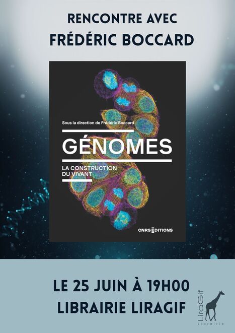
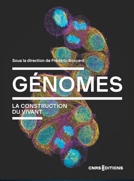
Rencontre-Dédicace avec Frédéric Boccard, Mireille Bétermier, Sarah Lambert et Bernard Dujon Le 1er Juillet 2026 de 19h00 à 20h00 Librairie Liragif 15 Square de la Mairie , 91190 Gif-sur-Yvette Pour Génomes, la construction du vivant paru aux éditions CNRS Une présentation des génomes, de leur séquençage et des innovations permises par leur analyse en biologie. Les auteurs exposent comment l'étude de cette information génétique permet de dévoiler les mécanismes de l'évolution, de retracer l'histoire adaptative des espèces ou encore de décrypter le fonctionnement intime des virus. Frédéric Boccard est directeur de recherche CNRS, directeur de l’Institut de Biologie Intégrative de la Cellule – I2BC (CNRS/CEA/UPSaclay, Gif-sur-Yvette). Il sera accompagné de Mireille Bétermier directrice de recherche à l'I2BC, Sarah Lambert et Bernard Dujon. -> Contact : frederic.boccard@i2bc.paris-saclay.fr
Via Life Sciences UPSaclay
|
|
Scooped by
I2BC Paris-Saclay
May 13, 4:22 AM
|
A coherent structural picture of the interaction of Tau with tubulin provides a link to its aggregation.
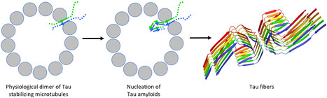
Tau is a protein regulating microtubule dynamics which also forms neurofibrillary tangles in pathological conditions. Recent results suggest that a physiological dimer of Tau could serve as a nucleus for its aggregation. Tauopathies are a group of neurodegenerative diseases characterized by the presence of insoluble filaments of the Tau protein in the brain. In physiological conditions, Tau is involved in the regulation of microtubule dynamics. The study of its interaction with different tubulin assemblies, using various experimental approaches, leads to a seemingly disparate picture. In this opinion-type article, we integrate this information into a model of how Tau participates in microtubule assembly and stabilization. Related to its intrinsically disordered nature, the binding of Tau to microtubules involves both specific interactions, along protofilaments, and non-specific ones, with the C-terminal region of tubulin subunits. In addition, a Tau:tubulin structure that we recently determined leads to a model of a functional Tau dimer targeting a microtubule aperture between protofilaments. This model also provides a framework for a Tau aggregation that would be initiated on the microtubule. More information : https://www.jbc.org/article/S0021-9258(26)01958-7/fulltext Contact : Benoît Gigant benoit.gigant@i2bc.paris-saclay.fr
|
|
|
Scooped by
I2BC Paris-Saclay
October 8, 2:54 AM
|
L'I2BC met la Science en Fête !
Les 3 et 4 octobre 2026, l’I2BC était au rendez-vous de la Fête de la Science à l’ENS Paris-Saclay, avec une belle mobilisation de 34 volontaires, dont 15 doctorants. Au programme, des ateliers autour des cristaux, des stomates, des aliments fermentés du monde et la fabrication d’un microscope en carton. Manipuler, expérimenter, observer au microscope, bricoler… mais aussi échanger avec les scientifiques : il y en avait pour tous les goûts et tous les âges ! Une belle occasion de partager notre passion pour les sciences et de susciter curiosité et émerveillement. Grâce à l’enthousiasme et à l’engagement de tous les volontaires, ce week-end a été un joli moment de découverte, de partage et de convivialité !
|
|
Scooped by
I2BC Paris-Saclay
October 1, 5:31 AM
|
Unveiling a missing component of the atypical type IV secretion system required for natural transformation of Helicobacter pylori.
Identification of a Missing Component of the Exogenous DNA Internalisation System During Natural Transformation in Helicobacter pylori. Horizontal gene transfer plays a crucial role in the evolution of bacterial populations. In pathogens, it promotes the spread of antibiotic resistances and virulence factors. Helicobacter pylori, a bacterium infecting the stomach of 50% of the world's population and responsible for the majority of gastric ulcers and cancers, has a remarkable capacity to exchange genes via natural transformation. Through this pathway, bacteria can capture and incorporate into their genome DNA present in their environment. To do this, H. pylori uses an atypical DNA uptake system: a type 4 secretion system (T4SS) named ComB, rather than the pilus present in all other naturally transformable bacteria. Compared to classical T4SS, certain components appeared to be absent from this system. In a study published in PLoS Pathogens, researchers from the Department of Cellular and Molecular Radiobiology (François Jacob Institute of Biology, CEA/UPSaclay, UP Cité, Fontenay aux Roses) and I2BC (CEA/CNRS/UPSaclay, Gif-sur-Yvette), in collaboration with a laboratory at the Pasteur Institute, identified an ATPase essential for this secretion system. Homology searches detected a gene, located outside the known operons of the ComB complex, capable of encoding an ATPase associated with a T4SS. Genetics and biochemistry experiments demonstrated that the protein encoded by this gene, designated as ComB11, is indeed an ATPase essential for natural transformation of H. pylori. Structural modelling revealed an interaction between this ATPase and the ComB4 component of the ComB system, confirming its membership in this atypical T4SS. More information: https://journals.plos.org/plospathogens/article?id=10.1371/journal.ppat.1014140 Contact: Jessica Andreani jessica.andreani@i2bc.paris-saclay.fr
|
|
Scooped by
I2BC Paris-Saclay
September 25, 6:42 AM
|
How the polarisability of its environment shapes the electronic excited states of lutein, a key pigment of photosynthesis.
Lutein helps plants harvest sunlight and protect themselves from excess light. Ultrafast laser spectroscopy at I2BC shows that the polarisability of its environment reshapes its electronic excited states and their lifetimes. In plant leaves, lutein, a yellow carotenoid also abundant in spinach and egg yolk, is bound to the proteins that collect sunlight for photosynthesis. It helps capture light, stabilises these proteins and, under strong illumination, contributes to dissipating excess energy safely. All these functions rely on the electronic excited states that lutein reaches after absorbing a photon, which last only a few picoseconds (trillionths of a second). Inside these proteins, lutein is thought to sit in a highly polarisable environment, that is, one whose electron clouds readily deform in response to the pigment. How this polarisability affects its excited states was unknown. I2BC researchers compared lutein in two solvents of very different polarisability: tetrahydrofuran (THF, low) and carbon disulfide (CS2, high). Combining femtosecond transient absorption with femtosecond stimulated resonance Raman spectroscopy (FSRRS), which selectively records the vibrational fingerprint of each electronic state, they identified the same set of excited states in both solvents: a vibrationally 'hot' S1 state, the relaxed S1 state, a weak charge-transfer contribution and a 'dark' state called S*. High polarisability, however, changes how these states behave. The S1 absorption shifts to lower energy and broadens, and the molecule barely reorganises after excitation. The S* state lives almost twice as long (about 29 instead of 16 picoseconds), and its vibrational frequency deviates from the trend expected from the length of the conjugated chain. Polarisability therefore does not simply lower the energy of the excited states: it alters their nature and dynamics. These results provide a reference for understanding how the protein environment tunes the excited states of lutein, and thus its roles in light harvesting and photoprotection. More information : Contact : Manuel Llansola-Portoles manuel.llansola@i2bc.paris-saclay.fr
|
|
Scooped by
I2BC Paris-Saclay
September 14, 9:44 AM
|
Total synthesis and structural characterization of a novel protein scaffold from the snail Biomphalaria glabrata.
Total synthesis and structural characterization of a novel protein scaffold from the snail Biomphalaria glabrata, the host of the parasite schistosoma. Schistosomiasis, also known as bilharzia, remains one of the most widespread tropical diseases, affecting over 280 million people, particularly in Africa. This acute and chronic parasitic disease is caused by flatworms of the genus Schistosoma, whose complex life cycle alternates between vertebrate hosts and freshwater snails, the latter serving as intermediate hosts.
In collaboration with Oleg Melnyk and Jérôme Vicogne from the MinT team at the Institut Pasteur de Lille (CIIL/CNRS), Solange Moréra’s team at the I2BC studied miniproteins (schistosomins) of approximately 80 residues from the snail Biomphalaria glabrata. These miniproteins are conserved in gastropods and belong to a long-standing orphan family whose structure and biological roles had until now remained unknown.
A recently published study describes the total chemical synthesis and structural characterization of a schistosomin isoform. Using solid-phase peptide synthesis, chemoselective peptide ligation, and controlled oxidative folding, the miniprotein was crystallized and its structure resolved. This structure, unprecedented in the Protein Data Bank, reveals a novel disulfide-rich fold with remarkable thermal stability. In silico analyses suggest that the two natural isoforms (which differ by only a single residue) exhibit very similar structural and dynamic properties.
Transcript and protein analyses across different snail tissues provide the first spatial expression map of schistosomin. These findings establish schistosomin as the prototype of a new family of gastropod miniproteins, whose biological roles in mollusc-parasite interactions remain to be explored. More information : https://pubmed.ncbi.nlm.nih.gov/42554520/ Contact: Solange Morera solange.morera@i2bc.paris-saclay.fr
|
|
Scooped by
I2BC Paris-Saclay
September 10, 5:18 AM
|
A new path to Rubisco processing
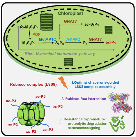
A unique plant machinery comprising three dedicated components ensures N-terminal processing of Rubisco’s catalytic subunit, enabling its proper accumulation and activity as Earth’s most abundant protein. RuBP carboxylase/oxygenase (Rubisco)—the central enzyme of carbon dioxide assimilation and plant autotrophy— is Earth’s most abundant protein and a major carbon and nitrogen reservoir, which can be remobilized during stress and development. The catalytic large subunit of Rubisco (RbcL) undergoes a highly conserved maturation process that results in N-terminal proline acetylation, a modification not reported in other biological systems. Until now, both the enzymatic machinery responsible for N-terminal maturation and its associated functional implications have remained unknown. In this study, we uncover a specialized chloroplast processing pathway, composed of two aminopeptidases and one N-terminal acetyltransferase, specifically evolved to catalyze RbcL maturation. Proper processing of RbcL by this machinery is tightly coupled to chaperone-mediated assembly of the hexadecameric holoenzyme, enhances activation by Rubisco activase, and safeguards the fully modified complex from premature degradation during senescence. Disruption of this pathway compromises both the reactivity and accumulation of Rubisco. These findings reveal a specialized maturation mechanism that underpins the exceptional abundance and persistence of Rubisco in photosynthetic organisms, and more broadly, its central role in sustaining life on Earth. This paves the way for enhanced photosynthesis, stronger crop yields, and more effective carbon capture. More information : https://www.science.org/doi/full/10.1126/sciadv.aeh5060 Contact : Thierry Meinnel thierry.meinnel@i2bc.paris-saclay.fr
|
|
Scooped by
I2BC Paris-Saclay
August 3, 9:33 AM
|
Confocal microscopy workshop 2026
|
|
Scooped by
I2BC Paris-Saclay
July 27, 4:36 AM
|
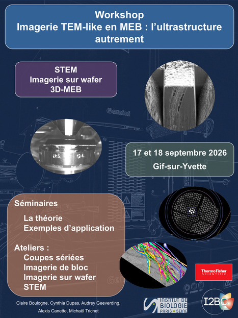
Workshop in Sept. @I2BC: Come & Explore the Many Facets of Scanning Electron Microscopy
|
|
Scooped by
I2BC Paris-Saclay
June 18, 10:35 AM
|
NMR-based detection of ultra-weak antibody–excipient interactions to optimize therapeutic monoclonal antibody formulations.
The aggregation and high viscosity of monoclonal antibodies at high concentrations restrict their administration by subcutaneous injection. We developed an NMR-based approach to detect ultra-weak interactions between mAbs and excipients, enabling rational screening of excipients and formulation conditions. The INTGEN team at I2BC, in collaboration with the NMR facilities at CEA Saclay and ICSN in Gif-sur-Yvette, and the formulation department at Sanofi's Research & Development center in Vitry-sur-Seine, investigated antibody–excipient interactions in highly concentrated monoclonal antibody (mAb) formulations designed for subcutaneous injection. At high concentrations, mAbs tend to self-associate, increasing viscosity and complicating administration. By combining NMR spectroscopy, rheology, and light scattering, the research team identified excipients capable of improving mAb solubility and characterized the relationship between excipient binding and its effect on viscosity. Lysine was identified as an excipient that significantly enhances mAb solubility and also directly binds to the antibody. In contrast, sucrose also binds to the mAb but does not reduce viscosity, demonstrating that binding alone is not sufficient to predict solubilizing efficacy. This work establishes an NMR-based approach for uderstanding the molecular mechanisms governing mAb viscosity and for rationally optimizing therapeutic antibody formulations. More information : https://www.tandfonline.com/doi/full/10.1080/19420862.2026.2685366 Contact : Sophie Zinn-Justin sophie.zinn@i2bc.paris-saclay.fr
|
|
Scooped by
I2BC Paris-Saclay
June 16, 4:26 AM
|
Structural analysis reveals how Ku recruits and stimulates WRN.
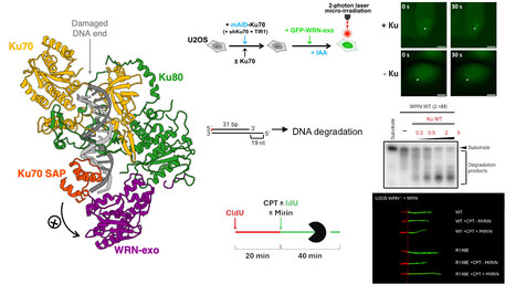
Combining cryo-EM, biochemistry and cell biology, this study reveals how the DNA repair factor Ku recruits and activates the WRN exonuclease, uncovering a new mechanism that protects genome stability during replication stress. The Werner syndrome protein (WRN) is a key factor in the maintenance of genome stability, but the molecular mechanisms regulating its exonuclease activity have remained unknown. In this study, we combine cryo-electron microscopy, biochemistry, and cell biology to uncover how the DNA repair factor Ku recruits and stimulates WRN.
Our structural analysis reveals the molecular interface between the Ku70–Ku80 heterodimer and the WRN exonuclease domain, explaining how Ku positions WRN at DNA ends to stimulate its exonuclease activity. Biochemical assays demonstrate that Ku directly enhances WRN-mediated DNA degradation in vitro. In cells, disruption of the Ku–WRN interaction impairs WRN recruitment to DNA double-strand break (DSB) sites and compromises replication fork remodeling under replication stress, highlighting the physiological importance of this interaction.
Together, these findings demonstrate that Ku directly regulates WRN exonuclease activity and clarify the molecular basis of WRN recruitment and activation at DNA ends, providing new insights into DNA end processing and the response to replication stress. More information : https://doi.org/10.1038/s41467-026-71888-w Contact : Virginie Ropars virginie.ropars@i2bc.paris-saclay.fr
|
|
Scooped by
I2BC Paris-Saclay
June 8, 9:21 AM
|
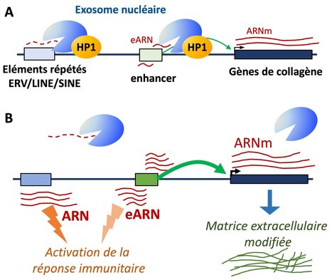
HP1 targets the RNA exosome to chromatin
A novel role for the HP1 proteins in targeting the RNA-exosome to chromatin for the turnover of repetitive RNAs and enhancer RNAs. Heterochromatin protein 1 (HP1), a hallmark of pericentromeric heterochromatin, is a chromatin-bound regulator of co-transcriptional processes including alternative splicing, but its role in RNA degradation remains unexplored. Here, we uncover a direct interaction between HP1 and nuclear RNA exosome complexes, major RNA decay machineries. In mouse embryonic liver cells, inactivation of all three HP1 isoforms leads to accumulation of retrotransposon-derived RNAs and stabilization of enhancer RNAs. These changes coincide with increased activity at a subset of liver enhancers particularly sensitive to reduced exosome activity, many of which regulate genes encoding extracellular matrix components such as collagen genes. Stratifying hepatocellular carcinoma samples by HP1 expression further reveal that tumors with low HP1 are marked by reduced RNA degradation, and increased expression of a similar subset of genes encoding extracellular matrix components and possibly contributing to tumor stiffness. These results suggest that HP1’s impact on RNA turnover contributes to its function in cancer biology. More information: https://www.nature.com/articles/s41467-026-72504-7 Contact : Carl Mann carl.mann@i2bc.paris-saclay.fr
|
|
Scooped by
I2BC Paris-Saclay
June 5, 3:49 AM
|
New Release: Génomes: la construction du vivant
From DNA sequencing and studies of genome function and organization—covering viruses, bacteria, archaea and eucaryotes, this collective book explores genomes and their role in life, evolution, and diversity. A collective book that traces the history of genetics and the major discoveries surrounding genomes. The genome, the complete set of genetic information, builds, operates, and reproduces all living beings, serving as the engine of their evolution. Thanks to DNA, genetic information is stored, decoded, duplicated, and transmitted. But the study of the genome goes much further: it allows us to understand life, trace the history of species, and identify non-coding DNA regions that are nonetheless essential for life, the protection of genetic material, and its diversity. From DNA sequencing and genome function to the mechanisms of evolution—covering viruses, bacteria, archaea and eucaryotes, —the book explores how scientists today are striving to understand genome function and evolution. Coordinated by Frédéric Boccard, Director of the I2BC, this book brings together chapters written by several French scientists, including Mireille Bétermier, Group Leader at the I2BC. More information: https://www.cnrseditions.fr/catalogue/biologie-et-sante/genomes/ Contact: Frédéric Boccard frederic.boccard@i2bc.paris-saclay.fr
Le pôle des plateformes de biophysique de l’Institut de Biologie Intégrative de la Cellule (I2BC, CNRS/CEA/UPSaclay, Gif-sur-Yvette) a le plaisir d’accueillir Viola Caroline D’mello, qui a rejoint le CEA en mars 2025 en tant qu’ingénieure-chercheuse. Après une licence en chimie à l’université de Mumbai et un master en chimie analytique à l’université de Mangalore, elle a réalisé sa thèse au Tata Institute of Fundamental Research (TIFR) à Mumbai. Ses travaux portaient sur l’étude en phase gazeuse de liaisons hydrogène dans des molécules aromatiques azotées par spectroscopies dans les domaines de l’ultraviolet (UV) et de l’infrarouge (IR) nanoseconde – ces liaisons hydrogène étant similaires à celles présentes dans l’ADN et l’ARN. En 2019, elle a rejoint en tant que chercheuse postdoctorante le groupe de recherche LIDYL de l’institut IRAMIS au CEA Saclay, où elle a étudié les paires d’ions et le repliement de peptides en phase gazeuse. Après un court passage dans l’industrie à Bangalore (Inde), elle est revenue à la recherche académique comme postdoctorante au Département de Physique de l’Université de Göteborg, où elle a contribué à la mise en service d’un spectromètre de masse haute résolution couplé à une source à jet supersonique. Depuis son arrivée au sein du Pôle de biophysique de l’I2BC, elle est responsable des plateformes de spectroscopies électroniques, de spectroscopie Raman de résonance et de spectroscopie infrarouge : elle supervise l’accès des utilisateurs et veille à ce que les expériences soient conçues et réalisées dans des conditions optimales. Elle accompagne et conseille régulièrement les utilisateurs sur un large éventail de techniques, incluant l’absorption UV-Visible, l’absorption transitoire ultrarapide (de la femtoseconde à la milliseconde), la spectroscopie infrarouge à transformée de Fourier (FTIR) et la spectroscopie Raman, dont la spectroscopie Raman femtoseconde (plateforme LUMA). L’étendue de son expertise en méthodes photophysiques, associée à sa fiabilité et à son attitude ouverte et collaborative, fait d’elle un pilier central du Pôle de biophysique. -> Contact : Viola-Caroline D'mello (Viola.Dmello@cea.fr) Plug In Labs Université Paris-Saclay : cliquer ICI I2BC / Plateforme de spectroscopies électroniques. La plateforme de Spectroscopies Électroniques (Institut de Biologie Intégrative de la Cellule) offre ses services appliqués aux biomolécules à des équipes de recherche françaises et internationales. Nous sommes capables de suivre des changements spectroscopiques au niveau de la protéine dans des cellules intactes. La plateforme est équipée de plusieurs spectromètres d'absorption et de fluorescence (y compris un certain nombre de spectromètres PAM spécialisés) ainsi que des spectromètres à thermoluminescence. Pour certaines configurations, des cryostats sont disponibles pour les études à basse température, jusqu'à 77K ou 4K. La plateforme a développé (et continue à améliorer) un montage unique de spectroscopie optique résolue dans le temps (ca. 300 ps), surpassant les montages conventionnels (commerciaux) en sensibilité et en résolution temporelle. Ce type de méthodologie est particulièrement déterminant pour l'élucidation de processus irréversibles et/ou de processus qui se produisent dans des fenêtres temporelles allant de quelques centaines de picosecondes à des dizaines de nanosecondes, où les montages conventionnels performent mal ou ne peuvent pas être utilisées du tout. Cette plateforme fait partie du pôle des plateformes de Biophysiques de l'I2BC qui comprend les plateformes de RPE, FTIR, Résonance Raman, Spectroscopies Electroniques et Microscopie de fluorescence à super-résolution. I2BC / Plateforme de spectroscopie RAMAN de résonance. Cette plateforme met à disposition des équipements de spectroscopies avancées Raman et FLN (7 spectromètres, avec plusieurs accessoires, large gamme de température possible (thermostats 273-320 K, cryostats 4-250 K)). Analyses faisables sur échantillons de toutes formes physiques, en particulier ceux qui contiennent des molécules pigmentées (voir ci-dessous). Le laboratoire se spécialise sur les propriétés physico-chimiques des cofacteurs pigmentés en biologie (caroténoïdes, chlorophylles, hèmes, flavines, …), y compris des études in vivo des réactions biochimiques, photo-induites et régulatrices. L'état de l'échantillon (liquide, poudre, gel, solide, …) limité seulement par la taille du signale (présence de molécules pigmentées nécessaires pour des mesures en milieux complexes). Exemples récents : processus régulatoires dans des membranes photosynthétiques in vivo (feuilles entières et micro-organismes), structure moléculaire des caroténoïdes et opsines dans la rétine humaine ex vivo. Cette plateforme fait partie du pôle des plateformes de Biophysiques de l'I2BC qui comprend les plateformes de RPE, FTIR, Résonance Raman, Spectroscopies Electroniques et Microscopie de fluorescence à super-résolution. I2BC / Plateforme de spectroscopie IRTF. La plateforme de spectroscopie IRTF est située au Laboratoire des Mécanismes Fondamentaux en Bioénergétique (UMR 9198). Elle met à disposition des utilisateurs des spectromètres IRTF avancés et elle est équipée pour répondre à la plus grande partie des besoins des analyses IRTF. La plateforme comprend 4 spectromètres avec plusieurs accessoires : cellule à transmission, accessoires ATR, cellule électrochimique, thermostats, cryostats pour expériences à basse température... Elle permet l'étude d'échantillons sous différentes formes (liquide, solide, poudre..). Le laboratoire est spécialisé dans la spectroscopie IRTF différentielle, résolue dans le temps, à basse température, et possède une bonne expertise dans l'étude de réactions biochimiques et photo-induites. La plateforme de spectroscopie IRTF fait partie du pôle des plateformes de Biophysiques de l'I2BC qui comprend les plateformes de RPE, FTIR, Résonance Raman, Spectroscopies Electroniques et Microscopie de fluorescence à super-résolution. A propos de l’Institut de Biologie Intégrative de la Cellule (I2BC - UMR 9198). L’I2BC est une Unité Mixte de Recherche (CEA, CNRS, Université Paris-Saclay), accueillant une soixantaine d’équipes de recherche et hébergeant 17 plateformes technologiques, réparties en 6 pôles. 2025 a aussi été une année clé pour l’I2BC : cette unité a fêté ses 10 ans !
Via Life Sciences UPSaclay
|