Your new post is loading...

|
Scooped by
I2BC Paris-Saclay
October 29, 5:19 AM
|
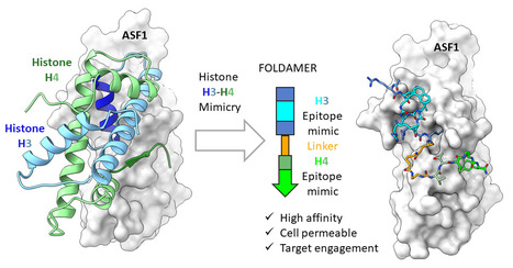
One foldamer, two histone epitopes: mimicking an extended protein interface.
Can a synthetic molecule mimic an extended protein interface? Peptide-oligourea foldamers mimic two histone epitopes recognized by anti-silencing function 1, an emerging and challenging target in oncology, with nanomolar affinity and cell permeability. Extended protein-protein interfaces are challenging targets for synthetic ligands competing with endogenous partners, as most peptidomimetics target a single recognition motif. The histone chaperone anti-silencing function 1 (ASF1), involved in chromatin assembly and genome maintenance and an emerging target in oncology, illustrates this challenge: its essential interaction with the H3-H4 histone dimer spans two epitopes, an H3 α-helix and an H4 β-strand. To target this interface, the Molecular Assemblies and Genome Integrity team at I2BC (Université Paris-Saclay, CEA, CNRS) joined forces with Gilles Guichard’s team at CBMN (Laboratoire de Chimie et Biologie des Membranes et Nanoobjets) and IECB (Institut Européen de Chimie et Biologie) in Bordeaux. Their approach combines peptide-oligourea foldamer chemistry and synthesis with molecular design, structural biology, and biophysical characterization. Foldamers are synthetic molecules mimicking structural features of proteins. High-resolution crystal structures obtained with researchers at Synchrotron SOLEIL guided the design of peptidomimetics combining both histone epitopes. These foldamers reproduce the natural H3-H4 binding mode on ASF1, with affinities reaching 6 nanomolar, and resist degradation in human plasma. This optimized compound binds endogenous ASF1 in cell extracts. Cell biology studies with the PICCEL facility at the CEA Fontenay aux Roses (formerly PARi) demonstrated their cell permeability and antiproliferation activity. Selected as a Very Important Paper (VIP) in Angewandte Chemie International Edition, this collaborative study highlights a strategy for targeting extended protein-protein interfaces. The resulting foldamers provide tools to investigate ASF1 function and may help target other challenging intracellular protein-protein interactions. More information : https://onlinelibrary.wiley.com/doi/10.1002/ange.4112426 Contact : Françoise Ochsenbein francoise.ochsenbein@i2bc.paris-saclay.fr
|
|
Scooped by
I2BC Paris-Saclay
October 8, 2:54 AM
|
L'I2BC met la Science en Fête !
Les 3 et 4 octobre 2026, l’I2BC était au rendez-vous de la Fête de la Science à l’ENS Paris-Saclay, avec une belle mobilisation de 34 volontaires, dont 15 doctorants. Au programme, des ateliers autour des cristaux, des stomates, des aliments fermentés du monde et la fabrication d’un microscope en carton. Manipuler, expérimenter, observer au microscope, bricoler… mais aussi échanger avec les scientifiques : il y en avait pour tous les goûts et tous les âges ! Une belle occasion de partager notre passion pour les sciences et de susciter curiosité et émerveillement. Grâce à l’enthousiasme et à l’engagement de tous les volontaires, ce week-end a été un joli moment de découverte, de partage et de convivialité !
|
|
Scooped by
I2BC Paris-Saclay
September 25, 6:42 AM
|
How the polarisability of its environment shapes the electronic excited states of lutein, a key pigment of photosynthesis.
Lutein helps plants harvest sunlight and protect themselves from excess light. Ultrafast laser spectroscopy at I2BC shows that the polarisability of its environment reshapes its electronic excited states and their lifetimes. In plant leaves, lutein, a yellow carotenoid also abundant in spinach and egg yolk, is bound to the proteins that collect sunlight for photosynthesis. It helps capture light, stabilises these proteins and, under strong illumination, contributes to dissipating excess energy safely. All these functions rely on the electronic excited states that lutein reaches after absorbing a photon, which last only a few picoseconds (trillionths of a second). Inside these proteins, lutein is thought to sit in a highly polarisable environment, that is, one whose electron clouds readily deform in response to the pigment. How this polarisability affects its excited states was unknown. I2BC researchers compared lutein in two solvents of very different polarisability: tetrahydrofuran (THF, low) and carbon disulfide (CS2, high). Combining femtosecond transient absorption with femtosecond stimulated resonance Raman spectroscopy (FSRRS), which selectively records the vibrational fingerprint of each electronic state, they identified the same set of excited states in both solvents: a vibrationally 'hot' S1 state, the relaxed S1 state, a weak charge-transfer contribution and a 'dark' state called S*. High polarisability, however, changes how these states behave. The S1 absorption shifts to lower energy and broadens, and the molecule barely reorganises after excitation. The S* state lives almost twice as long (about 29 instead of 16 picoseconds), and its vibrational frequency deviates from the trend expected from the length of the conjugated chain. Polarisability therefore does not simply lower the energy of the excited states: it alters their nature and dynamics. These results provide a reference for understanding how the protein environment tunes the excited states of lutein, and thus its roles in light harvesting and photoprotection. More information : Contact : Manuel Llansola-Portoles manuel.llansola@i2bc.paris-saclay.fr
|
|
Scooped by
I2BC Paris-Saclay
September 14, 9:44 AM
|
Total synthesis and structural characterization of a novel protein scaffold from the snail Biomphalaria glabrata.
Total synthesis and structural characterization of a novel protein scaffold from the snail Biomphalaria glabrata, the host of the parasite schistosoma. Schistosomiasis, also known as bilharzia, remains one of the most widespread tropical diseases, affecting over 280 million people, particularly in Africa. This acute and chronic parasitic disease is caused by flatworms of the genus Schistosoma, whose complex life cycle alternates between vertebrate hosts and freshwater snails, the latter serving as intermediate hosts.
In collaboration with Oleg Melnyk and Jérôme Vicogne from the MinT team at the Institut Pasteur de Lille (CIIL/CNRS), Solange Moréra’s team at the I2BC studied miniproteins (schistosomins) of approximately 80 residues from the snail Biomphalaria glabrata. These miniproteins are conserved in gastropods and belong to a long-standing orphan family whose structure and biological roles had until now remained unknown.
A recently published study describes the total chemical synthesis and structural characterization of a schistosomin isoform. Using solid-phase peptide synthesis, chemoselective peptide ligation, and controlled oxidative folding, the miniprotein was crystallized and its structure resolved. This structure, unprecedented in the Protein Data Bank, reveals a novel disulfide-rich fold with remarkable thermal stability. In silico analyses suggest that the two natural isoforms (which differ by only a single residue) exhibit very similar structural and dynamic properties.
Transcript and protein analyses across different snail tissues provide the first spatial expression map of schistosomin. These findings establish schistosomin as the prototype of a new family of gastropod miniproteins, whose biological roles in mollusc-parasite interactions remain to be explored. More information : https://pubmed.ncbi.nlm.nih.gov/42554520/ Contact: Solange Morera solange.morera@i2bc.paris-saclay.fr
|
|
Scooped by
I2BC Paris-Saclay
September 4, 4:22 AM
|
Different Photons, Same Chemistry: Identical Mechanism and Efficiency of Fatty Acid Photodecarboxylase under Blue and UV Excitation.
Blue or violet, FAP plays by the same rules: both wavelengths trigger the same photocycle with identical efficiency, intermediates, and products, overturning the proposed model of violet-enhanced activity. Fatty acid photodecarboxylase (FAP) is a promising photoenzyme for light‑driven hydrocarbon synthesis and a versatile chassis for photobiocatalysis. A recent study proposed that UV/violet illumination enhances FAP activity relative to blue light by accessing a distinct, more efficient photochemical regime. Here, steady-state and time‑resolved fluorescence, transient absorption spectroscopy under single‑turnover conditions, and product quantification under continuous irradiation show that the quantum yield of decarboxylation, the identities and kinetics of reaction intermediates, and the overall chemical yield are identical for blue and UV/violet excitation. These findings establish that FAP operates through a single, wavelength‑independent photocycle, overturning the proposed “violet‑enhanced” reactivity model. More information : https://doi.org/10.1021/acscatal.6c04891 Contact : pavel.muller@i2bc.paris-saclay.fr
|
|
Scooped by
I2BC Paris-Saclay
June 18, 10:35 AM
|
NMR-based detection of ultra-weak antibody–excipient interactions to optimize therapeutic monoclonal antibody formulations.
The aggregation and high viscosity of monoclonal antibodies at high concentrations restrict their administration by subcutaneous injection. We developed an NMR-based approach to detect ultra-weak interactions between mAbs and excipients, enabling rational screening of excipients and formulation conditions. The INTGEN team at I2BC, in collaboration with the NMR facilities at CEA Saclay and ICSN in Gif-sur-Yvette, and the formulation department at Sanofi's Research & Development center in Vitry-sur-Seine, investigated antibody–excipient interactions in highly concentrated monoclonal antibody (mAb) formulations designed for subcutaneous injection. At high concentrations, mAbs tend to self-associate, increasing viscosity and complicating administration. By combining NMR spectroscopy, rheology, and light scattering, the research team identified excipients capable of improving mAb solubility and characterized the relationship between excipient binding and its effect on viscosity. Lysine was identified as an excipient that significantly enhances mAb solubility and also directly binds to the antibody. In contrast, sucrose also binds to the mAb but does not reduce viscosity, demonstrating that binding alone is not sufficient to predict solubilizing efficacy. This work establishes an NMR-based approach for uderstanding the molecular mechanisms governing mAb viscosity and for rationally optimizing therapeutic antibody formulations. More information : https://www.tandfonline.com/doi/full/10.1080/19420862.2026.2685366 Contact : Sophie Zinn-Justin sophie.zinn@i2bc.paris-saclay.fr
|
|
Scooped by
I2BC Paris-Saclay
June 16, 4:26 AM
|
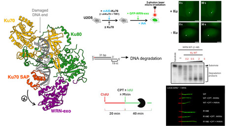
Structural analysis reveals how Ku recruits and stimulates WRN.
Combining cryo-EM, biochemistry and cell biology, this study reveals how the DNA repair factor Ku recruits and activates the WRN exonuclease, uncovering a new mechanism that protects genome stability during replication stress. The Werner syndrome protein (WRN) is a key factor in the maintenance of genome stability, but the molecular mechanisms regulating its exonuclease activity have remained unknown. In this study, we combine cryo-electron microscopy, biochemistry, and cell biology to uncover how the DNA repair factor Ku recruits and stimulates WRN.
Our structural analysis reveals the molecular interface between the Ku70–Ku80 heterodimer and the WRN exonuclease domain, explaining how Ku positions WRN at DNA ends to stimulate its exonuclease activity. Biochemical assays demonstrate that Ku directly enhances WRN-mediated DNA degradation in vitro. In cells, disruption of the Ku–WRN interaction impairs WRN recruitment to DNA double-strand break (DSB) sites and compromises replication fork remodeling under replication stress, highlighting the physiological importance of this interaction.
Together, these findings demonstrate that Ku directly regulates WRN exonuclease activity and clarify the molecular basis of WRN recruitment and activation at DNA ends, providing new insights into DNA end processing and the response to replication stress. More information : https://doi.org/10.1038/s41467-026-71888-w Contact : Virginie Ropars virginie.ropars@i2bc.paris-saclay.fr
|
|
Scooped by
I2BC Paris-Saclay
June 8, 9:21 AM
|
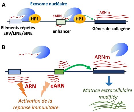
HP1 targets the RNA exosome to chromatin
A novel role for the HP1 proteins in targeting the RNA-exosome to chromatin for the turnover of repetitive RNAs and enhancer RNAs. Heterochromatin protein 1 (HP1), a hallmark of pericentromeric heterochromatin, is a chromatin-bound regulator of co-transcriptional processes including alternative splicing, but its role in RNA degradation remains unexplored. Here, we uncover a direct interaction between HP1 and nuclear RNA exosome complexes, major RNA decay machineries. In mouse embryonic liver cells, inactivation of all three HP1 isoforms leads to accumulation of retrotransposon-derived RNAs and stabilization of enhancer RNAs. These changes coincide with increased activity at a subset of liver enhancers particularly sensitive to reduced exosome activity, many of which regulate genes encoding extracellular matrix components such as collagen genes. Stratifying hepatocellular carcinoma samples by HP1 expression further reveal that tumors with low HP1 are marked by reduced RNA degradation, and increased expression of a similar subset of genes encoding extracellular matrix components and possibly contributing to tumor stiffness. These results suggest that HP1’s impact on RNA turnover contributes to its function in cancer biology. More information: https://www.nature.com/articles/s41467-026-72504-7 Contact : Carl Mann carl.mann@i2bc.paris-saclay.fr
|
|
Scooped by
I2BC Paris-Saclay
May 13, 4:22 AM
|
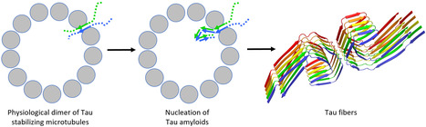
A coherent structural picture of the interaction of Tau with tubulin provides a link to its aggregation.
Tau is a protein regulating microtubule dynamics which also forms neurofibrillary tangles in pathological conditions. Recent results suggest that a physiological dimer of Tau could serve as a nucleus for its aggregation. Tauopathies are a group of neurodegenerative diseases characterized by the presence of insoluble filaments of the Tau protein in the brain. In physiological conditions, Tau is involved in the regulation of microtubule dynamics. The study of its interaction with different tubulin assemblies, using various experimental approaches, leads to a seemingly disparate picture. In this opinion-type article, we integrate this information into a model of how Tau participates in microtubule assembly and stabilization. Related to its intrinsically disordered nature, the binding of Tau to microtubules involves both specific interactions, along protofilaments, and non-specific ones, with the C-terminal region of tubulin subunits. In addition, a Tau:tubulin structure that we recently determined leads to a model of a functional Tau dimer targeting a microtubule aperture between protofilaments. This model also provides a framework for a Tau aggregation that would be initiated on the microtubule. More information : https://www.jbc.org/article/S0021-9258(26)01958-7/fulltext Contact : Benoît Gigant benoit.gigant@i2bc.paris-saclay.fr
|
|
Scooped by
I2BC Paris-Saclay
April 17, 4:52 AM
|
The tRNA moieties of both aminoacyl-tRNA substrates of a cyclodipeptide synthase share a common binding site, as revealed by RNA microhelices mimicking tRNA acceptor arms
Cyclodipeptide synthases (CDPSs) utilize two aminoacyl-tRNAs as substrates to produce diverse natural products. Here, we demonstrate that CDPSs efficiently recognize aminoacylated microhelices (miHxs) that mimic the tRNA acceptor arm. Structural and enzymological analyses using unacylated, misacylated, and engineered miHxs reveal a shared RNA-binding mode for both substrates. These findings establish miHxs as versatile tools to investigate CDPS function and, more broadly, other aminoacyl-tRNA–dependent enzymes. Two teams from the I2BC, in collaboration with the ICSN, combined enzymological and structural approaches to investigate cyclodipeptide synthases (CDPSs), enzymes involved in natural product biosynthesis. CDPSs sequentially use two aminoacyl-tRNAs (AA-tRNAs) to catalyse cyclodipeptide formation. We previously showed that microhelices (miHxs), mimicking the tRNA acceptor arm, are as efficient as full-length AA-tRNAs when aminoacylated by flexizymes.
Here, we generated a diverse set of miHxs (acylated, unacylated, misacylated, mutated, or shortened) and analysed their interactions with CDPSs. We focused on the Nocardia brasiliensis CDPS (Nbra-CDPS), which synthesizes cyclo(L-Ala–L-Glu) from Ala-tRNAAla and Glu-tRNAGlu. Crystal structures of Nbra-CDPS bound to analogues of its first substrate, including unacylated and acylated miHxAla, were determined. Cryo-EM analysis confirmed that miHxs mimic the acceptor stem of full-length tRNAs.
We also solved the structure of Nbra-CDPS bound to unacylated miHxGlu, an analogue of the second substrate, and found that it superimposes well with miHxAla despite sequence differences. Together with results obtained using misacylated substrates, these data reveal a shared RNA-binding mode for both substrates. Our findings establish miHxs as powerful tools to dissect CDPS function and to study other AA-tRNA–dependent enzymes. More information: https://academic.oup.com/nar/article-abstract/doi/10.1093/nar/gkag307/8625897?utm_source=authortollfreelink&utm_campaign=nar&utm_medium=email Contacts: Muriel Gondry muriel.gondry@i2bc.paris-saclay.fr https://www.i2bc.paris-saclay.fr/enzymology-and-non-ribosomal-peptide-biosynthesis/ Jean-Baptiste Charbonnier jb.charbonnier@i2bc.paris-saclay.fr https://www.i2bc.paris-saclay.fr/nuclear-enveloppe-telomeres-and-dna-repair/
|
|
Scooped by
I2BC Paris-Saclay
February 16, 10:02 AM
|
Snapshots of cotranslational N-myristoylation reveal NMT as a ribosome-associated chaperone.
NMT in a new light: associated with the ribosome via the NAC complex, this enzyme not only adds lipid tags to the N-termini of nascent proteins but also acts as a chaperone, cooperating with MetAPs to ensure proper folding and delivery. N-myristoylation is an essential cotranslational lipid modification catalyzed by N-myristoyltransferases (NMTs). Structural and cellular analyses reveal that NMT1 associates with the ribosomal tunnel exit via the nascent polypeptide–associated complex (NAC) and acts sequentially after MetAP-mediated initiator methionine removal, in contrast to previously described simultaneous cotranslational modification assemblies. Unexpectedly, NMT1 also exhibits chaperone-like activity, expanding its functional repertoire in cotranslational protein biogenesis. More information : https://www-nature-com.insb.bib.cnrs.fr/articles/s41467-025-67962-4 Contact : Thierry Meinnel and Carmela Giglione thierry.meinnel@i2bc.paris-saclay.fr and carmela.giglione@i2bc.paris-saclay.fr
|
|
Scooped by
I2BC Paris-Saclay
December 8, 2025 5:52 AM
|
A tripartite protein complex promotes DNA transport during natural transformation in Firmicutes.
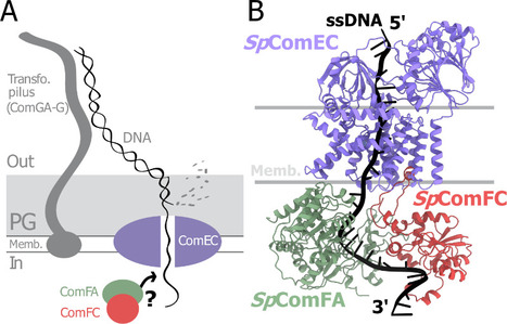
From structural modeling to natural transformation in bacteria: identification of a key protein complex for DNA transport across the membrane of Firmicutes. Transformation is a key mechanism of horizontal gene transfer, central to bacterial adaptation. This evolutionarily conserved process allows bacteria to integrate exogenous genetic material into their genome, thereby facilitating, for example, the spread of antibiotic resistance.
In a study published in the journal PNAS, scientists from I2BC (CEA/CNRS/UPSaclay, Gif-sur-Yvette) and DRCM/IBFJ (CEA/UPSaclay/UP Cité, Fontenay-aux-Roses), in collaboration with a laboratory at CBI (CNRS/University of Toulouse), identified a protein complex involved in the transport of single-stranded DNA across the membrane during transformation. Using AlphaFold, they structurally modeled this three-protein complex, which is highly conserved in the phylum Firmicutes, in interaction with single-stranded DNA. The structural model allowed them to identify a possible path for DNA through a conserved channel in one of the three proteins, a transmembrane protein, and then along a groove formed by the other two proteins. This model was validated by a robust experimental strategy in the bacterium Streptococcus pneumoniae, by measuring the impact on transformation efficiency of disruptive mutations in several protein–protein and protein–DNA interfaces. The transmembrane channel was found to be conserved in a structural model in Helicobacter pylori, and its importance for transformation was also experimentally confirmed in this bacterium. This study sheds light on the molecular mechanisms of bacterial transformation and demonstrates the power of macromolecular structure prediction to generate molecular hypotheses and guide functional experiments. More information: https://doi.org/10.1073/pnas.2511180122 Contact : Jessica Andreani jessica.andreani@i2bc.paris-saclay.fr
|
|
Scooped by
I2BC Paris-Saclay
September 5, 2025 3:50 AM
|
Restriction of Ku translocation protects telomere ends
How Ku inward translocation can be restricted to protect telomere ends from NHEJ without altering other important functions. Safeguarding chromosome ends against fusions via nonhomologous end joining (NHEJ) is essential for genome integrity. Paradoxically, the conserved NHEJ core factor Ku binds telomere ends. How it is prevented from promoting NHEJ remains unclear, as does the mechanism that allows Ku to coexist with telomere-protective DNA binding proteins, Rap1 in Saccharomyces cerevisiae. Here, we find that Rap1 directly inhibits Ku’s NHEJ function at telomeres. A single Rap1 molecule near a double-stand break suppresses NHEJ without displacing Ku in cells. Furthermore, Rap1 and Ku form a complex on short DNA duplexes in vitro. Cryo-EM shows Rap1 blocks Ku’s inward translocation on DNA – an essential step for NHEJ at DSBs. Nanopore sequencing of telomere fusions confirms this mechanism protects native telomere ends. These find- ings uncover a telomere protection mechanism where Rap1 restricts Ku’s inward translocation. This switches Ku from a repair-promoting to a protective role preventing NHEJ at telomeres. More information : https://doi.org/10.1038/s41467-025-61864-1 Contact : Philippe Cuniasse philippe.cuniasse@i2bc.paris-saclay.fr
|
|
|
Scooped by
I2BC Paris-Saclay
October 29, 4:43 AM
|
BSI@Paris-Saclay 2026: 5th Integrative Structural Biology Meeting – Register Now!
The 5th BSI@Paris-Saclay Meeting will take place on Monday, November 30, 2026, at the I2BC Auditorium (Institut de Biologie Intégrative de la Cellule, building 21, CNRS Campus, Gif-sur-Yvette). This annual event gathers the local structural biology and biophysics community to share recent scientific advances and discuss future perspectives. Program Highlights: -a session dedicated to new recruits, featuring a keynote by Julien Robert-Paganin -a young researchers session with 4 oral presentations by PhD students and post-docs -a platform session highlighting the latest advances in local platforms -a round table on industry-academia interactions. Registration & abstract submission deadline: October 31, 2026 - Informations and program - Free but mandatory registration https://bsi-psay-2026.sciencesconf.org/ The BSI@Paris-Saclay 2026 event is organized with the generous support of I2BC (https://www.i2bc.paris-saclay.fr/), the French Infrastructure for Integrated Structural Biology (FRISBI) (https://frisbi.eu/), the interdisciplinary project “Living Machines at Work” (https://www.universite-paris-saclay.fr/objets-interdisciplinaires/living-machines-work) of Université Paris-Saclay, and the B3S department of I2BC (https://www.i2bc.paris-saclay.fr/disciplinary-department/b3s/).
|
|
Scooped by
I2BC Paris-Saclay
October 1, 5:31 AM
|
Unveiling a missing component of the atypical type IV secretion system required for natural transformation of Helicobacter pylori.
Identification of a Missing Component of the Exogenous DNA Internalisation System During Natural Transformation in Helicobacter pylori. Horizontal gene transfer plays a crucial role in the evolution of bacterial populations. In pathogens, it promotes the spread of antibiotic resistances and virulence factors. Helicobacter pylori, a bacterium infecting the stomach of 50% of the world's population and responsible for the majority of gastric ulcers and cancers, has a remarkable capacity to exchange genes via natural transformation. Through this pathway, bacteria can capture and incorporate into their genome DNA present in their environment. To do this, H. pylori uses an atypical DNA uptake system: a type 4 secretion system (T4SS) named ComB, rather than the pilus present in all other naturally transformable bacteria. Compared to classical T4SS, certain components appeared to be absent from this system. In a study published in PLoS Pathogens, researchers from the Department of Cellular and Molecular Radiobiology (François Jacob Institute of Biology, CEA/UPSaclay, UP Cité, Fontenay aux Roses) and I2BC (CEA/CNRS/UPSaclay, Gif-sur-Yvette), in collaboration with a laboratory at the Pasteur Institute, identified an ATPase essential for this secretion system. Homology searches detected a gene, located outside the known operons of the ComB complex, capable of encoding an ATPase associated with a T4SS. Genetics and biochemistry experiments demonstrated that the protein encoded by this gene, designated as ComB11, is indeed an ATPase essential for natural transformation of H. pylori. Structural modelling revealed an interaction between this ATPase and the ComB4 component of the ComB system, confirming its membership in this atypical T4SS. More information: https://journals.plos.org/plospathogens/article?id=10.1371/journal.ppat.1014140 Contact: Jessica Andreani jessica.andreani@i2bc.paris-saclay.fr
|
|
Scooped by
I2BC Paris-Saclay
September 22, 8:11 AM
|
JIP3 and JIP4 proteins: much more than simple adapters for vesicle transport.
The JIP3 and JIP4 proteins (JIP3/4) do not merely serve as adapters linking the molecular motors kinesin-1 and dynein to their vesicular cargoes for bidirectional transport along microtubules. They also act as key regulators of this transport, capable of activating these motors by relieving their autoinhibited state. In a recent study published in the Journal of Biological Chemistry, scientists from I2BC (CNRS/CEA-Jacob/Université Paris-Saclay, Gif-sur-Yvette) characterized the interaction between JIP3/4 and KIF5B in vitro. To do this, they employed molecular biology techniques alongside a biophysical approach known as MicroScale Thermophoresis (PIM platform, I2BC). The study identified the minimal regions of these proteins required for their interaction: the N-terminal N-RH1 domain of JIP3/4 and the CC4 region of KIF5B. An unexpected discovery emerged from the study: JIP3 and JIP4 exhibit a 40-fold difference in affinity for KIF5B. To further investigate these mechanisms, the researchers conducted extensive site-directed mutagenesis on the N-RH1 region of JIP3/4, targeting accessible charged residues and sequence differences between JIP3 and JIP4. Their results reveal the existence of multiple negatively charged, low-affinity binding sites and the involvement of the unstructured N-terminal domain; together, these elements define an extensive interaction surface with KIF5B on the JIP3 N-RH1 domain.
This work sheds new light on how JIP3/4 might relieve kinesin-1 auto-inhibition. Indeed, in its auto-inhibited form, kinesin-1 is stabilized by the CC4 region. By interacting extensively with the latter, JIP3/4 destabilize this conformation, thereby enabling activation of the molecular motor. More information : https://www.jbc.org/article/S0021-9258(26)02227-1/fulltext Contact : Paola Llinas paola.llinas@i2bc.paris-saclay.fr and Julie Ménétrey julie.menetrey@i2bc.paris-saclay.fr
|
|
Scooped by
I2BC Paris-Saclay
September 10, 5:18 AM
|
A new path to Rubisco processing
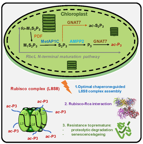
A unique plant machinery comprising three dedicated components ensures N-terminal processing of Rubisco’s catalytic subunit, enabling its proper accumulation and activity as Earth’s most abundant protein. RuBP carboxylase/oxygenase (Rubisco)—the central enzyme of carbon dioxide assimilation and plant autotrophy— is Earth’s most abundant protein and a major carbon and nitrogen reservoir, which can be remobilized during stress and development. The catalytic large subunit of Rubisco (RbcL) undergoes a highly conserved maturation process that results in N-terminal proline acetylation, a modification not reported in other biological systems. Until now, both the enzymatic machinery responsible for N-terminal maturation and its associated functional implications have remained unknown. In this study, we uncover a specialized chloroplast processing pathway, composed of two aminopeptidases and one N-terminal acetyltransferase, specifically evolved to catalyze RbcL maturation. Proper processing of RbcL by this machinery is tightly coupled to chaperone-mediated assembly of the hexadecameric holoenzyme, enhances activation by Rubisco activase, and safeguards the fully modified complex from premature degradation during senescence. Disruption of this pathway compromises both the reactivity and accumulation of Rubisco. These findings reveal a specialized maturation mechanism that underpins the exceptional abundance and persistence of Rubisco in photosynthetic organisms, and more broadly, its central role in sustaining life on Earth. This paves the way for enhanced photosynthesis, stronger crop yields, and more effective carbon capture. More information : https://www.science.org/doi/full/10.1126/sciadv.aeh5060 Contact : Thierry Meinnel thierry.meinnel@i2bc.paris-saclay.fr
|
|
Scooped by
I2BC Paris-Saclay
July 20, 9:20 AM
|
Study on protein–bicelle interactions using switchSENSE technology with the PIM facility at I2BC
Using bicelles as membrane mimetics and switchSENSE technology available at the PIM facility, we uncover new insights into the dynamic mechanisms governing weak protein–membrane interactions, revealing how proteins recognize and interact with membrane environments. To investigate the association of human dystrophin with biomimetic phospholipid bicelles, we employed switchSENSE®, a fluorescence-based biosensing technology that relies on dynamic DNA nanolevers operating in a microfluidic environment while requiring only minimal sample amounts. We demonstrate that this technology enables real-time kinetic analysis of weak and transient interactions, providing a significant advantage over conventional biophysical methods, which often struggle to detect low-affinity protein–lipid binding events. Focusing on two functionally relevant fragments from the central rod domain of dystrophin (R1-3 and R11-15), we quantified their interactions with zwitterionic and anionic bicelles using two complementary experimental configurations: either the proteins were immobilized and the bicelles served as analytes, or vice versa. Both approaches yielded consistent micromolar affinities, in agreement with our previous microscale thermophoresis data, while additionally providing association and dissociation rate constants. This study represents the first kinetic characterization of dystrophin–bicelle interactions. Our results reveal that the observed low affinity is largely driven by the rapid dissociation of the resulting complexes. Notably, the R11-15 fragment displayed a faster association rate and a slower dissociation rate than R1-3, indicating a more stable and long-lasting interaction with lipid assemblies. Beyond providing new mechanistic insights into the reversible membrane association of dystrophin—a process thought to contribute to maintaining sarcolemmal integrity during cycles of muscle contraction and stretching—our work highlights switchSENSE® as a versatile platform for quantifying weak protein–lipid interactions. By delivering kinetic resolution for interactions that lie at the detection limits of conventional techniques, switchSENSE® offers a powerful approach for investigating the dynamic interplay between structural proteins and lipid assemblies. For more information: Article: https://link.springer.com/article/10.1007/s00249-026-01829-4 Contact: sophie.combet@i2bc.paris-saclay.fr ; paloma.fernandez-varela@polytechnique.edu Facility web site: https://www.i2bc.paris-saclay.fr/structural-biology/pim/
|
|
Scooped by
I2BC Paris-Saclay
June 16, 4:34 AM
|
Reassessing Carotenoid Photophysics: Shedding Light on Dark States.
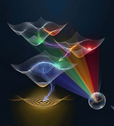
The dark excited states of carotenoids have been debated for decades. Researchers at I2BC now use femtosecond stimulated resonance Raman spectroscopy to determine the nature and symmetry of three of them, resolving controversies that have long obscured our understanding of photosynthetic light harvesting. Carotenoid molecules are critical in photosynthesis, performing functions at the heart of both light-harvesting and photoprotection. As both these processes involve excitation energy transfer, fully understanding them requires a precise description of the electronic states involved. The excited-state manifold of carotenoids is not yet fully characterised, and includes several dark electronic states that remain elusive. Using femtosecond stimulated resonance Raman spectroscopy, where the vibrational contributions of each excited state can be observed selectively as a function of the Raman excitation, we reveal the nature and symmetry of no less than three different dark states. These results end long-standing controversies in carotenoid research, shining new light on the photophysics of these essential molecules and establishing a spectroscopic framework for characterising their multiple roles. The figure is an artistic representation of the excited states in carotenoids. More information : https://pubs.acs.org/doi/10.1021/jacs.6c03864 Contact : Manuel Llansola-Portoles manuel.llansola-portoles@i2bc.paris-saclay.fr
|
|
Scooped by
I2BC Paris-Saclay
June 10, 9:28 AM
|
The final of the interuniversity 3-Minute Thesis (3MT®) competition
Linnéa Strandberg, PhD student from Photobiology, Photosynthesis, Photocatalysis team in I2BC, will present her thesis project “When breaking the heart of a plant”. The Institut Polytechnique de Paris will host the final of the interuniversity 3-Minute Thesis (3MT®) competition. Developed by The University of Queensland, the 3MT® is more than a competition—it’s a platform for PhD students to strengthen their communication skills in english and share their research with a non-specialist audience in a clear and engaging manner.
Join on June 25, from 2:00 pm to 4:00 pm at Télécom Paris (Thévenin lecture hall) to support the finalists and vote for the Audience Award (free registration required): https://www.ip-paris.fr/form/3-minute-thesis-registration.
|
|
Scooped by
I2BC Paris-Saclay
June 8, 9:13 AM
|
3R 2026: A Great Edition in La Grande-Motte
The 3R Congress brings together researchers working on DNA Replication, Repair and Recombination, from fundamental molecular mechanisms to clinical applications, promoting scientific exchange within the community. Twenty-two participants from the B3S and Genome departments of I2BC attended the 16th 3R Congress, which took place in La Grande-Motte from May 18 to 21, 2026. High-quality science, lively discussions and a friendly atmosphere once again made this edition a great success. Many thanks to all our sponsors, and especially to I2BC, for their valuable support.
Portrait Jeune Chercheur – Thibault Tubiana, Chercheur en bioinformatique structurale
Thibault Tubiana est chargé de recherche (CRCN) au CNRS depuis la fin de l'année 2024. Il exerce ses fonctions au sein de l’Institut de Biologie Intégrative de la Cellule - I2BC (CNRS/CEA/UPSaclay, Gif-sur-Yvette), dans l'équipe dirigée par le Dr Stéphane Bressanelli. Son parcours illustre une volonté constante de faire le pont entre la bioinformatique structurale, la virologie moléculaire et l'étude des membranes cellulaires. Après avoir obtenu une licence et un master en bioinformatique à l'Université Paris Diderot, où il nourrit déjà un fort attrait pour la virologie, il réalise sa thèse de doctorat sous la co-direction des Drs Stéphane Bressanelli et Yves Boulard. Ses travaux portent alors sur la dynamique d'assemblage de la capside du norovirus, lui permettant d'acquérir une solide expertise en modélisation moléculaire et en approches intégratives (combinant dynamique moléculaire, données SAXS et Cryo-EM). Il poursuit sa carrière par un premier post-doctorat au sein de l'Institut de Recherche Servier (IdRS). Cette immersion dans le monde de l'industrie pharmaceutique lui permet d'acquérir de solides compétences en drug design. Afin d'approfondir ensuite ses connaissances sur les interactions protéines-membranes, il rejoint le groupe de la Pr Nathalie Reuter à l'Université de Bergen (Norvège). Il y mène un vaste projet de cartographie biostatistique et bioinformatique visant à décrypter et redéfinir les interfaces de liaison des protéines membranaires périphériques. Fort de ces expertises pluridisciplinaires, il choisit de revenir en France au sein de son ancienne équipe à l'I2BC. Soutenu dans un premier temps par des financements postdoctoraux de l'ANRS-MIE, il y déploie des approches innovantes en bioinformatique structurale, contribuant activement à l'étude des virus à ARN positif simple brin. Ce travail structurant conduit à son recrutement au CNRS fin 2024. Dans le contexte de la révolution de l'intelligence artificielle appliquée à la biologie (AlphaFold), ses recherches actuelles portent sur la modélisation des complexes de réplication des virus de l'hépatite E (HEV) et de l'hépatite C (HCV). Ses travaux visent à comprendre à l'échelle atomique l'organisation de ces protéines virales et leurs interactions avec les cellules hôtes. Très impliqué dans la communauté scientifique nationale sur ces pathogènes, il a récemment intégré le bureau de l'Action Coordonnée 42 sur les hépatites virales de l'ANRS-MIE. Parallèlement à ses recherches, Thibault Tubiana a toujours eu à cœur de transmettre ses connaissances. Au sein de l'I2BC, en plus de l'encadrement d'étudiants, il s'investit fortement dans la formation continue de son institut. Il a notamment créé et mis en place, en lien avec la plateforme BioI2, une nouvelle formation dédiée à la modélisation et à la visualisation moléculaires, offrant ainsi un outil précieux pour l'ensemble des chercheurs de l'université. « Nous sommes tous des poussières d'étoile. » - Pr. André Brahic -> Contact : thibault.tubiana@i2bc.paris-saclay.fr
Via Life Sciences UPSaclay
|
|
Scooped by
I2BC Paris-Saclay
March 9, 8:48 AM
|
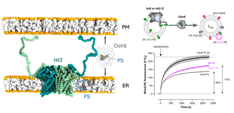
Phosphatidylserine dynamics between the endoplasmic reticulum and the plasma membrane in Saccharomyces cerevisiae.
A functional partnership between a lipid scramblase and a lipid transfer protein in the regulation of phosphatidylserine homeostasis. Phosphatidylserine is synthesised in the endoplasmic reticulum (ER), but this negatively charged lipid is highly concentrated in the plasma membrane (PM). There, it plays numerous roles in processes such as cell signalling and cell fusion, and it can also mediate apoptosis and synaptic pruning when exposed in the outer leaflet of the PM. In the yeast Saccharomyces cerevisiae, selective PS transport between the ER and the PM by the lipid transfer protein Osh6 is required for PS enrichment in the plasma membrane. Osh6 operates at membrane contact sites (MCS), where it interacts with the Ist2 protein embedded in the ER, tethering the ER to the PM via its long, disordered C-terminal tail. In two studies conducted in collaboration with the CRBM (CNRS/University of Montpellier), the IPMC (CNRS/University of Nice Côte d'Azur), and the IBCP (CNRS/University of Lyon), we show that Ist2 is a membrane transporter that catalyses rapid lipid exchange across the two leaflets of the ER – acting as a lipid ‘scramblase’ – and that this scrambling activity sustains Osh6-mediated PS transfer between the ER and the PM. First, we demonstrated that Ist2 catalyses the scrambling of different lipids in vitro after reconstituting purified Ist2 in proteoliposomes. Molecular dynamics simulations were then used to identify a cavity through which the lipid headgroup passes during transport from one leaflet to another. Further cellular studies revealed a close relationship between the COPII complex and the Ist2 protein. This was manifested by impaired yeast growth and vacuolar trafficking, as well as disruption of ER exit sites. We also found that Ist2 deletion stimulates the formation of ER-derived lipid droplets and changes their composition. Finally, using artificial, reconstituted ER-PM contact sites, we demonstrated that Ist2-mediated lipid scrambling sustains Osh6-mediated lipid transfer. Together, our studies identify Ist2 as a lipid scramblase and establish that lipid scrambling in the ER by Ist2 controls various cellular functions, such as vesicular transport and lipid droplet homeostasis. This highlights the importance of lipid dynamics for ER function. Furthermore, we reveal a functional partnership between Ist2-mediated lipid scrambling and Osh6-mediated lipid transfer at MCS. More information : https://doi-org.insb.bib.cnrs.fr/10.1083/jcb.202502112 https://www-science-org.insb.bib.cnrs.fr/doi/10.1126/sciadv.adz2217 Contact : Guillaume Lenoir guillaume.lenoir@i2bc.paris-saclay.fr
|
|
Scooped by
I2BC Paris-Saclay
January 12, 7:46 AM
|
Cross-regulation of [2Fe–2S] cluster synthesis by ferredoxin-2 and frataxin.
Tight regulation of Fe-S clusters biosynthesis via a mutually antagonistic binding of frataxin and ferredoxin-2 to the assembly machinery, with several important implications for the Friedreich’s ataxia disease caused by frataxin deficiency. Iron-sulfur (Fe-S) clusters are essential metallocofactors that perform a multitude of biological functions. They are synthesized de novo by multi-proteins machienries and any defect in their synthesis leads to severe diseases such as Friedreich’s ataxia (FRDA), caused by defective expression of frataxin (FXN). Here, we uncover that efficient [2Fe-2S] cluster assembly requires a fine-tuned balanced ratio of FXN and Ferredoxin-2 (FDX2), an essential enzyme of the assembly process. [2Fe-2S] clusters are assembled on the scaffold protein ISCU2 with sulfur provided as a persulfide by NFS1, which is cleaved into sulfide by FDX2. FXN stimulates the whole process by accelerating persulfide transfer to ISCU2. Using an in vitro reconstituted human system, we show that any deviation from a close-to-equal amount of FXN or FDX2 downregulates Fe-S cluster synthesis. We performed a structure-function investigation, which revealed that this is due to competition between FXN and FDX2 for the same binding site on the NFS1-ISCU2 complex. We found that higher levels of FXN impair the persulfide-reductase activity of FDX2 and higher levels of FDX2 slow FXN-accelerated persulfide transfer to ISCU2. We also discovered that FDX2 directly hinders persulfide generation and transfer to ISCU2 by interacting with the persulfide-carrying mobile loop of NFS1. We further found that knocking-down FDX2 expression in a FRDA drosophila model, increases fly lifespan. Altogether, this work highlights a direct regulation of Fe-S cluster biosynthesis through antagonistic binding of FXN and FDX2 and suggests that decreasing FDX2 in the context of FXN deficiency in FRDA might constitute a novel therapeutic axis. More information: https://www.nature.com/articles/s41586-025-09822-1 Contact : Benoit D'Autréaux benoit.dautreaux@i2bc.paris-saclay.fr
|
|
Scooped by
I2BC Paris-Saclay
October 15, 2025 9:07 AM
|
Moss BRCA2 lacking the canonical DNA-binding domain promotes homologous recombination and binds to DNA
Despite having low homology with BRCA2 proteins from other organisms and no folded domain, the newly discovered BRCA2 protein from Physcomitrium patens (the green yeast) promotes homologous recombination by binding to recombinases and DNA. BRCA2 is crucial for mediating homology-directed DNA repair (HDR) through its binding to single-stranded DNA (ssDNA) and the recombinases RAD51 and DMC1. Most BRCA2 orthologs have a canonical DNA-binding domain (DBD) with the exception of Drosophila melanogaster. It remains unclear whether such a noncanonical BRCA2 variant without DBD possesses a DNA-binding activity. Here, we identify a new noncanonical BRCA2 in the model plant Physcomitrium patens (PpBRCA2). We establish that PpBRCA2 is essential for genome integrity maintenance, somatic DNA double-strand break (DSB) repair, HDR-mediated gene targeting, and RAD51 foci recruitment at DNA break sites. PpBRCA2 is also critical for DSB repair during meiosis. Interestingly, PpBRCA2 interacts strongly with RAD51 but weakly with DMC1, suggesting a distinct meiotic function compared to other BRCA2 homologs. Despite lacking the canonical DBD, PpBRCA2 binds ssDNA through its disordered N-terminal region and efficiently promotes HDR. Our work highlights that the ssDNA binding capacity of BRCA2 homologs is conserved regardless of the presence of a canonical DBD and provides a deeper understanding of BRCA2’s functional diversity across species. More information: https://pmc.ncbi.nlm.nih.gov/articles/PMC12412785/ Contact: Sophie Zinn sophie.zinn@i2bc.paris-saclay.fr
|