 Your new post is loading...

|
Scooped by
Gilbert C FAURE
December 30, 2020 1:24 PM
|
See resources to maximize your annual meeting experience: mobile app, networking events, contacts, frequently asked questions, and more.
|
|
Scooped by
Gilbert C FAURE
December 14, 2020 2:50 PM
|
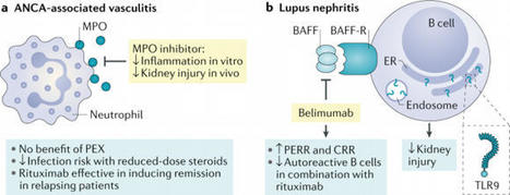
In 2020 a number of clinical trials have provided insights into therapeutic approaches for the treatment of anti-neutrophil cytoplasmic autoantibody (ANCA)-associated vasculitis and lupus nephritis. Moreover, mechanistic insights have potential to open new therapeutic strategies in the future.
|
|
Scooped by
Gilbert C FAURE
December 3, 2020 1:09 PM
|
Journal of Clinical Medicine, an international, peer-reviewed Open Access journal.
|
|
Scooped by
Gilbert C FAURE
November 17, 2020 4:56 AM
|
|
|
Scooped by
Gilbert C FAURE
November 14, 2020 1:38 PM
|
Antiphospholipid Syndrome Antiphospholipid syndrome (APS) is defined by the coexistence of obstetric morbidity (mainly pregnancy losses) and/or vascular thrombosis (venous or arterial) with the presence of aPL, namely lupus anticoagulant, anticardiolipin, or anti-β2-glycoprotein I antibodies. From: Handbook of Systemic Autoimmune Diseases, 2018 Related terms: View all Topics Antiphospholipid Syndrome Lucia R. Wolgast MD, in Transfusion Medicine and Hemostasis (Third Edition), 2019 Introduction The antiphospholipid (aPL) syndrome (APS) is an autoimmune thrombophilic condition that is defined by a combination of clinical and laboratory criteria. In general terms, APS patients have developed circulating antibodies against plasma proteins that bind to phospholipids (i.e., aPL antibodies) with subsequent clinical morbidity of thrombosis and/or pregnancy complications. The investigational criteria for APS (often referred to as the Sydney Investigational Criteria) are detailed in Table 108.1 and require that patients have documented evidence for vascular thrombosis and/or obstetric complications, such as unexplained recurrent miscarriages, intrauterine growth restriction, intrauterine fetal demise, preeclampsia/toxemia, placental abruption, and preterm labor. The laboratory criteria require identifying the persistent aPL antibodies (i.e., at least two abnormal measurements at least 12 weeks apart), including elevated medium to high tiers of anticardiolipin (aCL) IgG or IgM antibodies, anti-β2-glycoprotein I (anti-β2GPI) IgG or IgM antibodies, and/or positive lupus anticoagulant (LA). The laboratory diagnosis of APS is discussed in Chapter 158. Table 108.1. Sydney Investigational Criteria for the Diagnosis of the Antiphospholipid Syndromea Clinical • Vascular thrombosis (one or more episodes of arterial, venous, or small vessel thrombosis). For histopathologic diagnosis, there should be no evidence of inflammation in the vessel wall. • Pregnancy morbidities attributable to placental insufficiency, including (a) three or more otherwise unexplained recurrent spontaneous miscarriages, before 10 weeks of gestation, (b) one or more fetal losses after the 10th week of gestation, (c) stillbirth, and (d) episode of preeclampsia, preterm labor, placental abruption, intrauterine growth restriction, or oligohydramnios that are otherwise unexplained. Laboratory • Medium- or high-titer aCL or anti-β2GPI IgG and/or IgM antibody present on two or more occasions, at least 12 weeks apart, measured by standard ELISAs. • Lupus anticoagulant in plasma, on two or more occasions, at least 12 weeks apart, detected according to the guidelines of the ISTH SSC Subcommittee on Lupus Anticoagulants and Phospholipid-Dependent Antibodies. β2GPI, β2-glycoprotein I; aCL, anticardiolipin; aPL, antiphospholipid; ELISA, enzyme-linked immunosorbent assay; Ig, immunoglobulin. a “Definite APS” is considered to be present if at least one of the clinical criteria and one of the laboratory criteria are met. Modified from Miyakis, S., Lockshin, M. D., Atsumi, T., et al. (2006). International consensus statement on an update of the classification criteria for definite antiphospholipid syndrome (APS). J Thromb Haemost, 4, 295–306. It is important for the reader to understand that these criteria were not designed to be requirements for the clinical diagnosis of APS. Rather, they were intended to provide a uniformly rigorous definition of APS for the purpose of standardizing research on the disorder. In “real-world” clinical practice, some patients may be appropriately diagnosed for presumptive APS without meeting the strict investigational criteria. Some APS patients may be positive for other “noncriteria” clinical laboratory tests—see Chapter 158—that have not been included by consensus panels as diagnostic criteria for the disorder, but these positive noncriteria laboratory tests may indicate a clinical risk and warrant treatment. Furthermore, APS patients may have positive APS criteria assays but not the typical criteria manifestations. These “noncriteria manifestations” include thrombocytopenia, livedo reticularis, skin ulcers, nephropathy, migraine, cognitive defects, diffuse alveolar hemorrhage, and valvular heart disease (Libman–Sacks endocarditis), and these clinical manifestations may need to be managed differently. Occasional patients may even test entirely negative for the APS criteria assays but have typical clinical manifestations of the disorder—a situation referred to as seronegative APS (termed SNAPS). At present, APS may be divided into the following subcategories: (1) Primary APS is the “stand alone” disorder, in the absence of systemic lupus erythematosus (SLE), (2) secondary APS occurs in the presence of APS, (3) catastrophic APS (CAPS) manifests as disseminated thrombosis in large and small vessels with resulting multiorgan failure (Table 108.2), and (4) SNAPS includes patients whose diagnostic tests are entirely negative but who, on clinical grounds, are still suspected to have the disorder. Table 108.2. Proposed Criteria for the Classification of Catastrophic Antiphospholipid Syndrome (APS) 1. Evidence of involvement of three or more organs, systems, and/or tissuesa 2. Development of manifestations simultaneously or in less than a week 3. Confirmation by histopathology of small vessel occlusion in at least one organ or tissueb 4. Laboratory confirmation of the presence of antiphospholipid antibodies (lupus anticoagulant and/or anticardiolipin antibodies)c Definite Catastrophic APS • All four criteria Probable Catastrophic APS • All four criteria, except for only two organs, systems and/or tissues involvement • All four criteria, except for the absence of laboratory confirmation at least 6 weeks apart due to the early death of a patient never previously tested for aPL before the catastrophic APS event • Criteria 1, 2, and 4 • Criteria 1, 3, and 4 and the development of a third event in more than a week but less than a month, despite anticoagulation a Usually, clinical evidence of vessel occlusions, confirmed by imaging techniques when appropriate. Renal involvement is defined by a 50% rise in serum creatinine, severe systemic hypertension (N180/100 mm Hg), and/or proteinuria (N500 mg/24 hours). b For histopathological confirmation, significant evidence of thrombosis must be present, although, in contrast to Sydney criteria, vasculitis may coexist occasionally. c If the patient had not been previously diagnosed as having an APS, the laboratory confirmation requires that the presence of antiphospholipid antibodies must be detected on two or more occasions at least 6 weeks apart (not necessarily at the time of the event), according to the proposed preliminary criteria for the classification of definite APS. Modified from Asherson, R. A., Cevera, R., de Groot, P. G., et al. (2003). Catastrophic antiphospholipid syndrome: international consensus statement on classification criteria and treatment guidelines. Lupus, 12, 530–534. Antiphospholipid Syndrome Nancy Agmon-Levin, ... Yehuda Shoenfeld, in The Autoimmune Diseases (Fifth Edition), 2014 The antiphospholipid syndrome (APS) is an autoimmune disorder characterized by thrombosis, pregnancy losses, and the presence of at least one of three antiphospholipid antibodies. However, it has been acknowledged that the clinical spectrum of APS is much wider, including systemic and organ-specific manifestations. Moreover, the presence of more than 20 antiphospholipid antibodies has been recognized, some of which may participate in the pathogenesis of APS although not included as criteria. Our knowledge and understanding of the etio-pathogenesis of APS has increased tremendously, as the effects of antiphospholipid antibodies, mainly the anti-B2GPI–B2GPI immune complexes, were elucidated. The latter bind to different cells (i.e., monocytes, endothelial cells, and trophoblasts), recruit cell surface receptors, and subsequently induce intracellular signaling. Additionally an arsenal of environmental factors as infectious agents and vitamin D has been associated with the presence of antiphospholipid antibodies and overt APS. It seems therefore that in APS both thrombotic and immune-mediated mechanisms play a part and may be considered as candidates for targeted therapeutic interventions. Antiphospholipid Syndrome ROBERT A.S. ROUBEY, in The Autoimmune Diseases (Fourth Edition), 2006 Publisher Summary The antiphospholipid syndrome (APS) is the association of autoantibodies having an apparent specificity for anionic phospholipids with one or more clinical manifestations that include venous and arterial thrombosis and pregnancy loss and morbidity. APS is an under-recognized autoimmune disease that accounts for a significant proportion of thromboembolic disease and recurrent pregnancy loss. Animal models of APS are important tools for investigating the cellular basis of the autoimmune response, the contribution of genetic factors, and mechanisms by which antiphospholipid antibodies (aPL) contribute to thrombosis and pregnancy loss Anticoagulation rather than immunosuppression is the current mainstay of therapy. Although effective in most cases, full anticoagulation carries a significant risk of adverse side effects. Future research characterizing the underlying autoimmune response, the spectrum of APS autoantibodies, and the relevant mechanisms of hypercoagulability will lead to the identification of novel therapeutic targets and approaches. These new approaches raise hope for safer and more effective treatment of APS. Antiphospholipid syndrome Munther A. Khamashta, Mary-Carmen Amigo, in Rheumatology (Sixth Edition), 2015 Antiphospholipid syndrome: definition and history Antiphospholipid syndrome (APS) is an autoimmune multisystemic disorder characterized by recurrent arterial and venous thrombosis and/or pregnancy morbidity associated with the presence of persistent antiphospholipid antibodies (aPLs). aPLs are a heterogeneous group of antibodies directed against anionic phospholipids or protein-phospholipid complexes. Laboratory tests to identify aPL includes solid-phase immunoassays (enzyme-linked immunosorbent assay [ELISA]) to detect anticardiolipin (aCL) and anti–β2-glycoprotein I (anti-β2-GPI) antibodies and functional assays to detect the so-called lupus anticoagulants (LACs) that demonstrate the ability of aPL to prolong phospholipid-dependent clotting reactions.1 The term lupus anticoagulant is a misnomer. The LAC phenomenon results in prolonged plasma clotting times; however, patients with LAC are subject to thrombosis, not to hemorrhage, except in uncommon cases such as in hypoprothrombinemia-LAC syndrome. Although initially described in patients with systemic lupus erythematosus (SLE), APS was soon recognized also to occur in patients without underlying autoimmune disease. APS was then classified as “secondary” in the presence of SLE and “primary” in the absence of SLE or other autoimmune disorders. Primary APS is the most common cause of acquired thrombophilia and accounts for 15% to 20% of all episodes of deep vein thrombosis with or without pulmonary embolism, one third of new strokes occurring in patients younger than age 50, and 10% to 15% of women with recurrent fetal loss.2 APS also accounts for a significant proportion of thromboembolic disease and recurrent fetal loss in patients with SLE. aPLs are present in 30% to 40% of SLE patients, and 10% to 15% of all SLE patients have clinical manifestations of APS.3 It is now generally accepted that aPLs are the most frequent acquired risk factor for a treatable cause of recurrent pregnancy loss and for pregnancy complications. In 1999, an international consensus meeting formulated the first classification criteria (known as the Sapporo criteria) for patients with APS.4 In 2006, these criteria were updated1 (Box 139.1). Antiphospholipid Syndrome Tadej Avčin, Kathleen M. O'Neil, in Textbook of Pediatric Rheumatology (Seventh Edition), 2016 Other Manifestations Cardiac manifestations are frequent in adult patients with APS but have not been extensively investigated in childhood. The most prominent cardiac manifestations include valvular disease, occlusive coronary artery disease, cardiomyopathy, and intracardiac thrombosis.22,248 Nonbacterial (Libman–Sacks) vegetations were disclosed by echocardiographic studies in 11% of adult patients with APS,22 but were only rarely observed in patients with pediatric APS.29,249 Several cases of pediatric patients with aPL-related myocardial infarction have been reported, often in association with underlying SLE or congenital heart disease.32,250-253 Multiple small vascular occlusions are responsible for APS cardiomyopathy, especially in CAPS, in which it is one of the most common causes of death.254,255 Pulmonary embolism and infarction constitute the most frequent pulmonary manifestation of APS.45,182,256-258 aPLs were reported in 30% to 40% of children with pulmonary embolism who were referred for hematology evaluation.256,258 Rarely, recurrent pulmonary embolism may lead to pulmonary hypertension.196,259,260 The kidney is a major target organ of pediatric APS with manifestations that include renal vascular occlusion, thrombotic glomerular microangiopathy, and hypertension.46,261-265 The term antiphospholipid syndrome-associated nephropathy (APSN) was proposed to describe thrombotic microangiopathy involving both arterioles and glomerular capillaries that cause hypertension, acute renal failure, proteinuria, and poor renal function with a tendency to develop end-stage renal disease.2,266-268 This entity was reported in 16% of Thai patients with childhood-onset SLE who underwent renal biopsy and was significantly more frequent in adult SLE patients (41%).268 aPLs are also associated with hepatic, digestive, and adrenal manifestations resulting from occlusive vascular disease of intraabdominal vessels.269-272 Osteoarticular manifestations such as avascular necrosis of bone, nontraumatic fractures, and bone marrow necrosis are rarely seen in APS patients.273,274 Adult patients with primary APS and no prior glucocorticoid treatment appear to have an increased risk of avascular necrosis.275 aPLs were reported as one of the most common prothrombotic alterations in patients with multifocal osteonecrosis, which is present in 20% of cases.276 Perthes disease has been linked with aPLs in two pediatric studies,274,277 but the association does not appear to be strong. Antiphospholipid Syndrome Tatsuya Atsumi, ... Takao Koike, in Systemic Lupus Erythematosus (Fifth Edition), 2011 Antiphospholipid antibodies (aPLs) include a heterogeneous group of circulating immunoglobulins present in a wide range of infectious and autoimmune diseases. In particular, anticardiolipin antibodies (aCL) and lupus anticoagulants (LA) are associated with the antiphospholipid syndrome (APS). APS is recognized as a condition that is a frequent cause of acquired thrombophilia. They react with a complex of phospholipid and plasma proteins, including β2GPI, prothrombin, annexin V, high- and low-molecular-weight kininogen, protein S, and protein C, previously known as “cofactors.” Because of their strong relationship with clinical symptoms, aPLs have been considered pathogenic antibodies. It is recognized that the inhibition of the natural anticoagulant systems, the impairment of fibrinolytic activity, and the direct effect of aPLs on cell functions are some of the mechanisms of aPL-mediated thrombosis. Studies on the pathogenicity of aPLs have been carried out mainly on the corresponding antigens, especially on the function of β2GPI and their modifications by the antibodies. Evidence suggests that complement activation is also required for aPL-mediated tissue injury, and findings have revealed that aPLs play a role in the T-cell responses. Pathogenesis of Antiphospholipid Antibody Syndrome Ian Giles, ... Yiannis Ioannou, in Dubois' Lupus Erythematosus and Related Syndromes (Ninth Edition), 2019 Clinical Importance of the Antiphospholipid Syndrome in Patients With SLE APS has a wide range of clinical manifestations.5 Initially, APS was identified in patients with SLE, in whom a positive test for either aPLs occurred in the presence of thrombosis or pregnancy morbidity. The existence, however, of primary APS (PAPS) in patients lacking another autoimmune rheumatic disease (ARD) was identified in ~50% of APS patients. The original description of the remaining cases as secondary APS, occurring in the presence of another ARD, most commonly SLE, has now been dropped because the clinical and serologic features of APS are similar in both groups. In this regard, the 2006 APS classification criteria recommended that any associated disorder should be reported, such as SLE-associated APS.4 A catastrophic variant occurs in less than 1% of patients with APS who develop multiple (often small vessel) thromboses resulting in multiorgan failure, and 40% of these patients have SLE.6 Antiphospholipid Syndrome Wendy Lim MD, MSc, in Transfusion Medicine and Hemostasis (Second Edition), 2013 Future Directions APS is a complex disorder with evolving diagnostic criteria. Insight into the pathophysiology of APS may lead to novel treatments for APS, which to date have focused on antithrombotic therapy. Complement activation may play an important role in fetal loss, and although there is currently no specific complement-targeted therapy approved for patients with APS, complement inhibition may prove to be a novel upstream treatment for APS. Other investigational treatments include the use of rituximab, other immunosuppressive agents and autologous stem cell transplantation, all of which require further study. Management of patients with aPL and APS can be challenging, as many patients with APS have risk factors for bleeding and have a high risk of recurrent thromboembolism. The clinical trials and observational studies performed to date have yielded valuable information on the management of patients with APS, but further research is required to address the ongoing controversies that exist in the management of these patients. The Clinical Evaluation of Kidney Disease in Systemic Lupus Erythematosus Brad H. Rovin, Isabelle Ayoub, in Systemic Lupus Erythematosus, 2016 Antiphospholipid Syndrome and the Kidney APS (Chapters 56–58Chapter 56Chapter 57Chapter 58) can result in kidney injury by causing noninflammatory occlusions of renal blood vessels, including the intrarenal microvasculature. APS is seen in about 30% of patients with SLE, often (but not always) accompanied by LN and antiphospholipid antibodies such as anticardiolipin and anti-β2-glycoprotein I antibodies, or lupus anticoagulants.41 It is important to consider the diagnosis of renal APS and verify with a kidney biopsy, because the usual immunosuppression used for LN does not treat renal APS, which requires anticoagulation. Failure to treat APS can lead to CKD or ESRD. Antiphospholipid Syndrome in Systemic Autoimmune Diseases Roger A. Levy, ... Ricard Cervera, in Handbook of Systemic Autoimmune Diseases, 2017 1.1 Introduction Antiphospholipid syndrome (APS) is defined by the occurrence of venous and arterial thromboses (often multiple) and pregnancy morbidity (abortions, foetal deaths, premature births), in the presence of antiphospholipid antibodies (aPL); namely, lupus anticoagulant (LA), anticardiolipin antibodies (aCL), or anti-β2 glycoprotein-I (anti-β2GPI) antibodies. APS can occur in patients having neither clinical nor laboratory evidence of another definable condition (primary or isolated APS), or it may be associated with other diseases, mainly systemic lupus erythematosus (SLE), and occasionally with other autoimmune conditions, infections, drugs, and malignancies. Rapid chronological occlusive events, occurring over days to weeks, have been termed as catastrophic APS (CAPS). Other postulated APS subsets include microangiopathic APS (MAPS) and seronegative APS.
Researchers from the Nationwide Institute of Arthritis and Musculoskeletal and Pores and skin Ailments (NIAMS) have performed a research displaying that the...
Via Krishan Maggon
|
|
Scooped by
Gilbert C FAURE
October 6, 2020 5:03 AM
|
Transancestral mapping and genetic load in systemic lupus erythematosus
|
|
Scooped by
Gilbert C FAURE
July 3, 2020 2:39 PM
|
To investigate the fine epitope(s) of anti-C1q A08 antibodies and their roles in complement activation in lupus nephritis, C1q A08 and related peptides with various amino acid sequences around A08 were synthesized. Anti-C1q A08 antibodies from 10 lupus nephritis patients were purified from plasmapheresis samples, and four monoclonal antibodies against C1q A08 were screened and identified from mouse hybridoma cells, to study the fine epitope(s) of C1q A08 using ELISA and Biolayer Interferometry (BLI). The biofunction of anti-C1q A08 antibodies for complement classical pathway activation was investigated by C3 activation assay. Anti-C1q A08 antibodies and anti-C1q antibodies were also detected in the sera of female BALB/C mice immunized by C1q A08 peptides. None of the anti-C1q A08 antibodies, which were affinity purified from the 10 lupus nephritis patients, could bind intact C1q coated on microtitre plates, neither could the anti-C1q antibodies bind to C1q A08 peptides coupled on resin, indicating that the human anti-C1q antibodies and anti-C1q A08 antibodies may recognize different epitopes of C1q. One of the four C1q A08 mAbs (32-4) bound to the six amino acids of N-terminus of C1q A08, while another C1q A08 mAb (17-9) bound to eight or 10 amino acids of C-terminus of A08. The third and fourth C1q A08 mAb (1A12 and 4B11) bound to the whole sequence of A08. Only 32-4 mAb bound to the intact C1q coating on an ELISA plate, whereas 17-9 mAb, 1A12 mAb, and 4B11 mAb coul
|
|
Scooped by
Gilbert C FAURE
May 15, 2020 2:59 PM
|
Abstract Background The laboratory detection of lupus anticoagulants (LA) in anticoagulated patients represents a challenge and there is no consensus on the types of assays/procedures to be adopted...
BackgroundTubulointerstitial inflammation (TII) in lupus nephritis is associated with a worse prognosis. Vimentin, a filamental antigen, is commonly targeted by in situ activated B-cells in TII. Th...
Via Krishan Maggon
|
|
Scooped by
Gilbert C FAURE
April 10, 2020 2:50 PM
|
Celiac disease (CeD) is a common gastrointestinal disorder that can be diagnosed at any age. The disease is associated with intake of cereal gluten proteins, and the diagnostic scheme initially relied on elimination-provocation diets, typical of food intolerances. This has changed. Currently, diagnosis in pediatric patients can be made solely on the basis of the presence of high serum concentrations of autoantibodies (antibodies that recognize “self” antigens) to transglutaminase 2 (TG2, also known as TGM2 ), a cytosolic enzyme with broad tissue expression. Accumulating evidence indicates that these autoantibodies are formed as a result of an adaptive immune response to gluten and that interactions between gluten-specific T cells and TG2-specific B cells are important for development of CeD. No other human autoantibodies are better diagnostic markers for disease than TG2-specific antibodies. Without knowledge of disease dependence on dietary gluten, the presence of these autoantibodies would categorize CeD as an archetypical autoimmune disease rather than a food intolerance. Although autoimmune disorders are a collection of heterogeneous conditions, for which a single unifying mechanism is unlikely, some autoimmune diseases might share key pathogenic processes with CeD. Indeed, a provocative idea is that immune reactions to exogenous antigens can drive autoimmune diseases other than CeD (1). Environmental factors such as viral or bacterial infections have often been associated with development of autoimmunity. Yet, CeD is the only autoimmune disease for which pathogenic T cell epitopes originating from an exogenous antigen (gluten) have been identified. CeD shows strong association to certain genetic variants (allotypes) of major histocompatibility complex (MHC) class II molecules that mediate antigen presentation to CD4+ T cells. Hence, gluten-reactive CD4+ T cells are considered key pathogenic players in CeD. These CD4+ T cells reside in the gut lamina propria and release proinflammatory cytokines in response to gluten in the diet. As recently demonstrated in a mouse model of CeD, cytokines from CD4+ T cells control the action of cytotoxic intraepithelial lymphocytes (2). The result is release of cytotoxic molecules that kill epithelial cells and thus cause the typical destruction of the small intestinal tissue structure that is seen in CeD. The gluten epitopes that are recognized by the CD4+ T cells uniformly contain negatively charged glutamate residues that are important for binding to the CeD-associated MHC class II molecules. The glutamate residues are introduced into gluten peptides through deamidation, a posttranslational modification that is catalyzed by TG2. The dual role of TG2 in CeD as the target of autoantibodies and responsible for creating T cell epitopes can be explained in the context of T cell–B cell collaboration. TG2-specific B cells can take up TG2-gluten complexes through B cell receptor (BCR)–mediated endocytosis. Deamidated gluten peptides may then be presented to CD4+ T cells in a complex with MHC class II molecules on the surface of the B cells. The outcome is mutual activation of B cells and T cells, resulting in production of TG2-specific antibodies by the B cells and release of proinflammatory cytokines by the T cells (see the figure). The generation of autoantibodies in CeD implies breaking of B cell self-tolerance to TG2. However, a recent study in genetically modified mice expressing a CeD patient–derived, TG2-specific BCR suggested that there is no active induction of B cell tolerance to TG2 under normal conditions (3). The reason for the lack of tolerance induction is probably that TG2 is a cytosolic enzyme and that TG2-reactive B cells are therefore not exposed to extracellular antigen during their development. Hence, TG2-reactive naïve B cells are most likely continuously present both in CeD and in healthy individuals. In CeD, such B cells receive activation signals from gluten-reactive effector T cells. We hypothesize that once efficient T cell–B cell collaboration has been established, autoimmunity and tissue damage could ensue, in CeD and likely also in other autoimmune diseases. Although T cell–B cell interactions have been implicated in other autoimmune diseases, the well-characterized target epitopes in CeD offer distinct possibilities for studying pathogenic mechanisms. Hence, collaboration between TG2-specific B cells and gluten-specific T cells has been demonstrated both in vitro and in vivo (3). Gluten presentation by TG2-specific B cells was shown to depend on the TG2 epitope that is recgnized by the BCR (4). Thus, targeting of some TG2 epitopes allowed more efficient presentation of gluten on MHC class II than others, and antibody production against those epitopes correlated with the onset of clinical disease. Efficient T cell–B cell interactions therefore seem to be important for CeD development, and B cells are likely to be the main antigenpresenting cells (APCs) for pathogenic CD4+ T cells in inductive lymphoid structures. In addition, B-lineage cells may be involved in antigen presentation in nonlymphoid tissues. Plasma cells were recently identified as the main cell type presenting an immunodominant gluten epitope in gut biopsies of CeD patients (5). Plasma cells are terminally differentiated B cells whose main function is to secrete antibodies. However, plasma cells secreting immunoglobulin A (IgA) and IgM antibodies also express cell-surface immunoglobulins (6), which serve as functional BCRs (7), allowing receptor-mediated uptake of cognate antigen. Although the ability of plasma cells to stimulate CD4+ T cells has yet to be demonstrated, these observations suggest that they may act as APCs for tissue-resident CD4+ effector T cells in CeD. A role of B cells as the main APCs in CeD is supported by characteristics of gluten-reactive CD4+ T cells in blood and gut biopsies of CeD patients. These cells were recently described to have a distinct phenotype with features resembling those of T follicular helper (TFH) cells that are specialized for providing activation signals to B cells and that rely on B cell interactions for their differentiation (8). Thus, the CD4+ T cells expressed high amounts of the cytokine interleukin-21 and C-X-C motif chemokine ligand 13 (CXCL13), which are important for activating and attracting B cells. But, they lacked expression of the chemokine receptor CXCR5, which is required for homing to B cell follicles. A similar expression profile was observed in CD4+ T cells of patients with other autoimmune dieseases, including systemic lupus erythematosus (SLE) (8) and rheumatoid arthritis (9). The lack of CXCR5 expression suggests that inductive T cell–B cell interactions take place not in conventional germinal centers (GCs) in secondary lymphoid organs, but rather at extrafollicular sites such as the border between the T cell zone and the B cell follicle in lymph nodes or Peyer's patches of the gut. Extrafollicular activation of B cells in CeD is supported by the observation that TG2-specific antibodies rapidly disappear when patients start a gluten-free diet, indicating that GC-dependent long-lived plasma cells are not generated. Furthermore, these antibodies contain relatively few mutations, consistent with B cells being activated extrafollicularly rather than in GCs (6). Curiously, activated B cells in SLE patients were also found to lack CXCR5, consistent with a non–GC-dependent origin (10). Similarities between CeD and other autoimmune diseases may not be restricted to immune cell phenotypes and interactions. Common mechanisms could also guide the targeting of antigens. Similar to the deamidation of gluten in CeD, posttranslational modifications of antigens have been implicated in other autoimmune diseases. Yet, it remains to be established whether T cells specific to modified (self )-peptides are controlling tissue destruction in patients or if posttranslational modifications are a side effect of autoimmune reactions. Posttranslational modifications can potentially create neoepitopes that the immune system perceives as foreign, thereby facilitating escape of autoreactive cells from tolerance. The underlying mechanisms of neoepitope formation and their potential role in autoimmunity are poorly understood. Environmental factors such as smoking and viral infections are candidate triggers that may induce inflammatory tissue alterations, accompanied by dysregulation of posttranslational modifications and formation of neoantigens that could lead to autoimmunity. A prominent example of the connection between viral infections and autoimmunity is the association of Epstein-Barr virus (EBV) infection with development of multiple sclerosis (MS) (11)—a demyelinating autoimmune disease that affects the central nervous system. EBV persists in a latent state in memory B cells, which could serve as a permanent reservoir of viral antigens that can stimulate other immune cells. The disease is traditionally considered T cell mediated. Nevertheless, B cell depletion therapy with CD20-specific antibodies has a beneficial effect and limits relapses in MS, suggesting that B cells play an important role. If persistent viral antigens are important drivers of autoimmunity, a possible explanation for the clinical observations is that EBV-infected B cells are depleted by anti-CD20 therapy, thereby effectively removing the driving antigen (12). In this case, B cell depletion in MS would resemble the exclusion of gluten from the diet of CeD patients. Although intriguing, it has not been proven experimentally that viral antigens can drive autoimmune disease. Thus, the exact role of EBV in MS remains unclear, and it is not established whether EBV-specific T cells are pathogenic or if EBV-infected B cells in the central nervous system give rise to inflammation. Antibody production is typically considered the main function of B cells. However, B cells can play additional roles in regulation of immune reactions through secretion of cytokines or antigen presentation. Indeed, because anti-CD20 therapy does not deplete plasma cells, the clinical benefits of the treatment in MS strongly suggest that B cells have pathogenic involvement independent of antibody production. Circulating B cells in MS patients were shown to stimulate autoreactive, potentially pathogenic T cells that home to the brain (13). In addition, ablation of MHC class II expression specifically on B cells in mice resulted in amelioration of symptoms in experimental autoimmune encephalomyelitis, the primary mouse model of MS (14). Similar findings were also obtained in a mouse model of SLE (15), suggesting that antigen presentation by B cells to CD4+ T cells plays a key role in development of destructive immune reactions, at least in some autoimmune diseases. Dendritic cells are usually credited as the main APCs for T cells during an immune response. However, B cells are receiving increased attention as potent APCs in several autoimmune diseases. The main limitation to our understanding of pathogenic T cell–B cell interactions is the lack of well-defined target antigens in most autoimmune disorders. Characterization of T cell and B cell specificities will allow the study of disease-relevant immune cells that potentially can be targeted. Another major challenge is to understand why some people develop autoimmunity. Genetic predisposition is part of the answer, but environmental factors also play a role, possibly both by triggering and driving autoimmune reactions. Defining such factors is crucial for efficient treatment and prevention of autoimmune diseases. It is important to note that autoimmune disorders are a heterogeneous group of diseases with different manifestations and etiologies. Nevertheless, the mechanisms that are beginning to be unraveled in CeD could be relevant for other autoimmune conditions. http://www.sciencemag.org/about/science-licenses-journal-article-reuse This is an article distributed under the terms of the Science Journals Default License. References and Notes ↵ L. M. Sollid, B. Jabri, Nat. Rev. Immunol. 13, 294 (2013).OpenUrlCrossRefPubMed ↵ V. Abadie et al., Nature 578, 600 (2020).OpenUrl ↵ M. F. du Pré et al., J. Exp. Med. 217, e20190860 (2020).OpenUrl ↵ R. Iversen et al., Proc. Natl. Acad. Sci. U.S.A. 116, 15134 (2019). ↵ L. S. Høydahl et al., Gastroenterology 156, 1428 (2019).OpenUrl ↵ R. Di Niro et al., Nat. Med. 18, 441 (2012).OpenUrlCrossRefPubMed ↵ D. Pinto et al., Blood 121, 4110 (2013). ↵ A. Christophersen et al., Nat. Med. 25, 734 (2019).OpenUrlCrossRef ↵ D. A. Rao et al., Nature 542, 110 (2017).OpenUrlCrossRef ↵ S. A. Jenks et al., Immunity 49, 725 (2018).OpenUrl ↵ L. I. Levin, K. L. Munger, E. J. O'Reilly, K. I. Falk, A. Ascherio, Ann. Neurol. 67, 824 (2010). ↵ U. C. Meier et al., Clin. Exp. Immunol. 167, 1 (2012).OpenUrlCrossRefPubMed ↵ I. Jelcic et al., Cell 175, 85 (2018).OpenUrlCrossRef ↵ N. Molnarfi et al., J. Exp. Med. 210, 2921 (2013). ↵ J. R. Giles et al., J. Immunol. 195, 2571 (2015). Acknowledgments: The authors are supported by the University of Oslo World-leading research program on human immunology (WL-IMMUNOLOGY) and by grants from the South-Eastern Norway Regional Health Authority (project 2016113), the European Commission (project ERC-2010-Ad-268541), and Stiftelsen KG Jebsen (SKGJ-MED-017).
|
|
Scooped by
Gilbert C FAURE
March 26, 2020 2:48 PM
|
Lupus nephritis (LN) is a common manifestation of systemic lupus erythematosus that can lead to irreversible renal impairment. Although the prognosis of LN has improved substantially over the past 50 years, outcomes have plateaued in the USA in the past 20 years as immunosuppressive therapies have failed to reverse disease in more than half of treated patients. This failure might reflect disease complexity and heterogeneity, as well as social and economic barriers to health-care access that can delay intervention until after damage has already occurred. LN progression is still poorly understood and involves multiple cell types and both immune and non-immune mechanisms. Single-cell analysis of intrinsic renal cells and infiltrating cells from patients with LN is a new approach that will help to define the pathways of renal injury at a cellular level. Although many new immune-modulating therapies are being tested in the clinic, the development of therapies to improve regeneration of the injured kidney and to prevent fibrosis requires a better understanding of the mechanisms of LN progression. This mechanistic understanding, together with the development of clinical measures to evaluate risk and detect early disease and better access to expert health-care providers, should improve outcomes for patients with LN. Lupus nephritis is a serious and currently irreversible complication of systemic lupus erythematosus that is a leading cause of mortality. New biomarkers and therapies are being developed to improve the monitoring and treatment of this disease.
|
|
Scooped by
Gilbert C FAURE
March 11, 2020 3:18 PM
|
View and download Interferon and lupus 2020.pdf on DocDroid
|
|
|
Scooped by
Gilbert C FAURE
December 28, 2020 12:33 PM
|
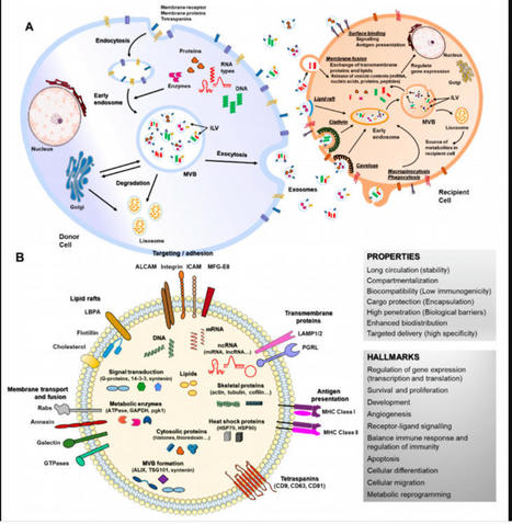
The review highlights the key features of exosomes as drug delivery vehicles, such as therapeutic cargo, use of targeting peptide, loading method and more...

|
Suggested by
LIGHTING
December 3, 2020 1:30 PM
|
Abstract. Lupus nephritis (LN) affects a large proportion of patients with systemic lupus erythematosus (SLE). LN can lead to end-stage renal disease depending
|
|
Scooped by
Gilbert C FAURE
November 25, 2020 10:43 AM
|
Lupus nephritis (LN) is one of the most common manifestations of systemic lupus erythematosus (SLE), affecting approximately 40% of patients with lupus.It represents a major risk factor for morbidity...
|
|
Scooped by
Gilbert C FAURE
November 16, 2020 1:34 PM
|
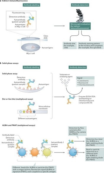
Antinuclear antibodies (ANAs) are valuable laboratory markers to screen for and support the diagnosis of various rheumatic diseases (known as ANA-associated rheumatic diseases). The importance of ANA testing has been reinforced by the inclusion of ANA positivity as an entry criterion in the 2019 systemic lupus erythematosus classification criteria. In addition, specific ANAs (such as antibodies to Sm, double-stranded DNA (dsDNA), SSA/Ro60, U1RNP, topoisomerase I, centromere protein B (CENPB), RNA polymerase III and Jo1) are included in classification criteria for other rheumatic diseases. A number of techniques are available for detecting antibodies to a selection of clinically relevant antigens (such as indirect immunofluorescence and solid phase assays). In this Review, we discuss the advantages and limitations of these techniques, as well as the clinical relevance of the differences between the techniques, to provide guidance in understanding and interpreting ANA test results. Such understanding not only necessitates insight into the sensitivity and specificity of each assay, but also into the importance of the disease context and antibody level. We also highlight the value of titre-specific information (such as likelihood ratios). Antinuclear antibodies (ANAs) are valuable biomarkers for various autoimmune rheumatic diseases and can be detected using various assays. This Review discusses the advantages and disadvantages of different ANA assays, to help in the understanding and interpretation of ANA test results.
|
|
Scooped by
Gilbert C FAURE
November 7, 2020 6:22 AM
|
Background/Purpose: ANA testing as an approach to diagnosing and classifying SLE, now embedded in the EULAR/ACR Criteria, is more important than ever. Cross-sectional studies indicate that as few as 70% are ANA-positive, although ANA status may change over time.
|
|
Scooped by
Gilbert C FAURE
October 7, 2020 2:52 PM
|
To the Editor, Recently in the Journal of Thrombosis and Haemostasis, Harzallah and colleagues report the results of antiphospholipid antibody testing in a series of 56 patients with confirmed or suspected SARS‐CoV‐2 infection. 1 Twenty‐five patients were found to be positive for lupus anticoagulants, while five patients had either anticardiolipin or anti–β2‐glycoprotein 1 antibodies. The isotypes for the anticardiolipin and anti–β2‐glycoprotein 1 antibodies were reportedly IgG and IgM, although specific antibody titers and details of which were found in combination with a lupus anticoagulant in three overlap patients were not reported. The authors also reference published work by Zhang and colleagues, who reported three patients with SARS‐CoV‐2 infection, coagulopathy, thrombocytopenia, and the presence of anticardiolipin IgA and anti–β2‐glycoprotein 1 IgA and IgG antibodies who developed cerebral infarcts. 2 Harzallah and colleagues suggest the presence of these antibodies should be used as evidence for early anticoagulation of patients with COVID‐19. Antiphospholipid antibodies are common in the general population, especially during infection. 3 , 4 Whether the IgA isotype alone, noted by Zhang and colleagues, invokes thrombosis remains controversial, with only high titer IgG and IgM isotypes included as diagnostic criteria for the antiphospholipid syndrome. 5 Lack of IgG and/or IgM titers in these case series precludes any evaluation of their role in the thrombotic sequelae described. Thrombosis is common during critical illness and all patients in the Zhang series had preexisting cardiovascular disease, further increasing risk for arterial thrombosis. A key question remains whether COVID‐19 patients experience arterial thrombotic events at a higher rate compared to critically ill patients without SARS‐CoV‐2. The findings presented by Zhang and colleagues cannot confirm anticardiolipin antibodies as the causal agent for the arterial thrombosis observed in their series. False positive lupus anticoagulant testing might be expected in patients with COVID‐19 given the marked elevation in measured C‐reactive protein (CRP) levels seen in patients with significant pulmonary or systemic inflammation. Many assays to detect lupus anticoagulants are sensitive to the presence of CRP, resulting in false positive results, further limiting interpretation of this test in the acute inflammatory state. 6 COVID‐19 appears to induce a hypercoagulable state, with elevated fibrinogen, and minimal prolongation of prothrombin time and activated partial thromboplastin time, as seen in these patients. The exact mechanisms underlying the coagulopathy are unclear. 7 We urge clinicians who are evaluating coagulation parameters in patients with COVID‐19 to be cognizant of the pre‐analytic and analytic variables that affect the validity and interpretation of coagulation testing and to adhere to established anticoagulation protocols and guidelines until clinical studies demonstrating efficacy and safety of various anticoagulation strategies are published.
|
|
Scooped by
Gilbert C FAURE
September 6, 2020 4:03 AM
|
Systemic lupus erythematosus is a multisystem autoimmune disease that commonly affects
the kidneys. Lupus nephritis (LN) is the most common cause of kidney injury in systemic
lupus erythematosus and a major risk factor for morbidity and mortality. The pathophysiology
of LN is heterogeneous. Genetic and environmental factors likely contribute to this
heterogeneity. Despite improved understanding of the pathogenesis of LN, treatment
advances have been few and risk for kidney failure remains unacceptably high.
|
|
Scooped by
Gilbert C FAURE
June 30, 2020 6:21 AM
|
AbstractBackground. Anti-CD20 B-cell depletion has not shown superior efficacy to standard immunosuppression in patients with systemic lupus erythematosus (SLE
|
|
Suggested by
LIGHTING
April 30, 2020 3:48 AM
|
B cell therapy in lupus nephritis
Abstract Properdin is the only one positive regulator of the complement system. In this study, we characterize the prevalence, functional consequences and disease associations of autoantibodies aga...
Via Krishan Maggon
|
|
Scooped by
Gilbert C FAURE
March 29, 2020 4:36 AM
|
PubMed comprises more than 30 million citations for biomedical literature from MEDLINE, life science journals, and online books. Citations may include links to full-text content from PubMed Central and publisher web sites.
The metabolism of healthy murine and more recently human immune cells has been investigated with an increasing amount of details. These studies have revealed the challenges presented by immune cell
Via Krishan Maggon
|


 Scoop.it!





















RA slide deck inside