 Your new post is loading...
The iliopsoas muscle is a primary hip flexor that assists in the femur's external rotation and maintains the hip joint's strength and integrity. It also helps to stabilize the lumbar spine and pelvis. Athletes often overuse these muscles with all the sprinting, jumping, kicking, and changing directions when running, causing strains and/or tears. Repetitive hip flexion can result in chronic degenerative tendon changes. Chiropractic care and physical therapy can assist in the early phases of healing, safely transitioning to rehabilitation, and returning to physical activities. Iliopsoas Muscle The hip flexors are the group of muscles, including the iliacus and psoas major muscles/iliopsoas and the rectus femoris/quadriceps. One of the largest and thickest muscles in the body, the psoas, extends from the lumbar vertebrae, crosses in front of each hip, and attaches to the inside top of the thigh bone. The muscle works by flexing the hip joint and lifting the upper leg towards the body. These fibers can tear if tension is more than the muscle can bear. An iliopsoas strain occurs when one or more of these hip flexor muscles become overly stretched or begin to tear. Injury The injury can occur from sports or everyday physical activities. This leads to inflammation, pain, and scar tissue formation. An iliopsoas injury is commonly caused by sudden movements, including sprinting, kicking, and changing direction fast while running. Individuals participating in any sports, especially cycling, running, dance, tennis, martial arts, and soccer, are more likely to experience this injury. Other contributing factors include: - Muscle tightness
- Joint stiffness
- Muscle weakness
- Inadequate core stability
- Not warming up correctly
- Improper biomechanics
- Decreased fitness and conditioning
Individuals will feel a sudden stinging pain or pulling sensation, usually on the front of the hip, groin, or abdominal area. Other symptoms include: - Stiffness after resting.
- Swelling
- Tenderness
- Bruising around the area.
- Anterior hip pain and/or burning sensation.
- Groin discomfort sensations.
- Hip snapping or a catching sensation.
- Discomfort when flexing the leg.
- Walking problems and discomfort.
- Lower stomach and/or back symptoms.
Healing and recovery depend on the severity of the injury. A minor iliopsoas muscle injury can take around three weeks to recover fully. More serious strains and tears take six to eight weeks before returning to activity, as the tissue needs time to repair before starting rehabilitation. Chiropractic Rehabilitation and Recovery The first steps when dealing with this injury should be P.R.I.C.E. protection, rest, ice, compression, and elevation. It is important to rest and seek treatment immediately; if left untreated, the condition could worsen, lead to a chronic condition, and require surgery. A chiropractic treatment and rehabilitation plan will consist of the following: - Soft tissue massage
- Joint mobilization
- A chiropractor may recommend crutches to keep the weight off the hip.
- A brace can help compress and stabilize the hip flexor to expedite healing.
- A flexibility and strengthening program will be implemented to target the muscles around the hip.
- Core strengthening exercises will improve the stability of the pelvis area to prevent any further overuse problems.
- Wearing compression clothing could also be recommended, as the clothing helps maintain muscle temperature.
General Disclaimer * The information herein is not intended to replace a one-on-one relationship with a qualified healthcare professional or licensed physician and is not medical advice. We encourage you to make healthcare decisions based on your research and partnership with a qualified healthcare professional. Our information scope is limited to chiropractic, musculoskeletal, physical medicines, wellness, sensitive health issues, functional medicine articles, topics, and discussions. We provide and present clinical collaboration with specialists from a wide array of disciplines. Each specialist is governed by their professional scope of practice and their jurisdiction of licensure. We use functional health & wellness protocols to treat and support care for the injuries or disorders of the musculoskeletal system. Our videos, posts, topics, subjects, and insights cover clinical matters, issues, and topics that relate to and directly or indirectly support our clinical scope of practice.* Our office has reasonably attempted to provide supportive citations and identified the relevant research study or studies supporting our posts. We provide copies of supporting research studies available to regulatory boards and the public upon request. We understand that we cover matters that require an additional explanation of how it may assist in a particular care plan or treatment protocol; therefore, to further discuss the subject matter above, don't hesitate to get in touch with Dr. Alex Jimenez or contact us at 915-850-0900. Dr. Alex Jimenez DC, MSACP, CCST, IFMCP*, CIFM*, ATN* email: coach@elpasofunctionalmedicine.com Licensed in: Texas & New Mexico* References Dydyk AM, Sapra A. Psoas Syndrome. [Updated 2022 Oct 24]. In: StatPearls [Internet]. Treasure Island (F.L.): StatPearls Publishing; 2022 Jan-. Available from: https://www.ncbi.nlm.nih.gov/books/NBK551701/ Lifshitz, Liran BPt, MSc, PT; Bar Sela, Shlomo BPt MPE; Gal, Noga BPt, MSc; Martin, RobRoy PhD, PT; Fleitman Klar, Michal BPt. Iliopsoas the Hidden Muscle: Anatomy, Diagnosis, and Treatment. Current Sports Medicine Reports 19(6):p 235-243, June 2020. | DOI: 10.1249/JSR.0000000000000723 Rauseo, Carla. "THE REHABILITATION OF A RUNNER WITH ILIOPSOAS TENDINOPATHY USING AN ECCENTRIC-BIASED EXERCISE-A CASE REPORT." International journal of sports physical therapy vol. 12,7 (2017): 1150-1162. doi:10.26603/ijspt20171150 Rubio, Manolo, et al. "Spontaneous Iliopsoas Tendon Tear: A Rare Cause of Hip Pain in the Elderly." Geriatric orthopedic surgery & rehabilitation vol. 7,1 (2016): 30-2. doi:10.1177/2151458515627309
Concussions are a fairly common injury in the world of sports medicine. As hard as it might be to admit it, the CDC reported 1.7 million concussions within the U.S. population last year. Furthermore, this type of mild traumatic brain injury (mTBI) is not exclusively seen in sports medicine. All the population can be exposed to this, and 90% of concussion patients have a resolution of symptoms after four weeks. Nevertheless, the remaining debilitating post-concussion symptoms can lead to neurodegenerative consequences later in life. Nowadays, integrating an anti-inflammatory nutritional approach after a concussion is integral to preventing post-concussion syndrome (PCS). Concussions and Leaky Gut - A patient with PCS can develop increased intestinal permeability within 4 hours after the injury. Indeed, this is due to the connection between the brain and the enteric nervous system.
- Dehydrated patients are more likely to suffer from post-concussion complications.
- In addition to intestinal permeability, patients with PCS are more likely to develop small intestine bacterial overgrowth, known as SIBO.
- A concussion is rapidly followed by brain hypometabolism, and studies have shown that a moderate caloric reduction may improve cognitive impairment.
Brain hypometabolism: Indeed, the brain is a highly metabolic organ that requires an elevated and uninterrupted energy supply, usually glucose. After a concussion injury, the brain can be susceptible to diminished glucose uptake resulting in a bioenergetic deficit. Recently, research has shown that a ketogenic diet has potential therapeutic applications to reverse the energetic deficit. In this one-arm study, post-concussion patients taking a very high fat-ketogenic diet reported improved visual memory and decreased concussion-related symptoms. The researchers noted that a ketogenic diet could promote multiple benefits to PCS patients. Ketone Diet Benefits: - Ketone body metabolism produces lower amounts of reactive oxygen species (ROS).
- A high-fat ketogenic diet upregulates mitochondrial antioxidant systems that scavenge ROS.
- This dietary approach increases the activity of the Nrf2 anti-inflammatory pathway.
- Overall improvement of neurotransmitter levels.
Supplements and Herbs vs. Concussion: The recovery of an injury is affected by the patient’s nutritional status. Indeed, there are specific nutrients in the particular case of a concussion: - Boswellia: The use of this herbal component has proven to enhance the cognitive outcome in patients with diagnosed diffuse axonal injury (DAI). In a double-blind, randomized study, 38 patients with DAI experienced an increased cognitive function when Boswellia was applied to their treatment.
-
- N-Acetyl Cysteine: Treatments with NAC after ischemic injuries is associated with brain cell damage prevention and lower death. In addition, NAC stimulates and promotes glutathione peroxidase activity. Furthermore, this effect is linked with NAC’s potent ROS scavenging function.
- Fish oil: The application of omega-3 fatty acids is linked to the conservation of the brain’s protection mechanisms and maintenance of the integrity of brain cells.
- Vitamin D: vitamin D increases resilience to TBI due to its role during the conversion of tryptophan to serotonin. Furthermore, the genetic impact of vitamin D modulates serotonin levels, which controls mood, decision-making, and social behavior.
- Curcumin: Curcumin is a potent anti-inflammatory agent linked to reversing behavioral deficits in locomotion and memory. In addition, curcumin acts as an antioxidant reducing oxidative stress and thus improving synaptic simplicity and cognition.
The overall objective of using herbs and supplements to improve TBI and concussion secondary effects is to increase the body’s resilience. In addition, the recovery process will always be joined by a depletion of the vitamins and antioxidants pool, making us susceptible to other conditions. It is vital to support our patients with antioxidants, anti-inflammatory compounds, and enough vitamins to accelerate this curing process. – Ana Paola Rodríguez Arciniega, MS Bibliography: Ellis, E F et al. “Restoration of cerebrovascular responsiveness to hyperventilation by the oxygen radical scavenger N-acetylcysteine following experimental traumatic brain injury.” Journal of neurosurgery vol. 75,5 (1991): 774-9. doi:10.3171/jns.1991.75.5.0774 -
Moein, Payam et al. “The effect of Boswellia Serrata on neuro recovery following diffuse axonal injury.” Brain injury vol. 27,12 (2013): 1454-60. doi:10.3109/02699052.2013.825009 General Disclaimer * The information herein is not intended to replace a one-on-one relationship with a qualified health care professional, licensed physician, and is not medical advice. We encourage you to make your own health care decisions based on your research and partnership with a qualified health care professional. Our information scope is limited to chiropractic, musculoskeletal, physical medicines, wellness, sensitive health issues, functional medicine articles, topics, and discussions. We provide and present clinical collaboration with specialists from a wide array of disciplines. Each specialist is governed by their professional scope of practice and their jurisdiction of licensure. We use functional health & wellness protocols to treat and support care for the injuries or disorders of the musculoskeletal system. Our videos, posts, topics, subjects, and insights cover clinical matters, issues, and topics that relate to and support, directly or indirectly, our clinical scope of practice.* Our office has made a reasonable attempt to provide supportive citations and has identified the relevant research study or studies supporting our posts. We provide copies of supporting research studies available to regulatory boards and the public upon request. We understand that we cover matters that require an additional explanation of how it may assist in a particular care plan or treatment protocol; therefore, to further discuss the subject matter above, please feel free to ask Dr. Alex Jimenez or contact us at 915-850-0900. Dr. Alex Jimenez DC, MSACP, CCST, IFMCP*, CIFM*, ATN* email: coach@elpasofunctionalmedicine.com Licensed in: Texas & New Mexico*
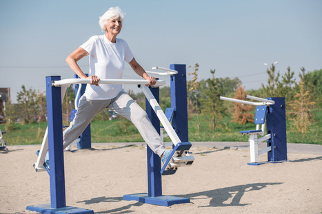
As we age, staying active keeps us healthy, our lives are lengthened and we feel great! Older individuals are discovering that exercise, sports and being physically fit does not mean having to do hardcore workouts and hard-to-maintain exercise/s and schedules.
Many of these individuals get their exercise from active pastimes like biking, Crossfit, and tennis. Others participate in less active recreational activities like walking, gardening or golf. Regardless of which activity they get into, they are all getting relaxation and fun while securing a healthy future.
Exercise helps us feel better because it improves our health. Spending just a little time each day doing some type of physical activity, will bring these benefits: - Longer
- Healthier life
- Stronger bones
- Reduced joint
- Reduced muscle pain
- Improved mobility
- Improved balance
- Lower risk of falls
- Lower risk of serious injuries e.g. hip fractures
- Slower loss of muscle mass
Fortunately, individuals are living longer but their quality of life means staying healthy and active to remain independent. Staying active will lower the risk of many common diseases, relieve arthritis pain and help you recover faster when illness hits. Activity and Safety Keeping active means that it's also important to be safe during these activities/exercises. With more older individuals participating in physical activities, there is an equal increase in sports-related injuries. This is true for bicyclists, skiers, weight lifters and those that use exercise machines. A recent study by the U.S. Consumer Product Safety Commission (CPSC), showed an estimated 53,000 people ages 65 and up were treated in U.S. emergency rooms for sports, physical activity-related injuries. Additional injuries were treated in doctor's clinics/offices. The increase comes from more older individuals engaging in active sports. However, most of these injuries were not severe but more importantly, they could've been prevented. An example was cyclists treated in emergency rooms for head injuries were not wearing helmets. Wearing a helmet reduces the risk of serious head injury up to 85 percent. Regular exercise along with doing it safely means you can enjoy yourself a lot more. Activity log Medium physical activity for 30 minutes a day is beneficial for everyone but especially those with chronic bone/joint conditions.
The 30 minutes of activity can be broken up into shorter periods of different activities, like 15 minutes of gardening and 15 minutes of stretching exercises. This can help not getting bored with a routine by mixing it up.
Activity log to keep track of the time you spend on each. Injury Prevention Tips When exercise/participating in an activity, doctors recommend following these tips: - Wear the proper safety gear for whichever activity/sport you choose.
- Wear the right shoes for each sport/activity.
- Warm-up before engaging in physical activity. This could be moderate walking at your normal pace with an emphasis on arm movements.
- Exercise at least 30 minutes a day. Break the activities into shorter periods of 10 or 15 minutes throughout the day.
- Follow the 10 percent rule, which means never increasing the program like walking/running distance or weight-lifting more than 10 percent a week.
- Try not to do the same routine two days in a row.
- Mix it up so as not to sprain/strain the same muscles and allow the other muscles to get a workout. So walk, swim, tennis or lift weights, as this keeps the exercise more interesting.
- Read instructions carefully when working with exercise equipment, and if needed, ask a qualified professional to help you.
- Check exercise equipment making sure it's in proper working order.
- If weight training interests you but you have never done it, make sure to get professional consultation before starting.
- Stop exercising if there is severe pain or swelling and get checked by your doctor.
There are plenty of ways to enhance our lives as we age, and staying fit and active along with the proper diet are a few of the most important. Our clinical focus and personal goals are to help your body heal itself naturally quickly and effectively. At times, it may seem like a long path; nevertheless, with our commitment to you, it’s sure to be an exciting journey. The commitment to you in health is to, never lose our deep connection to each one of our patients on this journey. When your body is truly healthy, you will arrive at your optimal fitness level proper physiological fitness state. We want to help you live a new and improved lifestyle. Over the last two decades, while researching and testing methods with thousands of patients, we have learned what works effectively at decreasing pain while increasing human vitality.
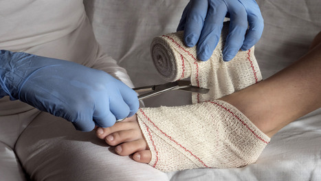
Ankle sprains can be frustrating and painful. But not all ankle sprains are equal in their severity. When diagnosing an ankle sprain, you will hear a doctor classify the injury in one of three ways—first degree, second degree, and third degree. Knowing what each classification means can help you understand the nature of your injury and how careful you need to be with it. Classification of ankle sprain degrees will help to focus on the best treatment options. The 3 Degrees of Ankle Sprains and What They Mean A sprain is an injury involving the stretching and/or tearing of your ligaments. With the ankle, it is possible to suffer an injury to either side of the joint, the interior or the exterior. You may have an inversion sprain or an eversion sprain. Whichever type you suffer from, it will fall into one of three categories: First Degree A first-degree sprain is one where the ligaments have not torn, only stretched further than normal. Symptoms of a first-degree sprain include discomfort when jumping, jogging or sometimes walking. Pain is usually mild and swelling is minimal. The joint may be a little stiff and slightly less stable than normal. Second Degree A second-degree sprain is the most common type of sprain people seek medical care for. The ligament is partially torn, which can lead to several uncomfortable symptoms. The sprain will make walking difficult, be moderately painful and make it hard to use the ankle. The injury will result in noticeable swelling and often bruising. Third Degree A third-degree sprain is the most severe and involves a full tear of the ligament. Pain is often severe and swelling is significant. The torn ligament makes the joint unstable, so it is not only painful but also very hard to use. How Your Chiropractor Can Help Whether you are walking, running, jumping or just standing, the ankle plays a vital role in how you move and use your body. That is why it can be so frustrating when you find yourself with an ankle injury. Fortunately, ankle sprains do heal with enough rest and the right treatment. Your chiropractor can help you recover from an ankle injury, both reducing pain and speeding up recovery time. There is research to support the use of chiropractic for ankle sprains. One study showed that patients with ankle sprains experienced less pain when chiropractic was added to their treatment program. Chiropractic also focuses on improving mobility and range of motion, which can be quite beneficial when trying to recover from a sprain and get back to your normal life. Some of the most common chiropractic treatments for ankle sprains include: Adjustments Adjustments can be made to more than just the spine. The bones, ligaments, and tendons in the ankle are designed to work in a certain way. A sprain can knock the ankle out of alignment, something that most traditional doctors and rehabs do not consider. An ankle adjustment will work to put things back into their proper places. Strength Exercises There are a variety of useful exercises that your chiropractor can take you through to improve strength and mobility in the ankle. Things like a wobble board, where you stabilize yourself on a wobbly platform and drawing on a board with a market held in your toes can be surprisingly effective at strengthening your ankle. Get Treatment Today Please contact us today to schedule an appointment with a chiropractor for your sprained ankle. Let us help you get stronger and heal faster!
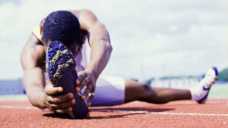
Why do people automatically assume that endless amounts of static stretching and foam rolling will provide them with transferable benefits in the gym, sport and their daily lives? The stretching myths need to be dispelled, especially as it pertains to the most commonly stretched area of the body, the hip. From attempting to get out of lower back pain to targeting more and more hip mobility to improve squatting and other functional movements, people gravitate towards stretching, but is it really doing them any good? We have Dr. Zach Long here on DrJohnRusin.com because he wants to put a stop to static stretching of the hips, and rightfully so! He has a better way to improve positions and alleviate pain, and it doesn’t involve sticking lacrosse balls in your butt or holding painful static stretches for minutes at a time. Enjoy. Here’s What You Need To Know…1. Many people use foam rolling and static stretching to alleviate tight muscles, but the fact of the matter is that these practices make little to no useable change to the the muscles or movements they control in the short and long term. 2. If you really want to improve your “hip mobility” you better look outside the hip and start targeting stability of the pelvis and spine instead of adding endless mobility to one of the most mobile joints in the body. 3. When it comes to alleviating lower back pain and hip dysfunction, gaining and maintaining pelvic alignment in the neutral zone is pivotal. And guess what, it will improve your strength performances as well. 4. Dynamic stability is the new mobility, so here’s the most effective program that will open up your hips without ever holding a static stretch ever again. Stupid Static StretchingThe fitness world has come a long way in our understanding of the importance of mobility work and the impact it has on athletic performance and training. It’s safe to say that “mobility” exercises and tools like the foam roller are becoming commonplace in gyms, CrossFit boxes, and on the playing field worldwide. Unfortunately, most athletes performing these mobility drills have yet to figure out that much of their mobility work does not actually result in real performance gains! Sadly, their mobility work does not address the true root of their problems and their constant stretching, foam rolling, and joint band distractions get them nowhere. The hips are the greatest example of this problem, with athletes everywhere wasting hours on useless mobility work! Let’s stop wasting time and start seeing objective results from corrective exercise and training, you know, the kind that shows up in PRs instead of in fluffy feel good effects. It’s time to quit the static stretching of the hips and start refocusing your “mobility” work on more effective exercises and techniques that will not only improve your mobility, but your athletic performance as well! And did I mention that we can achieve this in a fraction of the time? Yeah, better listen up. Where Hip Mobility Exercises Have Gone WrongBecause of the inherent stability of the hip joint provided by the ball-and-socket, many athletes and coaches spend far too much time trying to improve hip mobility by performing stretching and soft tissue work. Months of intense stretching techniques provide little actual change in available motion and only serve to waste time and create pain in the athletes. Instead, small stability changes at the pelvis can provide drastically fast improvements in performance. Contraction of the muscles around the pelvis can result in changes in pelvic positioning and thus available hip range of motion. For example, a posterior tilt of the pelvis will put the hips in a position advantageous to improve hip flexion mobility, while an anterior pelvic tilt will result in increased hip extension. It’s important to remember that it’s not all about the hips. The hamstrings are the perfect example of the effects pelvic positioning can have on mobility. I’ve yet to meet an athlete who doesn’t claim to have “tight hamstrings”. Even the elite gymnasts, dancers, runners, and yogis believe that their hamstrings should be further stretched in order to maintain and enhance mobility. In an anteriorly rotated pelvis, the hamstrings will have increased tension placed on them, thus resulting in a perceived decrease in flexibility. More often than not, simple core stabilization movements will provide immediate improvement in their perceived tightness or chronic hamstring muscle strains. To test this directly, have athletes perform a straight leg raise while lying on their back. Quite often, simply cueing the athlete to “push your rib cage down” or “flatten your lower back into the floor” will result in a posterior pelvic tilt that instantly improves “hamstring mobility” and decreases the perception of hamstring tightness and muscle strain. This simple repositioning of the pelvis can provide more gains almost instantly than months of static stretching, manual therapy, and self-myofascial work combined. This effect can also be seen when working to improve hip extension, rotation, and functional patterns such as the squat. The Hip Mobility Solution The self-sufficient solutions to hip mobility deficits are simple. Perform movements that challenge the available active hip range of motion, while engaging the core to stabilize the pelvis. As the athlete learns to better control the core and pelvis, mobility will drastically improve and be transferable into function. Hip “mobility” work done this way will have two effects. First, it will reposition the pelvis to a more neutral position, allowing for improved mobility within the hip socket. Secondly, it will serve as a “reset” to muscle tone around the hips. Often times, the body realizes it does not have the needed stability around a joint due to muscle weakness. The body’s response is to increase the tone in a muscle to provide some false-stabilization. The hip flexors (like the hamstrings) are another muscle group that often feels tight in athletes but when proper core stabilization movements are performed, this increased muscle tone instantly vanishes and mobility problems are gone! Your New Hip Mobility RegimenThe following exercises should be a strong component of any athlete’s hip mobility work, as they will produce faster results than the typically prescribed foam rolling and stretching routines. Lets break these down one by one with a video and my notes on what makes each movement so powerful: The Reverse Active Straight Leg Raise Coaching Notes: The Reverse Active Straight Leg Raise is an excellent movement to improve hip flexion and active hamstring mobility. The athlete begins lying on his or her back with both legs vertical and knees straight. One leg is kept in this vertical position (this can be done by using a stretch strap or not using one to increase the challenge) while the other leg is slowly lowered to the floor. The key point of performance is that the lower back remains flat on the ground, ensuring that the core is actively engaged to stabilize the spine and pelvis. Single Leg Hip Lift Coaching Notes: Up next for those with hip flexor tightness, the Single Leg Hip Lift and Psoas March variations can be incredible exercises. To perform the Single Leg Hip Lift, the athlete lies on their back with one foot flat on the floor and that knee bent to approximately 90 degrees. The other leg is pulled towards the chest and held in the athlete’s arms. Next, the athlete lifts his or her hips up as high as possible without arching their lumbar spine. The athlete should consciously focus on activating his or her glutes throughout the entire movement. The Psoas March Coaching Notes: The Psoas March is an amazing exercise for quickly eliminating hip flexor tightness as it retrains the psoas’ role in spinal stability. The athlete lies supine with a resistance band around both feet. While focusing on maintaining a neutral spine, the athlete lifts one knee towards his or her chest, stopping at ~90 degrees of hip flexion, and then returns to supine. This is then repeated on the opposite leg. The Goblet Squat The Goblet Squat may be the most powerful mobility exercise specific to the squat. By holding a weight in front of the body, the athlete is able to better sit back into the squat, maintain a neutral spine and pelvis, and reach better squat depths. When performing Goblet Squats to work hip mobility, we suggest performing a slow negative and pausing for several seconds in the bottom of the squat. 90/90 Breathing with Hip Internal Rotation Coaching Notes: The 90/90 Breathing with Hip Internal Rotation is another fantastic drill for quickly changing hip mobility. This has repeatedly helped improve rock-bottom squat depth and decreased hip pinching in the elite Olympic weightlifters and CrossFit athletes that I work with. Start with the athlete lying on his or her back, their feet up on a wall and their hips and knees bent to ninety degrees. After raising the hips slightly off the ground, they inhale through their nose, focusing on filling their stomach with air before allowing the check to rise. As they exhale, the rib cage is pushed down. This movement puts the spine in a neutral position and the pelvis slightly posteriorly rotated. After several breaths, the athlete then lifts one leg off the wall and repeatedly internally rotates it while continuing the breath cycles. The Anti-Stretch Hip Mobility ProgramFor an athlete looking to optimize hip mobility and performance, I highly recommend that they do each of the above exercises three to four times weekly, usually as part of their warm up. And hell, if more attention is needed to improve these positions, work this exact program into a cool down after a workout or a stand alone session later on that day. - 90/90 Breathing with Hip Internal Rotation
- 5 breaths followed by 20 internal rotations per side
- Reverse Active Straight Leg Raise
- 2 sets to moderate fatigue each leg
- Single Leg Hip Lifts
- 2 sets to moderate fatigue
- Psoas March
- 2 sets to moderate fatigue
- Goblet squats
- 2 sets of 10 reps with 5 second negative and three second pause in the bottom
Alright guys, there you have it! A full “hip mobility” program that is geared towards improving your motor control, stability and of course your movement abilities as a whole, without the need to stretch out that always tight piriformis!! Source:
|
Older and elderly individuals have an increased risk of developing a herniated disc/s. The age of the intervertebral discs/cushions causes deflation, drying out, and shifting, making it easier for discs to herniate. Muscle mass also reduces/lessens with age; specifically, the muscles parallel to the spinal column are responsible for stability. When the spine loses strength, the risk of injuries like slips and falls can damage the spine and the rest of the body. Herniated disc decompression will keep the vertebral cushions healthy, functioning, and properly aligned. Symptoms of Disc Herniation A herniated disc bulge or tear/s will press on the spinal nerves causing discomfort that can range from mild to severe pain and can last for weeks to months. The symptoms of disc herniation vary and depend on the injury angle, how much of the disc ruptured and if it is touching or has leaked out on the nerve roots. The most common symptoms include: - Restricted hip and waist flexion.
- Continuous back pain that radiates.
- Sciatica symptoms
- Back muscles contract/spasm
- The pain can worsen by sudden body movements caused by coughing, sneezing, or hiccups.
- Numbness in the affected area
- Numbness or tingling in the leg or foot
- Decreased knee or ankle reflexes
- Weakness
- Bladder or bowel function changes like difficulty moving waste through the colon or large intestine.
Herniated Disc Decompression Nonsurgical herniated disc decompression therapy can help heal the herniation by: - Stretching the spine to the total capacity.
- Removing the pressure.
- Pulls the herniated disc back into its correct position.
- Fills the injured/damaged areas and the rest of the spine with blood, oxygen, nutrients, and lubricating fluids.
- Helping to rebuild joint and muscle strength.
- Increasing flexibility in the muscles that support the affected area of the spine.
The therapy duration depends on the herniation, injury, and damage severity. The objective is to bring significant improvement that will last. Chiropractic, Physical/Massage Therapy, and Health Coaching A chiropractor and physical massage therapy team will develop a personalized herniated disc decompression treatment plan with specific goals. The therapy will include: - Mechanical decompression.
- Manual chiropractic adjustments.
- Massage sessions.
- Health coaching.
- Exercises and stretches will be given that will help maintain pressure relief and flexibility.
- Core stabilization exercises will strengthen and stabilize the spine and muscles.
- Aerobic conditioning will help increase endurance.
General Disclaimer * The information herein is not intended to replace a one-on-one relationship with a qualified health care professional, or licensed physician, and is not medical advice. We encourage you to make your own healthcare decisions based on your research and partnership with a qualified healthcare professional. Our information scope is limited to chiropractic, musculoskeletal, physical medicines, wellness, sensitive health issues, functional medicine articles, topics, and discussions. We provide and present clinical collaboration with specialists from a wide array of disciplines. Each specialist is governed by their professional scope of practice and their jurisdiction of licensure. We use functional health & wellness protocols to treat and support care for the injuries or disorders of the musculoskeletal system. Our videos, posts, topics, subjects, and insights cover clinical matters, issues, and topics that relate to and support, directly or indirectly, our clinical scope of practice.* Our office has made a reasonable attempt to provide supportive citations and has identified the relevant research study or studies supporting our posts. We provide copies of supporting research studies available to regulatory boards and the public upon request. We understand that we cover matters that require an additional explanation of how it may assist in a particular care plan or treatment protocol; therefore, to further discuss the subject matter above, please feel free to ask Dr. Alex Jimenez or contact us at 915-850-0900. Dr. Alex Jimenez DC, MSACP, CCST, IFMCP*, CIFM*, ATN* email: coach@elpasofunctionalmedicine.com Licensed in: Texas & New Mexico* References Carla Vanti, PT, MSc, OMPT, Alice Panizzolo, PT, OMPT, Luca Turone, PT, OMPT, Andrew A Guccione, PT, Ph.D., DPT, FAPTA, Francesco Saverio Violante, MD, Paolo Pillastrini, PT, MSc, Lucia Bertozzi, PT, MSc, Effectiveness of Mechanical Traction for Lumbar Radiculopathy: A Systematic Review and Meta-Analysis, Physical Therapy, Volume 101, Issue 3, March 2021, pzaa231, https://doi.org/10.1093/ptj/pzaa231 Dydyk AM, Ngnitewe Massa R, Mesfin FB. Disc Herniation. [Updated 2022 Jan 18]. In: StatPearls [Internet]. Treasure Island (FL): StatPearls Publishing; 2022 Jan-. Available from: https://www.ncbi.nlm.nih.gov/books/NBK441822/ Maistrelli, G L et al. “Lumbar disc herniation in the elderly.” Spine vol. 12,1 (1987): 63-6. doi:10.1097/00007632-198701000-00012 Suri, Pradeep, et al. “Nonsurgical treatment of lumbar disk herniation: are outcomes different in older adults?.” Journal of the American Geriatrics Society vol. 59,3 (2011): 423-9. doi:10.1111/j.1532-5415.2011.03316.x
It's the weekend in the backyard doing some chores, grilling, etc, while several kids play and jump on a nearby trampoline. The joviality from all this fun is filled with laughter, screams of joy, and other sounds or game instructions from one child to another. Then there is silence. The kids are huddled around their friend. One of the children fell off flat on their back. The paramedics arrive and immobilize the head, neck, and spine strapping the child to a backboard and off to the hospital where there is no severe damage, just some mild bruising but everything turned out ok. This was a made-up scenario but unfortunately, emergency room doctors are seeing and treating this type of injury more and more. Statistics The statistics of the number of trampoline-related injuries treated in emergency rooms. A simple analysis of the above stats is shocking. This means that since 1995, the number of injuries has increased between 30 and 45 percent. The CDC states that around 10 percent of trampoline injuries affect the head and neck. Many injuries are minor like bruising, scrapes and whatnot but some can be serious like broken bones, blunt-force trauma, and even paralysis. For Home Most of us see trampolines as a toy but are not aware of the dangers that come with it. Most injuries take place on trampolines purchased for home use. The American Academy of Pediatrics recommends that parents never let their children use a friend's trampoline. While the American Medical Association recommends children should not be allowed to play/jump on a trampoline, even with adult supervision. Trampoline Safety Equipment Tips A full-size trampoline consists of flexible fabric attached to a metal frame with springs, hooks, and a safety mesh/net. Most are around three feet off the ground. Consider the following safety tips: - Read the information and instructions provided by the trampoline manufacturer. Give this information to anyone who will be using the trampoline.
- The proper placement of the trampoline is very important. Look at the location and surrounding area. Don't place the trampoline close to a house/building, playground equipment, a swimming pool, the street, electrical lines, outdoor appliances, and plants/trees.
- Remember a child can bounce 10 feet or higher in the air from a trampoline.
- Make sure the springs, hooks, and frame are secured and covered with sturdy shock-absorbing pads.
- There should be shock-absorbing material all-around and under the trampoline. Use the owner’s manual for recommended materials.
- Stores where trampolines are sold often sell special padding.
- Check the trampoline for wear and tear often. Which include the frame's structure like the screws, bolts springs, hooks, and fabric.
Using the Trampoline - Step-ladders, boxes, and chairs to climb on the trampoline should be kept out of reach to prevent children from using the equipment without permission. And they should be moved out of the way once the individuals are on.
- Do not use the trampoline when darkness begins to take over like sunset. Individuals cannot see and when up in the air judging distance and where the trampoline is can be difficult/impossible.
- Before use, warm-up with a few exercises. To make sure muscles are loose and ready to react.
- Children need adult supervision at all times. Judging distances, foreseeing danger and quick reaction in situations that can become dangerous require an adult.
- At least two adults are needed to spot the individuals and help prevent anyone from falling off.
- Most want to jump together but too many people on a trampoline can be dangerous. Limit the number of people to where they can bounce safely without bumping into someone else, or falling off from lack of space.
- CDC reports that half of all injuries happen when more than two people use a trampoline. Usually, this happens with children that are lightweight, still not fully coordinated, and can't control how they move, especially in the air.
- Learn how to land properly.
- Unless a professional or expertly trained do not try somersaults, backflips, stunts, or crazy acrobatics.
- Don't jump or bounce off the trampoline.
Professional Help There are professional gymnastic centers that have trampolines with padding all around and also give lessons/classes on proper usage. This could help a great deal, as it could be a safe alternative instead of home use. But if not then take the classes which could save a trip to an emergency room! As El Paso’s Chiropractic Rehabilitation & Integrated Medicine Center, we are focused on treating patients after debilitating injuries and chronic pain conditions. We focus on improving your ability through flexibility, mobility and agility programs tailored for all age groups and disabilities.
Athletes both recreational and fully competitive can be impacted by injuries to the muscles and ligaments around the hip. These injuries interfere with performance levels and sometimes end participation completely. Excessive pronation along with shoes with poor shock absorption has been found to be an underlying cause for various leg/hip injuries. Custom made Orthotics improve the biomechanics of the feet and reduce the extent of pronation helping to prevent sport-related leg/foot injuries. Over Foot Pronation Research has determined that athletes with more foot pronation have a higher probability of sustaining a leg injury, including iliotibial band syndrome that comes from excessive tightness of the hip muscles. People involved in sports or recreational activities lower their likelihood of developing traumatic and overuse hip injuries through chiropractic treatment and using custom foot orthotics. - The amount of pronation during standing and while running at a standard speed is determined by measuring the angles of the footprints.
- Athletes with more pronation have a higher likelihood of an overuse injury.
- Standing (static) and running (dynamic) prints show the amount of pronation and is a predictor of developing an overuse injury.
- Athletic performance and injury prevention involve regularly checking the alignment of patients’ feet in the standing position.
Hip Injuries & The Hamstring Many hip injuries develop from poor biomechanics and improper movement, especially when running. Smooth muscle coordination provides balance and support for the pelvis and is needed for optimum sports performance. This includes: - Hamstring muscles
- Hip abductor muscles
- Tensor fascia lata or the iliotibial band
When there is an issue with the feet and ankles, abnormal motion like over-rotating the entire leg is the perfect set-up for pulls, sprains, and strains. 50% of standing consists of heel strike and maximum pronation. The hamstring muscles function to control the knee and ankle when the heel strikes and absorb the impact. The theory behind orthotic support is that orthotics help the hamstrings control the position of the calcaneus and knee, so there is less stress on the hip and pelvis. Hip Injuries & Over-Pronation Orthotics can correct excessive pronation and treatment of hip problems. These are some of the problems/pathologies that can develop. Hip and pelvis pathologies: - Anterior pelvic tilt
- Hip adductor muscle strain
- Hip flexor muscle strain
- Hip joint capsulitis
- Iliotibial band syndrome
- Piriformis muscle strain
- Tensor fascia lata strain
These conditions develop in athletes who push their body's to the limit going for optimal performances. Conclusion Overpronation and poor shock absorption contribute to leg injuries — from: - Foot
- Lower leg
- Knee
- Thigh
- Hip
These conditions can be prevented with custom-made orthotics. - Foot biomechanics evaluation is a must
- To avoid hip injuries, athletes need regular evaluations of foot alignment and function
- Wear well-designed and solid-constructed shoes
Chiropractors can prevent arch breakdown and foot problems with custom orthotics, and also treat numerous injuries to the lower extremities, especially the hips. The following video discusses how excessive foot pronation can ultimately affect foot posture and mobility. Several factors can affect foot posture and mobility, such as excessive foot pronation. Excessive foot pronation is most prevalent among the general population, therefore, it is considered to be one of the most common factors for abnormal foot posture and mobility, which can lead to a variety of health issues like overuse injuries. Excessive foot pronation and even supination can ultimately affect overall health and wellness. Hip Labrum tears in athletes can occur from a single event or recurring trauma. Running may cause labrum tears due to the labrum being utilized more for weight-bearing and taking excess forces while at the end-range motion of the leg. Sporting activities are probable causes, specifically those that require frequent hip rotation or pivoting to a loaded femur as in ballet or hockey. Constant hip rotation places increased strain on the capsular tissue and harm to the iliofemoral ligament. This subsequently causes hip instability putting increased stress on the labrum and causing a hip labrum tear. What's Afoot Chiropractic seeks to find the cause of the conditions it is used to treat, including pain, instead of just treating symptoms. Because of this, the chiropractor will work to find the cause of the pain, in this case, overpronation and overpronation, and correct it – or the effects of the condition – in addition to treating the back pain. Overpronation and oversupination can cause a variety of injuries and conditions that affect not only the feet and ankles, but also the knees, hips, and back as well. Some of the more common injuries and conditions include: - Flat feet or posterior tibial tendon dysfunction
- Ankle Sprains
- Achilles tendinitis
- Arch pain
- Plantar fasciitis
- Corns
- Shin splints
- Heel pain
- Tight calves
- Calluses
- Knee pain
- Patellar tendonitis
- Tight hip flexors
- Back pain
- Sciatica
- Herniated disks
Science chiropractor, Dr. Alexander Jimenez explores ankle movement -- and discusses why it is so Important. IntroductionGiven that we're a bipedal bunch, the joint that connects the foot into the rest of our body is going to have a considerable influence on our movement. Most men and women get pretty good at compensating for any motion deficiencies they might have. It seems, however, that one of the most troublesome moves to compensate for, without hurting oneself, is a lack of ankle dorsiflexion (DF) range of motion (RoM). There is some evidence to suggest that a deficiency of DF will cause compensatory moves elsewhere. Understandably the research showing that these movement patterns lead directly to injury are few and far between, but there are a few. So the logic seems sound, the evidence in 'normals' is suggestive and you'll find a small number of research pertaining directly to injury. On the other hand, the literature to demonstrate that enhancing deficiencies will really reduce following injury is nonexistent. I feel this should not stop us from pursuing our own logic, and therefore in many circumstances this will mean trying to 'normalize' an athlete's DF. Identifying HypomobilityMeasurement of DF is straightforward enough (Figures 1 and 2), also supplying subjects keep their heels on the floor and foot nicely aligned I'd suggest it's also fairly reliable; one screening study working with sports folks agrees(1), and so does a study on validity and reliability from 2012(2). The test in Figure 1 is far more prone to cheating by athletes, as it is too tempting to push 'just that little bit farther'; nonetheless, the test from Figure 2 is somewhat slower. I find a camera may be helpful here; it is good for illustrating issues to athletes, and can be more compelling than a numerical measurement on a page when you return. This is very helpful when an athlete has a subsequent trauma, as you can both see where their older 'normal' was; if that is different to 'normal' in their other hand, this will provide you a much more valid dimension to use. In a clinical sports setting, we seldom let athletes operate until they are within 2cm of normal, so if they had a pre-injury side-to-side difference in RoM this can be quite relevant. Lining up the camera lens with the front of the knee is imperative to be able to prevent parallax error. It is simpler, but to decide where 'normal' lies. I have a tendency to see 'normal' DF RoM as approximately 8cm plus, unless an athlete is particularly tall. 1 cricket study found a higher risk of harm if DF RoM was significantly less than 14cm(3). A study of patella tendinopathy in young basket ballers identified a DF angle of less than 36.5 deg to be a risk factor for injury(4) Naturally a angle is a more practical and legitimate measure but sadly they are far more difficult to measure. There are certainly more functional DF tests with good reliability on the market (once testers are satisfactorily trained(5)), if that's a degree of screening you'd like to extend to. However, a very simple dimension of DF (like Figures 1 and 2) may add additional detail to the big picture if these functional tests (Figures 3 and 4) highlight deficiencies. The RationaleThe rationale here is straightforward enough; athletes utilize triple flexion (in the hip, shoulder and knee) to attenuate the forces of landing, during running, jumping and cutting. When there is inadequate DF RoM from the ankle to get this done, another joint may then need to compensate, and musculotendinous structures will be functioning in a less than optimum position. To be able to attenuate more pressure, the knee will tend to use either more flexion, more valgus/frontal airplane knee excursion (FKPE)(Figure 5) or both. (see SIB posts 102 and 110 to learn much more on FKPE and its own role in harm). Any structure above or below the ankle could of course end up bearing the additional weight here; consider (or see SIB 113) the spinal column (Figure 6), or the feet(6). A lot of the literature is focused on how altered movement patterns will affect the knee as we see below: - Poorer movement patterns are associated with reduced DF RoM during a lateral step down task in healthy subjects(7);
- Normal subjects with artificially limited DF on a double squat show increased FPKE(8);
- In a study of soccer players, those with less DF RoM exhibited higher FPKE during a drop landing task(9) ;
- A 2013 study showed that subjects with lower DF RoM altered their kinematics during vertical jump by lifting their heels and changing their trunk position(10).
Another possible factor here is that the institution of decreased DF RoM with poor balance. Put the last two studies together and you've got a scenario where an athlete with poor DF RoM is now raising WB through their feet during landing with decreased balance. This will place the knee at a particularly vulnerable position. There are a variety of interesting studies showing restricted DF RoM to be associated with, or a risk factor for, injury. For example, away from the knee specifically, this is seen for lower limb injuries in Aussie rules players and Naval Trainers, and standard injury risk in cricketers(12,13,3). Soldiers with reduced DF RoM also had 4.6:1 odds of developing a metatarsal stress fracture compared to those without(6). Decreased DF was also implicated in the development of patellofemoral pain syndrome (PFP) at runners(15); this could possibly be clarified by the current study previously(8) in which increased knee valgus/FPKE and decreased quadriceps activation were seen when DF was restricted, very similar to the typical demonstration of PFP. CorrectionI have encounter no noteworthy evidence either way for whether 'normalizing' that this dorsiflexion will lead to an actual reduced injury risk in our athletes. One 2013 study, however, did reveal that in chronic foot injuries, mobilization of DF facilitated improved joint angles during landing, resulting in a more positive foot position(16). So, with avoidance in mind, we will need to look at what we do understand and apply some logic. It appears sensible that where any shortage is marked and unilateral (maybe the end result of an old/recent injury) there should be good profits to be made. Anecdotally, not just if changes come more easily, but asymmetries generally have a greater impact on motion patterns and injury risks. Where shortages are bilateral and more standing in character I feel that the profits will be harder to come by and less inclined to be effective, in order a preventative/screening problem it may be bad value for money. The listing of structures that could be impacted by these compensations is infinite. I would include in this group any athlete having an greater strain or strain in any areas round the ankle. Any athlete with a famous DF deficit ought to be persuaded to have this sorted out to the good of not just their ankle but a lot besides. Consequently, should you wish to enhance an athlete's DF, what resources do we have available to us? First let us talk my favorite techniques. Patient-Generated TechniquesThis ought to be the key muscle here, as it is the key musculotendinous restraint of DF. As always, very good alignment of the knee and foot (and buttocks, to prevent that habitual rectal pelvic tilt when possible)is essential. The accession of great toe extension (easiest in running sneakers) provides quite a marked shift to the elongate and may target another potential restrictor of DF RoM. Plantar Fascia StretchExtending the knee and feet should have the effect of tightening up the fascial structures (shallow back lineup) and helping place a bias on the plantar fascia. The foam roller or the athlete's own knee will offer some firm pressure to target some particular areas of tightness that stretching won't reach. Both methods will allow active moves to be added as a progression. A golf ball for the brave or something softer for yours truly. Again, this strikes any distinct troublesome spots and will also permit you to add in knee, knee, ankle or toe motions as a development. Therapist-Generated TechniquesMy favorite techniques are the ones which allow you to access a number of constructions in the 1 position and to add active motion whenever possible. This is a good place to start, as it allows you to select the gastrocnemius off stretch and receive at the true ankle RoM. From here you are able to bring in a stretch of the delicate tissue and get the planar fascia and ankle soft tissue structures. You can even apply your torso to provide a force and leave both hands free to provide any variety of PNF, acupressure or myofascial release methods. Step LungingThis position is much more of a progression. Not only does this add weight-bearing to the mix, but it also allows for a few more practical and lively motion to be added. From facing the athlete this provides a fantastic place for Mulligan's MWMs to be implemented. The talocrural and distal tibiofibular joints can both be mobilized here. With the ‘convex/ concave rule’ applied, an AP glide of the talus should (and does(18)) improve DF, adding some active DF will create an MWM. Equally a posterior glide of the fibula may well(19) correct Mulligan’s favorite positional fault(20) and allow more movement into DF. As an extra progression a seat belt can be added in lieu of a third hand. From behind the patient (Figure 15), we get better access to the myofascial structures and the athlete can control any movement, leaving both of your hands free to provide any number of PNF, acupressure or myofascial release techniques. ConclusionFor my money, these simple methods of identification and therapy are well worth the attempt. The tests only take a moment and once a deficit is identified the treatments are only a logical development. The remedies also have the benefit of between the athlete and keeping them active, which makes this a team effort rather than being overly passive. They also lead us nicely into a 'test, re-test' scenario so both you and the athlete could determine which techniques work to them and how they then change their more functional tests. References
1. Dennis RJ, Finch CF, Elliott BC, Farhart PJ. The reliability of musculoskeletal screening tests used in cricket. Phys Ther Sport. 2008 Feb;9(1):25-33. Epub 2007 Nov 8.
2. Chisholm MD, Birmingham TB, Brown J, Macdermid J, Chesworth BM. Reliability and validity of a weight-bearing measure of ankle dorsiflexion range of motion. Physiother Can. 2012 Fall;64(4):347-55.
3. Dennis RJ, Finch CF, McIntosh AS, Elliott BC. Use of field-based tests to identify risk factors for injury to fast bowlers in cricket. Br J Sports Med. 2008 Jun;42(6):477-82. Epub 2008 Apr 7.
4. Backman LJ, Danielson P. Low range of ankle dorsiflexion predisposes for patellar tendinopathy in juniorelite basketball players: a 1-year prospective study. Am J Sports Med. 2011 Dec;39(12):2626-33. 2011 Sep 14.
5. Minick KI, Kiesel KB, Burton L, Taylor A, Plisky P, Butler RJ. Interrater reliability of the functional movement screen. J Strength Cond Res. 2010 Feb;24(2):479-86.
6. Hughes LY. Biomechanical analysis of the foot and ankle for predisposition to developing stress fractures. J Orthop Sports Phys Ther. 1985;7(3):96-101.
7. Rabin A, Kozol Z. Measures of range of motion and strength among healthy women with differing quality of lower extremity movement during the lateral step-down test. J Orthop Sports Phys Ther. 2010 Dec;40(12):792-800. Epub 2010 Oct 22.
8. Macrum E, Bell DR, Boling M, Lewek M, Padua D. Limiting Ankle Dorsiflexion Range of Motion Alters Lower Extremity Kinematics and Muscle Activation Patterns during a Squat. J Sport Rehabil. 2011 Nov 15. (Epub ahead of print).
9. Sigward SM, Ota S, Powers CM. Predictors of frontal plane knee excursion during a drop land in young female soccer players. J Orthop Sports Phys Ther. 2008 Nov;38(11):661-667.
10. Papaiakovou G.Kinematic and kinetic differences in the execution of vertical jumps between people with good and poor ankle joint dorsiflexion. J Sports Sci. 2013;31(16):1789-96.
11. Basnett CR, Hanish MJ, Wheeler TJ, Miriovsky DJ, Danielson EL, Barr JB, Grindstaff TL. Ankle dorsiflexion range of motion influences dynamic balance in individuals with chronic ankle instability. Int J Sports Phys Ther. 2013 Apr;8(2):121-8.
12. Gabbe BJ, Finch CF, Wajswelner H, Bennell KL. Predictors of lower extremity injuries at the community level of Australian football. Clin J Sport Med. 2004 Mar;14(2):56-63.
13. Kaufman KR, Brodine SK, Shaffer RA, Johnson CW, Cullison TR. The effect of foot structure and range of motion on musculoskeletal overuse injuries. Am J Sports Med. 1999 Sep-Oct; 27(5):585-93.
14. Malliaras P, Cook JL, Kent P.Reduced ankle dorsiflexion range may increase the risk of patellar tendon injury among volleyball players. J Sci Med Sport. 2006 Aug;9(4):304-9. Epub 2006 May 2.
15. Lun V, Meeuwisse WH, Stergiou P, Stefanyshyn D. Relation between running injury and static lower limb alignment in recreational runners. Br J Sports Med. 2004 Oct;38(5):576-80.
16. Delahunt E, Cusack K, Wilson L, Doherty C. Joint mobilization acutely improves landing kinematics in chronic ankle instability. Med Sci Sports Exerc. 2013 Mar;45(3):514-9.
17. Loudon JK, Reiman MP, Sylvain J. The efficacy of manual joint mobilisation/manipulation in treatment of lateral ankle sprains: a systematic review. Br J Sports Med. 2014 Mar;48(5):365-70. doi: 10.1136/bjsports-2013-092763. Epub 2013 Aug 26.
18. Landrum EL, Kelln CB, Parente WR, Ingersoll CD, Hertel J. Immediate effects of anterior-toposterior talocrural joint mobilization after prolonged ankle immobilization: a preliminary study. J Man Manip Ther. 2008;16(2):100-5.
19. Fujii M, Suzuki D, Uchiyama E, Muraki T, Teramoto A, Aoki M, Miyamoto S.J Man Manip Ther. 2008;16(2):100-5. Does distal tibiofibular joint mobilization decrease limitation of ankle dorsiflexion? Man Ther. 2010 Feb;15(1):117-21.
Epub 2009 Oct 17.
20. Mulligan, BR. 1. Manual Therapy: NAGs, SNAGs, MWMs, etc. 6th Edition. New Zealand: Plane View Services Ltd.; 2010
|


Athletic iliopsoas muscle injury. Chiropractic care can assist in the early stages of recovery, rehabilitation, and return to activities. For answers to any questions you may have, please call Dr. Jimenez at 915-850-0900 or 915-412-6677