Your new post is loading...

|
Scooped by
Gilbert C FAURE
November 18, 2020 2:45 PM
|
Objective To evaluate the role of immune cells and their effector cytokines in the pathogenesis and progression of knee osteoarthritis (OA) in matched OA synovial fluid (SF) and synovial tissue sam...
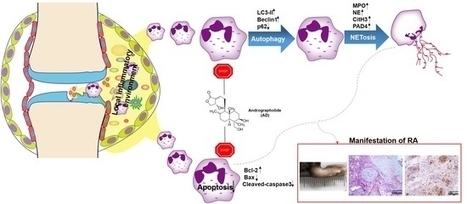
Aims Rheumatoid arthritis (RA) is a systematic autoimmune disorder, characterized by synovial inflammation, bone and cartilage destruction, and disease involvement in multiple organs. Although nume...
Via Krishan Maggon
|
|
Scooped by
Gilbert C FAURE
May 20, 2020 5:30 AM
|
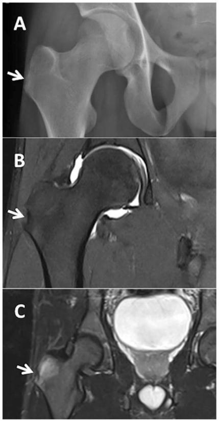
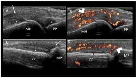
Arthritis Rheumatol. Author manuscript; available in PMC 2016 Dec 28. Published in final edited form as: doi: 10.1002/art.39458 PMCID: PMC5195265 NIHMSID: NIHMS835582 PMID: 26473401 Enthesitis New Insights Into Pathogenesis, Diagnostic Modalities, and Treatment The publisher's final edited version of this article is available free at Arthritis Rheumatol See other articles in PMC that cite the published article. Introduction Enthesitis is a central feature of spondyloarthritis (SpA). Although enthesitis has traditionally been considered to be a focal insertional disorder, advanced imaging and pathologic findings suggest that enthesitis is a diffuse process with effects on adjacent bone and soft tissue. As a result of repeated biomechanical stress, it appears that microdamage at the enthesis triggers an inflammatory response in the synovium, leading to synovitis. Along with mechanical stress, exogenous bacteria may play a role in activating the immune response, especially in genetically predisposed individuals whose major histocompatibility locus encodes the class I molecule HLA–B27. Recent studies in animal models suggest that autoimmunity against versican and fibrocartilage proteins, and bone morphogenetic protein (BMP) signaling play roles in enthesitis development. Finally, interleukin-23 (IL-23) has been implicated in enthesitis with inflammatory effects mediated through IL-17 and tumor necrosis factor (TNF), and new bone formation driven by IL-22. Although prior therapeutic choices were limited to nonsteroidal antiinflammatory drugs (NSAIDs) and activity modification, in recent years TNF inhibitors have proven to be useful. Further research on the effects of IL-22 and IL-23 blockade is needed to understand the effects on the treated patient. While enthesitis is underdiagnosed by physical examination alone, the use of ultrasound has proven to be highly sensitive for the detection of enthesitis, with utility in monitoring response to therapy, and will be an invaluable tool for assessing the efficacy of newer treatments. This review summarizes the substantial progress that has been made in addressing the pathophysiology, molecular mechanisms, genetic associations, clinical features, diagnostic modalities, and treatment of enthesitis. Definitions and evolution of the enthesis concept Historic definition Although the adjective “enthetic” derives from the ancient Greek word “enthetikos,” meaning “introduced into the body from without,” in the nineteenth century the adjective was increasingly used to refer to diseases that were “implanted into the body from external sources” (1). It was not until the twentieth century that the term “enthesis” was used as it is today, referring to focal insertional abnormalities at sites of bony attachments to tendons, ligaments, fascia, muscles, or joint capsules (2,3). The first suggestion that the enthesis is centrally affected in SpA was made by Ball in 1971 and was substantiated after a review of pathologic tissues from both patients with rheumatoid arthritis (RA) and patients with ankylosing spondylitis (AS), where he noted the presence of a unique inflammatory enthesopathy that could help to distinguish SpA from RA (2). Broadening the definition of enthesis with the concept of the “enthesis organ” Magnetic resonance imaging (MRI) and ultrasound findings have suggested that enthesopathy encompasses pathologic changes extending to the adjacent bone and soft tissues (4). Likewise, it has been argued that this entity should be considered an “enthesis organ” encompassing not only the enthesis itself, but also the fibrocartilage, bursa, fat pad, adjacent trabecular bone networks, and deeper fascia (5) (Figure 1). Representing areas where hard and soft tissues meet, entheses are sites of concentrated stress with effects not only on the bony attachment interface and the enthesis itself, but also on these neighboring tissues (4–7). Entheses and mechanical stress The concept of an enthesis organ was extended to that of a synovioentheseal complex (8,9), which refers to the relationship between the proinflammatory synovium and the avascular enthesis. In contrast to other skeletal locations, the enthesis is a site of repetitive biomechanical forces. High biomechanical stress at the enthesis triggers an inflammatory cascade with cytokine production by infiltrating monocytes and lymphocytes in the adjacent synovial tissue, resulting in an articular inflammatory response, and clinically leading to synovitis adjacent to attachment sites (8,9). Support for this theory of a dynamic response to biomechanical stress at the enthesis originates from animal models. In one experiment, botulinum toxin A injection delayed fibrocartilage development, suggesting that enthesis development is sensitive to mechanical environmental factors (10). In a mouse model that overexpresses TNF, enthesitis was reduced when the hind legs of the mice were made non–weight-bearing through tail suspension (11). Those authors proposed that triggering of mechanoreceptors via the MAPK pathway stimulates the production of inflammatory mediators. As a result of biomechanical stress, adjacent bone reacts with formation of surface spurs or enthesophytes, observed both radiographically and on histologic examination (12). In early disease, there is destruction of superficial fibrocartilage, with vascular invasion and inflammatory cell infiltration, predominantly with macrophages (13). This leads to another important microanatomical feature, which is the presence of blood vessels at sites where synovium, subchondral bone, and bone marrow are close to each other. In early experiments using labeled phosphorus, Ball identified capillary-like vessels that pass through the enthesis to the marrow (2). Later studies described the presence of vascular channels penetrating cortical bone in the knees of mice adjacent to the cruciate ligaments with associated subclinical changes, including subchondral bone damage and microcyst formation. In the rat adjuvant-induced arthritis model, vascular channels provided a site for inflammatory tissue entry and osteoclast activation (14). Whether enthesitis is a primary central lesion or a secondary process remains a matter of debate. Studies that have implicated enthesitis as the primary process include studies of TNF-transgenic mice, in which the earliest lesion appears to be in the enthesis (11). However, this may be model specific, and a number of reports have challenged the idea of enthesitis as the primary inflammatory lesion (15,16). In one study examining different stages of spontaneous tail spondylitis and peripheral arthritis in HLA–B27/hβ2m–transgenic mice, histologic samples displayed destructive synovitis with neutrophils and multinucleated giant cells rather than by enthesitis or osteitis (16). Among human studies examining biopsy specimens and MRIs of sacroiliac joints, synovitis and subchondral bone marrow changes were more prominent features while enthesitis was not (17,18). In a subsequent study, in patients with early untreated knee or ankle arthritis, analyses revealed a higher synovitis score by MRI in SpA than in RA, whereas there were no differences in the prevalence of enthesitis as assessed by perientheseal focal tissue, entheseal enhancement, and bone marrow edema (15). However, in light of substantial data in animal models highlighting 3 stages of tendon response to injury that have been defined by distinct pathologic changes, determining the initiating event in the enthesis may be confounded by the timing of the analysis (19–21). Contributing cellular and molecular mechanisms Genetic susceptibility It has long been known that AS susceptibility is largely genetically determined. The strongest genetic association is with the major histocompatibility complex (MHC)–encoded class I molecule, HLA–B27, and it is postulated that HLA–B27 contributes to ~40% of the overall risk for SpA (22). Protein misfolding of nascent HLA–B27 in the endoplasmic reticulum has been hypothesized to trigger an unfolded protein response with aberrant recognition by natural killer cell receptors (23). The HLA–B27–induced unfolded protein response in macrophages has been demonstrated in HLA–B27–transgenic rats and is associated with an increase in IL-23 production by these cells (24). Although HLA–B27 remains the dominant risk factor for susceptibility to the AS phenotype, other important influences of the MHC have been observed (25). More recently, Haroon et al (26) found a positive association of B*27:05:02 with enthesitis, dactylitis, and symmetric sacroiliitis in a cohort of psoriatic arthritis (PsA) patients, whereas B*44 haplotypes were associated with a decreased frequency of enthesitis, dactylitis, and joint fusion. Finally, investigators have recently focused on genes outside of the MHC region, such as ERAP1 and ERAP2, which code for aminopeptidases that are involved in MHC class I presentation (25,27). Although additional HLA class I and class II alleles have also been implicated, the scale and scope of gene identification to date have not yet matched the putative total genetic risk for SpA. Microbial factors Microbial infection with virulent organisms remote from affected joints, as well as gastrointestinal dysbiosis without a directly invading pathogen, are known features of certain phenotypes of SpA, and it has long been appreciated that microbial factors can lead to immune activation (28). Clinically, reactive arthritis (ReA) is known to follow infections with Chlamydia, Campylobacter, Shigella, or Yersinia. AS patients consistently have been found to have subclinical gut inflammation and increased gastrointestinal permeability (29,30). In animal models, HLA–B27–transgenic rats raised in germ-free environments do not develop intestinal inflammatory or peripheral joint disease, yet the disease recurs if rats are reconstituted with Bacteroides, supporting the role of gut flora in the development of joint inflammation (31). In a more recent study, colonoscopic biopsies of the terminal ileum of AS patients showed a discrete microbial signature as revealed by sequencing and quantitative polymerase chain reaction analysis of the 16S ribosomal RNA (16S rRNA) gene, exhibiting higher levels of 5 families of bacteria as compared to healthy controls (32). In that study there was no significant difference in the 16S rRNA copy number between patients with AS and controls, indicating that the observed differences were not due to bacterial overgrowth. It has been postulated that the combination of bacterial adjuvants and mechanical factors act synergistically to activate the immune response, particularly in genetically predisposed individuals (5). Fibrocartilage and versican autoimmunity A number of studies have indicated that autoimmunity against fibrocartilage proteins, including aggrecan, may underlie enthesitis and spondylitis (33). A model of SpA induced by immunizing BALB/c mice with the G1 globular domain of versican, leading to spondylitis and enthesitis, suggests that versican autoimmunity may also play a role in enthesitis (34). The inflammatory lesions are characterized by mononuclear cell infiltration at the entheseal insertions to the vertebrae, as is seen with AS, and are associated with angiogenesis which then progresses to cause destructive discitis (35). Role of bone morphogens In the DBA/1 mouse model, where mice develop spontaneously occurring arthritis that culminates in bone formation and joint ankylosis, male mice in crowded conditions developed arthritis in the hind paws that was entheseal, but not synovially based, with new bone formation driven by BMP-7 signaling (36). In that experiment, the incidence of arthritis was increased in mice that were caged together in crowded conditions, yet decreased when the mice were placed in larger cages (37). Thus, in addition to a genetic predisposition for enthesitis, this observation points to the role of environmental factors in the development of arthritis. Finally, immunohistochemical studies in SpA show increased synovial expression of BMP-2 and BMP-6, which is up-regulated by proinflammatory cytokines such as IL-1 and TNF, suggesting that synovial molecules contribute to chronic arthritis and joint ankylosis (36,38). Role of proinflammatory cytokines The role of IL-23 has been addressed as a major driver of cascades that lead to inflammation and bone remodeling in SpA. Alterations in AS susceptibility are related to the existence of single-nucleotide polymorphisms in the IL-23 receptor as demonstrated in genome-wide association studies, and serum levels of the IL-12/23 p40 subunit have been shown to be significantly higher in patients with PsA compared with controls (39,40). IL-23 is produced in the gut, suggesting that the intestinal mucosa is a key site of IL-23 production in SpA. Additionally, Chlamydia trachomatis also leads to induction of IL-23 via CHOP10. Taken together, these findings indicate that IL-23 is a pivotal cytokine and potentially central to the pathogenesis of SpA (41). Increased IL-17 expression by innate immune cells such as mast cells and neutrophils in SpA has been shown to target the facet joints and synovial tissue (42,43). In a subsequent set of investigations, Sherlock et al found that IL-23 could induce SpA by acting on an isolated population of CD3+CD4−CD8− entheseal resident lymphocytes, leading to increased expression of TNF and IL-6 in the enthesis. When IL-23 was overexpressed, mice developed enthesitis with inflammation, which spread into the adjacent synovium (41). Enthesitis was associated with new bone erosion. IL-23 promoted inflammation through IL-17 and TNF, whereas new bone formation was associated with overproduction of IL-22 (41,44) (Figure 2). Additional support for the role of IL-23 comes from the SKG mouse model, in which curdlan (β-1,3-glucan) injections induce enthesitis and dactylitis. Arthritis and spondylitis were IL-23 dependent and were transferable to SCID mouse recipients with CD4+ T cells (45). In this model, disease severity was dependent on the external microbial environment and the host immunogenetic background. More recent work illustrates the differential impact of microbiota on specific pathologic features of SpA; ileitis development, ileal IL-23 expression, and lymph node IL-17A production were microbiota dependent, but arthritis was not (46). In curdlan-treated SKG mice, enthesitis was specifically dependent on IL-17A and IL-22 (47). The role of up-regulation of the IL-23/Th17 pathway in promoting joint inflammation and bone turnover is further supported by recent murine studies, with inhibition of the PsA phenotype after neutralization of IL-17A (48,49). Clinical enthesitis in SpA SpA is by definition a heterogeneous group of clinical entities long recognized as having unique phenotypes that include AS, ReA, PsA, enteropathic arthritis, and what has traditionally been referred to as undifferentiated arthritis. However, with advances in imaging and careful long-term followup observations, it appears that these diseases share common features, including subclinical spinal and peripheral joint inflammation, along with associations with microbes and gene identifications. In attempting to develop a model for an underlying unifying anatomical basis for SpA, an “enthesitis-based model” has been proposed as the basis for the osteitis, periostitis, and new bone formation that are seen in SpA (5). The association between enthesitis and adjacent osteitis has been further supported by imaging and cadaver studies, primarily in patients with PsA (50–52). Regional sites Patients with SpA have a remarkable propensity for inflammation at certain enthesis sites that are ubiquitous and numerous. Clinically, peripheral enthesitis is observed not only in all forms of SpA, but particularly frequently in juvenile-onset SpA. A number of patients with juvenile SpA are classified as having enthesitis-related arthritis (ERA), a heterogeneous subtype that includes some patients who predominantly have enthesitis, enthesitis and arthritis, or juvenile AS. Compared to other subtypes of juvenile idiopathic arthritis, ERA is associated with worse function, worse quality of life, and increased pain (53,54). Enthesitis can be seen in 33–58% of patients with ReA and may be the only clinical manifestation in some whose disease has been triggered by an enteric infection (55). In SpA, the entheses of the lower extremities are more frequently involved than those of the upper limbs, and the heel is the most frequent site (55). In addition to the Achilles and plantar fascia insertions, identified sites of enthesitis include muscle attachments to the greater and lesser trochanters, the insertion of the quadriceps tendon at the upper patellar pole, the insertions of the patellar ligament at the lower patellar pole and the tibial tubercle, acromial and clavicular insertions of the deltoid muscle, and the insertions of the flexor and extensor tendons at the phalanges (55–57). It is unknown why there is a predilection for the entheses at the lower parts of the lower limbs, although it has been hypothesized that this may be due to the length, anatomy, and higher mechanical load at these sites. Given the presumed role of repetitive biomechanical forces discussed above, it is not surprising that in patients with longstanding AS, those with occupational activities that required more bending, twisting, and stretching had more functional limitations and radiographic damage than those whose jobs required little or no dynamic flexibility (58). A recently published computer-based method that fully quantified syndesmophyte heights and volumes on computed tomography scans has revealed that syndesmophytes grow at different rates over time in AS patients, suggesting that mechanical factors local to the disc space may influence syndesmophyte formation (59). Clearly, there are sites that are not associated with SpA despite being sites of significant biomechanical stress, and perhaps it is the compressive and shear force nature of the stress as well as the putative role of antigen expression adjacent to the enthesis that may underlie this apparent discrepancy (5). Additionally, it cannot be discounted that the increased detection of enthesitis at the lower limbs is explained by the accessibility of these sites to ultrasound. Diagnostic criteria and outcome measures Enthesitis is often underdiagnosed in the clinic; clinical assessment and quantification of peripheral enthesitis in daily practice lacks sensitivity and specificity (56,60,61). Although both the Amor criteria (62) and the European Spondylarthropathy Study Group criteria (63) for SpA include peripheral enthesitis, there are limitations to these criteria with regard to the exact quantification of enthesitis. Two clinical methods have been designed and often implemented for evaluating enthesopathy in AS: Mander’s Entheseal Index and the Maastricht Ankylosing Spondylitis Enthesitis Score (MASES) (64,65). Both rely on pain elicited by local pressure of entheseal points. The intraarticular and deep location of entheseal insertions, however, makes quantification of enthesitis by physical examination alone difficult, and not surprisingly, these scoring systems have only moderate sensitivity and specificity for predicting positive sonogram results, depending on the entheseal site (66). Imaging of the enthesis Because of the clinical limitations described above and the poor sensitivity of markers of inflammation, it is necessary to rely on typical abnormalities seen on various imaging techniques to diagnose SpA. Plain radiographs are limited by their inability to show inflammation or soft tissue changes, although late chronic bony changes such as enthesophyte formation or occasional erosions can be seen at the attachment of the Achilles tendon or plantar aponeurosis. More sensitive methods such as ultrasound and MRI, which are useful in their ability to detect both inflammatory and chronic changes in enthesitis at both early and late stages, can be used. MRI MRI has changed the way we approach both the diagnosis and classification of SpA; it is particularly useful in detecting spinal disease in early AS when conventional radiographs are still normal (67). The use of fat-suppressed, fat-saturated, and water-sensitive MRI sequences has demonstrated that the extracapsular inflammation of joints quite often represents enthesitis with variable degrees of soft tissue and bone marrow edema (68,69) (Figures 3–5). The typical appearance of enthesitis on MRI includes soft tissue inflammatory changes outside the joint capsule and perientheseal bone marrow edema (70). Recent studies have examined the utility of whole-body MRI, which has shown promise in the detection of subclinical axial and peripheral enthesitis (71). Of course, MRI has limitations; structures that make up entheses have a low signal on conventional MRI, with low water accumulation in the areas where fibroblasts are tightly cross-linked. MRI is further limited by its cost and availability, and therefore ultrasound remains the preferred modality for the detection of enthesitis both in the clinical setting as well as in research. Ultrasound Ultrasound has indeed proven to be a highly useful and sensitive tool in the evaluation of enthesitis and improves the ability of the clinical examination to detect enthesopathy. In one study of 92 patients with PsA, ultrasound was useful in detecting subclinical entheseal involvement, independent of clinical examination and symptoms (72). In another study of 600 lower limb entheses, at least 1 ultrasound sign of enthesopathy was detected in 60% of clinically asymptomatic cases of enthesitis, thus demonstrating a higher sensitivity than physical examination (73). Ultrasound may be most useful in the early diagnosis of SpA, and likewise, entheseal abnormalities can be detected prior to overt clinical disease. Nevertheless, in an older cross-sectional single-center study of 51 SpA patients and 24 controls, neither MRI nor power Doppler ultrasound (PDUS) discriminated between SpA and controls (74). In a prospective single-center cohort study of 118 patients with symptoms suggestive of SpA conducted by D’Agostino and colleagues (57), vascularization at cortical bone detected by PDUS of at least one enthesis provided good predictive value for diagnosing SpA with a sensitivity of 76.5% and a specificity of 81.3%. Indeed, PDUS is a sensitive and reliable technique used to detect increased blood flow in the enthesis revealing neovascularity and subclinical active inflammation (56,75) (Figure 6). Recent studies have indicated that ultrasound may accurately predict which patients will go on to develop SpA (51,57,75). In one investigation, ultrasound examination of Achilles erosions correlated with objective activity-based measurements of SpA outcomes, and was sensitive to change (76). In the study by D’Agostino and colleagues described above, vascularized enthesis as detected by PDUS combined with Amor’s criteria proved to be the only independent contributors to a diagnosis of SpA (57). Finally, ultrasound may be used to monitor response to therapeutic interventions. A few studies have illustrated improvement in enthesitis shown on ultrasound after the use of TNF antagonists (77,78). In one investigation of 327 patients with active SpA who were treated with anti-TNF therapy for 6 months, cumulative entheseal morphologic abnormalities, intraenthesis and perienthesis, and bursitis were all significantly decreased on PDUS after 6 months of treatment (77). In another study, D’Agostino et al monitored regression of enthesitis using PDUS after treatment with infliximab (78), providing confirmatory evidence for the utility of ultrasound in a clinical research setting. Treatment of enthesitis Historically, treatment of clinical enthesitis had been limited to NSAIDs. Continuous use of NSAIDs not only controls symptoms of disease, but may also slow progression of bony changes in AS (79,80). Therefore, Assessment of SpondyloArthritis international Society/European League Against Rheumatism guidelines place optimal NSAID therapy as a cornerstone of the management plan for AS (81). Treatment with TNF inhibitors is indicated in patients that do not respond to NSAID therapy. TNF inhibition with adalimumab, etanercept, infliximab, and golimumab has been shown to be efficacious in the treatment of enthesitis (82–87). Olivieri et al (88) have reported that adalimumab and etanercept are effective treatments of MRI-documented refractory heel enthesitis, with progressive improvement of bone edema in a 6-month period (88). Agents that block IL-23 have the potential to inhibit both inflammation and altered bone remodeling, although further analysis of the effect of IL-22 and IL-23 blockade on bone pathologies in animal models and patients with PsA are needed to address this important therapeutic issue (89). Entheseal inflammation in a passive-transfer model of collagen antibody-induced arthritis was reduced by an antibody to the p19 subunit of IL-23, which was also associated with the down-regulation of several inflammatory mediators, such as IL-6 and IL-1β, and genes such as Rankl, Ctsk, and matrix metalloproteinases known to be involved in bone erosion (41). Both ustekinumab, a monoclonal antibody directed against the common p40 subunit of IL-12 and IL-23, and secukinumab, a human anti–IL-17A monoclonal antibody, have already demonstrated promise in PsA, with significant improvements in enthesitis (90,91). Apremilast, an oral inhibitor of phosphodiesterase 4, which increases cAMP and thus modulates multiple proinflammatory mediators, has demonstrated efficacy in PsA, with significant improvements in the severity of both enthesitis and dactylitis evidenced by reductions in MASES over a 52-week period (92). Finally, bisphosphonates may also have a role in peripheral enthesitis felt to be refractory to NSAID therapy. In a 6-month randomized controlled comparison of intravenous pamidronate treatment of NSAID-refractory AS, patients treated with pamidronate showed symptomatic improvement with significant reductions in Bath Ankylosing Spondylitis Functional Index and Bath Ankylosing Spondylitis Disease Activity Index measurements together with regression of periarticular osteitis documented by MRI with gadolinium (93). Treatment of patients with SpA enthesitis with currently available agents has not had universal success. In placebo-controlled trials of methotrexate and leflunomide in PsA, enthesitis measures were not assessed (94,95). In a randomized controlled trial, sulfasalazine was not effective for enthesitis (96). Other agents that have not demonstrated clinical efficacy in AS include tocilizumab, and lymphocyte-targeted therapies such as abatacept (97,98). Rituximab only showed modest therapeutic efficacy in SpA (99,100). Conclusions In summary, investigations and clinical observations uniformly point out with increasing clarity that the enthesis is much more than a simple attachment site. A number of studies have shown that it functions as a unit comprising adjacent tissues, including bone and fibro-cartilage linked to synovium, and serves as a way of dissipating stress over a wide area. Inflammation at the enthesis manifests in the adjacent synovium presumably via immunity to common antigens or via release of proinflammatory cytokines at the enthesis. Although work by Benjamin and McGonagle (9) suggests that the enthesis is the primary SpA lesion, the precise role of the enthesis in early stages of disease, especially regarding issues of cause or effect, remains an area of continued debate and discovery. Improved imaging modalities may in the future be able to detect enthesitis at different stages of disease. However, this will require a clinically diverse and large sample size to help address this question. Inflammation at the enthesis is likely modulated by multiple factors. A more complete role for genetic predisposition will require additional advances in gene sequencing and discovery. Repeated biomechanical stress with the resultant inflammatory response regulated by IL-17, IL-22, and IL-23 now provide clues as to why certain areas of the body are affected, and perhaps why others are not. The spine itself (the clinical hallmark of the disease) remains inaccessible to traditional enthesitis-focused research methodologies thus far. However, newer imaging techniques are on the horizon. Further examination into the role of the inflammatory mediators, including IL-17, IL-22, and IL-23 as well as potentially others, in driving enthesitis and bone formation will be important to direct our attention toward future therapeutic targeted pathways in patients with SpA. Acknowledgments Supported in part by the NIH (National Institute of Arthritis and Musculoskeletal and Skin Diseases grant P01-AR-052915 and National Center for Advancing Translational Sciences grant UL1-TR-000124). The authors wish to thank Joseph Robinson, MD (Cedars-Sinai Medical Center Department of Radiology) for assistance with MRI acquisition and interpretation. Footnotes AUTHOR CONTRIBUTIONS All authors were involved in drafting the article or revising it critically for important intellectual content, and all authors approved the final version to be published. References 1. Lampman JH. Origin of enthesopathy. J Rheumatol. 1985;12:1030–1. [PubMed] [Google Scholar] 2. Ball J. Enthesopathy of rheumatoid and ankylosing spondylitis. Ann Rheum Dis. 1971;30:213–23. [PMC free article] [PubMed] [Google Scholar] 3. Francois RJ, Braun J, Khan MA. Entheses and enthesitis: a histopathologic review and relevance to spondyloarthritides. Curr Opin Rheumatol. 2001;13:255–64. [PubMed] [Google Scholar] 4. Benjamin M, Moriggl B, Brenner E, Emery P, McGonagle D, Redman S. The “enthesis organ” concept: why enthesopathies may not present as focal insertional disorders. Arthritis Rheum. 2004;50:3306–13. [PubMed] [Google Scholar] 5. McGonagle D, Stockwin L, Isaacs J, Emery P. An enthesitis based model for the pathogenesis of spondyloarthropathy: additive effects of microbial adjuvant and biomechanical factors at disease sites. J Rheumatol. 2001;28:2155–9. [PubMed] [Google Scholar] 6. Benjamin M, McGonagle D. The anatomical basis for disease localisation in seronegative spondyloarthropathy at entheses and related sites. J Anat. 2001;199:503–26. [PMC free article] [PubMed] [Google Scholar] 7. McGonagle D. Enthesitis: an autoinflammatory lesion linking nail and joint involvement in psoriatic disease. J Eur Acad Dermatol Venereol. 2009;23(Suppl 1):9–13. [PubMed] [Google Scholar] 8. McGonagle D, Lories RJ, Tan AL, Benjamin M. The concept of a “synovio-entheseal complex” and its implications for understanding joint inflammation and damage in psoriatic arthritis and beyond. Arthritis Rheum. 2007;56:2482–91. [PubMed] [Google Scholar] 9. Benjamin M, McGonagle D. The enthesis organ concept and its relevance to the spondyloarthropathies. Adv Exp Med Biol. 2009;649:57–70. [PubMed] [Google Scholar] 10. Thomopoulos S, Kim HM, Rothermich SY, Biederstadt C, Das R, Galatz LM. Decreased muscle loading delays maturation of the tendon enthesis during postnatal development [published erratum appears in J Orthop Res 2009;27:141] J Orthop Res. 2007;25:1154–63. [PubMed] [Google Scholar] 11. Jacques P, Lambrecht S, Verheugen E, Pauwels E, Kollias G, Armaka M, et al. Proof of concept: enthesitis and new bone formation in spondyloarthritis are driven by mechanical strain and stromal cells. Ann Rheum Dis. 2014;73:437–45. [PubMed] [Google Scholar] 12. Benjamin M, Rufai A, Ralphs JR. The mechanism of formation of bony spurs (enthesophytes) in the Achilles tendon. Arthritis Rheum. 2000;43:576–83. [PubMed] [Google Scholar] 13. McGonagle D, Marzo-Ortega H, O’Connor P, Gibbon W, Hawkey P, Henshaw K, et al. Histological assessment of the early enthesitis lesion in spondyloarthropathy. Ann Rheum Dis. 2002;61:534–7. [PMC free article] [PubMed] [Google Scholar] 14. Binks D, Matzelle M, Bergin D, Hodgson RJ, Tan AL, Gravallese EM, et al. The frequency of bone marrow oedema adjacent to the cruciate ligament peri-entheseal vascular channels in inflammatory and degenerative arthritis [abstract] Arthritis Rheum. 2013;65(Suppl):S25–6. [Google Scholar] 15. Paramarta JE, van der Leij C, Gofita I, Yeremenko N, van de Sande MG, de Hair MJ, et al. Peripheral joint inflammation in early onset spondyloarthritis is not specifically related to enthesitis. Ann Rheum Dis. 2014;73:735–40. [PubMed] [Google Scholar] 16. Van Duivenvoorde LM, Dorris ML, Satumtira N, van Tok MN, Redlich K, Tak PP, et al. Relationship between inflammation, bone destruction, and osteoproliferation in the HLA–B27/human β2-microglobulin–transgenic rat model of spondylarthritis. Arthritis Rheum. 2012;64:3210–9. [PMC free article] [PubMed] [Google Scholar] 17. Francois RJ, Gardner DL, Degrave EJ, Bywaters EG. Histopathologic evidence that sacroiliitis in ankylosing spondylitis is not merely enthesitis: systematic study of specimens from patients and control subjects. Arthritis Rheum. 2000;43:2011–24. [PubMed] [Google Scholar] 18. Muche B, Bollow M, Francois RJ, Sieper J, Hamm B, Braun J. Anatomic structures involved in early- and late-stage sacroiliitis in spondylarthritis: a detailed analysis by contrast-enhanced magnetic resonance imaging. Arthritis Rheum. 2003;48:1374–84. [PubMed] [Google Scholar] 19. Cook JL, Purdam CR. Is tendon pathology a continuum? A pathology model to explain the clinical presentation of load-induced tendinopathy. Br J Sports Med. 2009;43:409–16. [PubMed] [Google Scholar] 20. Aspenberg P. Stimulation of tendon repair: mechanical loading, GDFs and platelets: a mini-review. Int Orthop. 2007;31:783–9. [PMC free article] [PubMed] [Google Scholar] 21. Apostolakos J, Durant TJ, Dwyer CR, Russell RP, Weinreb JH, Alaee F, et al. The enthesis: a review of the tendon-to-bone insertion. Muscles Ligaments Tendons J. 2014;4:333–42. [PMC free article] [PubMed] [Google Scholar] 22. Reveille JD. The genetic basis of spondyloarthritis. Ann Rheum Dis. 2011;70(Suppl 1):i44–50. [PubMed] [Google Scholar] 23. Colbert RA, Tran TM, Layh-Schmitt G. HLA-B27 misfolding and ankylosing spondylitis. Mol Immunol. 2014;57:44–51. [PMC free article] [PubMed] [Google Scholar] 24. DeLay ML, Turner MJ, Klenk EI, Smith JA, Sowders DP, Colbert RA. HLA–B27 misfolding and the unfolded protein response augment interleukin-23 production and are associated with Th17 activation in transgenic rats. Arthritis Rheum. 2009;60:2633–43. [PMC free article] [PubMed] [Google Scholar] 25. Cortes A, Pulit SL, Leo PJ, Pointon JJ, Robinson PC, Weisman MH, et al. Major histocompatibility complex associations of ankylosing spondylitis are complex and involve further epistasis with ERAP1. Nat Commun. 2015;6:7146. [PMC free article] [PubMed] [Google Scholar] 26. Haroon M, Winchester R, Giles JT, Heffernan E, FitzGerald O. Certain class I HLA alleles and haplotypes implicated in susceptibility play a role in determining specific features of the psoriatic arthritis phenotype. Ann Rheum Dis. 2014 E-pub ahead of print. [PubMed] [Google Scholar] 27. Breban M, Costantino F, Andre C, Chiocchia G, Garchon HJ. Revisiting MHC genes in spondyloarthritis. Current Rheumatology Reports. 2015;17:516. [PubMed] [Google Scholar] 28. Hacker G, Redecke V, Hacker H. Activation of the immune system by bacterial CpG-DNA. Immunology. 2002;105:245–51. [PMC free article] [PubMed] [Google Scholar] 29. Jacques P, Van Praet L, Carron P, Van den Bosch F, Elewaut D. Pathophysiology and role of the gastrointestinal system in spondyloarthritides. Rheum Dis Clin North Am. 2012;38:569–82. [PubMed] [Google Scholar] 30. Matzkies FG, Targan SR, Berel D, Landers CJ, Reveille JD, McGovern DP, et al. Markers of intestinal inflammation in patients with ankylosing spondylitis: a pilot study. Arthritis Res Ther. 2012;14:R261. [PMC free article] [PubMed] [Google Scholar] 31. Taurog JD, Richardson JA, Croft JT, Simmons WA, Zhou M, Fernandez-Sueiro JL, et al. The germfree state prevents development of gut and joint inflammatory disease in HLA-B27 transgenic rats. J Exp Med. 1994;180:2359–64. [PMC free article] [PubMed] [Google Scholar] 32. Costello ME, Ciccia F, Willner D, Warrington N, Robinson PC, Gardiner B, et al. Intestinal dysbiosis in ankylosing spondylitis. Arthritis Rheumatol. 2015;67:686–91. [PubMed] [Google Scholar] 33. Guerassimov A, Zhang Y, Banerjee S, Cartman A, Webber C, Esdaile J, et al. Autoimmunity to cartilage link protein in patients with rheumatoid arthritis and ankylosing spondylitis. J Rheumatol. 1998;25:1480–4. [PubMed] [Google Scholar] 34. Shi SL, Ciurli C, Cartman A, Pidoux I, Poole AR, Zhang Y. Experimental immunity to the G1 domain of the proteoglycan versican induces spondylitis and sacroiliitis, of a kind seen in human spondylarthropathies. Arthritis Rheum. 2003;48:2903–15. [PubMed] [Google Scholar] 35. Zhang YP, Guerassimov A, Leroux JY, Cartman A, Webber C, Lalic R, et al. Arthritis induced by proteoglycan aggrecan G1 domain in BALB/c mice. Evidence for T cell involvement and the immunosuppressive influence of keratan sulfate on recognition of T and B cell epitopes. J Clin Invest. 1998;101:1678–86. [PMC free article] [PubMed] [Google Scholar] 36. Lories RJ, Derese I, Luyten FP. Modulation of bone morphogenetic protein signaling inhibits the onset and progression of ankylosing enthesitis. J Clin Invest. 2005;115:1571–9. [PMC free article] [PubMed] [Google Scholar] 37. Braem K, Carter S, Lories RJ. Spontaneous arthritis and ankylosis in male DBA/1 mice: further evidence for a role of behavioral factors in “stress-induced arthritis” Biol Proced Online. 2012;14:10. [PMC free article] [PubMed] [Google Scholar] 38. Lories RJ, Derese I, Ceuppens JL, Luyten FP. Bone morphogenetic proteins 2 and 6, expressed in arthritic synovium, are regulated by proinflammatory cytokines and differentially modulate fibroblast-like synoviocyte apoptosis. Arthritis Rheum. 2003;48:2807–18. [PubMed] [Google Scholar] 39. Wellcome Trust Case Control Consortium, Australo-Anglo-American Spondylitis Consortium (TASC) Burton PR, Clayton DG, Cardon LR, Craddock N, et al. Association scan of 14,500 nonsynonymous SNPs in four diseases identifies autoimmunity variants. Nat Genet. 2007;39:1329–37. [PMC free article] [PubMed] [Google Scholar] 40. Reveille JD. Genetics of spondyloarthritis-beyond the MHC. Nat Rev Rheumatol. 2012;8:296–304. [PubMed] [Google Scholar] 41. Sherlock JP, Joyce-Shaikh B, Turner SP, Chao CC, Sathe M, Grein J, et al. IL-23 induces spondyloarthropathy by acting on ROR-γt+ CD3+CD4−CD8− entheseal resident T cells. Nat Med. 2012;18:1069–76. [PubMed] [Google Scholar] 42. Noordenbos T, Yeremenko N, Gofita I, van de Sande M, Tak PP, Canete JD, et al. Interleukin-17–positive mast cells contribute to synovial inflammation in spondylarthritis. Arthritis Rheum. 2012;64:99–109. [PubMed] [Google Scholar] 43. Appel H, Maier R, Wu P, Scheer R, Hempfing A, Kayser R, et al. Analysis of IL-17+ cells in facet joints of patients with spondyloarthritis suggests that the innate immune pathway might be of greater relevance than the Th17-mediated adaptive immune response. Arthritis Res Ther. 2011;13:R95. [PMC free article] [PubMed] [Google Scholar] 44. Lories RJ, McInnes I. Primed for inflammation: enthesis resident cells. Nat Med. 2012;18:1018–9. [PubMed] [Google Scholar] 45. Ruutu M, Thomas G, Steck R, Degli-Esposti MA, Zinkernagel MS, Alexander K, et al. β-glucan triggers spondylarthritis and Crohn’s disease–like ileitis in SKG mice. Arthritis Rheum. 2012;64:2211–22. [PubMed] [Google Scholar] 46. Rehaume LM, Mondot S, Aguirre de Carcer D, Velasco J, Benham H, Hasnain SZ, et al. ZAP-70 genotype disrupts the relationship between microbiota and host, leading to spondyloarthritis and ileitis in SKG mice. Arthritis Rheumatol. 2014;66:2780–92. [PubMed] [Google Scholar] 47. Benham H, Rehaume LM, Hasnain SZ, Velasco J, Baillet AC, Ruutu M, et al. Interleukin-23 mediates the intestinal response to microbial β-1,3-glucan and the development of spondyloarthritis pathology in SKG mice. Arthritis Rheumatol. 2014;66:1755–67. [PubMed] [Google Scholar] 48. Yamamoto M, Nakajima K, Takaishi M, Kitaba S, Magata Y, Kataoka S, et al. Psoriatic inflammation facilitates the onset of arthritis in a mouse model. J Invest Dermatol. 2015;135:445–53. [PubMed] [Google Scholar] 49. Khmaladze I, Kelkka T, Guerard S, Wing K, Pizzolla A, Saxena A, et al. Mannan induces ROS-regulated, IL-17A-dependent psoriasis arthritis-like disease in mice. Proc Natl Acad Sci U S A. 2014;111:E3669–78. [PMC free article] [PubMed] [Google Scholar] 50. Tan AL, Benjamin M, Toumi H, Grainger AJ, Tanner SF, Emery P, et al. The relationship between the extensor tendon enthesis and the nail in distal interphalangeal joint disease in psoriatic arthritis—a high-resolution MRI and histological study. Rheumatology (Oxford) 2007;46:253–6. [PubMed] [Google Scholar] 51. Naredo E, Moller I, de Miguel E, Batlle-Gualda E, Acebes C, Brito E, et al. High prevalence of ultrasonographic synovitis and enthesopathy in patients with psoriasis without psoriatic arthritis: a prospective case-control study. Rheumatology (Oxford) 2011;50:1838–48. [PubMed] [Google Scholar] 52. Yasser R, Yasser E, Hanan D, Rasker JJ. Enthesitis in seronegative spondyloarthropathies with special attention to the knee joint by MRI: a step forward toward understanding disease pathogenesis. Clin Rheumatol. 2011;30:313–22. [PubMed] [Google Scholar] 53. Weiss P, Beukelman T, Schanberg LE, Kimura Y, Colbert RA CARRAnet Investigators. Enthesitis is a significant predictor of decreased quality of life, function, and arthritis-specific pain across juvenile idiopathic arthritis (JIA) categories: preliminary analyses from the CARRAnet registry. Arthritis Rheum. 2011;63(Suppl):S105. [Google Scholar] 54. Weiss PF. Evaluation and treatment of enthesitis-related arthritis. Curr Med Lit Rheumatol. 2013;32:33–41. [PMC free article] [PubMed] [Google Scholar] 55. D’Agostino MA, Olivieri I. Enthesitis. Best Pract Res Clin Rheumatol. 2006;20:473–86. [PubMed] [Google Scholar] 56. D’Agostino MA, Said-Nahal R, Hacquard-Bouder C, Brasseur JL, Dougados M, Breban M. Assessment of peripheral enthesitis in the spondylarthropathies by ultrasonography combined with power Doppler: a cross-sectional study. Arthritis Rheum. 2003;48:523–33. [PubMed] [Google Scholar] 57. D’Agostino MA, Aegerter P, Bechara K, Salliot C, Judet O, Chimenti MS, et al. How to diagnose spondyloarthritis early? Accuracy of peripheral enthesitis detection by power Doppler ultrasonography. Ann Rheum Dis. 2011;70:1433–40. [PubMed] [Google Scholar] 58. Ward MM, Reveille JD, Learch TJ, Davis JC, Jr, Weisman MH. Occupational physical activities and long-term functional and radiographic outcomes in patients with ankylosing spondylitis. Arthritis Rheum. 2008;59:822–32. [PMC free article] [PubMed] [Google Scholar] 59. Tan S, Yao J, Flynn JA, Yao L, Ward MM. Quantitative syndesmophyte measurement in ankylosing spondylitis using CT: longitudinal validity and sensitivity to change over 2 years. Ann Rheum Dis. 2015;74:437–43. [PMC free article] [PubMed] [Google Scholar] 60. Wiell C, Norregaard J, Szkudlarek M, Hasselquist M, Moller JM, Terslev L, et al. Ultrasonography of finger joints, tendons and entheses in patients with spondyloarthropathy: a comparison with clinical examination and MRI. Ann Rheum Dis. 2005;64(Suppl III):327. [Google Scholar] 61. Wiell C, Norregaard J, Szkudlarek M, Hasselquist M, Moller JM, Terslev L, et al. Ultrasonography of lower extremity tendons and entheses in patients with spondyloarthropathy: a comparison with clinical examination and MRI. Ann Rheum Dis. 2005;64(Suppl III):378. [Google Scholar] 62. Amor B, Dougados M, Mijiyawa M. Criteria for the classification of spondylarthropathies. Rev Rhum Mal Osteoartic. 1990;57:85–9. In French. [PubMed] [Google Scholar] 63. Dougados M, van der Linden S, Juhlin R, Huitfeldt B, Amor B, Calin A, et al. the European Spondylarthropathy Study Group. The European Spondylarthropathy Study Group preliminary criteria for the classification of spondylarthropathy. Arthritis Rheum. 1991;34:1218–27. [PubMed] [Google Scholar] 64. Mander M, Simpson JM, McLellan A, Walker D, Goodacre JA, Dick WC. Studies with an enthesis index as a method of clinical-assessment in ankylosing spondylitis. Ann Rheum Dis. 1987;46:197–202. [PMC free article] [PubMed] [Google Scholar] 65. Heuft-Dorenbosch L, Spoorenberg A, van Tubergen A, Landewe R, van ver Tempel H, Mielants H, et al. Assessment of enthesitis in ankylosing spondylitis. Ann Rheum Dis. 2003;62:127–32. [PMC free article] [PubMed] [Google Scholar] 66. Klauser AS, Wipfler E, Dejaco C, Moriggl B, Duftner C, Schirmer M. Diagnostic values of history and clinical examination to predict ultrasound signs of chronic and acute enthesitis. Clin Exp Rheumatol. 2008;26:548–53. [PubMed] [Google Scholar] 67. Braun J, Bollow M, Eggens U, Konig H, Distler A, Sieper J. Use of dynamic magnetic resonance imaging with fast imaging in the detection of early and advanced sacroiliitis in spondylarthropathy patients. Arthritis Rheum. 1994;37:1039–45. [PubMed] [Google Scholar] 68. McGonagle D, Marzo-Ortega H, O’Connor P, Gibbon W, Pease C, Reece R, et al. The role of biomechanical factors and HLA–B27 in magnetic resonance imaging–determined bone changes in plantar fascia enthesopathy. Arthritis Rheum. 2002;46:489–93. [PubMed] [Google Scholar] 69. Marzo-Ortega H, McGonagle D, O’Connor P, Emery P. Efficacy of etanercept in the treatment of the entheseal pathology in resistant spondylarthropathy: a clinical and magnetic resonance imaging study. Arthritis Rheum. 2001;44:2112–7. [PubMed] [Google Scholar] 70. Aquino MR, Tse SML, Gupta S, Rachlis AC, Stimec J. Whole-body MRI of juvenile spondyloarthritis: protocols and pictorial review of characteristic patterns. Pediatr Radiol. 2015;45:754–62. [PubMed] [Google Scholar] 71. Poggenborg RP, Eshed I, Ostergaard M, Sorensen IJ, Moller JM, Madsen OR, et al. Enthesitis in patients with psoriatic arthritis, axial spondyloarthritis and healthy subjects assessed by ‘head-to-toe’ whole-body MRI and clinical examination. Ann Rheum Dis. 2014;74:823–9. [PubMed] [Google Scholar] 72. Bandinelli F, Prignano F, Bonciani D, Bartoli F, Collaku L, Candelieri A, et al. Ultrasound detects occult entheseal involvement in early psoriatic arthritis independently of clinical features and psoriasis severity. Clin Exp Rheumatol. 2013;31:219–24. [PubMed] [Google Scholar] 73. Ruta S, Gutierrez M, Pena C, Garcia M, Arturi A, Filippucci E, et al. Prevalence of subclinical enthesopathy in patients with spondyloarthropathy: an ultrasound study. J Clin Rheumatol. 2011;17:18–22. [PubMed] [Google Scholar] 74. Feydy A, Lavie-Brion MC, Gossec L, Lavie F, Guerini H, Nguyen C, et al. Comparative study of MRI and power Doppler ultrasonography of the heel in patients with spondyloarthritis with and without heel pain and in controls. Ann Rheum Dis. 2012;71:498–503. [PubMed] [Google Scholar] 75. De Miguel E, Munoz-Fernandez S, Castillo C, Cobo-Ibanez T, Martin-Mola E. Diagnostic accuracy of enthesis ultrasound in the diagnosis of early spondyloarthritis. Ann Rheum Dis. 2011;70:434–9. [PubMed] [Google Scholar] 76. De Miguel E, Falcao S, Castillo C, Plasencia C, Garcia M, Branco JC, et al. Enthesis erosion in spondyloarthritis is not a persistent structural lesion. Ann Rheum Dis. 2011;70:2008–10. [PubMed] [Google Scholar] 77. Naredo E, Batlle-Gualda E, Garcia-Vivar ML, Garcia-Aparicio AM, Fernandez-Sueiro JL, Fernandez-Prada M, et al. Power Doppler ultrasonography assessment of entheses in spondyloarthropathies: response to therapy of entheseal abnormalities. J Rheumatol. 2010;37:2110–7. [PubMed] [Google Scholar] 78. D’Agostino MA, Breban M, Said-Nahal R, Dougados M. Refractory inflammatory heel pain in spondylarthropathy: a significant response to infliximab documented by ultrasound [letter] Arthritis Rheum. 2002;46:840–1. [PubMed] [Google Scholar] 79. Poddubnyy D, Rudwaleit M, Haibel H, Listing J, Marker-Hermann E, Zeidler H, et al. Effect of non-steroidal anti-inflammatory drugs on radiographic spinal progression in patients with axial spondyloarthritis: results from the German Spondyloarthritis Inception Cohort. Ann Rheum Dis. 2012;71:1616–22. [PubMed] [Google Scholar] 80. Kroon F, Landewe R, Dougados M, van der Heijde D. Continuous NSAID use reverts the effects of inflammation on radiographic progression in patients with ankylosing spondylitis. Ann Rheum Dis. 2012;71:1623–9. [PubMed] [Google Scholar] 81. Braun J, van den Berg R, Baraliakos X, Boehm H, Burgos-Vargas R, Collantes-Estevez E, et al. 2010 update of the ASAS/EULAR recommendations for the management of ankylosing spondylitis. Ann Rheum Dis. 2011;70:896–904. [PMC free article] [PubMed] [Google Scholar] 82. Marzo-Ortega H, McGonagle D, Jarrett S, Haugeberg G, Hensor E, O’Connor P, et al. Infliximab in combination with methotrexate in active ankylosing spondylitis: a clinical and imaging study. Ann Rheum Dis. 2005;64:1568–75. [PMC free article] [PubMed] [Google Scholar] 83. Baraliakos X, Davis J, Tsuji W, Braun J. Magnetic resonance imaging examinations of the spine in patients with ankylosing spondylitis before and after therapy with the tumor necrosis factor α receptor fusion protein etanercept. Arthritis Rheum. 2005;52:1216–23. [PubMed] [Google Scholar] 84. Antoni C, Krueger GG, de Vlam K, Birbara C, Beutler A, Guzzo C, et al. Infliximab improves signs and symptoms of psoriatic arthritis: results of the IMPACT 2 trial. Ann Rheum Dis. 2005;64:1150–7. [PMC free article] [PubMed] [Google Scholar] 85. Genovese MC, Mease PJ, Thomson GT, Kivitz AJ, Perdok RJ, Weinberg MA M02-570 Study Group. Safety and efficacy of adalimumab in treatment of patients with psoriatic arthritis who had failed disease modifying antirheumatic drug therapy [published erratum appears in J Rheumatol 2007;34:1439] J Rheumatol. 2007;34:1040–50. [PubMed] [Google Scholar] 86. Kavanaugh A, McInnes I, Mease P, Krueger GG, Gladman D, Gomez-Reino J, et al. Golimumab, a new human tumor necrosis factor α antibody, administered every four weeks as a subcutaneous injection in psoriatic arthritis: twenty-four–week efficacy and safety results of a randomized, placebo-controlled study [published erratum appears in Arthritis Rheum 2010;62:2555] Arthritis Rheum. 2009;60:976–86. [PubMed] [Google Scholar] 87. Van der Heijde D, Schiff MH, Sieper J, Kivitz AJ, Wong RL, Kupper H, et al. Adalimumab effectiveness for the treatment of ankylosing spondylitis is maintained for up to 2 years: long-term results from the ATLAS trial. Ann Rheum Dis. 2009;68:922–9. [PMC free article] [PubMed] [Google Scholar] 88. Olivieri I, Giasi V, Scarano E, Gigliotti P, D’Angelo S, Padula A. A brief course of anti-TNF-α therapy can cure recurrent episodes of HLA-B27-associated severe and refractory heel enthesitis [letter] Clin Exp Rheumatol. 2009;27:1057. [PubMed] [Google Scholar] 89. Hreggvidsdottir HS, Noordenbos T, Baeten DL. Inflammatory pathways in spondyloarthritis. Mol Immunol. 2014;57:28–37. [PubMed] [Google Scholar] 90. McInnes IB, Kavanaugh A, Gottlieb AB, Puig L, Rahman P, Ritchlin C, et al. Efficacy and safety of ustekinumab in patients with active psoriatic arthritis: 1 year results of the phase 3, multicentre, double-blind, placebo-controlled PSUMMIT 1 trial. Lancet. 2013;382:780–9. [PubMed] [Google Scholar] 91. McInnes IB, Mease PJ, Kirkham B, Kavanaugh A, Ritchlin CT, Rahman P, et al. Secukinumab, a human anti-interleukin-17A monoclonal antibody, improves active psoriatic arthritis: 24-week efficacy and safety data from a phase 3 randomized, multicenter, double-blind, placebo-controlled study using subcutaneous dosing [abstract] Arthritis Rheumatol. 2014;66:3529. [Google Scholar] 92. Kavanaugh A, Mease PJ, Gomez-Reino JJ, Adebajo AO, Wollenhaupt J, Gladman DD, et al. Longterm (52-week) results of a phase III randomized, controlled trial of apremilast in patients with psoriatic arthritis. J Rheumatol. 2015;42:479–88. [PubMed] [Google Scholar] 93. Maksymowych WP, Lambert R, Jhangri GS, Leclercq S, Chiu P, Wong B, et al. Clinical and radiological amelioration of refractory peripheral spondyloarthritis by pulse intravenous pamidronate therapy. J Rheumatol. 2001;28:144–55. [PubMed] [Google Scholar] 94. Kingsley GH, Kowalczyk A, Taylor H, Ibrahim F, Packham JC, McHugh NJ, et al. A randomized placebo-controlled trial of methotrexate in psoriatic arthritis. Rheumatology (Oxford) 2012;51:1368–77. [PMC free article] [PubMed] [Google Scholar] 95. Kaltwasser JP, Nash P, Gladman D, Rosen CF, Behrens F, Jones P, et al. for the Treatment of Psoriatic Arthritis Study Group. Efficacy and safety of leflunomide in the treatment of psoriatic arthritis and psoriasis: a multinational, double-blind, randomized, placebo-controlled clinical trial. Arthritis Rheum. 2004;50:1939–50. [PubMed] [Google Scholar] 96. Clegg DO, Reda DJ, Mejias E, Cannon GW, Weisman MH, Taylor T, et al. Comparison of sulfasalazine and placebo in the treatment of psoriatic arthritis: a Department of Veterans Affairs Cooperative Study. Arthritis Rheum. 1996;39:2013–20. [PubMed] [Google Scholar] 97. Song IH, Heldmann F, Rudwaleit M, Haibel H, Weiss A, Braun J, et al. Treatment of active ankylosing spondylitis with abatacept: an open-label, 24-week pilot study. Ann Rheum Dis. 2011;70:1108–10. [PubMed] [Google Scholar] 98. Lekpa FK, Poulain C, Wendling D, Soubrier M, De Bandt M, Berthelot JM, et al. Is IL-6 an appropriate target to treat spondyloarthritis patients refractory to anti-TNF therapy? A multicentre retrospective observational study. Arthritis Res Ther. 2012;14:R53. [PMC free article] [PubMed] [Google Scholar] 99. Song IH, Heldmann F, Rudwaleit M, Listing J, Appel H, Haug-Rost I, et al. One-year follow-up of ankylosing spondylitis patients responding to rituximab treatment and re-treated in case of a flare. Ann Rheum Dis. 2013;72:305–6. [PubMed] [Google Scholar] 100. Wendling D, Dougados M, Berenbaum F, Brocq O, Schaeverbeke T, Mazieres B, et al. on behalf of the French Society of Rheumatology and the Club Rhumatismes et Inflammation. Rituximab treatment for spondyloarthritis. A nationwide series: data from the AIR registry of the French Society of Rheumatology. J Rheumatol. 2012;39:2327–31. [PubMed] [Google Scholar]
|
|
Scooped by
Gilbert C FAURE
February 28, 2020 2:07 PM
|
JOURNAL ARTICLE Establishment of anti-C1q monoclonal antibodies to measure serum C1q levels discriminating disease severity subsets of rheumatoid arthritis within 5 years of onset Chikako Yukioka, Kosuke Ebina, Yasunori Shimaoka, Masao Yukioka, Hideki Yoshikawa, Ken Nakata, Takahiro Ochi Modern Rheumatology 2019 September 19, : 1-5 Objectives: To establish anti-C1q monoclonal antibodies which can measure serum C1q levels discriminating disease severity subsets of rheumatoid arthritis (RA) within 5 years of onset. Methods: In this multi-centre, longitudinal, observational study, 122 RA patients [102 females, baseline age 58.5 years, rheumatoid factor (RF) positivity 78.7%, serum C-reactive protein (CRP) 1.2 mg/dl, and concomitant methotrexate (MTX) 4.9 mg/week (29.5%)] within 5 years of onset (disease duration 21.0 months) were enrolled from 1985 to 2000. Patients were not treated by more than 8 mg/week of MTX or biologics which may strongly affect the course of joint destruction. Disease severity at 10-15 years of onset was classified according to the number of destructed joints of overall 68 joints on plain radiographs (36 patients were mild RA group involving only peripheral joints and 86 were severe RA group involving large axial joints). Baseline serum C1q levels were evaluated by ELISA with newly developed 4 monoclonal anti-C1q antibodies, and compared between two groups as well as conventional RA disease activity markers. Results: There were no significant differences between two groups in baseline conventional RA disease activity markers such as RF, erythrocyte sedimentation rate, CRP, and matrix metalloproteinase-3. However, compared to mild RA group, severe RA group showed higher baseline serum C1q levels (μg/ml) evaluated by anti-C1q monoclonal antibodies of no.33 (104.8 ± 22.3 vs. 118.3 ± 19.3; p = .0024), no. 40 (102.6 ± 21.9 vs 121.2 ± 22.3; p = .000069), no. 54 (102.1 ± 22.5 vs. 119.3 ± 26.9; p = .00052), and no. 76 (105.6 ± 21.8 vs. 122.6 ± 26.4; p = .00043). Receiver operating characteristic curve analysis revealed that in patients with serum C1q levels of ≥110.5 μg/ml (measured by antibody no. 40), 78.9% (75/95) belonged to severe RA group. Conclusion: Measuring serum C1q levels of RA within 5 years of onset by newly developed anti-C1q antibodies may be useful in predicting the prognosis of disease severity evaluated by the extent of joint destruction. Discussion You are not logged in. Sign Up or Log In to join the discussion. Related Papers Serum matrix metalloproteinase-3 as predictor of joint destruction in rheumatoid arthritis, treated with non-biological disease modifying anti-rheumatic drugs. Akira Mamehara, Takeshi Sugimoto, Daisuke Sugiyama, Sahoko Morinobu, Goh Tsuji, Seiji Kawano, Akio Morinobu, Shunichi Kumagai Kobe Journal of Medical Sciences 2010 September 30, 56 (3): E98-107 Effect of Sanhuangyilong decoction plus methotrexate on tumor necrosis factor alpha and interferon gamma in serum and synovial fluid in rheumatoid arthritis patients with symptom pattern of damp heat obstruction. Defang Liu, Jiao Yan, Mingdong Yun, Ming Yang, Yong Luo, Jun Zhang, Mingyang Guo, Mei Yang, Weili Yuan, Wei Zou, Hua Li, Yonghe Hu Journal of Traditional Chinese Medicine 2016, 36 (5): 625-33 The diagnostic utility of anti-cyclic citrullinated peptide antibodies, matrix metalloproteinase-3, rheumatoid factor, erythrocyte sedimentation rate, and C-reactive protein in patients with erosive and non-erosive rheumatoid arthritis. O Shovman, B Gilburd, G Zandman-Goddard, Y Sherer, H Orbach, R Gerli, Y Shoenfeld Clinical & Developmental Immunology 2005, 12 (3): 197-202 Unique correlation between mutated citrullinated vimentine IgG autoantibodies and markers of systemic inflammation in rheumatoid arthritis patients. Walid E Zahran, Magda I Mahmoud, Kamal A Shalaby, Manal H Abbas Indian Journal of Clinical Biochemistry: IJCB 2013, 28 (3): 272-6 Baseline serum level of matrix metalloproteinase-3 as a biomarker of progressive joint damage in rheumatoid arthritis patients. Sahar Mahfouz Abdel Galil, Abeer Mohamed El-Shafey, Hoda A Hagrass, Faten Fawzy, Ahmed El Sammak International Journal of Rheumatic Diseases 2016, 19 (4): 377-84 Rheumatoid Factor Is Associated With the Distribution of Hand Joint Destruction in Rheumatoid Arthritis. Chikashi Terao, Noriyuki Yamakawa, Koichiro Yano, Iris M Markusse, Katsunori Ikari, Shinji Yoshida, Moritoshi Furu, Motomu Hashimoto, Hiromu Ito, Takao Fujii, Koichiro Ohmura, Kosaku Murakami, Meiko Takahashi, Masahide Hamaguchi, Yasuharu Tabara, Atsuo Taniguchi, Shigeki Momohara, Soumya Raychaudhuri, Cornelia F Allaart, Hisashi Yamanaka, Tsuneyo Mimori, Fumihiko Matsuda Arthritis & Rheumatology 2015, 67 (12): 3113-23 The natural history and prognosis of rheumatoid arthritis: association of radiographic outcome with process variables, joint motion and immune proteins. Niels Graudal Scandinavian Journal of Rheumatology. Supplement 2004, 118: 1-38 Leucine-rich α2 -glycoprotein as a potential biomarker for joint inflammation during anti-interleukin-6 biologic therapy in rheumatoid arthritis. Minoru Fujimoto, Satoshi Serada, Katsuya Suzuki, Ayumi Nishikawa, Atsushi Ogata, Toshihiro Nanki, Kunihiro Hattori, Hitoshi Kohsaka, Nobuyuki Miyasaka, Tsutomu Takeuchi, Tetsuji Naka Arthritis & Rheumatology 2015, 67 (8): 2056-60 Matrix metalloproteinase-3 levels in relation to disease activity and radiological progression in rheumatoid arthritis. Turkan Tuncer, Arzu Kaya, Arif Gulkesen, Gul Ayden Kal, Dilara Kaman, Gurkan Akgol Advances in Clinical and Experimental Medicine: Official Organ Wroclaw Medical University 2019 February 8 Periodontal Disease as a Risk Factor for Rheumatoid Arthritis: A Systematic Review. Sushil Kaur, Sarahlouise White, Mark Bartold JBI Library of Systematic Reviews 2012, 10 (42 Suppl): 1-12
|
|
Scooped by
Gilbert C FAURE
November 8, 2019 1:27 PM
|
Abstract Background Synovial membrane inflammation is common in osteoarthritis (OA) and increases cartilage injury. However, synovial fluid and histology studies suggest that OA inflammatory respon...
|
|
Scooped by
Gilbert C FAURE
October 20, 2019 12:31 AM
|
Mast cells are present in limited numbers in normal human synovium, but in rheumatoid arthritis and other inflammatory joint diseases this population can expand to constitute 5% or more of all synovial cells.
|
|
Scooped by
Gilbert C FAURE
October 2, 2019 6:42 AM
|
Abstract Lack of understanding of the nature and physiological regulation of γδ T cell ligands has considerably hampered full understanding of the function of these cells. We developed an unbiased approach to identify human γδ T cells ligands by the production of a soluble TCR-γδ (sTCR-γδ) tetramer from a synovial Vδ1 γδ T cell clone from a Lyme arthritis patient. The sTCR-γδ was used in flow cytometry to initially define the spectrum of ligand expression by both human tumor cell lines and certain human primary cells. Analysis of diverse tumor cell lines revealed high ligand expression on several of epithelial or fibroblast origin, whereas those of hematopoietic origin were largely devoid of ligand. This allowed a bioinformatics-based identification of candidate ligands using RNAseq data from each tumor line. We further observed that whereas fresh monocytes and T cells expressed low to negligible levels of TCR-γδ ligands, activation of these cells resulted in upregulation of surface ligand expression. Ligand upregulation on monocytes was partly dependent upon IL-1β. The sTCR-γδ tetramer was then used to bind candidate ligands from lysates of activated monocytes and analyzed by mass spectrometry. Surface TCR-γδ ligand was eliminated by treatment with trypsin or removal of glycosaminoglycans, and also suppressed by inhibition of endoplasmic reticulum–Golgi transport. Of particular interest was that inhibition of glycolysis also blocked TCR-γδ ligand expression. These findings demonstrate the spectrum of ligand(s) expression for human synovial Vδ1 γδ T cells as well as the physiology that regulates their expression.
|
|
Scooped by
Gilbert C FAURE
June 14, 2019 9:10 AM
|
To identify a synovial fluid (SF) biomarker profile characteristic of individuals with an inflammatory osteoarthritis (OA) endotype. A total of 48 knees (of 25 participants) were characterized for an extensive array of SF biomarkers quantified by Rules Based Medicine using the high-sensitivity...
|
|
Scooped by
Gilbert C FAURE
May 20, 2019 1:46 PM
|
Despite ground-breaking clinical success in the treatment of different cancers, immune checkpoint inhibitors can cause profound inflammatory and immune-related adverse events. Autoimmune inflammatory arthritis following immune checkpoint inhibitor treatment has been reported; however, to date, no...
|
|
Scooped by
Gilbert C FAURE
April 20, 2019 5:30 AM
|
Cellular signatures define pathobiological endotypes in early rheumatoid arthritis, informing prognosis and treatment.

|
Suggested by
Société Francaise d'Immunologie
March 9, 2019 6:31 AM
|
Research ArticleBone biologyNeuroscience Free access | 10.1172/JCI121561 Subchondral bone osteoclasts induce sensory innervation and osteoarthritis pain Shouan Zhu,1,2 Jianxi Zhu,1,3 Gehua Zhen,1 Yihe Hu,3 Senbo An,1,3 Yusheng Li,1,3 Qin Zheng,4 Zhiyong Chen,5 Ya Yang,5 Mei Wan,1 Richard Leroy Skolasky,1 Yong Cao,1 Tianding Wu,1 Bo Gao,1 Mi Yang,1 Manman Gao,1 Julia Kuliwaba,6 Shuangfei Ni,1 Lei Wang,1 Chuanlong Wu,1 David Findlay,6 Holger K. Eltzschig,7 Hong Wei Ouyang,2,8 Janet Crane,1 Feng-Quan Zhou,1 Yun Guan,5 Xinzhong Dong,4 and Xu Cao1 First published December 11, 2018 - More info Abstract Joint pain is the defining symptom of osteoarthritis (OA) but its origin and mechanisms remain unclear. Here, we investigated an unprecedented role of osteoclast-initiated subchondral bone remodeling in sensory innervation for OA pain. We show that osteoclasts secrete netrin-1 to induce sensory nerve axonal growth in subchondral bone. Reduction of osteoclast formation by knockout of receptor activator of nuclear factor kappa-B ligand (Rankl) in osteocytes inhibited the growth of sensory nerves into subchondral bone, dorsal root ganglion neuron hyperexcitability, and behavioral measures of pain hypersensitivity in OA mice. Moreover, we demonstrated a possible role for netrin-1 secreted by osteoclasts during aberrant subchondral bone remodeling in inducing sensory innervation and OA pain through its receptor DCC (deleted in colorectal cancer). Importantly, knockout of Netrin1 in tartrate-resistant acid phosphatase–positive (TRAP-positive) osteoclasts or knockdown of Dcc reduces OA pain behavior. In particular, inhibition of osteoclast activity by alendronate modifies aberrant subchondral bone remodeling and reduces innervation and pain behavior at the early stage of OA. These results suggest that intervention of the axonal guidance molecules (e.g., netrin-1) derived from aberrant subchondral bone remodeling may have therapeutic potential for OA pain. Graphical Abstract Introduction Osteoarthritis (OA) is a common musculoskeletal disease in adults, and it is estimated that it will affect 78 million people by 2040 (1), leading to disability and reduced quality of life. Joint pain is the defining symptom of OA, and yet there is little understanding of its etiology (2). Currently, OA pain is inadequately controlled by analgesics and nonsteroidal antiinflammatory drugs, with unsustained pain relief and substantial adverse effects (3). More recently, humanized nerve growth factor (NGF) mAb has shown great potential in alleviating pain in patients with severe OA (4). However, side effects including association with rapid progression of OA and osteonecrosis in a clinical trial, and autonomic nervous system toxicity in a preclinical model (5) were detected. These observations suggest that a better understanding of OA pain pathomechanisms is essential to develop disease-modifying therapy for OA pain. Evidence from both clinical and preclinical studies suggests that continuous nociceptive input from the osteoarthritic joint drives sensitization of both central and peripheral nervous systems (6, 7). Central sensitization in the spinal cord and dysregulation of the ascending and descending pathways from the brain through the spinal cord, at least partially explains widespread pain sensitivity in OA patients (8, 9). Additionally, modulation of an integrated network among glial cells, neurons, and immune cells in the dorsal root ganglion (DRG) and central nervous system has been shown to correlate with arthritis pain (10, 11). On the other hand, locally in osteoarthritic joints, cytokines, chemokines, and inflammatory factors, including tumor necrosis factor (TNF) (12), interleukin-1 (IL-1) (13, 14), IL-6 (15, 16), IL-17 (17, 18), NGF (19–22), and prostaglandin E2 (23, 24), can lead to hypersensitivity with exaggerated pain (hyperalgesia) by noxious stimuli or innocuous stimuli that are perceived as painful (allodynia). Peripheral sensitization has been evaluated by behavioral testing in ample preclinical models to indicate OA pain (25). As a base for peripheral sensitization, multiple tissues including the synovium (26), ligament (27), osteochondral junction (28, 29), and meniscus (30) in the joint are densely innervated by perivascular sensory and sympathetic nerves. Examination of innervation changes in either animal models of OA or human specimens has reached inconsistent conclusions, probably because different disease stages were observed. Two studies in the collagenase-induced model reported either a transient (31) or a permanent (32) decrease in synovial innervation, while another study using surgical destabilization of the medial meniscus (DMM) and Pkcδ-null models reported increased synovial innervation (26). Neural elements revealed by gold chloride staining were initially reported to decrease in osteoarthritic posterior cruciate ligament (PCL) (33), while another study using immunohistochemistry to detect calcitonin gene–related peptide (CGRP) showed constant nociceptive sensory innervation in osteoarthritic PCL (27). In particular, perivascular sensory and sympathetic nerve fibers have been observed breaching the osteochondral junction in OA (28, 29). Subchondral bone may also be an important source of pain in OA; specifically, subchondral bone marrow edema–like lesions visualized by magnetic resonance imaging (MRI) highly correlated with OA pain (34, 35). Zoledronic acid, a drug that inhibits osteoclast activity, was effective in reducing osteoarthritic knee pain and bone marrow edema–like lesion size (36). Analysis of a comprehensive data set from the NIH Osteoarthritis Initiative showed that bisphosphonate users experienced significantly reduced knee pain at 2 and 3 years (37). Increased subchondral bone remodeling occurs during OA progression (38). We reported previously that aberrant subchondral bone remodeling initiates joint articular cartilage degeneration (39). Specifically, elevated osteoclast activity activates excessive TGF-β1 production to recruit mesenchymal stem cells in the marrow, where they undergo aberrant subchondral bone formation. Systemic or local administration of TGF-β1–neutralizing antibody (1D11) attenuated OA progression by targeting subchondral bone pathological features (40). The subchondral bone changes in the early stage of OA further suggest a potential pathogenesis of OA pain. In the mammalian nervous system, wiring of neuronal axons into tissues is directed by specific cues in the extracellular environment, a process called axon guidance (also called axon pathfinding). Guidance cues come in 4 types: netrins, Slits, ephrins, and semaphorins. These signals can be fixed in place or diffusible, and they can attract or repel axons. Neurite outgrowth includes elongation and branching (41), and is required by both attractive and repulsive cues to control, respectively, axon outgrowth and disassembly of adhesive structures together with cytoskeletal dynamics (42, 43). Interestingly, researchers using a combination of genetic and biochemical methods have found that axon guidance molecules, such as semaphorins, netrins, and ephrins, are also involved in differentiation and communication between osteoclasts and osteoblasts (44–51) essential for bone formation and skeletal homeostasis. Semaphorin 3A (Sema3A) has also been shown to regulate bone remodeling indirectly by modulating sensory nerve innervation (45). Here, we investigated the role of osteoclast-initiated subchondral bone remodeling in sensory innervation for pain hypersensitivity during OA progression. We found that an increase in osteoclasts in early OA was strongly related to the appearance and persistence of sensory nerves in the subchondral bone, with evidence for a role of osteoclast-derived netrin-1 in mediating OA pain. Results Sensory nerve innervation in subchondral bone correlates with osteoclast activity during OA progression. We first examined the potential role of osteoclasts in sensory innervation in subchondral bone, because we have shown an increase in osteoclasts at the early stage of OA and angiogenesis induced by preosteoclasts (39, 52). Mouse anterior cruciate ligaments (ACLs) were transected to create an ACL transection (ACLT) OA model. The ACLT OA joints were harvested at different time points for immunohistological analysis of subchondral bone. At 2 weeks after surgery, we observed decreased proteoglycan staining (red) and a rough surface in articular cartilage, suggesting cartilage matrix degeneration. At 4 weeks after surgery, there were small cartilage lesions across the tibial articular cartilage with big lesions deep within calcified cartilage at 8 weeks (Figure 1A). Tartrate-resistant acid phosphatase–positive (TRAP-positive) osteoclasts were increased in subchondral bone as early as 1 week after ACLT surgery and were maintained at a high level for 2 weeks (Figure 1, A and B). Osteoclastic bone resorption generated large bone marrow cavities at 8 weeks (Figure 1A). We then examined the neurons that innervate subchondral bone. Both posterior and anterior areas of tibial subchondral bone were imaged and analyzed (Supplemental Figure 2, whole-joint CGRP immunostaining; see supplemental material available online with this article; https://doi.org/10.1172/JCI121561DS1). Immunostaining of CGRP, a potent vasodilator that causes pain sensitization, showed aberrant distribution of peptidergic nociceptive nerve fibers adjacent to the trabecular bone surface beginning 1 week after surgery. The numbers and density of nerve endings remained increased at 8 weeks after ACLT surgery (Figure 1, A and C). Very few TRAP+ osteoclasts and CGRP+ sensory nerve endings in subchondral bone were observed in sham groups at corresponding time points (Supplemental Figure 1, A and D). Based on a newly proposed classification of sensory neurons (53), we also stained another 3 markers of nociceptive neurons: NF200, P2X2, and PIEZO2. Interestingly, the density of P2X2 and PIEZO2 was also increased, while NF200 remained constant in subchondral bone marrow of ACLT mice (Supplemental Figure 3, A and B). Staining for PGP9.5 and β tubulin to detect other subsets of neuronal fibers in subchondral bone marrow showed minimal alterations by ACLT surgery (Supplemental Figure 3, A and B). Together, these results suggest that the overall innervation of different subgroups of nociceptive neurons is increased in osteoarthritic subchondral bone. Because it has been shown that cartilage degeneration and subchondral bone destruction seem to develop preferentially at the posterior part of the knee (54), we further analyzed the distribution of CGRP+ nerves in the 2 different compartments of subchondral bone. Interestingly, no significant difference was observed between the posterior and anterior compartments (Supplemental Figure 3C). Figure 1 CGRP+ sensory nerves in subchondral bone increased along with an increase in osteoclast activity and DRG neuron hypersensitivity during OA progression. (A) Safranin orange/fast green (SO/FG) staining (first row), TRAP staining (second row, magenta), and immunofluorescence analysis of CGRP+ sensory nerve fibers (third row, green) in mouse tibial subchondral bone after ACLT surgery at different time points. Scale bars: 500 μm (first row), 100 μm (second row), and 50 μm (third row). Excitability (fourth row) of L4 DRG in Pirt-GCaMP3 mice at different time points after surgery. Scale bar: 100 μm. n = 7 per time point (see neuronal hyperactivity in Supplemental Videos 1–6). (B and C) Quantitative analysis of density of TRAP+ and CGRP+ sensory nerves in subchondral bone marrow. *P < 0.05 compared with the sham-operated group at the corresponding time points. n = 7 per time point. (D) Quantification of activated DRG neurons. *P < 0.05, **P < 0.01 compared with the sham-operated group at the corresponding time points. n = 7 per group. (E) Representative photomicrographs of CGRP and Dil double-labeled neurons in L4 DRG. Scale bar: 50 μm. n = 6 per group. (F) Percentage of L4 DRG neurons retrogradely labeled with Dil in all CGRP+ neurons 10 weeks after sham or ACLT surgery. **P < 0.01 compared with the sham-operated group at the corresponding time points. Statistical significance was determined by multifactorial ANOVA, and all data are shown as means ± standard deviations. To evaluate whether sensory nerve innervation in subchondral bone is associated with OA pain, we analyzed DRG neuronal activity in Pirt-GCaMP3 mice. In Pirt-GCaMP3 mice, the entire coding region of the phosphoinositide-interacting regulator of TRP (Pirt) gene (55), which is expressed predominantly in nociceptive neurons, was replaced with a Ca2+ indicator (GCaMP3) in frame with the Pirt promoter so that DRG neurons expressed the genetically encoded Ca2+-sensitive indicator (56). This mouse model allows for the detection of increased peripheral neuronal activity in primary sensory neurons in the DRG. We observed significantly increased numbers of activated DRG neurons in response to mechanical force generated by a rodent pincher analgesia meter on the knee at 1 week after ACLT surgery, which had increased to 70 ± 5 neurons at 4 weeks and remained steady at 8 weeks (Figure 1, A and D). In contrast, an average of 5–8 neurons were activated by the same mechanical force in sham-operated mice (Supplemental Figure 1, B and C). Similar neuronal hyperexcitability in a DMM OA mouse model was also recently reported by Miller and colleagues (57). To validate the increased number of DRG neurons responding to knee pinch through CGRP+ sensory innervation in subchondral bone, we conducted a retrograde labeling experiment using 1,1′-dioctadecyl-3,3,3′,3′-tetramethylindocarbocyanine perchlorate (Dil) in rats (rats were used instead of mice because of the technical difficulty of injecting dye into subchondral bone in mice). Indeed, the number of CGRP+ neurons labeled with Dil in L4-L5 DRG neurons in the ACLT group was significantly greater than that in the sham-surgery group (Figure 1, E and F, and Supplemental Figure 10). The number of IB4+ neurons labeled with Dil was not significantly different between the 2 groups (Supplemental Figure 3, D and E). The total number of neurons labeled with Dil in the sham group was 25 ± 5 (~5.2% were CGRP+). The total number of neurons labeled with Dil in the ACLT group was 31 ± 3 (~78.3% were CGRP+) (Figure 1, E and F). To define the specific type of neurons that responded to knee pinch, we assessed the size distribution of the neurons activated by knee pinch during the time course of OA development. Before ACLT, a few neurons with areas greater than 600 μm2 were activated by approximately 20-g knee pinch, consistent with the size of non-nociceptive neurons. The number of small- to medium-sized neurons (area < 600 μm2) increased continuously in response to knee pinch after surgery and became the majority of activated neurons at 8 weeks, consistent with the size of C- and Aδ-fiber neurons, which function primarily as nociceptors (Figure 2A). Figure 2 In OA, most DRG neurons responding to knee pinch are nociceptive neurons. (A) Relative frequency distributions of the areas of neurons responding to 20-g knee pinch in ACLT mice at different time points. Mean ± standard deviation. (B) Excitability of L4 DRG in Pirt-GCaMP3 mice responding to knee pinch or direct drop of 1 μM capsaicin. White arrows indicate neurons responding only to capsaicin; yellow arrows indicate neurons responding to both knee pinch and capsaicin. Scale bar: 250 μm. (C) Number of DRG neurons responding to knee pinch or capsaicin. To examine whether the increased number of DRG neurons responding to knee pinch were the neuronal population responsible for OA pain, we tested whether they are also capsaicin sensitive. We performed both mechanical force–evoked, as well as capsaicin-evoked in vivo DRG imaging experiments on the same L4 DRG of the same OA mice. With knee pinch, 54 neurons were activated, 76% of which were also activated by a direct drop of capsaicin (1 μM) onto the DRG (Figure 2, B and C). Because capsaicin can activate a subset of primary afferent neurons associated with both pain and thermoreception, some large neurons with greater brightness were activated only by capsaicin (Figure 2B, white arrow). Together, these findings suggest that an increase in osteoclast-mediated bone resorption induces sensory innervation in the subchondral bone and hyperexcitability of DRG neurons. The high correlation between bone remodeling and innervation of nociceptive neurons in subchondral bone suggests that nociceptive neurons could potentially mediate OA pain and be targeted locally. Sprouting of sensory nerves in subchondral bone and OA pain decreased in Dmp1-Ranklfl/fl mice. We next tested whether sensory innervation is initiated by osteoclasts and associated with OA pain. Dentin matrix acidic phosphoprotein 1 (Dmp1)-Cre mice were crossbred with receptor activator of nuclear factor kappa-B ligand–floxed (Ranklfl/fl) mice to knock out Rankl in DMP1+ osteocytes. DMP1+ osteocytes are the primary source of Rankl for osteoclast differentiation (58, 59). Deficiency of Rankl in osteocytes leads to a decrease in osteoclast number and a severe osteopetrotic phenotype (58, 59). TRAP+ osteoclasts were decreased in the subchondral bone surface in Dmp1-Rankfl/fl-ACLT mice relative to Ranklfl/fl-ACLT controls (Figure 3, A and C). Importantly, the density of CGRP+ neurofilaments was markedly decreased in Dmp1-Ranklfl/fl-ACLT mice (Figure 3, A and B), suggesting that osteoclast activity was associated with CGRP+ sensory innervation in the subchondral bone. Moreover, the articular cartilage was protected in Dmp1-Ranklfl/fl-ACLT mice, as indicated by proteoglycan staining (Figure 3A) and significantly lower Osteoarthritis Research Society International (OARSI) scores (51) than those of Ranklfl/fl-ACLT controls (Figure 3E). The tibial subchondral bone volume in the Ranklfl/fl-ACLT mice was 20% higher than that of sham-surgery controls at 2 months after surgery by micro–computed tomography (μCT) analysis. Subchondral bone tissue volume increased slightly (not significant) in Dmp1-Ranklfl/fl-ACLT mice (Figure 3, A and D). The thickness of the subchondral bone plate was decreased in the Ranklfl/fl-ACLT mice but remained the same in Dmp1-Ranklfl/fl-ACLT mice at 2 months after surgery compared with the sham-surgery controls (Supplemental Figure 4A). The trabecular pattern factor was increased in Dmp1-Ranklfl/fl-ACLT mice, but not as much as that in Ranklfl/fl-ACLT mice (Supplemental Figure 4B). Immunostaining showed that the numbers of osterix+ osteoblast progenitors and pSMAD2/3+ cells, which are indicators of increased bone remodeling (39, 60), also increased significantly in Ranklfl/fl-ACLT controls but not in Dmp1-Ranklfl/fl-ACLT mice, indicating minimal subchondral bone remodeling in the knockout (KO) mice (Supplemental Figure 4, C–E). Microfil contrast-enhanced angiography also demonstrated abrogation of the increase in subchondral blood vessels in Dmp1-Ranklfl/fl mice relative to Ranklfl/fl mice after ACLT (Supplemental Figure 4, F–H). These results suggest that uncoupled bone remodeling was arrested in Dmp1-Ranklfl/fl-ACLT mice and led to decreased sprouting of sensory nerves. Figure 3 Decreased sprouting of CGRP+ sensory nerves in the subchondral bone and pain amelioration in Dmp1-Ranklfl/fl mice. (A) TRAP staining (first row, magenta) and immunofluorescence analysis of CGRP+ sensory nerve fibers (second row, green) in mouse tibial subchondral bone after ACLT surgery. Scale bars: 100 μm. Third row: Safranin orange and fast green (SO/FG) staining of articular cartilage in sagittal sections of tibial medial compartment of Ranklfl/fl and Dmp1-Ranklfl/fl mice with or without ACLT surgery. Scale bar: 500 μm. Fourth row: 3-Dimensional μCT image of tibial subchondral bone medial compartment (sagittal view) of Ranklfl/fl and Dmp1-Ranklfl/fl mice with or without ACLT surgery. Scale bar: 1 mm. n = 9/group. (B and C) Quantitative analysis of the density of TRAP+ osteoclasts and CGRP+ nerve fibers in subchondral bone marrow. (D and E) Quantitative analysis of total tissue volume (TV) (D) and OARSI scores 8 weeks after surgery (E). n = 9/group. (F) In vivo calcium imaging in whole L4 DRG primary sensory neurons after mechanical press to knees of Ranklfl/fl;Pirt-GCaMP3 and Dmp1-Ranklfl/fl;Pirt-GCaMP3 mice. Scale bar: 50 μm. n = 10/group. (G) Number of neurons activated by mechanical press. (H) ΔF/Fo for neurons in a representative DRG responding to an approximately 20-g paw pinch in Ranklfl/fl (black) and Dmp1-Ranklfl/fl (red) mice after ACLT. (I) Paw withdrawal threshold (PWT) was tested at the right hind paw of Ranklfl/fl-sham, Ranklfl/fl-ACLT, Dmp1-Ranklfl/fl-sham, and Dmp1-Ranklfl/fl-ACLT mice. (J) Representative images of ink blotting trial of Ranklfl/fl and Dmp1-Ranklfl/fl mice after ACLT surgery on right knees. RH, right hind (orange); LH, left hind (orange); RF, right front (black); LF, left front (black). (K and L) Percentage RH ipsilateral intensity (K) and percentage RH ipsilateral contact area (L) were calculated using ImageJ software. n = 10/group. All data are shown as means ± standard deviations. *P < 0.05, **P < 0.01 by multifactorial ANOVA. NS, no significant difference. To examine whether sensory nerves in subchondral bone mediate OA pain, we next crossbred Dmp1-Ranklfl/fl with Pirt-GCaMP3 mice. Compared with that in Ranklfl/fl;Pirt-GCaMP3-ACLT mice, the number of DRG neurons activated by knee pinch was significantly decreased in Dmp1-Ranklfl/fl;Pirt-GCaMP3-ACLT mice (Figure 3, F and G). The intensity of responding neurons was then analyzed. The maximum magnitude and duration of response between wild-type (WT) and conditional KO mice remained the same in response to mechanical force (Figure 3H). Secondary allodynia assessed by von Frey test (61) showed that there was a significant decrease in the paw withdrawal thresholds (PWTs) induced by ACLT in Ranklfl/fl control mice by 1 week that persisted through 16 weeks (Figure 3I). Dmp1-Ranklfl/fl mice had a significant decrease in PWT 1 week after ACLT, but PWT was soon upregulated and was similar to that of sham-surgery controls by 2 weeks (Figure 3I). Furthermore, ink blot analysis revealed a significant disparity between the percentage of right hind paw ipsilateral intensity (Figure 3, J and K) and contact area (Figure 3, J and L) of the 2 limbs at 1 month after ACLT surgery in Ranklfl/fl controls relative to sham-surgery controls, which was not observed in Dmp1-Ranklfl/fl-ACLT mice. No significant changes were observed between Ranklfl/fl and Dmp1-Ranklfl/fl mice in ipsilateral stride length or hind paw base of support (BOS) (Supplemental Figure 4, I and J). Together, these results indicate that sensory innervation induced by subchondral bone osteoclasts may mediate OA pain. Netrin-1 secreted by osteoclasts and axonal growth. To examine the molecular mechanism by which osteoclasts regulate axonal growth, we cultured macrophages/monocytes to differentiate into osteoclasts, as evidenced by TRAP+ staining and the number of nuclei (Supplemental Figure 5). The conditioned media of macrophages/monocytes and osteoclasts were collected to screen potential factors from osteoclasts that could promote axonal growth. Primary DRG neurons were collected from adult mice and cultured on the cellular side of a microfluidic culture platform, an in vitro method used widely in studies of axonal injury and regeneration by probing axons independently from cell bodies (62). The wells on the axonal side were filled with different conditioned media. Osteoclast-conditioned media induced growth of axons across the microchannels into the axonal side. However, macrophage/monocyte-conditioned media had little effect on axonal growth (Figure 4A). This finding suggests that one or more diffusible factors were secreted in the osteoclast-conditioned media and promoted axonal growth. To identify the potential secreted factor(s), we added functional blocking antibodies against Slit3, ephrinB2, Sema3A, and netrin-1 to the conditioned media. The antibody against netrin-1 inhibited the axonal growth induced by the osteoclast-conditioned media, whereas the other antibodies were ineffective (Figure 4, A and B). Consistent with this finding, the addition of mouse recombinant netrin-1 peptide promoted axonal outgrowth (Figure 4, C and D). Figure 4 Netrin-1 from osteoclasts induces axonal growth. (A) Microfluidics assay of osteoclast-conditioned medium promoting DRG neuron axonal growth with treatment of functional blocking antibodies. Mono-CM, monocyte-conditioned medium; OC-CM, osteoclast-conditioned medium; ab, antibody. Scale bar: 100 μm. (B) Quantification of the length of axons that protruded into axonal side. **P < 0.01 compared with mono-CM group; #P < 0.05 compared with OC-CM group. n = 3/group. (C) Microfluidics assay of recombinant mouse netrin-1 promoting DRG neuron axonal growth. Scale bar: 100 μm. (D) Quantification of the length of axons that protruded into axonal side. **P < 0.01 compared with BSA control group. n = 3/group. (E) Western blots of the phosphorylation of FAK and AKT in DRG neurons treated with netrin-1 for 0–150 minutes (m). (F) Western blots of netrin-1 expression in monocytes, preosteoclasts, and osteoclasts. (G) ELISA analysis of netrin-1 concentration in conditioned media during osteoclast differentiation. **P < 0.01 compared with mono-CM group. (H) Immunohistochemical staining of netrin-1 and costaining of netrin-1 and TRAP in subchondral bone of WT mice at different time points after surgery. Scale bar: 100 μm. (I) Quantitative analysis of density of netrin-1 in subchondral bone marrow. *P < 0.05 compared with the sham-operated group. (J) ELISA analysis of netrin-1 concentration in subchondral bone marrow of Ranklfl/fl and Dmp1-Ranklfl/fl with or without ACLT surgery. *P < 0.05. All data are shown as means ± standard deviations. Statistical significance was determined by multifactorial ANOVA. NS, no significant difference. To examine the signaling mechanisms of netrin-1–induced axon growth, we tested whether netrin-1 activates focal adhesion kinase (FAK) and PI3K/AKT pathways (63). Notably, netrin-1 induced phosphorylation of FAK and AKT at 30 minutes, peaking at 90 minutes (Figure 4E). Interestingly, netrin-1 expression was noted primarily in protein extracted from mature osteoclasts, as shown in Western blot analysis (Figure 4F) and further confirmed by enzyme-linked immunosorbent assay (ELISA) in the osteoclast-conditioned media (Figure 4G). Furthermore, immunostaining demonstrated that netrin-1 colocalized with TRAP staining and was significantly higher on the bone surface 2 weeks after ACLT surgery, decreasing to baseline level at 4 and 8 weeks after ACLT surgery (Figure 4, H and I). We then measured the concentrations of netrin-1 in subchondral bone marrow in Dmp1-Ranklfl/fl and Ranklfl/fl mice. ACLT-operated Ranklfl/fl control mice had increased concentrations of netrin-1 in subchondral bone marrow relative to sham-surgery Ranklfl/fl mice (Figure 4J). The concentration of netrin-1 was higher in Dmp1-Ranklfl/fl mice relative to Ranklfl/fl controls but did not increase significantly after ACLT surgery relative to sham-operated controls (Figure 4J). In addition, we examined netrin-1 expression in the subchondral bone of human knee joints with OA. There were more TRAP+ osteoclasts expressing netrin-1 in osteoarthritic subchondral bone than there were in healthy controls (Figure 5, A and B, and Table 1). Taken together, these findings demonstrate that osteoclast-induced subchondral bone remodeling mediates OA pain, with a possible role for netrin-1 in promoting sensory innervation progression. Figure 5 Osteoclast-derived netrin-1 is elevated in human OA subchondral bone. (A) Top: Safranin orange staining of human normal and OA cartilage and subchondral bone. Scale bar: 100 μm. Bottom: Immunofluorescence staining of TRAP and netrin-1 in human subchondral bone. Scale bar: 50 μm. (B) Quantitative analysis of relative intensity of TRAP and netrin-1 double-positive cells in human subchondral bone. *P < 0.05, compared with healthy control by unpaired, 2-tailed Student’s t test. Table 1 Information for the human samples KO of Netrin1 in TRAP+ osteoclasts reduced sensory innervation in osteoarthritic subchondral bone and OA pain. We then examined the functions of netrin-1 secreted by osteoclasts in subchondral sensory innervation in vivo. We crossbred Netrin1-floxed mice (Ntnfl/fl mice) with Trap-Cre mice to generate Trap-Ntnfl/fl mice with the deletion of Netrin1 in the TRAP+ cell lineage. The concentration of netrin-1 decreased significantly in the subchondral bone of Trap-Ntnfl/fl mice relative to their WT littermates with ACLT according to ELISA (Figure 6A). Additionally, in vitro Western blot assay and immunostaining showed significantly decreased netrin-1 in the subchondral bone of Trap-Ntnfl/fl mice (Supplemental Figure 6, A and B). Safranin orange and fast green staining showed similar cartilage degeneration in WT and Trap-Ntnfl/fl mice after ACLT (Figure 6B), as also reflected in OARSI scores (Figure 6C). The tibial subchondral bone also showed similar changes in Trap-Ntnfl/fl mice and WT littermates (Figure 6D and Supplemental Figure 6, C and D) after ACLT surgery. Moreover, the subchondral bone remodeling rate (as indicated by number of osterix+ osteoblast progenitors and pSMAD2/3+ cells) increased similarly in Ntnfl/fl and Trap-Ntnfl/fl mice after ACLT (Supplemental Figure 6, G–I), suggesting that Netrin1 does not mediate OA progression. Importantly, although the number of TRAP+ osteoclasts increased after ACLT in the Trap-Ntnfl/fl mice (Figure 6, E and F), the density of CGRP+ sensory nerves was similar to that of sham-surgery controls (Figure 6, E and G). These findings suggest that netrin-1 secreted by osteoclasts plays an important role for sensory nerve innervation into subchondral bone. Figure 6 Knockout of Netrin1 in osteoclast-lineage cells reduces sprouting of CGRP+ sensory nerves in subchondral bone and ameliorates OA pain. (A) ELISA analysis of netrin-1 concentration in subchondral bone marrow of Ntnfl/fl and Trap-Ntnfl/fl mice with or without ACLT surgery. n = 5/group. (B) Left: 3-Dimensional μCT image of the tibial subchondral bone medial compartment (sagittal view) of Ntnfl/fl and Trap-Ntnfl/fl with or without ACLT surgery. Middle and right: Safranin orange and fast green staining of articular cartilage in sagittal sections of tibial medial compartment of mice. Scale bars: 1 mm (left), 500 μm (middle), and 100 μm (right). (C) OARSI scores 8 weeks after surgery. n = 8/group. (D) Quantitative analysis of total tissue volume (TV) in subchondral bone determined by μCT. n = 8/group. (E) TRAP staining (top, magenta) and immunofluorescence analysis of CGRP+ sensory nerve fibers (bottom, green) in mouse tibial subchondral bone after ACLT surgery. Scale bars: 50 μm. (F and G) Quantitative analysis of relative density of TRAP+ osteoclasts and CGRP+ nerve fibers in subchondral bone marrow. (H) In vivo calcium imaging in whole L4 DRG primary sensory neurons after mechanical press to knees of Ntnfl/fl;Pirt-GCaMP3 and Trap-Ntnfl/–;Pirt-GCaMP3 ACLT mice. Scale bars: 50 μm. (I) Number of neurons activated by mechanical press. (J) ΔF/Fo for neurons in a representative DRG responding to approximately 20-g knee pinch in Ntnfl/fl (black) and Trap-Ntnfl/– (red) mice after ACLT. (K) Paw withdrawal threshold (PWT) was tested at the right hind paw of Ntnfl/fl and Trap-Ntnfl/fl mice with or without ACLT. (L) Representative copies of ink blotting trial of Ntnfl/fl and Trap-Ntnfl/fl mice after ACLT surgery on right knees. RH, right hind (orange); LH, left hind (orange); RF, right front (black); LF, left front (black). (M and N) Percentage RH ipsilateral intensity (M) and percentage RH ipsilateral contact area (N) determined by ImageJ software. n = 10/group. All data are shown as means ± standard deviations. *P < 0.05 by multifactorial ANOVA. NS, no significant difference. We also measured DRG neuron activation in response to mechanical force. Trap-Ntnfl/fl mice were crossed with Pirt-GCaMP3 mice to yield Trap-Ntnfl/–;Pirt-GCaMP3 mice with calcium indicator expression in DRG neurons. Because mouse Pirt (chromosome 11, NC_000077.6, 66911910.66929877) and Netrin1 (chromosome 11, NC_000077.6, 68209364.68386826, complement) are in close proximity, per the law of linkage and crossing over, no homozygous Ntnfl/fl;Pirt-GCaMP3 mice were obtained. ELISA analysis confirmed that 1-allele deletion of Netrin1 in osteoclasts was sufficient to significantly decrease the netrin-1 concentration in osteoarthritic subchondral bone (Supplemental Figure 7C). Consistently, although some CGRP+ sensory fibers could be seen in the subchondral bone of the heterozygous Netrin1-KO mice after ACLT, the density of CGRP+ nerve endings (Supplemental Figure 7, A and B) and the number of activated DRG neurons (Figure 6, H and I) in Trap-Ntnfl/–;Pirt-GCaMP3 mice were significantly lower than those in Ntnfl/fl;Pirt-GCaMP3 mice after ACLT. The intensity of neuronal responses was then analyzed. The maximum magnitude and duration between Ntnfl/fl;Pirt-GCaMP3 and Trap-Ntnfl/–;Pirt-GCaMP3 remained the same in response to mechanical force (Figure 6J). To test whether sensory innervation in subchondral bone mediates OA pain, we measured PWTs in Trap-Ntnfl/fl mice. A significantly decreased PWT was sustained in Ntnfl/fl mice at 1–16 weeks after ACLT surgery (Figure 6K). However, in Trap-Ntnfl/fl mice, the decreased PWT did not persist, becoming upregulated after the acute phase of 1 week (Figure 6K). A similar though less effective upregulation of PWT was also seen in Netrin1 heterozygous Trap-Ntnfl/– mice (Supplemental Figure 7D). Ink blot analysis revealed a significant disparity in the percentage of right hind paw ipsilateral intensity (Figure 6, L and M) and contact area (Figure 6, L and N) in WT mice after ACLT surgery that was abrogated in Trap-Ntnfl/fl-ACLT mice. No differences in right hind paw ipsilateral stride length nor percentage of hind paw BOS were observed between Ntnfl/fl and Trap-Ntnfl/fl ACTL and sham-surgery groups (Supplemental Figure 6, E and F). Thus, netrin-1 secreted from osteoclast-lineage cells stimulates sensory innervation into osteoarthritic subchondral bone to mediate chronic OA pain but has no effect on OA progression. Netrin-1 promoted sensory innervation through its receptor DCC. To identify the receptor for netrin-1 that promotes neuronal growth, we first used in vitro microfluidic assays with DRG neurons treated with scramble, anti-Dcc, or anti-Unc5 small interfering RNAs (siRNAs). Knockdown of the expression of Dcc, but not Unc5, blocked the axonal protrusion induced by osteoclast-conditioned media (Figure 7A), suggesting that netrin-1 exerts its attractive functions through DCC. We next tested the requirement of DCC for sensory nerve fiber innervation into subchondral bone in vivo. We administered Ambion in vivo siRNA by tail vein injection. Knockdown of Dcc by injection of siRNA into the WT mouse tail vein did not halt the progression of OA, as indicated by similar degeneration of cartilage in the knee joint and OARSI score (Figure 7, B and C). However, the numbers of sensory fibers positive for DCC and CGRP (Figure 7, B and C) were decreased significantly in the ACLT group treated with siDcc compared with those treated with scramble siRNA. We further tested whether inhibition of sensory innervation by Ambion in vivo siDcc could ameliorate OA pain behavior. PWTs in siDcc-treated ACLT mice were significantly higher than in scramble siRNA–treated ACLT mice 4 weeks after surgery and persisted through 8 weeks (Figure 7D). Gait parameters were then measured using the CatWalk gait analysis system. In scramble siRNA–inoculated ACLT mice, left hind/right hind (LH/RH) paw pressure (light intensity), LH/RH print area ratio, and swing speed were significantly decreased, which were all abrogated in siDcc-treated ACLT mice (Figure 7E). Together, these findings suggest that inhibition of DCC expression in subchondral bone reduced OA pain after ACLT surgery. Figure 7 In vivo silencing of murine Dcc mRNA by siRNA systemic administration reduced CGRP+ sensory nerve subchondral bone innervation and OA pain. (A) Microfluidics assay of osteoclast-conditioned medium promoting DRG neuron axonal growth with treatment of siDcc and siUnc5. Scale bar: 100 μm. *P < 0.05. (B) Top: Safranin orange and fast green staining of articular cartilage in sagittal sections of the tibial medial compartment of mice. Scale bar: 100 μm. Immunofluorescence analysis of DCC+ (middle, red) and CGRP+ (bottom, green) sensory nerve fibers in mouse tibial subchondral bone 4 weeks after surgery. Scale bars: 50 μm. (C) Quantitative analysis of OARSI score (top), relative density of DCC+ (middle), and CGRP+ (bottom) nerve fibers in subchondral bone marrow. *P < 0.05. NS, no significant difference. (D) Paw withdrawal threshold (PWT) was tested at the left hind paw of sham, scramble ACLT, and siDcc ACLT mice at different time points after surgery. *P < 0.05, compared with sham mice; #P < 0.05, compared with ACLT-operated and scramble siRNA–administered mice. (E) Variations in the ipsilateral and contralateral hind limbs of gait parameters obtained from CatWalk analysis. *P < 0.05, compared with sham mice; #P < 0.05, compared with ACLT-operated and scramble siRNA–administered mice. Statistical significance was determined by multifactorial ANOVA, and all data are shown as means ± standard deviations. Inhibition of osteoclasts by alendronate ameliorated OA pain and disease progression. Bisphosphonates are an antiresorptive class of drugs that inhibit osteoclast resorptive activity and induce osteoclast apoptosis (64). The bisphosphonate alendronate (ALN) has been shown to be a potentially useful therapeutic agent for slowing the development of OA through chondroprotective effects and inhibition of subchondral bone remodeling in various surgical animal models (65–67). Compared with ACLT-induced OA, DMM-induced OA results in imbalanced joint biomechanics that lead to relatively slow disease progression and are usually considered more clinically relevant (68). Moreover, it has been demonstrated that standard analgesics can reverse pain in DMM mice, making this model ideal to test the effect of analgesics on OA pain development (10). Thus, for our intervention studies, the DMM model was used to test the effect of ALN on OA pain relief. Time-course analysis of netrin-1 levels in subchondral bone revealed that the density of netrin-1 staining was significantly higher 2 weeks after DMM compared with the sham group and peaked at 4 weeks (Supplemental Figure 8, A and B). This seems to be a delayed response compared with that in subchondral bone of ACLT mice, which is consistent with slower disease progression in the DMM model. Similar to the results in ACLT mice, immunostaining of CGRP in DMM mice showed an increasing distribution of peptidergic nociceptive nerve fibers adjacent to the trabecular bone surface beginning 1 week after surgery (Supplemental Figure 8, A and C). The numbers and density of nerve endings remained increased at 8 weeks after DMM surgery. Eight weeks after DMM surgery, vehicle-treated mice had loss of safranin orange staining, fibrous/defective surface cartilage, and significantly elevated OARSI scores (Figure 8, A and B). Similar to the ACLT OA model, DMM mice also had increased TRAP+ osteoclasts and CGRP+ sensory nerves (Figure 8, A and B) in the subchondral bone. ALN treatment attenuated OA progression, the number of TRAP+ osteoclasts, and the number of CGRP+ sensory nerves (Figure 8, A and B). ALN treatment also reduced the netrin-1 staining in subchondral bone (Figure 8C). Moreover, the decreased PWT after DMM was attenuated in the ALN-treated DMM mice at 4 weeks, which persisted at 8 weeks relative to vehicle-treated mice (Figure 8D). Using CatWalk gait analysis, we found that DMM surgery resulted in similar decreases of LH/RH print area, duty cycle, and swing speed and increased swing phase. These were prevented by ALN treatment (Figure 8E), suggesting that inhibition of osteoclast activity by ALN ameliorates OA pain. Figure 8 Effect of alendronate on DMM-induced OA pain. (A) Top: Safranin orange and fast green staining of articular cartilage in sagittal sections of the tibial medial compartment of mice. Scale bar: 100 μm. Middle and bottom: Immunohistochemical analysis of TRAP+ (middle) and immunofluorescence analysis of CGRP+ (bottom, green) sensory nerve fibers in mouse tibial subchondral bone after DMM surgery. Scale bars: 50 μm. (B) Quantitative analysis of OARSI score (top), relative density of TRAP+ osteoclasts (middle), and CGRP+ (bottom) nerve fibers in subchondral bone marrow. *P < 0.05. (C) Immunohistochemical staining and quantification of netrin-1 in subchondral bone of sham-operated mice and DMM-operated mice treated with either vehicle or ALN. Scale bar: 50 μm. *P < 0.05. (D) Paw withdrawal threshold (PWT) was tested at the left hind paw of sham, vehicle DMM, and ALN ACLT mice at different time points after surgery. *P < 0.05, compared with sham mice; #P < 0.05, compared with DMM-operated and vehicle-administered mice. (E) Variations in the ipsilateral and contralateral hind limbs of gait parameters obtained from CatWalk analysis. *P < 0.05, compared with sham mice; #P < 0.05, compared with DMM-operated and vehicle-administered mice. Statistical significance was determined by multifactorial ANOVA, and all data are shown as means ± standard deviations. Discussion Current OA pain management strategies have limited therapeutic effects, and progressive pathological joint changes are observed frequently with these treatments. The 2012 American College of Rheumatology guidelines recommend analgesics and nonsteroidal antiinflammatory drugs as the first-line pharmacologic therapies for OA of the hand, hip, and knee (69). However, these drugs provide insufficient and unsustained pain relief with considerable adverse effects. Our previous study revealed that excessive activation of TGF-β1 during subchondral bone remodeling recruits mesenchymal stem cells for aberrant bone formation and angiogenesis, which is a key step in the pathogenesis of OA (39, 70). In the current study, we report that osteoclast-initiated subchondral bone remodeling mediates OA pain, with a possible role for osteoclast-secreted netrin-1. For the first time to our knowledge, we revealed that nociceptors are generated during aberrant subchondral bone remodeling in the early phase of OA. Our findings suggest that inhibition of aberrant subchondral bone formation can reduce sensory innervation and attenuate articular cartilage degeneration. Articular cartilage and subchondral bone are not only a mechanical unit but also a biological functional unit (71). The structural alterations of subchondral bone in OA are believed to enhance its capacity for crosstalk with articular cartilage. In this study, we observed significant protection of articular cartilage from degeneration in Dmp1-Ranklfl/fl mice, suggesting that active subchondral bone remodeling in response to abnormal mechanical loading is critical for OA progression. Specifically, the decreased number of osteoclasts in Dmp1-Ranklfl/fl mice led to significantly reduced hyperexcitability of DRG neurons in response to mechanical stimulation applied to osteoarthritic joints. Consistent with previous reports (26, 28), an increased number of CGRP+ sensory nerves was seen in the synovium of both Dmp1-Ranklfl/fl (Supplemental Figure 9, A and B) and Trap-Ntnfl/fl (Supplemental Figure 9, C and D) mice after OA surgery in our study. However, the density of CGRP+ neurofilaments in subchondral bone was decreased in Dmp1-Ranklfl/fl and Trap-Ntnfl/fl mice. These results further indicate that osteoclasts in aberrant subchondral bone remodeling could be an important origin of OA pain. The various methods used to measure pain in this study are complementary. We examined mechanical hypersensitivity to von Frey filament stimulation applied to hind paws in OA animals. Mechanical hypersensitivity of hind paws may represent the secondary hyperalgesia developed after OA. Activation and sensitization of nociceptive neurons (peripheral sensitization) may contribute to the hyperalgesia at the knee joint (e.g., pinch-evoked pain hypersensitivity at the joint and movement-induced behavioral changes). Indeed, our in vivo GCaMP3 imaging experiments also indicated a hypersensitivity of DRG neurons to stimulation at the knee. It is important to note that continued nociceptive input may also induce central sensitization (e.g., increase of spinal dorsal horn neuron excitability) that would amplify the afferent input from the diseased joint and also lead to secondary hyperalgesia in other somatic body regions (e.g., hind paws). Central sensitization has been suggested by many clinical and animal studies to contribute to the lack of direct correlation between nociceptor activation and the pain. Primary hyperalgesia in osteoarthritic knees has recently been investigated by measuring withdraw threshold in response to direct knee pinching or press using a pressure application measurement (PAM) device in a mouse DMM model (72). Future studies integrating this method of pain measurement would further complement our current study. The increased remodeling rate in subchondral bone is initiated by osteoclasts and is a known pathological feature of OA, particularly during the early stage of disease (38). Osteoclast-lineage cells are the principal, if not exclusive, bone-resorbing cells essential for bone remodeling and skeletal development. Osteochondral junctions have long been implicated as early sites of new blood vessel growth, which is accompanied by extensions of sympathetic and sensory nerves in OA (28, 29). Blood vessel and nerve growth are linked by common pathways activated during the release of proangiogenic factors (73). Our study has identified a possible role for netrin-1 secreted by osteoclasts during aberrant subchondral bone remodeling in inducing sensory innervation and OA pain. In addition to its role in axon guidance, netrin-1 has been suggested to be a potent vascular mitogen (74–76). Netrin-1 was found to promote angiogenesis by controlling endothelial cell migration (75), tubal formation (77), and apoptosis blockade (78). Our previous study revealed that preosteoclasts secrete platelet-derived growth factor-BB during bone remodeling to induce angiogenesis coupled with osteogenesis during bone formation (52). Thus, osteoclast-lineage cells may promote both nerve and vessel growth in osteoarthritic subchondral bone, leading to disease progression and pain. We acknowledge that other mechanisms may exist for osteoclast activation–induced OA pain. For example, osteoclasts are believed to play multiple roles in cancer-associated bone pain (CABP) (79). Specifically, osteoclasts secrete protons from bone resorption sites via α3 vacuolar proton-ATPase acidifying the extracellular bone microenvironment. Acidosis is algogenic for nociceptive sensory neurons that innervate into bone. Acidic environments upregulate and activate pH-sensitive acid-sensing nociceptors, the transient receptor potential channel–vanilloid subfamily member 1 (TRPV1), and acid-sensing ion channels (ASIC3) to evoke CABP. In our study, we also observed activation of osteoclasts in the subchondral bone during early stages of OA. We think the activation of osteoclasts is bifunctional. On one hand, osteoclast-secreted netrin-1 facilitates peptidergic neurite growth. On the other hand, the osteoclastic resorption may also create an acidic environment in subchondral bone that peripherally sensitizes the nociceptive neurons. Moreover, netrin-1 was also found to activate TRPV1 in dorsal horn neurons (80). Thus, it is possible that netrin-1 itself could also peripherally sensitize nociceptive neurons. Neuroanatomical and molecular characterization of nociceptors demonstrates the heterogeneity of C-fibers (81). The peptidergic subpopulation of nociceptors releases neuropeptides, such as substance P and CGRP, and expresses tropomyosin receptor kinase A. The nonpeptidergic subpopulation of nociceptors expresses the c-Ret receptor. A large percentage of the c-Ret–positive population also binds the isolectin IB4 and expresses G protein–coupled receptors of the Mrg family (82). Our retrograde-labeling data showed a significant increase in the number of CGRP+ nociceptors that newly innervated into osteoarthritic subchondral bone marrow. Immunofluorescence studies showed that the density of NF200, PGP9.5, and β tubulin in subchondral bone marrow was not altered by ACLT. We observed that the density of CGRP+ and other nociceptive nerve endings (e.g., P2X3 and PIEZO2) increased, while PGP9.5+ nerve density remained unchanged in osteoarthritic subchondral bone. Protein gene product (PGP), also known as ubiquitin carboxy-terminal hydrolase 1 (UCHL1), is a pan neuronal marker that labels most peptidergic and nonpeptidergic, nociceptive, and non-nociceptive neurons. Our observation suggests that innervation of other non–CGRP-expressing neuronal populations might decrease, leaving the total density of PGP9.5+ nerves unchanged. Indeed, a recent study (83) using a UCHL1-eGFP reporter line found that approximately 64% of bright DRG eGFP+ neurons expressed CGRP, while the remaining approximately 36% of neurons did not, indicating that CGRP+ subpopulations exist in PGP9.5-expressing neurons. We acknowledge that determining the dynamic changes in subpopulations of neurons that innervate into subchondral bone in response to ACLT will require different genetic methods in our future work. Our findings agree with the observation in recent studies that the percentage of CGRP+ neurons innervating subchondral bone was significantly augmented after OA induction (84). Yet, the way in which these nociceptive neurons innervate subchondral bone marrow during OA progression remains to be investigated. Mice deficient in Netrin1 exhibited less OA pain and minimal alterations in gait, even though they developed rapid proteoglycan loss in articular cartilage, as did WT mice. Netrin1 conditional KO mice showed increases in subchondral bone volume and trabecular pattern factor after OA similar to those seen in WT mice. Accordingly, inhibition of innervation of CGRP+ sensory fibers in the subchondral bone reduced OA pain but did not affect subchondral bone remodeling or articular cartilage degeneration, suggesting a dissociation between pain perception and joint destruction. An extreme example of this dissociation is Charcot’s joint, a process marked by bony destruction, bone resorption, and eventual deformity caused by loss of sensation (85). One well-accepted explanation of this pathogenesis is neurotrauma. Loss of peripheral sensation and proprioception leads to repetitive microtrauma to the joint (86). Mediero and colleagues reported a similar increase of netrin-1 expression during osteoclast differentiation, with accompanying autocrine and paracrine stimulation of UNC5B to promote osteoclast differentiation (48). Interestingly, our Netrin1 conditional KO mice have a similar subchondral bone remodeling rate as that of WT mice. This could be attributable to different microenvironments in subchondral bone versus diaphyseal bone, especially in pathological conditions such as OA, in which a combination of cytokines, chemokines, and inflammatory factors affect osteoclast/osteoblast differentiation in joints locally. Netrin-1 has been shown to bind/interact with various receptors, including DCC, UNC5 homologs, and the adenosine A2B receptor. In agreement with other studies, our results showed that netrin-1 promoted DRG neuron axonal outgrowth and subchondral bone sensory innervation through the receptor DCC. The adenosine A2B receptor has been shown to bind directly to DCC and function as a netrin-1 co-receptor (87). In addition, recent studies have reported that netrin-1 signaling through the adenosine A2B receptor inhibits diabetic nephropathy (88) and hypoxia-induced inflammatory cell infiltration into mucosal organs (89). Inhibition of adenosine receptors by caffeine has been used frequently as an adjuvant analgesic in combination with nonsteroidal antiinflammatory drugs or opioids (90, 91). Thus, it would be of interest in future studies to further examine the expression of the adenosine A2B receptor in the peripheral nervous system and its involvement in netrin-1–induced axonal outgrowth in osteoarthritic subchondral bone. The inhibition of excessive TGF-β1 activity or osteoclast bone resorption, such as by using bisphosphonate, may interrupt aberrant subchondral bone remodeling and reduce innervation of CGRP+ sensory fibers in the subchondral bone to attenuate OA pain. Indeed, bisphosphonates have been tested in OA clinical trials (92–95) and achieved some beneficial effects on articular cartilage and marrow lesions, improved Western Ontario and McMaster Universities Osteoarthritis Index pain scores, and decreased prevalence of subchondral bone marrow lesions. However, the reported effects of bisphosphonates have been mixed (96). The lack of efficacy in some studies may be explained by the heterogeneity of pathogenesis. Our previous study showed that uncoupled aberrant subchondral bone formation led to articular cartilage degeneration. Active subchondral bone resorption releases excessive active TGF-β1, which recruits mesenchymal stem cells to the subchondral bone marrow for aberrant bone formation along with type H vessel formation during the early stage of OA (39, 70). During the middle and late stages of OA, uncoupled abnormal bone formation has largely finished, with limited osteoclast activity (39). Accordingly, the optimal time for bisphosphonate treatment would be during the early stage of OA because this is the period when sensory innervation is induced by osteoclasts. Given the increasing incidence of OA and the insufficient control of OA pain by current available medication, better understanding of the mechanisms of OA pain would potentially help develop more effective analgesics. Our study has determined that aberrant subchondral bone remodeling initiated by osteoclasts induces sensory innervation, with a possible role of netrin-1. Inhibition of osteoclast activity by ALN modifies aberrant subchondral bone remodeling and reduces innervation and pain behavior at the onset of OA. Our study suggests that intervention of the axonal guidance molecules (e.g., netrin-1) derived from aberrant subchondral bone remodeling may have therapeutic potential for OA pain. Methods Mice. We purchased C57BL/6J (WT) male mice from Charles River Laboratories. We anesthetized the mice at 2 months of age with ketamine and xylazine and then transected the ACL surgically to induce mechanical instability–associated OA of the right knee. Sham operations were performed on other groups of mice. In the sham groups, the knee capsule and infrapatellar fat pad were incised but no ACLT was performed. For the time-course experiments, mice were euthanized at 0, 1, 2, 4, or 8 weeks after surgery (n = 8 per group). DMM surgery was performed in the left knees of mice. Briefly, the surgery started with a 3-mm longitudinal incision over the distal patella to the proximal tibial plateau. The anterior medial meniscotibial ligament was identified and resected with the blade directed proximolaterally to destabilize the medial meniscus. Sham surgery followed the same procedure to expose the anterior medial meniscotibial ligament, but the ligament was left intact. Mice were not administered analgesia after surgery. ALN was injected intraperitoneally 3 times per week at a dose of 1 mg/kg for 8 weeks after DMM surgery. CatWalk analysis. Gait parameters of freely moving mice were measured using the CatWalk gait analysis system (Noldus Information Technology) as described previously (97). Briefly, the CatWalk instrument consists of an enclosed walkway with a glass plate floor, a fluorescent lamp that emits light inside the glass plate, a high-speed color video camera, and recording and analysis software to assess the gait of rodents. Each mouse was placed individually in the CatWalk walkway and allowed to walk freely and traverse from one side to the other of the walkway. Mice were trained as described previously (98). The recordings were made when the room was completely dark, except for the light from the computer screen. Where the mouse paws made contact with the glass plate, light was reflected down and the illuminated contact areas recorded with a high-speed color video camera that was positioned under the glass plate and connected to a computer running the CatWalk software, v7.1. The software automatically labeled all areas containing pixels above the set threshold (7 pixels). These areas were identified and assigned to the respective paws. The recording generated a wide range of parameters, the following 7 of which were analyzed: paw pressure, paw print area, stance phase, swing phase, duty cycle, stride length, and swing speed (please see detailed information about the 7 parameters in the supplemental methods). Statistics. Data are presented as means ± standard deviations. We used unpaired, 2-tailed Student’s t tests for comparisons between 2 groups, and 1-way ANOVA with Bonferroni’s post hoc test for multiple comparisons. For all experiments, P < 0.05 was considered to be significant (*P < 0.05, **P < 0.01). All inclusion/exclusion criteria were preestablished, and no samples or animals were excluded from the analysis. No statistical method was used to predetermine the sample size. The experiments were randomized, and the investigators were blinded to allocation during experiments and outcome assessment. Specifically, each animal was assigned an identification number using the animal’s litter number in combination with the ear tag number. The investigators who conducted experiments (e.g., ACLT/DMM surgery, siRNA injections, ALN injections) were blinded to animal genotypes. Outcome assessments (e.g., OARSI grading) were conducted by 2 independent graders who were not involved directly in the experiments, and outcomes were recorded in the order of animal identification number. Study approval. Human OA cartilage was obtained from patients undergoing total knee replacement surgery in the Department of Orthopaedic Surgery at Xiangya Hospital. Normal (control) cartilage was obtained postmortem from human subjects with no history of OA. The patients’ consent, as well as approval of the local ethics committees, were obtained before harvesting human tissue samples. We maintained all animals in the animal facility of Johns Hopkins University School of Medicine. The experimental protocol was reviewed and approved by the Institutional Animal Care and Use Committee of Johns Hopkins University. Author contributions SZ and JZ conceived the ideas for experimental designs, conducted most of the experiments, and prepared the manuscript. GZ, MW, and RLS provided some ideas and helped with behavior analysis. SA, YL, and BG helped with histology sections and animal surgery. QZ, ZC,
Objective To examine the effects of tofacitinib on metabolic activity, mitochondrial function, and proinflammatory mechanisms in rheumatoid arthritis (RA). Methods Ex vivo RA synovial explants and primary RA synovial fibroblasts (RASFs) were cultured with 1 μM tofacitinib. RASF bioenergetics were assessed using an XF24 analyzer, and key metabolic genes were assessed by reverse transcription–polymerase chain reaction (RT‐PCR) analysis. Mitochondrial function was assessed using specific cell fluorescent probes and by mitochondrial gene arrays. Mitochondrial mutagenesis was quantified using a mitochondrial random mutation capture assay, and lipid peroxidation was quantified by enzyme‐linked immunosorbent assay (ELISA). The effect of tofacitinib on spontaneous release of proinflammatory mediators from RA whole tissue synovial explants was quantified by ELISAs/MSD multiplex assays, and metabolic markers were quantified by RT‐PCR. Finally, RASF invasion, matrix degradation, and synovial outgrowths were assessed by transwell invasion/Matrigel outgrowth assays and ELISA. Results Tofacitinib significantly decreased mitochondrial membrane potential, mitochondrial mass, and reactive oxygen species production by RASFs and differentially regulated key mitochondrial genes. Tofacitinib significantly increased oxidative phosphorylation, ATP production, and the maximal respiratory capacity and the respiratory reserve in RASFs, an effect paralleled by a decrease in glycolysis and the genes for the key glycolytic enzymes hexokinase 2 (HK2), glycogen synthase kinase 3α (GSK‐3α), lactate dehydrogenase A, and hypoxia‐inducible factor 1α. Tofacitinib inhibited the effect of oncostatin M (OSM) on interleukin‐6 (IL‐6) and monocyte chemotactic protein 1 and reversed the effects of OSM on RASF cellular metabolism. Using RA whole tissue synovial explants, we found that tofacitinib inhibited the key metabolic genes for glucose transporter 1, 6‐phosphofructo‐2‐kinase/fructose‐2,6‐biphosphatase 3, 3′‐phosphoinositide–dependent protein kinase 1, HK2, and GSK‐3α, the proinflammatory mediators IL‐6, IL‐8, IL‐1β, intercellular adhesion molecule 1, vascular endothelial growth factor, and TIE‐2, and RASF outgrowth from synovial explants, RASF invasion, and matrix metalloproteinase 1 activity. Conclusion This study demonstrates that JAK/STAT signaling mediates the complex interplay between inflammation and cellular metabolism in RA pathogenesis.
Via Krishan Maggon
|
|
Scooped by
Gilbert C FAURE
December 13, 2018 8:53 AM
|
The aim of this global collaboration was to develop a consensual set of items for the analysis of synovial biopsies in clinical practice and translational research through the EULAR Synovitis Study Group (ESSG) and OMERACT Synovial Tissue Biopsy Group.
|
We aimed to explore the accuracy of physical examination (PE) to detect the synovial and extra-synovial pathologies in psoriatic arthritis (PsA) in comparison to ultrasonography (US). Twenty-nine PsA patients with hand pain were included in the study.
Via Krishan Maggon
|
|
Scooped by
Gilbert C FAURE
June 30, 2020 1:20 PM
|
The cytokine, interleukin-23 (IL-23), can be critical for the progression of inflammatory diseases, including arthritis, and is often associated with T lymphocyte biology. We previously showed that certain lymphocyte-independent, inflammatory arthritis and pain models have a similar requirement for tumour necrosis factor (TNF), granulocyte macrophage-colony stimulating factor (GM-CSF), and C-C motif ligand 17 (CCL17). Given this correlation in cytokine requirements, we explored whether IL-23 might interact with this cytokine cluster in the control of arthritic and inflammatory pain. The role of IL-23 in the development of pain-like behaviour was investigated using mouse arthritis models (zymosan-induced arthritis and GM-CSF-, TNF-, and CCL17-driven monoarticular arthritis) and inflammatory pain models (intraplantar zymosan, GM-CSF, TNF, and CCL17). Additionally, IL-23-induced inflammatory pain was measured in GM-CSF−/−, Tnf−/−, and Ccl17E/E mice and in the presence of indomethacin. Pain-like behaviour and arthritis were assessed by relative weight distribution in hindlimbs and histology, respectively. Cytokine mRNA expression in knees and paw skin was analysed by quantitative PCR. Blood and synovial cell populations were analysed by flow cytometry. We report, using Il23p19−/− mice, that innate immune (zymosan)-driven arthritic pain-like behaviour (herein referred to as pain) was completely dependent upon IL-23; optimal arthritic disease development required IL-23 (P < 0.05). Zymosan-induced inflammatory pain was also completely dependent on IL-23. In addition, we found that exogenous TNF-, GM-CSF-, and CCL17-driven arthritic pain, as well as inflammatory pain driven by each of these cytokines, were absent in Il23p19−/− mice; optimal disease in these mBSA-primed models was dependent on IL-23 (P < 0.05). Supporting this cytokine connection, it was found conversely that IL-23 (200 ng) can induce inflammatory pain at 4 h (P < 0.0001) with a requirement for each of the other cytokines as well as cyclooxygenase activity. These findings indicate a role for IL-23 in innate immune-mediated arthritic and inflammatory pain with potential links to TNF, GM-CSF, CCL17, and eicosanoid function.
The ontogeny of macrophages in most organs has already been established. Owing to the limited number and inaccessibility of synovial macrophages (SMs), the origin of SMs has not been fully elucidated.
Via Krishan Maggon
Objective Genetic associations imply a role for CD8+ T cells and the interleukin‐23 (IL‐23)/IL‐17 axis in psoriatic arthritis (PsA) and other spondyloarthritides (SpA). IL‐17A+CD8+ (Tc17) T cells a...
Via Krishan Maggon
|
|
Scooped by
Gilbert C FAURE
October 23, 2019 4:46 AM
|
KEY POINTS TCR-γδ tetramer identified ligand expression by flow cytometry. TCR-γδ ligands were induced on activated monocytes or T cells. Bioinformatics combined with mass spectrometry produced an overlapping list of 16 candidate ligands. Visual Abstract Abstract Lack of understanding of the nature and physiological regulation of γδ T cell ligands has considerably hampered full understanding of the function of these cells. We developed an unbiased approach to identify human γδ T cells ligands by the production of a soluble TCR-γδ (sTCR-γδ) tetramer from a synovial Vδ1 γδ T cell clone from a Lyme arthritis patient. The sTCR-γδ was used in flow cytometry to initially define the spectrum of ligand expression by both human tumor cell lines and certain human primary cells. Analysis of diverse tumor cell lines revealed high ligand expression on several of epithelial or fibroblast origin, whereas those of hematopoietic origin were largely devoid of ligand. This allowed a bioinformatics-based identification of candidate ligands using RNAseq data from each tumor line. We further observed that whereas fresh monocytes and T cells expressed low to negligible levels of TCR-γδ ligands, activation of these cells resulted in upregulation of surface ligand expression. Ligand upregulation on monocytes was partly dependent upon IL-1β. The sTCR-γδ tetramer was then used to bind candidate ligands from lysates of activated monocytes and analyzed by mass spectrometry. Surface TCR-γδ ligand was eliminated by treatment with trypsin or removal of glycosaminoglycans, and also suppressed by inhibition of endoplasmic reticulum–Golgi transport. Of particular interest was that inhibition of glycolysis also blocked TCR-γδ ligand expression. These findings demonstrate the spectrum of ligand(s) expression for human synovial Vδ1 γδ T cells as well as the physiology that regulates their expression. This article is featured in In This Issue, p.2353 Introduction Full understanding of γδ T cell biology has been handicapped by ignorance of the ligands for most TCR-γδ. γδ T cells reside at mucosal and epithelial barriers and often accumulate at sites of inflammation with autoimmunity, infections, or tumors (1). Evidence suggests that γδ T cells provide protection against infections with bacteria, viruses, and protozoans and are generally beneficial in autoimmunity (1–17). In addition, a role for γδ T cells in the immune response against tumors in humans is evident from a seminal study reporting that intratumoral γδ T cells are the most favorable prognostic immune population across 39 cancer types in humans (18). γδ T cells are often highly lytic against transformed proliferative cells, infected cells, and infiltrating CD4+ T cells in inflammatory arthritis (9, 17, 19). They can produce a variety of cytokines including IFN-γ, TNF-α, and IL-17 (20), as well as insulin-like growth factor-1 (IGF1) and keratinocyte growth factor (KGF) that promote epithelial wound repair (21). These collective studies indicate that a principal function of γδ T cells is in response to tissue injury of various causes. It is, thus, not surprising that γδ T cells are often suggested to react to host components that are upregulated or exposed during proliferation or cell injury (22). As such, γδ T cells may function in tissue homeostasis and immunoregulation as much as in protection from infection. Yet in the vast majority of cases, little if anything is known regarding the nature of these self-components or whether they actually engage the TCR-γδ. Whereas αβ T cells recognize proteins that are processed into peptides and presented on MHC molecules, the few proposed ligands for γδ T cells suggest that they recognize mostly intact proteins directly, without MHC restriction. This makes them highly attractive for immunotherapy. Despite the elaborate mechanisms that αβ T cells and B cells use to prevent autoreactivity, γδ T cells have been frequently reported to respond to autologous proteins. Furthermore, in contrast to other lymphocytes that maximize the potential diversity of their receptors, γδ T cells frequently show limitations in their diversity. Thus, human γδ T cells comprise a subset of Vδ2 T cells, the predominant γδ in peripheral blood that respond to prenyl phosphates and certain alkyl amines (23–25), and Vδ1 T cells, which do not respond to these compounds and often accumulate at epithelial barriers and sites of inflammation (1). A similar limited repertoire occurs in the mouse in which Vγ5Vδ1 cells colonize the epidermis, and a Vγ6Vδ1 subset colonizes the tongue, lung, and female reproductive tract (21, 26). This restricted repertoire implies that TCR-γδ ligands may also be limited. This may provide for a more rapid response and perhaps explain why, in contrast to αβ T cells and B cells, it is difficult to generate Ag-specific γδ T cells by immunization with a defined Ag. Various ligands for γδ T cells have been proposed, although only a few have been confirmed to bind to TCR-γδ, and these lack any obvious similarity in structure. γδ T cells for which ligands have been identified include the murine γδ T cell clone G8, which recognizes the MHC class I–like molecules T10 and T22 (27), γδ T cells from mice infected with HSV that recognize herpes glycoprotein gl (28), a subset of murine and human γδ T cells that bind the algae protein PE (20), a human γδ T cell clone G115 that recognizes ATP synthase complexed with ApoA-1 (28), a human γδ T cell clone (Vγ4Vδ5) from a CMV-infected transplant patient that recognizes endothelial protein C receptor (EPCR) (29), and some human Vδ1 T cells that recognize CD1d-sulfatide Ags (30). However, to date no systematic process has been reported for determining the spectrum of human TCR-γδ ligands. To provide an unbiased approach for the identification of candidate ligands for human γδ T cells, we produced a biotinylatable form of a soluble TCR-γδ (sTCR-γδ) from a synovial Vδ1 γδ T cell clone of a Lyme arthritis patient. The tetramerized sTCR-γδ was used in flow cytometry to identify various cell types that expressed candidate ligands. Initial analysis of 24 tumor cell lines identified a set of nine ligand-positive tumors, enriched for those of epithelial and fibroblast origin, and 15 ligand-negative tumors, largely of hematopoietic origin. In addition, ligand was not expressed by primary monocytes or T cells, although each could be induced to express ligand following their activation. Ligand expression was sensitive to trypsin digestion, revealing the protein nature of the ligands, and was also reduced by inhibition of glycolysis. These findings provide a framework and strategy for the identification of individual ligands for human synovial γδ T cells. Materials and Methods Production of a sTCR-γδ Human synovial γδ T cell clones from a Lyme arthritis patient were produced as previously described (9, 31). One of these clones, Bb15, was chosen for production of the sTCR-γδ using modification of a previously reported procedure (32, 33). Both TCR chains were produced as a single transcript in a baculovirus vector. The pBACp10pH vector used contains two back-to-back promoters, p10 and polyhedrin (Fig. 1A). The p10 promoter is followed by multiple cloning sites for the γ-chain, and the polyhedrin promoter is followed by multiple cloning sites for the δ-chain. Downstream of the γ-chain, we placed a hexa-His tag for nickel column purification, followed by a biotinylation sequence for tetramerization. The γ-chain and δ-chain were PCR amplified using high fidelity polymerase (Deep Vent Polymerase; New England Biolabs). Both TCR chain sequences were verified following the initial PCR amplification as well as after insertion into the pBACp10pH vector. Virus encoding the sTCR-γδ was generated by cotransfection of Sf21 moth cells using the Sapphire baculovirus DNA and Transfection kit (Orbigen) with the sTCR pBACp10pH construct. Virus was harvested 6 d later and used as primary stocks (P1 stock). Two additional rounds of viral amplification, P2 and P3, were completed using midlog phase Sf21 cells (∼1.6 × 10⁶ cells/ml) allowed to adhere for 1 h before infecting at a multiplicity of infection of 0.01 or 0.1 with P1 and P2 stock, respectively. After 72 h of infection, culture medium was clarified by centrifugation (1000 × g for 10 min) and filtration (VacuCap 90PF 0.8/0.2 μm Supor membrane filter units; Pall, Westborough, MA) before storing in the dark at 4°C until use. Protein production occurred in 12-l batches of midlog phase (∼1.6 × 10⁶ cells/ml) Hi5 cells growing in suspension (0.5 l of culture in 1 l spinner flasks) and infected with P3 stock at a 1:50 dilution. Following 72 h of infection, cells were removed by centrifugation and filtration as described above. The filtered supernatant (∼12 l) containing secreted sTCR-γδ was concentrated to ∼100 ml before dialyzing against 1 l of nickel column loading buffer (20 mM NaPhosphate buffer, pH 7.4, 20 mM imidazole, 0.5 M NaCl) using a Pellicon diafiltration system with two 10K MWCO membranes (MilliporeSigma, Burlington, MA) back down to ∼100 ml. After system flushing, the final sample volume was ∼200 ml. It was then loaded onto loading buffer–equilibrated His-Trap HP columns (GE Healthcare, Little Chalfont, U.K.) at 100 ml per 2 × 5 ml columns. Columns were washed with at least 10 column volumes of loading buffer until baseline absorption was achieved. Bound proteins were eluted using a gradient from 20 to 500 mM imidiazole over 20 column volumes. Elution was monitored by absorbance at 280 nM, and 1 ml fractions were collected. Fractions containing the target protein were identified using SDS-PAGE gel analysis using Coomassie Blue. High purity (>95%) sTCR-γδ fractions were pooled, dialyzed against PBS (pH 7.4), and frozen at −80°C until used in future studies. Yields were typically ∼1.0–2.5 mg/l of culture. Purified sTCR-γδ was then biotinylated using a biotin-protein ligase system (Avidity) and tetramerized with streptavidin-PE (BioLegend) for FACS staining. Verification of TCR-γδ protein was confirmed by SDS-PAGE gel analysis using Coomassie Blue as well as immunoblot using Abs to Vδ1 or Cγ (Endogen). Flow cytometry Cells were stained with either sTCR-γδ-PE (10 μg/ml) or negative controls that included streptavidin-PE (10 μg/ml), IgG-PE (10 μg/ml) (BioLegend), or a sTCRαβ-PE (a kind gift of Dr. M. Davis). Additional surface staining of T cells consisted of CD4, CD8, CD19, and CD25 (BioLegend). Live–Dead staining (BD Bioscience) was used to eliminate dead cells from analysis. Samples were run on an LSRII flow cytometer (Becton Dickinson). Purification and activation of human monocytes and T cells and cell lines Human monocytes were purified from human PBMC using CD14-labeled magnetic beads, followed by column purification (Miltenyi Biotec) and then cultured in RPMI complete medium with 10% FCS in the absence or presence of either a Borrelia burgdorferi sonicate (10 μg/ml) or LPS (1 μg/ml; Sigma-Aldrich) for 18 h. To some cultures were added TNF-α (10 ng/ml) (BioLegend), anti-TNF-α (10 μg/ml) (BioLegend), IL-1β (10 pg/ml) (Invitrogen), or anti–IL-1β (5 μg/ml) (R&D Systems). Cells were then stained with the sTCR-γδ tetramer. T cells from PBMC were either used fresh or were activated with anti-CD3/anti-CD28 (each 10 μg/ml; BioLegend) + IL-2 (50 U/ml; Cetus) and propagated for 3 d. Cells were then stained with the sTCR-γδ tetramer. Human PBMC were obtained using an approved protocol from The University of Vermont Human Studies Committee. Verified cell lines were obtained from American Type Culture Collection. CHO cells deficient for glycosaminoglycans (GAGs) were derived as previously described (34). Bioinformatics analysis Expression profiling (35) based on Illumina RNAseq technology (36) was used to characterize the transcriptomes of 22 of the 24 tumor cell lines examined (excluding bronchoepithelial cell line and 2fTGH). Expression data for all known genes (37) were generated, and those genes whose representation in tetramer-positive cell lines was significantly higher than in negative cell lines were considered as candidate ligands. Mass spectrometry analysis Biotinylated sTCR-γδ was bound to avidin magnetic beads and then incubated with cell lysates from monocytes activated with B. burgdorferi sonicate. Magnetic beads alone, without TCR-γδ tetramer, with monocyte lysates served as a negative control. After 4 h, beads were washed five times, and bound proteins were then separated on polyacrylamide gels. Gel lanes for each sample type were cut into 12 identical regions and diced into 1-mm cubes. In-gel tryptic digestion was conducted on each region as previously described (38). Extracted peptides were subjected to liquid chromatography tandem mass spectrometry (38), except that the analysis was performed using an LTQ linear ion trap mass spectrometer (Thermo Fisher Scientific, Waltham, MA). Tandem mass spectra were searched against the forward and reverse concatenated human IPI database using SEQUEST, requiring fully tryptic peptides, allowing a mass tolerance of 2 Da and mass additions of 16 Da for the oxidation of methionine and 71 Da for the addition of acrylamide to cysteine. SEQUEST matches in the first position were then filtered by XCorr scores of 1.8, 2, and 2.7 for singly, doubly, and triply charged ions, respectively. Protein matches made with more than two unique peptides were further considered. This list had a peptide false discovery rate of <0.01%. Inhibition of glycolysis, transcription, translation, and endoplasmic reticulum–Golgi transport or trypsin or heparinases I–III treatment Inhibition of glycolysis was performed using the 2-deoxyglucose (2-DG, 5 mM; Sigma-Aldrich) for 48 h. Transcription and translation were inhibited using, respectively, actinomycin D (5 μg/ml; ICN Biomedicals) or cycloheximide (1 μg/ml; MilliporeSigma) for 18 h. Endoplasmic reticulum (ER)–Golgi transport was blocked using brefeldin A (1:1000) or monensin (1:1400) (BD Bioscience) for 18 h. Cell surface protein digestion was performed using trypsin (Invitrogen) (1×; 5–10 min, 37°C.). GAGs were removed from cells by treatment with heparinases I–III (2 μU/ml) for 30 min in RPMI 1640 with no serum. The reaction was then stopped by the addition of PBS–BSA. Statistical analysis The following statistical tests were used: unpaired Student t test when comparing two conditions, and one-way ANOVA with Sidak test for correction for multiple comparisons when comparing multiple variables across multiple conditions. Results Production of a human synovial sTCR-γδ We previously produced a panel of synovial Vδ1 γδ T cells from Lyme arthritis patients (9, 31). A representative clone, Bb15 (Vδ1Vγ9), was selected from which to clone its TCR-γδ. The pBACp10pH vector has been used previously to produce murine sTCR-γδ tetramers (33). It contains two back-to-back promoters, p10 and polyhedrin, in which the p10 promoter is followed by multiple cloning sites for inserting the γ-chain, and the polyhedrin promoter is followed by multiple cloning sites for inserting the δ-chain (Fig. 1A). Downstream of the γ-chain we placed a hexa-His tag for purification, followed by a biotinylation BRP sequence for tetramerization with streptavidin-PE. Protein production was undertaken in Hi5 cells followed by purification using His-Trap HP columns. Fractions were analyzed by SDS-PAGE, and those with protein of the correct size were pooled, with yields typically of 1–2 mg/l of culture. A sample sTCR-γδ preparation is shown in Fig. 1B, stained with Coomassie Blue, showing bands of the expected size for the heterodimer under nonreducing (59 kDa) and reducing conditions (30/28 kDa for the γ- and δ-chains, respectively). The protein was stained by immunoblot with Abs to either Vδ1 or Cγ (Fig. 1C) and also blocked anti-γδ Ab staining of the synovial γδ T cell clones (Fig. 1D). The purified sTCR-γδ was then biotinylated and tetramerized with streptavidin-PE for use by flow cytometry. As an additional measure of specificity, sTCR-γδ tetramer staining of a fibrosarcoma tumor cell line (2fTGH) could be inhibited by anti-γδ Ab but not control IgG (Fig. 1E). Finally, staining of 2fTGH cells with the sTCR-γδ tetramer was dose dependent but did not increase with increasing dose on a negative tumor line, Daudi (Fig. 1F). FIGURE 1. Production of human synovial sTCR-γδ. (A) pBACp10pH vector containing the δ-chain driven by the polyhedrin promoter and the γ-chain with hexa-His and biotinylation BRP sequences driven by the p10 promoter from γδ T cell clone Bb15 (Vγ9Vδ1). (B) Sample of nickel NTA column-purified sTCR-γδ analyzed by SDS-PAGE under reducing and nonreducing conditions, and stained with Coomassie Blue. (C) Immunoblot of sTCR-γδ stained with anti-Vδ1 or anti-Cγ. (D) γδ T cell clone Bb15 was stained with anti–TCR-γδ Ab in the absence or presence of competing sTCR-γδ. (E) The fibrosarcoma cell line 2fTGH was stained with the sTCR-γδ in the absence or presence of the indicated concentrations of anti-γδ Ab or control IgG. (F) Titration of sTCR-γδ staining of the positively staining tumor line 2fTGH or negatively staining line Daudi. Number inserts indicate percent positively staining cells. Findings are representative of three experiments. Expression of sTCR-γδ candidate ligand(s) varies among cell lines We initially used the sTCR-γδ tetramer to screen a panel of 24 cell lines from a variety of cell types. None of the cell lines stained with the negative controls (IgG-PE, avidin-PE, or sTCR-αβ tetramer-PE), but the sTCR-γδ tetramer gave a spectrum of staining in which nine cell lines were strongly positive and the other cell lines manifested low to undetectable surface staining (Fig. 2). Of interest was that the positive group was enriched for cell lines of epithelial and fibroblast origin, cell types known to exist where γδ T cells are often found, such as skin, intestines, and synovium. With this information, expression profiling (35) using available RNAseq was used to characterize the transcriptomes of 22 of the 24 tumor cell lines (RNAseq on the bronchoepithelial and 2fTGH were not available). Expression data for all known genes (37) were generated, and those genes whose representation in tetramer-positive cell lines was significantly higher than in negative cell lines were considered to be candidate ligands. This produced an initial list of candidate ligands for sTCR-γδ (Supplemental Table I). FIGURE 2. sTCR-γδ tetramer staining of a cell line panel. A panel of 24 diverse cell lines was stained with either sTCR-αβ or sTCR-γδ, gated on live cells, and examined by flow cytometry. Shown are examples of tumors representing either (A) positive staining or (B) negative staining with sTCR-γδ, with the complete list summarized below each example. Number inserts indicate mean fluorescence intensity of entire histogram. Findings are representative of four experiments. Candidate sTCR-γδ ligands are sensitive to trypsin and reduced by inhibition of transcription, translation, ER–Golgi transport, or removal of GAGs We treated the positively staining cell lines with trypsin and noted a complete disappearance of surface staining, as exemplified for bronchoepithelial cells in Fig. 3A. Similar results were observed with two additional tumor lines. This supports the view that the TCR-γδ ligand contains a protein component essential for recognition by the receptor. We also observed no increase in sTCR-γδ tetramer staining of cells (C1R or HeLa) expressing CD1a, b, c, or d, nor with MICA/B (data not shown). Thus, at present there is no evidence that the synovial Vδ1 TCR-γδ ligand is one of these MHC class I–like molecules, at least bound to endogenous molecules from these particular cell lines. FIGURE 3. sTCR-γδ ligand is sensitive to protease, blockers of ER–Golgi transport, translation, or transcription and contains GAGs. The human bronchoepithelial cell line was either untreated or treated with (A) trypsin for 15 min, (B) untreated or treated for 18 h with cycloheximide or actinomycin D, or (C) untreated or treated for 18 h with brefeldin A or monensin. Cells were then stained with sTCR-γδ tetramer. (D) The 2fTGH fibrosarcoma cell line, wild-type CHO cells, or GAG-deficient CHO cells were either untreated or treated with a combination of heparinases I–III for 30 min and then stained with sTCR-γδ tetramer. Number inserts indicate mean fluorescence intensity of entire histogram. Findings are representative of three experiments. We further determined that surface TCR-γδ ligand expression was reduced by inhibition of protein translation or transcription with, respectively, cycloheximide or actinomycin D (Fig. 3B). Surface ligand was also considerably reduced by inhibition of transport from the ER to Golgi using either brefeldin A or monensin (Fig. 3C). This further demonstrated the protein nature of candidate TCR-γδ ligands. Finally, we examined the extent to which GAGs contribute to ligand binding by TCR-γδ. This was tested in two ways. Initially, the ligand-positive fibrosarcoma cell line 2fTGH was either treated or not with heparinases I–III, which removes most GAGs. This considerably reduced sTCR-γδ tetramer staining (Fig. 3D). This was further supported by the observation that sTCR-γδ stained wild-type but not GAG-deficient CHO cells (Fig. 3D). sTCR-γδ ligands are expressed by activated monocytes In considering what primary cells might express ligand(s) for the sTCR-γδ, we first examined fresh monocytes, as we had observed previously that following their activation with B. burgdorferi or LPS, monocytes could activate the synovial γδ T cell clones (31). Consistent with these earlier findings, we observed that the sTCR-γδ tetramer did not stain freshly isolated human monocytes, but following 24 h activation with a sonicate of B. burgdorferi or LPS, there was a robust upregulation of sTCR-γδ tetramer staining (Fig. 4). The same cells did not stain with negative controls that included avidin-PE, IgG-PE, or a human sTCR-αβ tetramer-PE. Because activated monocytes are known to produce certain cytokines, particularly TNF-α and IL-1β, we examined the possible influence of these cytokines on ligand expression. Curiously, the low level of sTCR-γδ tetramer staining of fresh monocytes was reduced further with TNF-α, whereas ligand expression by Borrelia-activated monocytes was not affected by the further addition of TNF-α or blocking anti–TNF-α Ab (Fig. 4B). By contrast, IL-1β increased ligand expression by fresh but not activated monocytes, and blocking anti–IL-1β Ab partially inhibited ligand expression by activated monocytes (Fig. 4C). Thus, sTCR-γδ ligand expression appears to be partly regulated by certain monocyte-derived cytokines. FIGURE 4. TCR-γδ ligand is induced on human monocytes following activation. (A) Freshly isolated monocytes were either unstimulated or activated with B. burgdorferi or LPS for 18 h and then stained with the indicated reagents and analyzed by flow cytometry. (B and C) Fresh monocytes or monocytes activated with Borrelia were incubated in the presence of medium alone or TNF-α or blocking anti-TNF-α (B) or IL-1β or blocking anti–IL-1β (C). Number inserts indicate percent positively staining cells. Error bars represent SEM. Findings are representative of four experiments. Given the induction of sTCR-γδ ligand expression by activated monocytes, we prepared lysates from Borrelia-activated monocytes and then used the biotinylated sTCR-γδ complexed with avidin magnetic beads as a bait. Following incubation with the monocyte lysates, the sTCR-γδ was isolated by magnetic purification and washed five times; bound proteins were separated on polyacrylamide gels, and gel slices were subjected to trypsin digestion and analyzed by mass spectrometry. Avidin magnetic beads alone incubated with monocyte lysates served as a negative control. This analysis yielded 291 unique proteins (Supplemental Table II). When compared with the list produced by the RNAseq bioinformatics approach of the tumor lines, 16 proteins were found in common (Supplemental Table III). Of interest is that two of these, Annexin A2 and heat shock protein 70, have previously been proposed as γδ ligands (39–41). sTCR-γδ ligands are expressed by activated T cells We further analyzed freshly isolated PBL from three individuals of various ages (28–66). This consistently revealed that fresh CD8+ T cells exhibited negligible sTCR-γδ staining, whereas a subset of fresh CD4+ T cells manifested modest levels of sTCR-γδ staining (Fig. 5A). In contrast to the freshly isolated T cells, following 3 d activation with anti-CD3/CD28 + IL-2, we observed that a subset of both CD4+ and CD8+ T cells now displayed high levels of sTCR-γδ staining (Fig. 5B). Both the proportion of cells expressing ligand and the density was higher on activated CD4+ T cells compared with CD8+ T cells. Given that in vitro–activated proliferating T cells express sTCR-γδ ligand, we considered that the subset of fresh CD4+ T cells expressing ligand might also represent a proliferative subset. One of the most rapidly proliferative T cell subsets in vivo is T regulatory cells (Treg) (42). Treg can be identified as a subset of fresh CD4+ T cells expressing CD25. Indeed, when we subset fresh human CD4+ T cells based on CD25 expression, sTCR-γδ tetramer staining was again observed preferentially by the CD25+ subset (Fig. 5C). FIGURE 5. sTCR-γδ tetramer stains a subset of activated human T cells and Treg. PBL were stained with Abs to CD4 and CD8 as well as with sTCR-αβ tetramer-PE or sTCR-γδ tetramer-PE either (A) freshly isolated or (B) 3 d after activation with anti-CD3/CD28 + IL-2. Number inserts indicate the percentages of T cells staining negatively or positively with sTCR-γδ tetramer, as a portion of the total CD4+ or CD8+ subsets, as well as mean fluorescence intensity (MFI) in some cases. Findings are representative of six experiments. (C) Freshly isolated PBL were stained with anti-CD4, anti-CD25 or isotype control, and streptavidin-PE (SA-PE) or sTCR-γδ-PE. Shown are cells gated on CD4 expression. Number inserts indicate MFI of sTCR-γδ-PE staining for CD25+ and CD25− subsets. Findings are representative of two experiments. TCR-γδ ligand expression is partly dependent upon glycolysis The finding that fresh monocytes and T lymphocytes expressed low to negligible levels of sTCR-γδ ligand(s), but upregulated expression following activation, raised the possibility that this might reflect the known induction of glycolysis following activation of T cells, monocytes, or dendritic cells (43, 44) and the resultant synthetic capacity promoted by glycolysis (45). This notion is supported by the fact that ligand-expressing Treg are also highly glycolytic (42). We thus examined this question in two ways. First, we exposed activated T cells to 2-DG, an inhibitor of glycolysis. This reduced expression of both CD25 and sTCR-γδ ligand (Fig. 6A). Second, we distinguished between activated T cells on day 3 based on their expression of CD25, as this identifies cells responsive to IL-2 and are hence most glycolytic (45). As shown in Fig. 6B, CD25+ T cells expressed sTCR-γδ ligand whereas the CD25− subset was devoid of ligand expression. Of further note is that within the CD25+ subset, CD4+ T cells again expressed more ligand than CD8+ T cells (Fig. 6B). We extended this analysis to the ligand-positive tumor 2fTGH and observed that 2-DG also resulted in reduced ligand expression in these cells (Fig. 6C). FIGURE 6. TCR-γδ ligand expression parallels glycolysis. (A and B) PBL were activated with anti-CD3/CD28 + IL-2 in the absence or presence of 2-DG (5 mM). On day 3, cells were stained with Abs to CD4, CD8, CD25, and sTCR-γδ tetramer-PE. Shown in (A) are the levels of CD25 and TCR-γδ ligand without or with 2-DG. Shown in (B) is the expression of TCR-γδ ligand in CD4+ or CD8+ subsets based on surface CD25. (C) 2fTGH cells were cultured for 48 h in either regular medium or medium plus 2-DG (5 mM). Cells were then stained with TCR-αβ or TCR-γδ. Number inserts indicate mean fluorescence intensity (MFI) of sTCR-γδ-PE staining. Findings are representative of three experiments. Discussion To our knowledge, the current findings provide the first unbiased characterization of the spectrum of ligand expression for human synovial Vδ1 γδ T cells. The range of ligand expression may reflect the various locations and seemingly diverse functions attributed to γδ T cells. For example, ligand induction by B. burgdorferi– or LPS-activated monocytes parallels their known ability to activate synovial γδ T cell clones (9, 31). In addition, ligand expression by fresh CD4+ but not CD8+ T cells also correlates with our previous observations that Lyme arthritis synovial γδ T cells suppress by cytolysis the expansion of synovial CD4+ but not CD8+ T cells in response to B. burgdorferi (9). Finally, defining the spectrum of tumor cell types that express TCR-Vδ1 ligands may help explain which tumors contain Vδ1 γδ T cells and impact their effectiveness as immunotherapy. The collective findings are also most consistent with the view that γδ T cells respond to self-proteins as much as or possibly more than foreign proteins. Although these results were obtained using a sTCR-γδ tetramer from a single synovial γδ T cell clone, the fact that it shares a common Vδ1 chain found on most synovial γδ T cells (9), as well as γδ T cells found in intestinal epithelium (1, 10, 21), several tumors (18), and cells expanded in PBL following certain infections such as HIV (46, 47) and CMV (29), suggests the possibility that Vδ1 γδ T cells from these other sources may share a common physiology of ligand expression. Previous studies of ligands for murine and human γδ T cells have come largely from the identification of individual molecules that activate a specific γδ T cell clone (27–30). Although this has been successful in some instances, the current study applied a broader approach of using a sTCR-γδ tetramer in an unbiased fashion to identify the spectrum of ligand expression and how they are regulated. This approach also provided two independent methods by which to identify candidate ligands. One method used RNAseq transcriptome analysis from 22 tumor cell lines to match genes increased in positively staining tumors and decreased in negatively staining tumors. The second approach used the sTCR-γδ tetramer as a bait to bind ligands from lysates of activated monocytes and then identify the bound proteins by mass spectrometry. It is of considerable intertest that among these two sets of candidate ligands were 16 in common, two of which, Annexin A2 and heat shock protein 70, have been previously proposed as ligands for γδ T cells (39–41). By contrast, surface sTCR-γδ tetramer binding was eliminated by treatment with trypsin or removal of GAGs, and also suppressed by inhibition of ER–Golgi transport, suggesting the involvement of a combination of protein and GAGs in tetramer binding. Future studies will explore through knockdown and transfection methods whether any of the candidate ligands we have identified activate the original γδ T cell clone and the extent to which GAG/glycoprotein binding may or may not be a confounder. Although the findings thus far have not determined whether there is one or several synovial Vδ1 TCR-γδ ligands, they do provide a framework for understanding the distribution and regulation of ligand expression, which is critical for better understanding of γδ T cell biology. For example, γδ T cells have been implicated in the defense against a variety of infections (2–7), which is consistent with our finding that different TLR agonists induce TCR-γδ ligand expression on monocytes. Similar studies using a murine sTCR-γδ also found ligands induced with bacterial infection (21). In addition, γδ T cells have been found to generally alleviate various autoimmune models (12–15), which may be consistent with the expression of ligand by a subset of activated CD4+ T cells. The induction of TCR-γδ ligand expression by activation of primary monocytes or T cells, as well as ligand expression by a variety of highly proliferative tumor cell lines, suggested that the metabolic state of cells may influence their ability to express TCR-γδ ligands. Activation of monocytes and T cells is known to induce a metabolic switch to glycolysis to provide the synthetic capacity for proliferation (43, 44). In addition, Treg, which are known to be glycolytic in vivo (42), spontaneously expressed ligand. Moreover, most tumors are highly glycolytic, and the inhibition of glycolysis in these cells also reduced ligand expression. Collectively, these findings suggest that some γδ T cells may function to survey and regulate highly proliferative cells. It is of some interest that the cell lines bearing high levels of TCR-γδ ligand expression were enriched for those of epithelial and fibroblast origin, because Vδ1 γδ T cells are typically found at epithelial barriers, such as skin or intestinal epithelium, as well as in inflamed synovium, which is rich in fibroblasts (48). By contrast, sTCR-γδ ligand expression was noticeably absent from most cell lines of hematopoietic origin. The spectrum of cell line staining with the human synovial sTCR-γδ also bears considerable similarity to previous results using a murine sTCR-γδ, which strongly stained epithelial and fibroblast tumors, and less well tumors of hematopoietic origin (33). These same murine sTCR-γδ also stained macrophages activated by TLR2 or TLR4 stimuli, similar to our findings with monocytes activated by Borrelia or LPS (49). Furthermore, staining of macrophages by the murine sTCR-γδ was also not affected by the absence of β2-microgloublin, suggesting little or no contribution of ligand by classical or nonclassical MHC class I molecules. This agrees with our findings that the human synovial sTCR-γδ tetramer staining was not affected by the presence or absence of CD1 or MICA/B molecules. The findings in this study were made using primary cells and tumor cell lines. Future studies will attempt to extend these results to analyses of sTCR-γδ tetramer histologic staining of primary tissues as well as tumors and inflamed synovium to determine the spectrum of TCR-γδ ligand expression at these sites. Screening primary tumors for binding of sTCR-γδ tetramer may also help identify tumors that may benefit from immunotherapy with Vδ1 γδ T cells. In addition, identifying the ligands in inflamed synovium or intestinal epithelium will provide therapeutic strategies for manipulating the function of infiltrating γδ T cells. Disclosures The authors have no financial conflicts of interest. Acknowledgments We thank Dr. Roxana del Rio-Guerra for technical assistance with flow cytometry, as well as the Harry Hood Bassett Flow Cytometry and Cell Sorting Facility at The University of Vermont Larner College of Medicine. We thank Drs. Mark Davis and Naresha Saligrama for providing the human soluble TCR-αβ. We also thank the Vermont Genetics Network National Institutes of Health IDeA Networks of Biomedical Research Excellence program and the Vermont Center for Immunology and Infectious Diseases National Institutes of Health Centers of Biomedical Research Excellence program for support of the mass spectrometry facility. Footnotes This work was supported by National Institutes of Health Grants AI107298, GM118228, and AI119979 (to R.C.B.), 8P20GM103449 (to B.A.B.), HL107152 (to K.B.), and by Wellcome Trust Grants 098274/Z/12/Z (to S.D.) and 206194 (to G.J.W.). The online version of this article contains supplemental material. Abbreviations used in this article: 2-DG 2-deoxyglucose ER endoplasmic reticulum GAG glycosaminoglycan sTCR-γδ soluble TCR-γδ Treg T regulatory cell. Received April 17, 2019. Accepted August 26, 2019. Copyright © 2019 The Authors This article is distributed under the terms of the CC BY 4.0 Unported license. References ↵Born, W., C. Cady, J. Jones-Carson, A. Mukasa, M. Lahn, R. O’Brien. 1999. Immunoregulatory functions of gamma delta T cells. Adv. Immunol. 71: 77–144.OpenUrlPubMed ↵Shi, C., B. Sahay, J. Q. Russell, K. A. Fortner, N. Hardin, T. J. Sellati, R. C. Budd. 2011. Reduced immune response to Borrelia burgdorferi in the absence of γδ T cells. Infect. Immun. 79: 3940–3946. Hiromatsu, K., Y. Yoshikai, G. Matsuzaki, S. Ohga, K. Muramori, K. Matsumoto, J. A. Bluestone, K. Nomoto. 1992. A protective role of gamma/delta T cells in primary infection with Listeria monocytogenes in mice. J. Exp. Med. 175: 49–56. Rosat, J. P., H. R. MacDonald, J. A. Louis. 1993. A role for gamma delta + T cells during experimental infection of mice with Leishmania major. J. Immunol. 150: 550–555.OpenUrlAbstract Kaufmann, S. H., C. H. Ladel. 1994. Role of T cell subsets in immunity against intracellular bacteria: experimental infections of knock-out mice with Listeria monocytogenes and Mycobacterium bovis BCG. Immunobiology 191: 509–519.OpenUrlCrossRefPubMed Tsuji, M., P. Mombaerts, L. Lefrancois, R. S. Nussenzweig, F. Zavala, S. Tonegawa. 1994. Gamma delta T cells contribute to immunity against the liver stages of malaria in alpha beta T-cell-deficient mice. Proc. Natl. Acad. Sci. USA 91: 345–349. ↵Mixter, P. F., V. Camerini, B. J. Stone, V. L. Miller, M. Kronenberg. 1994. Mouse T lymphocytes that express a gamma delta T-cell antigen receptor contribute to resistance to Salmonella infection in vivo. Infect. Immun. 62: 4618–4621. Brennan, F. M., M. Londei, A. M. Jackson, T. Hercend, M. B. Brenner, R. N. Maini, M. Feldmann. 1988. T cells expressing gamma delta chain receptors in rheumatoid arthritis. J. Autoimmun. 1: 319–326.OpenUrlCrossRefPubMed ↵Vincent, M. S., K. Roessner, D. Lynch, D. Wilson, S. M. Cooper, J. Tschopp, L. H. Sigal, R. C. Budd. 1996. Apoptosis of fashigh CD4+ synovial T cells by Borrelia-reactive fas-ligand(high) gamma delta T cells in lyme arthritis. J. Exp. Med. 184: 2109–2117. ↵Rust, C., Y. Kooy, S. Pena, M. L. Mearin, P. Kluin, F. Koning. 1992. Phenotypical and functional characterization of small intestinal TcR gamma delta + T cells in coeliac disease. Scand. J. Immunol. 35: 459–468.OpenUrlCrossRefPubMed Balbi, B., D. R. Moller, M. Kirby, K. J. Holroyd, R. G. Crystal. 1990. Increased numbers of T lymphocytes with gamma delta-positive antigen receptors in a subgroup of individuals with pulmonary sarcoidosis. J. Clin. Invest. 85: 1353–1361.OpenUrlCrossRefPubMed ↵Peterman, G. M., C. Spencer, A. I. Sperling, J. A. Bluestone. 1993. Role of gamma delta T cells in murine collagen-induced arthritis. J. Immunol. 151: 6546–6558.OpenUrlAbstract Pelegrí, C., P. Kühnlein, E. Buchner, C. B. Schmidt, A. Franch, M. Castell, T. Hünig, F. Emmrich, R. W. Kinne. 1996. Depletion of gamma/delta T cells does not prevent or ameliorate, but rather aggravates, rat adjuvant arthritis. Arthritis Rheum. 39: 204–215.OpenUrlCrossRefPubMed Peng, S. L., M. P. Madaio, A. C. Hayday, J. Craft. 1996. Propagation and regulation of systemic autoimmunity by gamma delta T cells. J. Immunol. 157: 5689–5698.OpenUrlAbstract ↵Mukasa, A., K. Hiromatsu, G. Matsuzaki, R. O’Brien, W. Born, K. Nomoto. 1995. Bacterial infection of the testis leading to autoaggressive immunity triggers apparently opposed responses of alpha beta and gamma delta T cells. J. Immunol. 155: 2047–2056.OpenUrlAbstract Girardi, M., D. E. Oppenheim, C. R. Steele, J. M. Lewis, E. Glusac, R. Filler, P. Hobby, B. Sutton, R. E. Tigelaar, A. C. Hayday. 2001. Regulation of cutaneous malignancy by gammadelta T cells. Science 294: 605–609. ↵Costa, G., S. Loizon, M. Guenot, I. Mocan, F. Halary, G. de Saint-Basile, V. Pitard, J. Déchanet-Merville, J. F. Moreau, M. Troye-Blomberg, et al. 2011. Control of Plasmodium falciparum erythrocytic cycle: γδ T cells target the red blood cell-invasive merozoites. Blood 118: 6952–6962. ↵Gentles, A. J., A. M. Newman, C. L. Liu, S. V. Bratman, W. Feng, D. Kim, V. S. Nair, Y. Xu, A. Khuong, C. D. Hoang, et al. 2015. The prognostic landscape of genes and infiltrating immune cells across human cancers. Nat. Med. 21: 938–945.OpenUrlCrossRefPubMed ↵Wilhelm, M., V. Kunzmann, S. Eckstein, P. Reimer, F. Weissinger, T. Ruediger, H. P. Tony. 2003. Gammadelta T cells for immune therapy of patients with lymphoid malignancies. Blood 102: 200–206. ↵Zeng, X., Y.-L. Wei, J. Huang, E. W. Newell, H. Yu, B. A. Kidd, M. S. Kuhns, R. W. Waters, M. M. Davis, C. T. Weaver, Y. Chien. 2012. Gamma delta T cells recognize a microbial encoded B cell antigen to initiate a rapid antigen-specific interleukin-17 response. Immunity 37: 524–534.OpenUrlCrossRefPubMed ↵Nielsen, M. M., D. A. Witherden, W. L. Havran. 2017. γδ T cells in homeostasis and host defence of epithelial barrier tissues. Nat. Rev. Immunol. 17: 733–745.OpenUrlCrossRef ↵Hirsh, M. I., W. G. Junger. 2008. Roles of heat shock proteins and gamma delta T cells in inflammation. Am. J. Respir. Cell Mol. Biol. 39: 509–513.OpenUrlCrossRefPubMed ↵Morita, C. T., E. M. Beckman, J. F. Bukowski, Y. Tanaka, H. Band, B. R. Bloom, D. E. Golan, M. B. Brenner. 1995. Direct presentation of nonpeptide prenyl pyrophosphate antigens to human gamma delta T cells. Immunity 3: 495–507.OpenUrlCrossRefPubMed Tanaka, Y., C. T. Morita, E. Nieves, M. B. Brenner, B. R. Bloom. 1995. Natural and synthetic non-peptide antigens recognized by human gamma delta T cells. Nature 375: 155–158.OpenUrlCrossRefPubMed ↵Bukowski, J. F., C. T. Morita, M. B. Brenner. 1999. Human gamma delta T cells recognize alkylamines derived from microbes, edible plants, and tea: implications for innate immunity. Immunity 11: 57–65.OpenUrlCrossRefPubMed ↵Witherden, D. A., K. Ramirez, W. L. Havran. 2014. Multiple receptor-ligand interactions direct tissue-resident γδ T cell activation. Front. Immunol. 5: 602.OpenUrlCrossRefPubMed ↵Adams, E. J., Y. H. Chien, K. C. Garcia. 2005. Structure of a gammadelta T cell receptor in complex with the nonclassical MHC T22. Science 308: 227–231. ↵Chien, Y. H., Y. Konigshofer. 2007. Antigen recognition by gammadelta T cells. Immunol. Rev. 215: 46–58.OpenUrlCrossRefPubMed ↵Willcox, C. R., V. Pitard, S. Netzer, L. Couzi, M. Salim, T. Silberzahn, J.-F. Moreau, A. C. Hayday, B. E. Willcox, J. Déchanet-Merville. 2012. Cytomegalovirus and tumor stress surveillance by binding of a human γδ T cell antigen receptor to endothelial protein C receptor. Nat. Immunol. 13: 872–879.OpenUrlCrossRefPubMed ↵Luoma, A. M., C. D. Castro, T. Mayassi, L. A. Bembinster, L. Bai, D. Picard, B. Anderson, L. Scharf, J. E. Kung, L. V. Sibener, et al. 2013. Crystal structure of V1 T cell receptor in complex with CD1d-sulfatide shows MHC-like recognition of a self-lipid by human T cells. Immunity 39: 1032–1042.OpenUrlCrossRefPubMed ↵Vincent, M. S., K. Roessner, T. Sellati, C. D. Huston, L. H. Sigal, S. M. Behar, J. D. Radolf, R. C. Budd. 1998. Lyme arthritis synovial gamma delta T cells respond to Borrelia burgdorferi lipoproteins and lipidated hexapeptides. J. Immunol. 161: 5762–5771. ↵Kappler, J., J. White, H. Kozono, J. Clements, P. Marrack. 1994. Binding of a soluble alpha beta T-cell receptor to superantigen/major histocompatibility complex ligands. Proc. Natl. Acad. Sci. USA 91: 8462–8466. ↵Aydintug, M. K., C. L. Roark, X. Yin, J. M. Wands, W. K. Born, R. L. O’Brien. 2004. Detection of cell surface ligands for the gamma delta TCR using soluble TCRs. J. Immunol. 172: 4167–4175. ↵Esko, J. D., T. E. Stewart, W. H. Taylor. 1985. Animal cell mutants defective in glycosaminoglycan biosynthesis. Proc. Natl. Acad. Sci. USA 82: 3197–3201. ↵‘t Hoen, P. A., Y. Ariyurek, H. H. Thygesen, E. Vreugdenhil, R. H. Vossen, R. X. de Menezes, J. M. Boer, G. J. van Ommen, J. T. den Dunnen. 2008. Deep sequencing-based expression analysis shows major advances in robustness, resolution and inter-lab portability over five microarray platforms. Nucleic Acids Res. 36: e141.OpenUrlCrossRefPubMed ↵Bentley, D. R., S. Balasubramanian, H. P. Swerdlow, G. P. Smith, J. Milton, C. G. Brown, K. P. Hall, D. J. Evers, C. L. Barnes, H. R. Bignell, et al. 2008. Accurate whole human genome sequencing using reversible terminator chemistry. Nature 456: 53–59.OpenUrlCrossRefPubMed ↵Hubbard, T. J., B. L. Aken, S. Ayling, B. Ballester, K. Beal, E. Bragin, S. Brent, Y. Chen, P. Clapham, L. Clarke, et al. 2009. Ensembl 2009. Nucleic Acids Res. 37: D690–D697.OpenUrlCrossRefPubMed ↵Ballif, B. A., Z. Cao, D. Schwartz, K. L. Carraway III., S. P. Gygi. 2006. Identification of 14-3-3epsilon substrates from embryonic murine brain. J. Proteome Res. 5: 2372–2379.OpenUrlCrossRefPubMed ↵Marlin, R., A. Pappalardo, H. Kaminski, C. R. Willcox, V. Pitard, S. Netzer, C. Khairallah, A. M. Lomenech, C. Harly, M. Bonneville, et al. 2017. Sensing of cell stress by human gammadelta TCR-dependent recognition of annexin A2. Proc. Natl. Acad. Sci. USA 114: 3163–3168. Born, W., L. Hall, A. Dallas, J. Boymel, T. Shinnick, D. Young, P. Brennan, R. O’Brien. 1990. Recognition of a peptide antigen by heat shock–reactive gamma delta T lymphocytes. Science 249: 67–69. ↵Chen, H., X. He, Z. Wang, D. Wu, H. Zhang, C. Xu, H. He, L. Cui, D. Ba, W. He. 2008. Identification of human T cell receptor gammadelta-recognized epitopes/proteins via CDR3delta peptide-based immunobiochemical strategy. J. Biol. Chem. 283: 12528–12537. ↵Galgani, M., V. De Rosa, A. La Cava, G. Matarese. 2016. Role of metabolism in the immunobiology of regulatory T cells. J. Immunol. 197: 2567–2575. ↵Kamiński, M. M., S. W. Sauer, M. Kamiński, S. Opp, T. Ruppert, P. Grigaravičius, P. Grudnik, H. J. Gröne, P. H. Krammer, K. Gülow. 2012. T cell activation is driven by an ADP-dependent glucokinase linking enhanced glycolysis with mitochondrial reactive oxygen species generation. Cell Rep. 2: 1300–1315.OpenUrlCrossRefPubMed ↵Everts, B., E. Amiel, S. C. Huang, A. M. Smith, C. H. Chang, W. Y. Lam, V. Redmann, T. C. Freitas, J. Blagih, G. J. van der Windt, et al. 2014. TLR-driven early glycolytic reprogramming via the kinases TBK1-IKKɛ supports the anabolic demands of dendritic cell activation. Nat. Immunol. 15: 323–332.OpenUrlCrossRefPubMed ↵Vander Heiden, M. G., L. C. Cantley, C. B. Thompson. 2009. Understanding the Warburg effect: the metabolic requirements of cell proliferation. Science 324: 1029–1033. ↵Li, Z., Y. Jiao, Y. Hu, L. Cui, D. Chen, H. Wu, J. Zhang, W. He. 2015. Distortion of memory Vδ2 γδ T cells contributes to immune dysfunction in chronic HIV infection. Cell. Mol. Immunol. 12: 604–614.OpenUrl ↵Li, Z., W. Li, N. Li, Y. Jiao, D. Chen, L. Cui, Y. Hu, H. Wu, W. He. 2014. γδ T cells are involved in acute HIV infection and associated with AIDS progression. PLoS One 9: e106064. ↵Firestein, G. S. 2005. Immunologic mechanisms in the pathogenesis of rheumatoid arthritis. J. Clin. Rheumatol. 11(3 Suppl.): S39–S44.OpenUrlCrossRefPubMed ↵Aydintug, M. K., C. L. Roark, J. L. Chain, W. K. Born, R. L. O’Brien. 2008. Macrophages express multiple ligands for gammadelta TCRs. Mol. Immunol. 45: 3253–3263.OpenUrlCrossRefPubMed
|
|
Scooped by
Gilbert C FAURE
October 18, 2019 4:52 AM
|
Rheumatoid arthritis (RA) is a chronic inflammatory disease characterized by symmetric polyarthritis with swelling and pain at synovial joints. In RA patients, delayed neutrophil apoptosis amplifies the inflammatory response and massively released neutrophil extracellular traps (NETs) induce...
|
|
Scooped by
Gilbert C FAURE
June 24, 2019 1:47 PM
|
The ontogeny of macrophages in most organ/tissues in human body has been proven. Due to the limited number and inaccessibility of synovial macrophages (SM), the origin of SM has not been illuminated. The objective of this study was designed to investigate the ontogeny of SM and to evaluate the role of SM from different origins in arthritis. SM was isolated from Cxcr1-EGFP mice and C57BL/6 mice by a strigent sorting strategy. Flow cytometry, immunostaining and bone marrow chimera model were used to identify different origins of SM. The comparison of cellular features of SM from different origins were evaluated by the total cell number, in situ proliferation, phagocytosis and expressions of pro-inflammatory and anti-inflammatory genes. In addition, collagen-induced arthritis (CIA) mice and rheumatoid arthritis (RA) patients’ synovium were used to validate the role of SM from different origins in arthritis. We identified two origins of SM, embryonic SMs (ESM) and bone marrow SMs (BMSM). The cellular features of ESM and BMSM were compared. Moreover, ESM and BMSM showed different expression patterns in RA patients’ synovium and during the developmental process of CIA mice. Taken together, these reuslts demonstrated that the SM has two origins, ESM and BMSM. The different cellular property and dynamic expression patterns in RA patient and CIA mice highlight the notion that ESM and BMSM might play differenti role in arthritis.
|
|
Suggested by
Société Francaise d'Immunologie
June 13, 2019 2:30 PM
|
Abstract Aminopeptidase N/CD13 is expressed by fibroblast-like synoviocytes (FLS) and monocytes (MNs) in inflamed human synovial tissue (ST). This study examined the role of soluble CD13 (sCD13) in angiogenesis, MN migration, phosphorylation of signaling molecules, and induction of arthritis. The contribution of sCD13 was examined in angiogenesis and MN migration using sCD13 and CD13-depleted rheumatoid arthritis (RA) synovial fluids (SFs). An enzymatically inactive mutant CD13 and intact wild-type (WT) CD13 were used to determine whether its enzymatic activity contributes to the arthritis-related functions. CD13-induced phosphorylation of signaling molecules was determined by Western blotting. The effect of sCD13 on cytokine secretion from RA ST and RA FLS was evaluated. sCD13 was injected into C57BL/6 mouse knees to assess its arthritogenicity. sCD13 induced angiogenesis and was a potent chemoattractant for MNs and U937 cells. Inhibitors of Erk1/2, Src, NF-κB, Jnk, and pertussis toxin, a G protein–coupled receptor inhibitor, decreased sCD13-stimulated chemotaxis. CD13-depleted RA SF induced significantly less MN migration than sham-depleted SF, and addition of mutant or WT CD13 to CD13-depleted RA SF equally restored MN migration. sCD13 and recombinant WT or mutant CD13 had similar effects on signaling molecule phosphorylation, indicating that the enzymatic activity of CD13 had no role in these functions. CD13 increased the expression of proinflammatory cytokines by RA FLS, and a CD13 neutralizing Ab inhibited cytokine secretion from RA ST organ culture. Mouse knee joints injected with CD13 exhibited increased circumference and proinflammatory mediator expression. These data support the concept that sCD13 plays a pivotal role in RA and acute inflammatory arthritis.
|
|
Scooped by
Gilbert C FAURE
May 14, 2019 5:28 AM
|
Nat Immunol. 2019 May 6. doi: 10.1038/s41590-019-0378-1. [Epub ahead of print]
|
|
Suggested by
Société Francaise d'Immunologie
April 9, 2019 2:13 PM
|
Research ArticleAgingTherapeutics Free access | 10.1172/jci.insight.125019 Senescence cell–associated extracellular vesicles serve as osteoarthritis disease and therapeutic markers Ok Hee Jeon,1 David R. Wilson,2 Cristina C. Clement,3 Sona Rathod,2 Christopher Cherry,2 Bonita Powell,4 Zhenghong Lee,5 Ahmad M. Khalil,6 Jordan J. Green,2 Judith Campisi,1,7 Laura Santambrogio,3 Kenneth W. Witwer,4 and Jennifer H. Elisseeff2 First published April 4, 2019 - More info Abstract Senescent cells (SnCs) are increasingly recognized as central effector cells in age-related pathologies. Extracellular vesicles (EVs) are potential cellular communication tools through which SnCs exert central effector functions in the local tissue environment. To test this hypothesis in a medical indication that could be validated clinically, we evaluated EV production from SnCs enriched from chondrocytes isolated from human arthritic cartilage. EV production increased in a dose-responsive manner as the concentration of SnCs increased. The EVs were capable of transferring senescence to nonsenescent chondrocytes and inhibited cartilage formation by non-SnCs. microRNA (miR) profiles of EVs isolated from human arthritic synovial fluid did not fully overlap with the senescent chondrocyte EV profiles. The effect of SnC clearance was tested in a murine model of posttraumatic osteoarthritis. miR and protein profiles changed after senolytic treatment but varied depending on age. In young animals, senolytic treatment altered expression of miR-34a, -30c, -125a, -24, -92a, -150, and -186, and this expression correlated with cartilage production. The primary changes in EV contents in aged mice after senolytic treatment, which only reduced pain and degeneration, were immune related. In sum, EV contents found in synovial fluid may serve as a diagnostic for arthritic disease and indicator for therapeutic efficacy of senolytic treatment. Introduction Osteoarthritis (OA) is an age-related and posttraumatic degenerative joint disease that is accompanied by cartilage degradation, persistent pain, and impairment of mobility (1). Senescent cells (SnCs) are a newly implicated factor in the development of OA (2–6). Cellular senescence is characterized by a proliferation arrest, which protects against cancer, as well as other changes that can also contribute to aging phenotypes and pathologies (7, 8). SnCs accumulate with age in many tissues, including articular cartilage, where they promote pathological age-related deterioration. These and other tissue pathologies are presumably mediated by the secretion of extracellular proteases, proinflammatory cytokines, chemokines, and growth factors, termed the senescence-associated secretory phenotype (SASP), by SnCs (9, 10). The local elimination of SnCs in a murine model of posttraumatic OA (PTOA) reduced pain and increased cartilage development (4). Bridging these results to human cells, the selective removal of senescent chondrocytes improved the cartilage-forming ability of chondrocytes isolated from human arthritic tissue. Recent findings suggest that SnCs can transmit limited senescent phenotypes to nearby cells, termed secondary or paracrine senescence (11, 12). Understanding the mechanisms of this SnC transmission may inform mechanisms of OA disease causation. Extracellular vesicles (EVs), including exosomes and microvesicles, are small membrane-limited particles (30 nm to 1 μm) that can participate in intercellular communication (13). EVs mediate local tissue development and homeostasis through the transfer of cargoes, such as proteins and microRNAs (miRs). For example, the EVs present in articular cartilage and synovial fluid can contribute to mineralization of the cartilage extracellular matrix (ECM) and formation of an inflammatory joint environment (14–16). Recently, it was reported that SnCs secrete more EVs compared with their nonsenescent counterparts (17, 18). These senescent-associated EVs may also induce senescence in neighboring cells (19). In the case of arthritis, SnCs can modulate the environment of the articular joint, increasing inflammation and ECM degradation. It is not known whether EVs secreted by SnCs in the articular joint are responsible for the progression of OA or whether they can be use as indicators of disease progression and treatment efficacy. In this study, we found that senescent chondrocytes isolated from OA patients secrete more EVs compared with nonsenescent chondrocytes. These EVs inhibit cartilage ECM deposition by healthy chondrocytes and can induce a senescent state in nearby cells. We profiled the miR and protein content of EVs isolated from the synovial fluid of OA joints from mice with SnCs. After treatment with a molecule to remove SnCs, termed a senolytic, the composition of EV-associated miR and protein was markedly altered. The senolytic reduced OA development and enhanced chondrogenesis, and these were attributable to several specific differentially expressed miRs (miR-30c, miR-92a, miR-34a, miR-24, miR-125a, miR-150, miR-186, and miR-223) and proteins (Serpina and aggrecan). In aged animals, treatment with senolytic modulated the inflammatory response by decreasing recruitment and activation of myeloid and phagocytic cells. Collectively, these findings suggest that altered levels of synovial EV miRs and proteins are a potential mechanism by which SnCs can transfer senescence, inhibit tissue formation, and promote OA development. When isolated from synovial fluid, EVs may also be used to predict therapeutic response to senolytic therapies in the articular joint. Results SnCs from OA patients impair cartilage ECM production by neighboring chondrocytes through secreted factors. To understand how senescent human chondrocytes might effect surrounding nonsenescent chondrocytes, we employed a coculture system in which cells are not in physical contact but are exposed to secreted factors. Since there are no known specific surface markers for SnCs, we sorted chondrocytes based on size (20) to isolate populations enriched in SnCs. Three relative concentrations (high, medium, low) of human senescent chondrocytes were enriched and validated by the presence of senescence-associated β-galactosidase (SA-β-Gal) activity, a marker of SnCs (21) (Supplemental Figure 1A; supplemental material available online with this article; https://doi.org/10.1172/jci.insight.125019DS1). To characterize the secretory phenotypes of nonsenescent and senescent chondrocytes isolated from arthritic human cartilages, we measured the levels of 36 secreted proteins using antibody arrays (Supplemental Figure 1B). Many proteins secreted by the high-senescent cultures were previously reported SASP factors (22, 23). These factors included growth-regulated oncogene α (GROA), IL-1A, IL-1B, and IL-6, intercellular adhesion molecule 1 (ICAM1), chemokine (C–C motif) ligand 5 (CCL5), and macrophage migration inhibitory factor (MIF). Notably, nonsenescent chondrocytes cocultured with the high-senescent chondrocyte group significantly increased expression levels of mRNAs encoding p16INK4a (also known as cyclin-dependent kinase inhibitor 2a [CDKN2A], which is a widely used SnC biomarker) (7, 8) and the SASP factor MMP3. Coculturing nonsenescent chondrocytes with SnCs also decreased chondrogenesis, as defined by Safranin-O staining for proteoglycans and type II collagen (COL2A1) mRNA levels (Supplemental Figure 1, C and D). Similar to that in the coculture experiments, nonsenescent chondrocytes cultured in SnC-conditioned medium for 7 days also reduced proteoglycan production, as confirmed by Safranin-O and Alcian blue staining. They also produced a dose-dependent increase in SA-β-Gal activity (Supplemental Figure 1, E and F). These findings indicate that SnCs from arthritic human cartilage impair the function of, and induce bystander senescence in, nonsenescent chondrocytes, both most likely through SASP factors. EVs secreted by senescent human chondrocytes induce bystander senescence. Since SnCs were found to secrete EVs (17, 18), we asked whether EVs secreted by senescent human chondrocytes mediated their effects on non-SnCs. We isolated vesicles with a characteristic EV diameter of ~100 nm by differential centrifugation (Supplemental Figure 2A), as visualized by electron microscopy (Figure 1A). Over 95% of the vesicles recovered from cultures with increasing amounts of senescent chondrocytes were <100 nm in diameter. Quantitative nanoparticle tracking analysis (NTA) showed that populations enriched with senescent chondrocytes released significantly more EVs than nonsenescent chondrocyte populations (Figure 1B). Figure 1 SnC-derived EVs inhibit cartilage ECM deposition of nonsenescent chondrocytes by transmitting cellular senescence. (A) Representative electron micrographs of nonsenescent chondrocyte-derived EVs. Scale bar: 200 nm. (B) Size distribution and concentration of EVs from nonsenescent primary chondrocytes (NS) and low, medium, or high levels of senescent OA chondrocytes, sorted by flow cytometry, determined by nanoparticle tracking analysis. (C) Uptake by nonsenescent chondrocytes of EVs labeled with SYTO RNASelect (arrows; dye-stained EVs). Scale bar: 100 μm. (D) Representative images of SA-β-Gal and Safranin-O staining of nonsenescent chondrocytes 6 days after incubation with EVs (8 × 10⁸) derived from conditioned medium (CM) from high, medium, or low SnC populations to measure the induction of senescence and proteoglycan levels (left). Scale bar: 100 μm. Quantification of the percentage of SA-β-Gal–positive cells (n = 3 for NS; n = 8 for low and medium; n = 15 for high) and Safranin-O–stained areas (n = 6 for NS; n = 8 for low, medium, and high) (right). (E) The expression of selected miRs (hsa-miR-140-3p, -34a-5p, -128a-3p, and -146a-5p) in EVs collected from CM of senescent and nonsenescent chondrocytes, detected by RT-qPCR (n = 3 per group). All data are expressed as mean ± SEM. Statistics in B, D, and E were performed using 1-way ANOVA and Tukey’s multiple-comparisons test. *P < 0.05, **P < 0.01, ***P < 0.001. We hypothesized that EVs from SnCs might be a mechanism for the transmission of senescence characteristics to non-SnCs. To test this idea, we exposed EVs isolated from senescent human chondrocytes to nonsenescent chondrocytes. The OA-derived EVs induced a senescence-like phenotype in nonsenescent chondrocytes, as determined by SA-β-Gal activity. They also reduced proteoglycan production, as measured by Safranin-O staining (Supplemental Figure 2, B and C). To track EVs and their uptake by the normal cells, we labeled EVs isolated from low, medium, and high-senescent OA chondrocyte populations using the green RNA-selective nucleic acid stain SYTO RNASelect. We then cultured the labeled EVs for 6 hours with nonsenescent chondrocytes and confirmed EV internalization (Figure 1C). We then cultured nonsenescent chondrocytes with enriched EVs secreted by the low, medium, or high SnC populations or control EVs from nonsenescent chondrocytes for 6 days. Nonsenescent chondrocytes exposed to enriched EVs secreted by SnCs developed more senescence and reduced proteoglycan production in a dose-responsive manner (Figure 1D). These findings suggest that senescent chondrocyte EVs can induce bystander senescence, spreading senescent characteristics to neighboring cells, and, moreover, contribute to the decline in chondrocyte matrix production. To identify EV components that may mediate the bystander effect and reduced cartilage production, we investigated extracellular miRs associated with senescence and OA. EVs from highly enriched senescent chondrocyte populations contained less human miR-140-3p (hsa-miR-140-3p) and more hsa-miR-34a-5p compared with nonsenescent chondrocyte EVs (Figure 1E). Changes in miR-140 are known to be associated with chondrocyte dysfunction and OA development (24). Low miR-140 expression impairs cartilage homeostasis, and miR-140 is regulated by SOX9, a cartilage master regulator transcription factor that promotes cartilage development and ECM production during development and repair (24, 25). miR-34a and miR-128a are associated with cellular senescence and target the CDKN1A and CDKN2A pathways, respectively (26). These data suggest that hsa-miR-34a and -140 in senescent chondrocyte-derived EVs contribute to human OA development and cartilage loss. SnC removal decreases the secretion of EVs from human OA chondrocytes. We previously demonstrated the ability of a senolytic small molecule (UBX0101) to clear SnCs in cultures of human OA chondrocytes. We therefore evaluated EV production after treating senescent OA chondrocytes with this molecule. Exposure to UBX0101 (43 μM) did not change EV size (Figure 2A). The mean size of EVs secreted by human OA chondrocytes 1, 2, 4, and 6 days after incubation with vehicle or UBX0101, measured by NTA, was similar (Figure 2B). However, treatment with the senolytic for 2 days significantly reduced the number of secreted EVs at all time points evaluated (Figure 2C). Figure 2 Decreased secretion of EVs from human OA chondrocytes after removing SnCs and alteration in miRs carried by synovial EVs from OA patients. (A) Representative electron micrographs of EVs derived from OA chondrocytes treated with vehicle (veh) and UBX0101. Scale bar: 200 nm. (B and C) Mean size and concentration of EV enrichments released per cell in human OA chondrocytes at 1, 2, 4, and 6 days after incubation with veh or 43 μM UBX0101, measured by nanoparticle tracking analysis (n = 3 per data point). The experiment was performed 2 independent times. Data are shown as mean ± SEM. Statistical analysis was performed using 2-tailed t tests (unpaired). **P < 0.01, ***P < 0.001. (D) Heatmap and hierarchical clustering depicting statistically significant (P < 0.1 by t test) differentially expressed miRs. Synovial fluid was obtained from normal (Nor) individuals (age 77.3 ± 6.8 years; n = 3) and OA patients (age 64.5 ± 2.1 years; n = 2). PC, predicted candidate. EVs from synovial fluid of aged normal and arthritic articular joints have different miR expression patterns. To determine whether EVs were also present in human disease, we isolated and evaluated EVs from synovial fluid from normal and OA patients at relatively advanced ages (70–80 years), when both groups are likely to harbor SnCs from aging and potentially previous trauma. The OA donors had clinical evidence of OA based on pain that led to total joint arthroplasty. EV size and concentration in synovial fluid from both OA and normal donors were similar. Sequencing identified a large number of known and potentially novel miRs that were differentially present in EVs isolated from OA patients compared with healthy controls (Supplemental Figure 3, A and B, and Supplemental Table 1). Twenty-two miRs were significantly upregulated or downregulated in OA synovial EV enrichments (Figure 2D and Table 1). Of the differentially expressed miRs, only 4 known miRs (hsa-miR-27b-3p, miR-199a-5p, miR-185-5p, and miR-23b-3p) exhibited changes in read counts when synovial EVs from OA patients were compared with those of normal patients. The predicted target genes for these differentially expressed EV-associated miRs and their biological function are shown in Supplemental Figure 3, C and D. It has been previously reported that pathways for mucin-type O-glycan biosynthesis and proteoglycans in cancer play important roles in the pathogenesis of OA (27). These data suggest that synovial fluid EV–derived miRs are significantly altered in OA compared with age-relate degeneration and can be used as potential biomarkers for OA diagnosis. Diseased human chondrocyte EVs changed with senolytic treatment that was previously demonstrated to increase cartilage production. The EVs isolated from synovial fluid were different than the cell-derived EVs. The content of the EVs produced by chondrocytes did not appear to overlap with EVs found in the synovial fluid, suggesting that EV production from cells may change with culture or EVs are produced by multiple different cell types in the articular joint, such as cells in the synovium. Table 1 Mean read count, P values, and fold change of annotated miR, which differed in synovial EVs between normal and OA patients by sequencing Specific EV-associated miRs in synovial fluid correlate with response to senolytic therapy in a murine posttraumatic OA model. We previously demonstrated that SnCs develop after PTOA created by anterior cruciate ligament transection (ACLT) and that their selective elimination reduced inflammation and pain in young and aged mice (4). Furthermore, in young mice, new cartilage formed on the articular surface after ACLT and senolytic treatment (Supplemental Figure 4A). We hypothesized that differences in the content of EV-associated miR and protein in joint fluids might correlate with OA and varying response to senolytics. To determine whether EVs are present in the joint space after trauma, we isolated EVs from synovial fluid of mice after PTOA and examined the presence of common EV markers by mass spectrometry and FunRich analysis (28, 29). Comparison of the total proteins found in synovial EVs isolated from young and aged OA mice treated with vehicle or UBX0101 against records in Vesiclepedia revealed that there was robust enrichment for proteins annotated as EV associated (Supplemental Figures 4 and 5 and Supplemental Tables 2 and 3). We found that miR-34a, miR-146a, and miR-128a were more abundant in EVs enriched from the synovial fluid of young PTOA mice compared with no surgery controls. This miR profile is similar to that of EVs produced by human OA chondrocytes (Figure 3A). Clearance of SnCs by senolytic treatment significantly decreased miR-34a in synovial EVs from young OA joints. No differences in miR-34a, -146a, and -128a expression were observed in aged OA animals with or without SnCs removal; however, since these animals have SnCs before injury, accumulation of extracellular miRs may have masked any treatment-related differences. Figure 3 miRs carried by EVs are differentially present in OA synovial fluid from young versus aged mice after clearance of SnCs. (A) Quantification of miR-34a-5p, miR-128a-3p, and miR-146a-5p in young and aged PTOA mice treated with vehicle (veh) or the senolytic UBX0101, which can mediate senescence and the SASP, 28 days after ACLT surgery. All data are expressed as mean ± SEM, and each data point represents an individual mouse. One-way ANOVA with Tukey’s multiple-comparisons test was used for statistical analysis (young, n = 4; aged, n = 5). *P < 0.05. (B) Workflow of analysis of EVs from the synovial fluid of PTOA mice treated with veh or UBX0101. (C and D) Plots illustrating the fold change (UBX0101/veh; x axis) and significance level expressed as the log P value (y axis). The blue circles represent miRs that were upregulated and red circles represent miRs that were downregulated by UBX0101 compared with veh-treated PTOA young (C) and aged (D) mice (n = 3 per group). Significance was determined based on a P value cutoff of 0.05. (E) The heatmap reveals significant correlations among mmu-miR-30c-5p, -92a-3p, -24-3p, -186-5p, -125a-5p, and -150-5p, expression of which was significantly altered by UBX0101 treatment in young PTOA mice and the signaling pathways in which they are predicted by the DIANA-miRPath (v3.0) to participate. To investigate potentially novel EV-associated miRs and proteins in young and old animals with PTOA treated with senolytic, we performed miR array and proteomic analyses on synovial fluid–derived EVs. The workflow for isolating and characterizing these EVs and the effects of senolytic treatment is illustrated in Figure 3B. Briefly, C57BL mice underwent ACLT of one rear limb to induce OA and were injected intra-articularly every other day with vehicle or UBX0101 (10 μl of a 1 mM solution) for 2 weeks starting 14 days after surgery. We collected synovial fluid on day 28 after surgery. EV enrichments were isolated through differential ultracentrifugation (30), followed by protein and RNA extraction for proteomics and miR analyses. To identify EV-associated miRs, we performed TaqMan mouse miR Low-Density Array analysis. Several miRs showed distinct profiles when we compared young mice treated with drug with vehicle-treated controls. Levels of mouse miR-92a-3p (mmu-miR-92a-3p), -186-5p, and -150-5p increased after senolytic treatment, while mmu-miR-30c-5p, -24-3p, and -125a-5p decreased (Figure 3, C and D). In aged animals, only mmu-miR-223-3p levels decreased significantly in synovial EVs after drug treatment. These findings suggest that the levels of miR-30c, -24, -125a, -92a, -150, -186, and -223 in synovial fluid–derived EVs correlate with senolytic response in mice. We next examined the target genes of the EV-derived miRs that are differentially expressed depending on the presence of SnCs. We applied the prediction algorithm DNA Intelligent Analysis (DIANA; DIANA-microT-CDS v5.0; ref. 31) to investigate the function of the extracellular miRs that are targets of the miRs. The analyses found that the increase in miR-92a-3p, -150-5p, and -186-5p combined with a decrease in miR-30c, -24, and -125a in the synovial EVs after senolytic treatment in young mice were enriched for target genes within pathways related to cartilage matrix formation (Figure 3E and Supplemental Figure 6). In contrast to that in young mice, analysis of EVs from aged mice identified only a decline in miR-223-3p after senolytic treatment. This miR participates in mucin-type O-glycan biosynthesis (Supplemental Table 4). These findings suggest that the miRs are differentially expressed in young and aged mice after UBX0101 treatment. These age-specific miRs identified target genes and signaling pathways that might explain the reduced cartilage regeneration observed in the older animals (4). Synovial fluid EVs from young OA mice treated with UBX0101 contain proteins associated with cartilage growth and cartilage protease inhibitors. To relate EV miRs and their predicted pathways to protein expression, we performed label-free quantitative (LFQ) proteomics and ingenuity pathway analysis (IPA) on synovial fluid–derived EVs isolated from young and aged mice after injury and standard senolytic treatment. The analysis identified 59 proteins that were differentially expressed between young OA mouse joints with and without the senolytic. Specifically, Serpina 1 and Serpina 3 — subtypes of serine protease inhibitors that regulate proteases involved in the cartilage degradation (32) — were increased in young PTOA joints after senolytic treatment. In addition, SnC removal decreased the level of Prss2 — a protease, serine 2 that degrades type II collagen–rich cartilage ECM (Figure 4A). In addition, synovial EVs from treated young mice contained increased levels of aggrecan, one of the major ECM components in cartilage. Overall, the top 5 molecular networks predicted by the IPA analysis were (a) metabolic disease and molecular transport; (b) energy production, nucleic acid metabolism, and small-molecule biochemistry; (c) immunological and inflammatory disease; (d) cellular movement, organismal injury and abnormalities, and tissue morphology; and (5) cell-to-cell signaling and interaction (Table 2). Figure 4 Identification of proteins with altered levels in synovial EVs derived from young OA mice after selective SnC clearance. (A) Venn diagram of the number of proteins quantified by mass spectrometry and heatmap of upregulated or downregulated proteins (P < 0.05 calculated by a right-tailed Fisher’s exact test, fold change > 2, n = 3 per group). (B and C) Classification of the significantly regulated proteins according to their roles in cellular components and molecular functions. (D) Significant function and disease roles were analyzed by ingenuity pathway analysis (IPA) from upregulated and downregulated proteins after treatment with UBX0101. Bars with positive Z-scores indicate that functional activity is increased, whereas negative Z-scores indicate decreased activity. Table 2 Physical function analysis using IPA-generated networks, ordered by a score denoting significance, in synovial EVs from young OA mouse joints after removal of SnCs To categorize the altered synovial EV–associated proteins after SnC clearance, we applied Gene Ontology analysis (Figure 4, B–D). As expected, the functions were predominantly in the ECM, protease inhibitor, and transporter categories (Figure 4C). The disease/functional analysis component of differentially expressed synovial EV–derived proteins in young OA mouse joints after SnC elimination showed increased development of body trunk, implicating cartilage growth (Figure 4D). Senolytic treatment in aged mice induced primarily immunological changes in EV proteins, and age-related synovial EVs can transfer arthritic disease to young animals. Synovial EVs from aged animals treated with only one round of senolytic therapy did not contain increased levels of cartilage ECM-related proteins, Serpins, or decreased serine proteases (Figure 5A). Gene Ontology analysis indicated that there were no significant changes in synovial EV–associated proteins after SnC clearance in aged animals (Figure 5, B and C). Most of the component changes after senolysis in aged animals were enriched in immunological responses, including the response of myeloid and phagocytic cells (Figure 5D). The top 5 molecular networks predicted by IPA were (a) cancer, cell death and survival, organismal injury, and abnormalities; (b) cellular assembly and organization, cell-to-cell signaling, and interaction; (c) cellular assembly and organization, cellular function and maintenance, cellular compromise, (d) cancer, hematological disease, immunological disease; and (e) humoral immune and inflammatory response (Table 3). These results are consistent with the in vivo observation of senolytic treatment reducing degeneration and pain in aged mice after traumatic injury. It also highlights the important of the immune system in disease and senolytic treatment in aged animals. Further studies on dosing and delivery of senolytics in aged mice exposed to articular trauma are needed. Figure 5 Differential expression of synovial EV–derived proteins altered by selective clearance of SnCs in aged PTOA mice. (A) Venn diagram of the number of proteins quantified by proteomics, and heatmap of upregulated and downregulated proteins that were present in PTOA joints and differentially present after UBX0101 treatment of 20-month-old mice (P < 0.05 calculated by a right-tailed Fisher’s exact test, fold change > 2, n = 3 per group). (B and C) Classification of the significantly differentially present proteins based on cellular components, molecular functions and cellular functions, and diseases. (D) Significant function and disease roles were analyzed by ingenuity pathway analysis (IPA) using quantitatively upregulated and downregulated EV proteins after treatment of aged PTOA mice with UBX0101. Table 3 Physical function analysis using IPA of networks associated with SnC clearance by UBX0101 in aged OA mouse joints Even without significant trauma, aging contributes broadly to the development of chronic diseases such as OA. Aged mice experience cartilage degeneration and a more severe degenerative response after traumatic injury in the joint. To determine whether EVs from an aged animal can transfer and induce disease, we isolated EVs from the synovial fluid of aged mice and injected them into the articular space of young mice. Young mice that received the aged EVs developed marked cartilage degeneration after 84 days, as demonstrated by decreased Safranin-O staining for proteoglycans. The animals also exhibited increased pain, as demonstrated by decreased weight bearing on the EV-injected leg between days 56 and 84 compared with control animals injected with saline (Supplemental Figure 7). This work demonstrates the physiological and pathologic functions of senescence-associated synovial EVs and their ability to transfer a specific age-related disease to young animals. Discussion Multiple miRs in synovial fluids relevant to OA progression were differentially expressed, including miR-27b, -199a, -185, and -23b. These miRs have been suggested to play a role in bone sclerosis along with catabolic and inflammatory response (27, 33–36), pathways that are relevant in OA disease and diagnosis. The effect of SnC clearance on the synovial EV contents was defined in a murine model of PTOA. Differential expression of miR-34a after senolytic treatment occurred in both murine synovial EVs and human EVs from senescent chondrocytes. Therefore, expression of this miR may be used to evaluate disease progression and response to drugs that target SnCs. Further studies are needed to validate this observation, since there were low numbers of clinical samples due to limitations in collection of clinical OA samples. Any disturbance of joint homeostasis is reflected in the levels of soluble factors (such as cytokines, enzymes, and growth factors) in the synovial fluid and possibly also in the number and content of EVs. Recent evidence suggests that synovial fluid–derived EV miRs vary with sex and disease state (27). miRs we found showing differential expression in OA (miR-27b, miR-199a, miR-185) were previously reported to contribute to OA progression by causing abnormal subchondral bone development, chondrogenesis, and catabolic and inflammatory gene expression (27, 33–36). Our studies revealed the presence of these OA-specific miRs in EVs, along with other unique miRs (e.g., hsa-miR-151a-3p_R+1, hsa-miR-652-3p_R+1, hsa-miR-4450-p3-1ss12TG, hsa-miR-3665-p5_1ss2CA, has-miR-4488_L+1R-4, and hsa-miR1262-p5-1ss11GC). We speculate that synovial EV–derived miRs contribute to dysregulated cartilage homeostasis and initiation/amplification of inflammation. miR expression profiling in human synovial fluid–derived EVs could provide a disease fingerprint. But future studies need to further examine EV profile differences at different timings and stages during the diseases process on a larger clinical study. Transfer of age-related pathologies (or conversely the more popular transfer of youthfulness to aged animals) has been demonstrated using a number of modalities. Parabiosis studies that demonstrated the improvement of age-related pathology by introducing young factors, also found that exposure to the aged environment had a negative effect on the young animals. In a specific example of senescence transfer, transplantation of SnCs or human aged adipose tissue to young animals produced deleterious effects (37, 38). Our data show that EVs secreted from senescent chondrocytes isolated from human arthritic patients can transfer features of senescence to nonsenescent (from healthy donor) chondrocytes and suppress cartilage tissue formation. The increased secretion of EVs from highly enriched senescent chondrocyte populations — and especially their miR-34a cargo — can also provoke senescence in a paracrine manner (39). The phenotypic changes may correlate with the cellular senescence response during OA progression that also affects EV secretion, partially through p53 and one of its targets (17, 40, 41). miR‑140 is one of the few miRs that is highly expressed in nonsenescent chondrocytes (42), and its expression is lower in human OA cartilage as well as in IL-1B–induced inflammation in articular cartilage (24, 43). To validate physiological relevance and age-related pathology transference to young animals, SnC-associated EVs alone were able to induce symptoms of OA (pain) and induced tissue degeneration in the young joint. There is mounting evidence that the immune system plays a role in the development of PTOA (44). The cellular sources of miRs that we found to be differentially expressed in synovial EVs after senolytic therapy have been defined previously (45). For example, miR-223 is significantly enriched in neutrophils and monocytes while miR-150 and -125 are exclusively expressed by lymphocytes. This suggests that clearance of SnCs modulates the response, recruitment, and activation of cells from the myeloid and lymphoid lineage. Just as the human chondrocyte EV production did not fully overlap with the EVs found in synovial fluid, these results provide evidence of other cell types that may be contributing to the EV population found in the synovial fluid. Future studies need to address the possibility of identifying the cellular origin of EVs and tissue-specific EV profiles in order to elucidate detailed mechanistic roles of EVs in the OA diseases. The findings also support the relevance of the immune system in PTOA, which when combined with age-related immune changes, may affect senolytic therapy design. Methods Cell isolation and culture. Explanted OA articular cartilage and synovial fluid from human patients undergoing total knee arthroplasty were received from the National Disease Resource Institution (Philadelphia, Pennsylvania, USA). The cartilage tissue was cut into 1-mm3 pieces, washed 3 times with PBS supplemented with 100 U/ml penicillin and 100 μg/ml streptomycin (Pen/Strep; Invitrogen, catalog 15140-122), and digested on a shaker for 16 hours at 37°C with 0.17% (w/v) type II collagenase (Worthington Biochemical, catalog 4176) in high-glucose DMEM (Gibco, catalog 11965-092) with 10% FBS (Hyclone, catalog SH30070.03). After the digestion, the filtrate was passed through a 70-μm strainer and cells were rinsed 3 times with growth media containing DMEM supplemented with 1% Pen/Strep and 10% FBS. For culture experiments in which EVs derived from SnCs were exposed to healthy chondrocytes, healthy chondrocytes were cultured with EVs (8 × 10⁸) in growth medium that was precentrifuged at 100,000 rcf for 20 hours to remove FBS-associated EVs for 6 days. Sorting senescent chondrocytes by flow cytometry. Human primary osteoarthritic chondrocytes were trypsinized, collected in the chondrocyte growth medium, and immediately used for sorting in a FACSAria IIu Sorter (BD Biosciences). Signals were analyzed using FACS Diva Version 6.1.3 software. There are currently no known markers of SnCs for live sort; therefore, the sort was based on examining the enlarged size and accumulated autofluorescence of the age pigment lipofuscin in SnCs (20, 46). SnCs were sorted based upon size in FL1 and autofluorescence (488 nm) by FSC as previously described (31). The FSC/SSC dot plot of autofluorescence versus size was then generated and used to arbitrarily set up gates to sort the chondrocytes into 3 groups to generate populations with different proportions of SnCs. Live cells were sorted in buffer composed of PBS with 1% FBS and collected in buffer composed of chondrocyte medium with 2× Pen/Strep. We sorted populations containing high- (65%), medium- (45%), and low-senescent (20%) chondrocytes using the upper 5.9%, middle 10.8%, and lower 9.9% quartiles, respectively, with respect to both FSC-A and FITC-A, which was confirmed by SA-β-Gal assay (Supplemental Figure 1). We collected sorted populations of SnCs in chondrocyte growth medium and used these for coculture experiments. Coculture of senescent chondrocytes with healthy chondrocytes. A 24-well Corning Transwell plate with 6.5-mm inserts (MilliporeSigma, catalog 3470) was used for coculture experiments. Transwell inserts (0.4-μm pore size) containing varying concentrations of senescent chondrocytes at a cell density of 20,000/insert were fitted into the 24-well containing primary nonsenescent chondrocytes at a cell density of 20,000/well. The coculture was incubated at 37°C and 5% CO2 in chondrocyte growth medium for 7 days; control cultures contained only nonsenescent chondrocytes. For SnC-conditioned media, high- (65%), medium- (45%), and low-senescent (20%) chondrocytes were plated in a 24-well culture plate at a cell density of 20,000/well and collected the conditioned media after 7 days of culture. After centrifugation (300 g for 10 minutes at 4°C), we aliquoted the conditioned medium, which was stored at –80°C. Nonsenescent chondrocytes at a density of 20,000/well were cultured in the conditioned medium for 7 days. The conditioned medium was changed on alternate days. Cytokine antibody array assay. Cytokines in conditioned media were assessed using the human cytokine array kit (R&D Systems, catalog ARY005) according to the manufacture’s instructions. Briefly, conditioned media were prepared by washing approximately 0.2 × 10⁵ cells once with PBS and incubating them in serum-free medium for 24 hours. The conditioned media were collected in 1.5-ml centrifuge tubes and clarified by centrifugation. The array membrane was incubated with 1.5 ml of 3-fold diluted conditioned media overnight at 4°C, washed, and incubated with biotin-conjugated antibody cocktail, washed, and then incubated with streptavidin-HRP conjugate. Cytokines were detected by Chemi reagent mix. The signals were developed on x-ray film and quantified relative to the average signal (pixel density) of a pair of duplicate spots representing each cytokine by ImageJ software (NIH). The experiments were performed in duplicate. SA-β-Gal staining. SA-β-Gal staining was done using a kit (BioVision Senescence Detection Kit, catalog K320-250) according to the manufacturer’s instructions. SnCs were identified as blue-stained cells under light microscopy. Total cells were counted using a nuclear DAPI counterstain in 10 random fields per culture dish to determine the percentage of SA-β-Gal–positive cells. Surgically induced OA mouse model. ACLT surgery was performed on 10-week-old or 19-month-old male C57BL/6 mice from Charles River. Mice were placed under general anesthesia with 3% isoflurane, and the hind limbs shaved and prepared for aseptic surgery. The knee joint was exposed following a medial capsular incision, and the ACL was transected with microscissors under a surgical microscope. After irrigation with saline to remove tissue debris, the skin incision was closed. EV enrichment by ultracentrifugation. EVs were isolated by differential ultracentrifugation according to established methods shown in Supplemental Figure 2A (30, 47). The 10,000 g step from that protocol was skipped because we did not see a need to focus on any particular class of EV in this initial study and thus did not try to deplete microvesicle-sized particles. For isolation of EVs from in vitro cultured cells, conditioned medium was collected and centrifuged at 2,000 rcf for 20 minutes at 4°C to remove cells and cell debris; the supernatant containing EVs was then centrifuged at 100,000 rcf for 70 minutes at 4°C. The EV pellet was suspended in PBS and centrifuged at 100,000 rcf again to eliminate contaminant proteins and nucleic acids. The pellet was then resuspended in PBS and store at –80°C until use. Isolation of EVs from synovial fluid was performed similarly, but synovial fluid was first diluted 1:4 with PBS prior to differential centrifugation. EV characterization via NTA and transmission electron microscopy. EVs isolated and resuspended in PBS were analyzed for size and concentration via NTA using a NanoSight NS300 (Malvern Panalytical) equipped with a 532-nm laser, low-volume flow-cell, syringe pump, and concentration upgrade. Thawed EV samples were further diluted in PBS to yield concentrations of between 20 and 100 particles/frame on NanoSight, and three 60-second videos were acquired per sample using the same camera settings and NTA 3.1 or 3.2 software. For transmission electron microscopy, EVs were incubated overnight on carbon film 400 square mesh transmission electron microscopy grids. Grids were briefly dipped in a droplet of ultrapure water, wicked dry, vacuum dried, and imaged on a Philips CM120 (Philips Research). Protein extraction from synovial fluid–derived EVs from young and aged OA joints. The final EV pellets were resuspended in 100 μl solubilization buffer (7 M urea, 2 M thiourea, in 30 mM Tris, pH 8.0). Aliquots from the solubilized EV extractions were used to determine the total protein concentration using the BCA method (Thermo Scientific). miR qPCR array. miR was harvested from EVs of conditioned media and synovial fluids of OA mouse joints using the miRCURY RNA isolation kit for biofluids (Exiqon, catalog 300112). A TaqMan low-density microRNA array A (TLDA; Thermo Fisher Scientific, catalog 4398967) was used for 375 miRs targets and for 6 common miRs as controls. Reverse transcription (TaqMan, PN 4366596), preamplification (TaqMan, PN 4391128), and TLDA card processing were done using manufacturer’s protocol. Data were extracted and processed as previously described (48). Normalization was performed to the geometric mean of 26 miRs detected in all samples. In-solution trypsin/LysC/Glu-C digestion of EVs proteomes. For proteomic analysis of proteins extracted from the EVs purified from synovial fluid from young and old OA joints of mice subjected to ACLT surgery, with or without UBX0101 treatment, equal aliquots (0.5–1 μg) were subjected to the in-solution reduction with 100 mM dithiothreitol for 50 minutes at 55°C followed by alkylation with 550 mM iodoacetamide for 1 hour at room temperature in the dark. The samples were further subjected to digestion for 18 hours at 37°C with endoproteinase Lys-C (sequencing grade, Promega) (1:50, protein/enzyme ratio) in 50 mM ammonium bicarbonate buffer, pH 8.5. Then, tryptic digestion was performed for 3 hours at 37°C, in 50 mM ammonium bicarbonate buffer, at pH 8.5 (1:50, protein/enzyme ratio). Finally, Glu-C was added (1:10, Glu-C/protein ratio) in ammonium 50 mM bicarbonate buffer (pH 7.5) at 37°C for 10 hours. Total peptides, extracted from all enzymatic digestions, were combined, desalted on C18 Prep clean columns, and further subjected to nanoLC/ESI/MS/MS on a Q Exactive HF quadrupole orbitrap mass spectrometer. NanoLC-ESI-MS/MS analysis of peptides generated from the digestion with LysC/trypsin/Glu-C enzymes. Each sample digest was analyzed by nano LC/MS/MS (liquid chromatography/mass spectrometry). For LFQ analysis, technical replicates (2 × 1μg) from each digested sample and corresponding EVs were analyzed on a Q Exactive HF quadrupole orbitrap mass spectrometer (Thermo Fisher Scientific) coupled to an Easy nLC 1000 UHPLC (Thermo Fisher Scientific) through a nanoelectrospray ion source. The mass spectrometer was operated in the positive ion mode and data-dependent acquisition mode. The full MS scans were obtained with a m/z range of 300–1600 and a mass resolution of 120,000 at m/z 200. Higher-energy collision-induced dissociation was performed on the 15 most significant peaks, and tandem mass spectra were acquired at a mass resolution of 30,000 at m/z 200 and a target value of 1.00 × 10⁵ with a maximum injection time of 100 ms. Protein identification and label-free relative peptide quantification (LFQ analysis). Raw files from each technical and biological replicate were filtered, de novo sequenced, and assigned with protein ID using PEAKS 7.0, 7.5, and 8.0 software (Bioinformatics Solutions) by searching against the mouse (Mus musculus) Swiss-Prot database (82,628 entries). The following search parameters were applied for LFQ analysis: trypsin, Lys-C, and GluC restriction for enzymes, with the allowance of one missing cleaved enzyme at one peptide end. The parent mass tolerance was set to 15–18 ppm using monoisotopic mass, and fragment ion mass tolerance was set to 0.05 Da. Carbamidomethyl cysteine (+57.0215 on C) was specified in PEAKS as a fixed modification. Methionine, lysine, proline, arginine, cysteine, and asparagine oxidations (+15.99 on CKMNPR) and deamidation of asparagine and glutamine (NQ-0.98) and pyro-Glu from glutamine (Q-18.01 N-term) were set as variable modifications. Data were validated using the FDR built into PEAKS 7.0–8.0, and protein identification was accepted at a confidence score (–10logP) > 15 for peptides and (–10logP) > 15 for proteins; a minimum of 1 peptide per protein after data was filtered for an FDR of less than 1.0% for peptides and less than 1.5% FDR for protein identifications (P < 0.05). LFQ analysis followed by quantitative IPA were performed as described previously (49). Gene ontology, pathway enrichment, and protein analysis of proteomics data. Networks, functional analyses, and biochemical and cellular pathways were generated by employing IPA (Ingenuity Systems). Specifically, experimentally determined protein ratios were used to calculate the fold changes by rescaling values using a log2 transformation, such that positive values reflected fold increases while negative values reflected fold decreases. For network generation, data sets containing gene identifiers (symbols) were uploaded into the IPA application together with their rescaled log2 transformation ratios. For all quantitative IPA, we used data sets that represent >2-fold expressed proteins across all analyzed samples. These molecules were overlaid onto a global molecular network in the Ingenuity Knowledge Base. The networks were then algorithmically generated based on their connectivity index using the IPA algorithm. The probability of having a relationship between each IPA-indexed function and the experimentally determined genes was calculated by a right-tailed Fisher’s exact test. The level of significance was set to P < 0.05. Accordingly, IPA identified the molecular and cellular pathways from the library of established biochemical pathways that were most significant to the data set (–log [P value] > 2.0). miR target selection and validation. To identify molecular pathways potentially altered by multiple miRs, we used DIANA-mirPath (v 3.0) (http://diana.imis.athena-innovation.gr/DianaTools/index.php), which performs an enrichment analysis of multiple miR target genes, comparing each set of miR targets to all known Kyoto encyclopedia of genes and genomes pathways. Library prep and miR sequencing. Sequencing was performed on the Illumina HiSeq 2500 platform in the 50 cycle SE configuration. In brief, single-end sequencing reads were cleaned with quality filter, adapter cutter, and length filter, using ACGT101_miR_v4.2g from LC Sciences. Considering that extremely low abundance might lead to false results, the known and new candidate miRs with less than 10 raw reads in the libraries were removed from the analysis. The cleaned reads were mapped to miRBase (miRBase v21) using Bowtie v1.1.1. The unmapped reads were mapped to multiple databases, including mRNA, Rfam11, and genomic DNA, using Bowtie v1.1.1. miR candidates we believe to be novel were identified from genome mapped reads based on the propensity of hairpin formation in corresponding genome positions using ACGT101_miR_v4.2g from LC Sciences. Data were normalized by dividing the sequence counts of individual samples by the corresponding normalization factors, which are the median values of the ratios between specific sample counts and geometric mean counts of all samples. The sequencing data presented in this study were submitted to the Gene Expression Omnibus (GSE126677). Labeling EVs with a lipophilic and near-infrared fluorescent cyanine dye. A 199-μl stock solution of DiR (Xenolight DiR, MW 1013.4, 25 mg, PerkinElmer, catalog 125964) was prepared by dissolving 25 mg in 3 ml 200 proof ethanol (~8.2 mM) and then diluting it in PBS to make a working solution of 320 μg/ml (0.31 mM). EVs from mouse synovial fluids were incubated with 320 μg/ml DiR at 37°C for 30 minutes. The EVs were then washed with 750 μl PBS and subjected to ultracentrifugation for 1 hour at 100,000 rcf. EVs were resuspend in 35 μl of 150 mM PBS. In vivo fluorescence imaging using the IVIS system was performed using 710 nm excitation and 760 nm emission at 10 minutes, 1 hour, 7 days, and 14 days after injecting DiR labeled EVs under the same imaging conditions. Weight bearing. Static incapacitance measurements were performed using an Incapacitance Tester (Columbus Instruments). Mice were first acclimated to the chamber at least 3 times before measurement. After acclimatization, mice were maneuvered inside the chamber such that they stood with 1 paw on each scale. Weight placed on each hind limb was measured over a 3-second interval for at least 3 separate measurements. Results are expressed as a percentage of weight placed on the operated limb versus contralateral control limb. The observer was blinded to the genotype and treatment of the mice. Histology. The mouse joints were fixed in 4% paraformaldehyde overnight, dehydrated in increasing concentrations of ethanol, and embedded in paraffin. Five-micrometer-thick sections were cut from the paraffin block and collected onto glass slides. The sections were stained for proteoglycans with aqueous Safranin-O (0.1%) for 5 minutes, and then specimens were mounted. Statistics. Data are expressed as mean ± SEM. All analyses were performed using Prism 8 (GraphPad software). Statistical tests used to calculate P values for each figure are indicated in the figure legends. One-way ANOVA and Tukey’s multiple-comparisons test were used for statistical analysis in Figures 1 and 3. Two-tailed t tests (unpaired) were performed for statistical analysis in Figure 2. A right-tailed Fisher’s exact test was performed to define statistical significance of functional analyses and biochemical and cellular pathways for proteomic studies in Figures 4 and 5. P < 0.05 was considered statistically significant (except P < 0.1 for differentially expressed miRs in synovial EVs between normal and OA patients in Figure 2D and Table 1). Study approval. All animal studies were performed with approval of and in accordance with the guidelines established by the Institutional Animal Care and Use Committee at Johns Hopkins University School of Medicine. Human cartilage and synovial fluid samples were received from the National Disease Resource Institution according to an IRB-approved protocol. Author contributions OHJ designed and carried out most of the experiments and analyzed data from experiments and wrote the manuscript with input from all coauthors. DRW, CCC, LS, and KWW designed experiments and analyzed and interpreted data from experiments. CC, SR, and BP performed experiments. JC and JJG analyzed and interpreted data and revised the manuscript. JHE conceived, designed, and supervised the study; analyzed and interpreted data; and wrote the manuscript. All authors discussed the results and commented on the manuscript. ZL and AMK carried out miR sequencing for human synovial EV samples. Supplemental material Acknowledgments The authors gratefully acknowledge financial support from the Morton Goldberg Chair (to JHE), the Bloomberg-Kimmel Institute for Cancer Immunotherapy (to JHE), the Office of the Assistant Secretary of Defense for Health Affairs through the Peer Reviewed Medical Research Program (W81XWH-17-1-0627) (to JHE), the NIH (AG009909) (to JC), National Institute of Biomedical Imaging and Bioengineering (1P41EB021911-01) (to AMK), and a fellowship from the Glenn Foundation for Medical Research (to OHJ). Footnotes Conflict of interest: JHE, OHJ, and JC are inventors on patents owned by Unity Biotechnology, Buck Institute, Mayo Clinic, and Johns Hopkins and licensed to Unity Biotechnology (9,849,218 and 9,855,266). JC is a founder of Unity Biotechnology. Copyright: © 2019 American Society for Clinical Investigation Reference information: JCI Insight. 2019;4(7):e125019. https://doi.org/10.1172/jci.insight.125019. References Wieland HA, Michaelis M, Kirschbaum BJ, Rudolphi KA. Osteoarthritis - an untreatable disease? Nat Rev Drug Discov. 2005;4(4):331–344. View this article via: PubMed CrossRef Google Scholar Loeser RF. Aging and osteoarthritis: the role of chondrocyte senescence and aging changes in the cartilage matrix. Osteoarthr Cartil. 2009;17(8):971–979. View this article via: PubMed CrossRef Google Scholar Martin JA, Brown T, Heiner A, Buckwalter JA. Post-traumatic osteoarthritis: the role of accelerated chondrocyte senescence. Biorheology. 2004;41(3-4):479–491. View this article via: PubMed Google Scholar Jeon OH, et al. Local clearance of senescent cells attenuates the development of post-traumatic osteoarthritis and creates a pro-regenerative environment. Nat Med. 2017;23(6):775–781. View this article via: PubMed CrossRef Google Scholar Price JS, et al. The role of chondrocyte senescence in osteoarthritis. Aging Cell. 2002;1(1):57–65. View this article via: PubMed CrossRef Google Scholar Jeon OH, David N, Campisi J, Elisseeff JH. Senescent cells and osteoarthritis: a painful connection. J Clin Invest. 2018;128(4):1229–1237. View this article via: JCI PubMed CrossRef Google Scholar Kuilman T, Michaloglou C, Mooi WJ, Peeper DS. The essence of senescence. Genes Dev. 2010;24(22):2463–2479. View this article via: PubMed CrossRef Google Scholar Campisi J, d’Adda di Fagagna F. Cellular senescence: when bad things happen to good cells. Nat Rev Mol Cell Biol. 2007;8(9):729–740. View this article via: PubMed CrossRef Google Scholar Coppé JP, et al. Senescence-associated secretory phenotypes reveal cell-nonautonomous functions of oncogenic RAS and the p53 tumor suppressor. PLoS Biol. 2008;6(12):2853–2868. View this article via: PubMed Google Scholar Baker DJ, et al. Naturally occurring p16(Ink4a)-positive cells shorten healthy lifespan. Nature. 2016;530(7589):184–189. View this article via: PubMed CrossRef Google Scholar Ferreira-Gonzalez S, et al. Paracrine cellular senescence exacerbates biliary injury and impairs regeneration. Nat Commun. 2018;9(1):1020. View this article via: PubMed CrossRef Google Scholar Acosta JC, et al. A complex secretory program orchestrated by the inflammasome controls paracrine senescence. Nat Cell Biol. 2013;15(8):978–990. View this article via: PubMed CrossRef Google Scholar Valadi H, Ekström K, Bossios A, Sjöstrand M, Lee JJ, Lötvall JO. Exosome-mediated transfer of mRNAs and microRNAs is a novel mechanism of genetic exchange between cells. Nat Cell Biol. 2007;9(6):654–659. View this article via: PubMed CrossRef Google Scholar Kato T, et al. Exosomes from IL-1β stimulated synovial fibroblasts induce osteoarthritic changes in articular chondrocytes. Arthritis Res Ther. 2014;16(4):R163. View this article via: PubMed CrossRef Google Scholar Withrow J, Murphy C, Liu Y, Hunter M, Fulzele S, Hamrick MW. Extracellular vesicles in the pathogenesis of rheumatoid arthritis and osteoarthritis. Arthritis Res Ther. 2016;18(1):286. View this article via: PubMed CrossRef Google Scholar Jubeck B, et al. Promotion of articular cartilage matrix vesicle mineralization by type I collagen. Arthritis Rheum. 2008;58(9):2809–2817. View this article via: PubMed CrossRef Google Scholar Lehmann BD, et al. Senescence-associated exosome release from human prostate cancer cells. Cancer Res. 2008;68(19):7864–7871. View this article via: PubMed CrossRef Google Scholar Effenberger T, et al. Senescence-associated release of transmembrane proteins involves proteolytic processing by ADAM17 and microvesicle shedding. FASEB J. 2014;28(11):4847–4856. View this article via: PubMed CrossRef Google Scholar Overhoff MG, Garbe JC, Koh J, Stampfer MR, Beach DH, Bishop CL. Cellular senescence mediated by p16INK4A-coupled miRNA pathways. Nucleic Acids Res. 2014;42(3):1606–1618. View this article via: PubMed CrossRef Google Scholar Passos JF, von Zglinicki T. Methods for cell sorting of young and senescent cells. Methods Mol Biol. 2007;371:33–44. View this article via: PubMed CrossRef Google Scholar Dimri GP, et al. A biomarker that identifies senescent human cells in culture and in aging skin in vivo. Proc Natl Acad Sci USA. 1995;92(20):9363–9367. View this article via: PubMed CrossRef Google Scholar Coppé JP, Desprez PY, Krtolica A, Campisi J. The senescence-associated secretory phenotype: the dark side of tumor suppression. Annu Rev Pathol. 2010;5:99–118. View this article via: PubMed CrossRef Google Scholar Cahu J, Bustany S, Sola B. Senescence-associated secretory phenotype favors the emergence of cancer stem-like cells. Cell Death Dis. 2012;3:e446. View this article via: PubMed Google Scholar Miyaki S, et al. MicroRNA-140 is expressed in differentiated human articular chondrocytes and modulates interleukin-1 responses. Arthritis Rheum. 2009;60(9):2723–2730. View this article via: PubMed CrossRef Google Scholar Miyaki S, Asahara H. Macro view of microRNA function in osteoarthritis. Nat Rev Rheumatol. 2012;8(9):543–552. View this article via: PubMed CrossRef Google Scholar Feliciano A, Sánchez-Sendra B, Kondoh H, Lleonart ME. MicroRNAs regulate key effector pathways of senescence. J Aging Res. 2011;2011:205378. View this article via: PubMed Google Scholar Kolhe R, et al. Gender-specific differential expression of exosomal miRNA in synovial fluid of patients with osteoarthritis. Sci Rep. 2017;7(1):2029. View this article via: PubMed CrossRef Google Scholar Pathan M, et al. FunRich: An open access standalone functional enrichment and interaction network analysis tool. Proteomics. 2015;15(15):2597–2601. View this article via: PubMed CrossRef Google Scholar Pathan M, et al. A novel community driven software for functional enrichment analysis of extracellular vesicles data. J Extracell Vesicles. 2017;6(1):1321455. View this article via: PubMed CrossRef Google Scholar Théry C, Amigorena S, Raposo G, Clayton A. Isolation and characterization of exosomes from cell culture supernatants and biological fluids. Curr Protoc Cell Biol. 2006;Chapter 3:Unit 3.22. View this article via: PubMed Google Scholar Paraskevopoulou MD, et al. DIANA-microT web server v5.0: service integration into miRNA functional analysis workflows. Nucleic Acids Res. 2013;41(Web Server issue):W169–W173. View this article via: PubMed Google Scholar Balakrishnan L, et al. Proteomic analysis of human osteoarthritis synovial fluid. Clin Proteomics. 2014;11(1):6. View this article via: PubMed CrossRef Google Scholar Akhtar N, Rasheed Z, Ramamurthy S, Anbazhagan AN, Voss FR, Haqqi TM. MicroRNA-27b regulates the expression of matrix metalloproteinase 13 in human osteoarthritis chondrocytes. Arthritis Rheum. 2010;62(5):1361–1371. View this article via: PubMed CrossRef Google Scholar Prasadam I, Batra J, Perry S, Gu W, Crawford R, Xiao Y. Systematic identification, characterization and target gene analysis of microRNAs involved in osteoarthritis subchondral bone pathogenesis. Calcif Tissue Int. 2016;99(1):43–55. View this article via: PubMed CrossRef Google Scholar Wang X, Ning Y, Zhou B, Yang L, Wang Y, Guo X. Integrated bioinformatics analysis of the osteoarthritis‑associated microRNA expression signature. Mol Med Rep. 2018;17(1):1833–1838. View this article via: PubMed Google Scholar Ham O, et al. Upregulation of miR-23b enhances the autologous therapeutic potential for degenerative arthritis by targeting PRKACB in synovial fluid-derived mesenchymal stem cells from patients. Mol Cells. 2014;37(6):449–456. View this article via: PubMed CrossRef Google Scholar Conboy IM, Conboy MJ, Wagers AJ, Girma ER, Weissman IL, Rando TA. Rejuvenat
|
|
Scooped by
Gilbert C FAURE
February 5, 2019 1:46 PM
|
Synovial membrane and CytOf
Improved understanding of cell migration in the synovial joint, including the associated cellular and environmental factors, might reveal new therapeutic strategies for joint diseases such as rheumatoid arthritis and osteoarthritis and have important implications for tissue engineering of the joint.
Via Krishan Maggon
|


 Scoop.it!