 Your new post is loading...
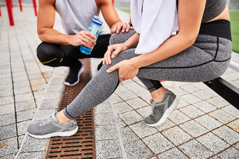
Compartment syndrome is a condition that causes pressure within a group of muscles to build up to dangerous levels. This pressure build-up begins to decrease blood flow, not allowing proper circulation, nutrients, and oxygen from getting to the nerves and muscle cells. The syndrome can be acute or chronic, and surgery can be required. Acute compartment syndrome is considered a medical emergency, usually caused by a severe injury and requires immediate treatment; otherwise, it can lead to permanent muscle damage. Chronic compartment syndrome or exertional compartment syndrome is usually not a medical emergency and is often caused by physical exertion. Muscle Compartment A compartment comprises a group of: The fascia does not stretch or expand because its job is to keep the tissues in place. If compartmental pressure builds up, swelling and bleeding may occur. When the tissues don't have enough blood to provide the proper amount of oxygen and nutrients, the tissues begin to die, leading to permanent damage. Because the fascia does not stretch if there is swelling or bleeding within a compartment, this increases pressure on the: - Capillaries
- Nerves
- Muscles in that compartment.
- Blood circulation does not reach the compartment to supply oxygen and nutrients.
- Nerve and muscle cells get damaged.
- Compartment syndrome most often takes place in the lower leg's anterior/front calf compartment.
However, it can also develop in other areas like the: - Legs
- Arms
- Hands
- Feet
- Buttocks
Acute The typical symptom is pain, specifically when the muscle in the compartment is stretched. - The pain is more intense than the injury itself.
- Flexing, contracting, or stretching the muscles increases the pain.
- Tingling or burning sensations may present.
- Muscle tightness or fullness sensation like bloating.
- Numbness or paralysis are late symptoms that usually indicate severe to permanent tissue injury.
The acute syndrome develops after a severe injury, like an automobile accident or from a broken bone. Injuries and conditions that can cause acute compartment syndrome include: - Fractures
- Muscle contusion/bruise that goes beyond just a bump. Two examples include a motorcycle falling on the rider's leg or a football player getting hit in the leg intensely.
- Crush injuries.
- Constricting bandages - Casts and bandages that are too tight can cause the blockage of blood. If symptoms develop, remove or loosen any constricting bandages. If it is from a cast, contact the doctor immediately.
- Anabolic steroids - Taking steroids is a possible factor in compartment syndrome.
Blood circulation restoration after a blockage. - When sleeping, a blood vessel can get blocked. Lying for a long time in a position that causes a limb to go to sleep, then shifting, moving, or getting up can contribute to the condition. This type of development can happen in individuals with neurological damage or who do not realize what is occurring. This can happen after intense intoxication with alcohol and/or drugs.
- Surgical repair of a damaged blood vessel that was blocked can result in compartment swelling.
- Permanent disability and tissue death can result unless the pressure is relieved.
Chronic Physical Exertion The pain and swelling from the chronic condition are caused by vigorous physical activity/exercise. It most often occurs in the leg. Individuals that participate in activities with repetitive motions have an increased risk. Physical activities/sports include: This is usually not dangerous and is often relieved by discontinuing the specific exercise/s or physical activity for a while. Symptoms include: - Pain during exercise.
- Cramping during exercise.
- Numbness
- Moving the foot is difficult.
- Muscle bulge can be seen.
Chiropractic Treatment Leg pain should not be ignored for long as the problems could escalate into severe/dangerous territory. Chiropractic treatment is highly effective in the detection and treatment of leg pain. Chiropractors are experts in the neuromusculoskeletal system. Their expertise in promoting physical function applies to the whole body's systems, including the: - Muscles
- Bones
- Ligaments
- Nerves
- Tendons
They are trained to diagnose and treat developing and chronic musculoskeletal problems and know when to seek specialized medical care when necessary. Can't Individuals Just Exercise More and Eat Whatever They Want? No individuals cannot just exercise/move more and eat whatever they want if they are serious about losing excess weight. A healthy diet and exercise are essential parts of the formula for effective weight loss. One study shows that being aware of diet in quality and quantity overtakes just exercising when achieving and maintaining healthy body composition changes as a vital part of maintaining a healthy lifestyle. Evaluating the effects of diet, exercise, or a combination of both revealed that long-term success was most significant in the mix of diet and exercise. Individuals can exercise vigorously, but losing weight can be very difficult if they have unhealthy eating habits or cannot stick to a healthy diet. The individual can develop other health problems from an unhealthy diet. General Disclaimer * The information herein is not intended to replace a one-on-one relationship with a qualified health care professional, licensed Physician and is not medical advice. We encourage you to make your own health care decisions based on your research and partnership with a qualified health care professional. Our information scope is limited to chiropractic, musculoskeletal, physical medicines, wellness, sensitive health issues, functional medicine articles, topics, and discussions. We provide and present clinical collaboration with specialists from a wide array of disciplines. Each specialist is governed by their professional scope of practice and their jurisdiction of licensure. We use functional health & wellness protocols to treat and support care for the injuries or disorders of the musculoskeletal system. Our videos, posts, topics, subjects, and insights cover clinical matters, issues, and issues that relate to and support, directly or indirectly, our clinical scope of practice.* Our office has made a reasonable attempt to provide supportive citations and has identified the relevant research study or studies supporting our posts. We provide copies of supporting research studies available to regulatory boards and the public upon request. We understand that we cover matters that require an additional explanation of how it may assist in a particular care plan or treatment protocol; therefore, to further discuss the subject matter above, please feel free to ask Dr. Alex Jimenez or contact us at 915-850-0900. Dr. Alex Jimenez DC, MSACP, CCST, IFMCP*, CIFM*, ATN* email: coach@elpasofunctionalmedicine.com Licensed in: Texas & New Mexico* References Braver, Richard T. "Chronic Exertional Compartment Syndrome." Clinics in podiatric medicine and surgery vol. 33,2 (2016): 219-33. doi:10.1016/j.cpm.2015.12.002 Joubert, Sonia V, and Manuel A Duarte. "Chronic Exertional Compartment Syndrome in a Healthy Young Man." Journal of chiropractic medicine vol. 15,2 (2016): 139-44. doi:10.1016/j.jcm.2016.04.007 Schmidt, Andrew H. "Acute compartment syndrome." Injury vol. 48 Suppl 1 (2017): S22-S25. doi:10.1016/j.injury.2017.04.024 Vajapey, Sravya, and Timothy L Miller. "Evaluation, diagnosis, and treatment of chronic exertional compartment syndrome: a review of current literature." The Physician and sportsmedicine vol. 45,4 (2017): 391-398. doi:10.1080/00913847.2017.1384289
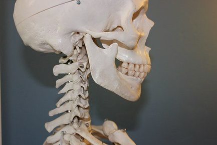
Tailbone pain begins in the coccyx, which is located at the bottom of the spine's sacrum. Coccydynia (kŏk′sĭ-dĭn′ē-ə) is the term for pain in the coccyx, or tailbone. Sitting and leaning back could be very uncomfortable. However, pretty much everybody responds well to conservative treatment. Spinal Anatomy of the Coccyx The coccyx, or tailbone, is the lowest area of the spine. It sits right below the sacrum. The tailbone is made up of 3 to 5 small bones fused together by around age 30. The coccyx helps support our weight while sitting. Risk Factors The prevalence of coccydynia is still unknown, but medical research has found that the condition affects: - Adolescents
- Adults
- Women
- People that are overweight
However, women and those that are overweight are the two highest risk factors. Women are affected five times more than men, which is likely due to injuries from childbirth. Also, a woman's coccyx is farther back than the male counterpart. This makes it more vulnerable to trauma. Obesity causes coccydynia because of the extra weight/pressure on the coccyx, which alters how a person sits. Causes There are several causes, the most common are: Trauma from: - Falling
- Getting bumped/hit
Repetitive/Extensive: and Childbirth Activities like these can bruise, break, and even dislocate the tailbone. If this happens there could be painful inflammation and muscle spasms in the tailbone area. Other possible causes: Bone spurs: A small bone spur on the lowest part of the coccyx can pinch the surrounding area, and cause pain and discomfort while sitting. Joint instability: Sacrococcygeal joint (which connects the coccyx and sacrum) allows too much or too little movement. Rare causes are: - Infection
- Metastatic cancer
- Chordomas
- Arachnoiditis
Coccydynia pain can mimic: - Lower back pain
- Sciatica
- Infection
- Pilonidal cysts (skin infection)
- Fractured bone
A doctor will rule out these causes to make a proper diagnosis. Diagnosing A doctor utilizes medical history and physical exam to diagnose coccydynia. Imaging scans are usually not necessary, but a doctor may order: - Computed tomography (CT) scan
- Magnetic resonance imaging (MRI) scan
If they believe a separate: Problem to be the cause of tailbone pain. Medical history is important, a doctor wants to know if a - Fall
- Accident
- Recent trauma
Might have caused the pain. A physical exam is next in line along with symptoms. The pain is usually localized in the tailbone, making it straightforward to diagnose. A doctor will want to know if the pain strikes when sitting or leaning back and which is worse. A doctor might ask the patient to point to where they're feeling the pain. Pointing could be enough for the doctor to tell if the pain is coccydynia when compared to other low back pain conditions. The doctor will want to palpate the tailbone area for signs of inflammation. Non-surgical Treatment Conservative treatment is extremely effective for coccydynia. Ninety percent of people experience pain reduction using non-surgical means or any medical intervention. It is first treated with noninvasive methods: - Ice or heating pad can help provide immediate, short-term relief.
- Donut/Wedge cushion provides extra padding taking the pressure off the coccyx when sitting. These cushions are available at pharmacies.
- Avoid sitting for long times, like a long flight will help prevent additional pain and injury.
- Rest is very important to help avoid further injury during recovery.
If there is still pain after these therapies, a doctor may recommend over-the-counter or prescription-strength non-steroidal anti-inflammatory drugs (NSAIDs) or other pain medications. If the medication doesn't work then a doctor may try cortisone injection or local spine blocker to send strong medicine directly to the tailbone area. Surgical Procedure If none of the conservative treatments work then surgery could be the next step. The procedure is called a coccygectomy, which means the removal of the tailbone. A surgeon will discuss the procedure in great detail before it is performed. These risks include: - Infection
- Hematoma (abnormal collection of blood outside an artery or vein)
- Perineal hernia (weakened pelvic muscles)
Chronic Coccydynia A small portion of people can develop chronic coccydynia, which means the pain lasts for more than 2 months. Chronic coccydynia can take a major toll on the quality of life. Talk to a doctor if symptoms are not resolving. They may refer you to a doctor that specializes in coccydynia management. Protecting the Tailbone There is no exact way or method to prevent coccydynia. However, reducing the risk of tailbone pain can be achieved by using caution when participating in sports like: - Skating - Ice, Inline, and Roller
- Biking
- Horseback riding
And take extra care when walking on icy/slippery/hazardous conditions so nobody falls. Gale Grijalva suffered from severe back pain as a result of an automobile accident injury. Where it was once very difficult to go about her regular daily tasks, Gale Grijalva is now able to participate in physical activities she wasn't able to engage in before thanks to Dr. Alex Jimenez, a chiropractor in El Paso, TX. Gale Grijalva describes how patient Dr. Jimenez is and she discusses how thoroughly he's been able to help her, including answering any concerns she may have. Gale Grijalva also experienced results through rehabilitation. Severe chronic back pain is a serious, recurring condition that affects a person's everyday life. Back pain lasting over three months is considered chronic. The spine is an essential component of the body. Severe chronic back pain might be the backbone's manner of telling the body that there is an issue. The spine is composed of bony vertebrae, soft spinal discs, facet joints, tendons, ligaments, and tendons. Within the bony vertebral artery lies the spinal cord, the delicate but effective nerve pathway of the central nervous system.
Osteopenia and osteoporosis are two similar conditions, but one is more severe. Both mean decreased bone density, but osteopenia is to a lesser degree than osteoporosis. However, osteopenia is still a problem because it can increase a person’s chances of breaking a bone. What are the symptoms? Osteopenia usually doesn't cause symptoms unless a bone is broken. However, some patients who present with osteopenia complain of dull back pain. Symptoms associated with osteoporosis include the following: - Back pain, caused by a fractured or collapsed vertebra
- Loss of height over time
- A stooped posture
- A bone fracture that occurs much more easily than expected
What are the causes and who is at risk? Women (primarily small-boned Caucasian and Asian) are most at risk for both conditions, primarily those who are age 65 or older as well as women who are postmenopausal. However, men can also be affected. Anyone who meets any of the criteria for being at risk for either of the bone conditions should be evaluated. Often, catching the conditions early can make a significant difference in the effects that they have on the body and in some cases, can even be arrested so that they don’t progress. Some of the common causes of both conditions include: - Lifestyle habits
- Smoking
- Insufficient calcium
- Sedentary lifestyle
- Excessive alcohol consumption
- Vitamin D deficiency
- Carbonated beverages
- Medical Situations
- Bulimia, anorexia, and other eating disorders
- Estrogen deficiency in women
- Certain hormone imbalances
- Overactive thyroid
- Certain treatments including radiation and chemotherapy
- Low testosterone in men
- Medications including anti-seizure, hydrocortisone, and steroids
- Health issues
- Tumors
- Cystic fibrosis
- Crohn’s disease
- Digestive issues
It should also be noted that certain types of diets, particularly those that advocate extremely low fat, or no fat can also cause problems. Vitamin D is necessary for calcium absorption in the body, but vitamin D is a fat-soluble vitamin meaning the body requires some fat in order to make use of it. When there is inadequate fat, the vitamin cannot be absorbed and in turn, calcium cannot be absorbed. A family history of osteopenia, osteoporosis, or low bone mass can increase a person risk by 50% to 85%. How is it diagnosed? Bone mineral density (BMD) tests are used to diagnose both osteopenia and osteoporosis by measuring the calcium levels in bone. This type of test can also provide an estimate of how much at risk a person is for bone fractures. This test is painless and non-invasive. It is usually performed on the heel, shin bone, wrist, spine, finger, or hip. Two common types of these tests are radiographs, a standard diagnostic tool for osteopenia, and Dual Energy X-ray Absorptiometry (DEXA). A DEXA scan is essentially a low energy x-ray so patients are not exposed to as much radiation as they would be if they had a regular x-ray. The results are attained by comparing the score (measurements were taken) to scans of individuals who do not have the condition. Once the score is measured and compared, it is assessed using a chart that identifies the level or risk: - +1.0 to -1.0 - Normal bone density
- -1.0 to -2.5 - Low bone density
- -2.5 or higher - At risk for osteoporosis
What are the treatments? As with most conditions, prevention is the most effective treatment. If you have a family history or fall under any of the risk factors, there are things you can do to minimize the effects or prevent the conditions completely. Your chiropractor can talk to you about lifestyle changes, exercise, and diet as well as supplements that you can take. Chiropractic adjustments can also be effective for many patients with osteopenia and osteoporosis as long as the chosen technique is a low force technique like Activator. Many patients find these natural treatments preferable to any medications that may be prescribed. The most important thing you should do, though, is get a bone density test if you are in an at-risk category, are a woman who is postmenopausal or age 65 or older.
Dr. Alex Jimenez collaborates with top rated diagnosticians and imaging specialists. We are blessed to have in our association, imaging specialists that provide fast, courteous & premiere board certified specialists. In collaboration with our offices we can provide the quality of service our patients mandate and deserve.
Who We AreDiagnostic Outpatient Imaging (DOI) is a state-of-the-art Radiology center in El Paso, TX. It is the only center of its kind in El Paso, owned and operated by a Radiologist. This means when you come to DOI for a radiologic exam, every detail, from the design of the rooms, the choice of the equipment, the hand-picked technologists, and the software which runs the office, is carefully chosen or designed by the Radiologist and not by an accountant. Our market niche is one center of excellence. Our values related to patient care are: We believe in treating patients the way we would treat our family and we will do our best to ensure that you have a good experience at our clinic. Dear Doctors,We are pleased to inform you of the arrival of our Titan 3-Tesla MRI at Diagnostic Outpatient Imaging. This is El Paso's only radiology imaging center that offers this technology. Patients do not always realize how important image quality is: It can make the difference in the diagnosis. 3-Tesla MRI is like HD TV and once you try it, you will not want to go back. The increased magnet strength gives us many benefits at no additional expense to the patient. It gives us the ability to scan faster or to scan with higher detail. An MRI of the brain can take 20 minutes and have exceptional quality, or we can perform the scan in less time, with better quality that is achieved on most 1.5 Tesla "high field" MRIs. This is incredibly useful for children. Our 3T MRI can perform Diffusion Tensor Imaging, MRI Spectroscopy and CSF flow studies to name just a few of its possibilities. This scanner is not only very fast, it is very large. Our open MRI has a clearance of 35 cm. The 3T has a diameter of 71 cm! This is welcome news for nervous or claustrophobic patients, and combined with its speed, it can actually eliminate the need for sedation for some patients. 3T MRI is faster, clearer, and has more diagnostic possibilities. We are certain you and your patients will notice the difference. Our Services MRI's:DOI has three MRI's under one roof. All are American College of Radiology (ACR) Certified. GoodOpen MRI (0.35 Tesla): This MRI perfect for claustrophobic and very large patients. There is no table weight limit on this MRI BetterHigh Field 1.5 Tesla MRI- This is a eight channel MRI with high end image quality. It is in a beautiful room and has 'pianissimo' technology, which makes the MRI relatively quiet. This machine has been the best MRI in private practice in El Paso for years. It will soon be eclipsed by our new 3.0 Tesla MRI. BestHigh Field 3.0 Tesla MRI- This is the only 3.0 Tesla MRI in private practice in El Paso. This technology can deliver stunning image quality, which can actually make a difference in your diagnosis. The increased magnet strength gives us many benefits at no additional expense to the patient.
It gives us the ability to scan faster, or to scan with higher detail. This is welcome news for nervous or claustrophobic patients, and as well as for children as it can actually eliminate the need for sedation in some patients. 3T is faster, clearer, more diagnostic for a better for MRI. It is like HD TV. Once you have tried it, you won't want to go back. This MRI effectively doubles our MRI capacity. If needed most exams can be completed in under 5 minutes, instead of the normal 30-45 minutes. Breast MRI:DOI began Breast MRI in July 2007, being the first facility in El Paso to perform the exam. We have now performed over 2500 breast MRI's and many MRI-guided breast biopsies. All have been interpreted and/or performed by Dr. Boushka, making him the most experienced radiologist in the city with this exam. This is the most powerful tool for the detection of Breast cancer to date. Hours:
Monday to Thursday 7 am to 9 pm
Friday 7 am to 5 pm
Saturday 8 am to 4 pm Prostate MRI:Guys, you need great medical care also. We are the only facility in El Paso performing this leading edge exam. MRI can see cancers when other imaging methods cannot. Not only can we see prostate cancers with MRI, we can perform MRI-guided prostate biopies for pathologic (definitive) diagnosis. Monday to Thursday 7 am to 9 pm
Friday 7 am to 5 pm
Saturday 8 am to 4 pm CT:We have a 16 slice Toshiba Aquillion CT scanner, with newly updated in Dec 2013. The upgrade allows for reduced X-ray dose, higher resolution, more patient comfort, shorter breath holds and doubles the speed of the scanner. This scanner performs CT X-ray exams as helical volume acquisitions in 3D from a single patient exam. Most exams are finished in under 60 seconds, unless delayed images with contrast are indicated. Additionally we have a powerful 3D post processing workstation. Hours
Monday to Friday 7 am to 6 pm Ultrasound:DOI has just doubled our Ultrasound capacity with newly purchased Philips 34 XRL scanner. We have Three certified Ultrasonographers with cumulative experience of 45 years. We are confident you will find them professional and compassionate. Beverly Bruner RDMS, Sonographer, formally of Desert Imaging has joined our team. 3D OB Ultrasounds:You better believe it. Available whenever our US department is open. No referral necessary. Images are reviewed by an actual radiologist. Ultrasound Hours:
Monday, Tuesday, Thursday 8 am to 5 pm
Wednesday 8 am to 8 pm
Friday 8 am to 5 pm
Saturday 8 am to 12 pm Digital MammographyDOI was the first facility in El Paso to acquire Hologic Full Field Digital Mammography and thus we have more experience with this technology than any facility in El Paso. Our Mammographer has 20 years of experience and has her own following of patents who seek her out to perform their mammograms because of her excellent and compassionate care. Our private pay screening mammography price of $90, including the interpretation is an unbeaten price in El Paso. Hours
Mon - Fri 8am to 4pm
Extended hours Wednesday until 8pm)
Saturdays 8am to 12pm Bone Denisity (DEXA)We have a brand new, Hologic Discovery CI bone densitometer scanner. This is the latest technology. X-RayOur digital computed radiography was just updated February 2014. No appointments are necessary. We look forward to serving you. Sincerely,
William M Boushka, MD
Boron is one of the main minerals used in the bone building process of the body which directly acts as an activator. Boron helps convert vitamin D into its active state by initiating estrogen production. The estrogen then improves the absorption of calcium. This mineral also helps ensure the metabolism within the bone can function properly. In fact, because of its role in the body, Boron can actually assist in preventing arthritis and tooth decay. The body depends on Boron to perform a large majority of its functions and when there is a Boron deficiency in the body, the parathyroid glands, small endocrine glands in the neck of humans, become overactive, causing the glands to release too much of the parathyroid hormone. As a result, the level of calcium in the blood increases as it’s released from the bones, joints, and teeth, into the blood stream. Several resulting symptoms of Boron deficiencies include, arthritis, brittle bones, carpal tunnel syndrome, degenerative joint disease, hormonal imbalances, loss of libido, memory loss, muscle pain, osteoporosis, receding gums, and weak joint cartilage. Bone analysis have previously shown that arthritis joints and nearby bones contain only half the Boron content of healthy joints. Boron supplements can help reduce the daily loss of calcium by nearly 50%. Because this calcium is mainly withdrawn from the bones and teeth, Boron deficiencies are considered to be the most important factors in causing arthritis, osteoporosis, and tooth decay today. Boron supplements are actually considered to be an effective treatment for up to 95% in the relief of arthritis, provided the joint has not completely deteriorated.
|
Medications can be lifesavers when it comes to the treatment of various conditions. But they can also open the door to other serious conditions. Medications fall into pharmacological drug classes. Certain medications can interfere with bone health, and induce bone density loss. Users of these medications could put them at risk for osteoporosis and possible spinal fracture/s. Medications that can potentially weaken bones and how to protect yourself is the focus. Not all of the medications listed are for treating spinal disorders or neck and back pain. Steroids Steroids taken by mouth are commonly prescribed for spinal conditions. This includes: - Low back pain
- Neck pain
- Spinal inflammatory arthritis
These medications carry anti-inflammatory compounds that are pretty powerful. These help the pain but can cause bone loss with long-term use. These types of steroids put the bones at risk because of how they slow down the osteoblasts, which are bone-building cells. As the osteoblasts are slowed, the work of the osteoclasts, which are bone-absorbing cells gets increased straining the system and ultimately leading to bone loss. Examples of steroids: - Dexamethasone
- Methylprednisolone
- Prednisone
Daily doses of more than 5 mg pose the biggest threat to the skeletal system. Ask a doctor about a short-term low-dose regimen, especially, if there is a heightened risk for osteoporosis or spinal fracture. Selective Serotonin Receptor Uptake Inhibitor Selective serotonin receptor uptake inhibitors help those with neck and low back pain in a variety of ways. These include reducing the mental and emotional effects of chronic pain. But, selective serotonin receptor uptake inhibitors can boost the fracture risk. This type of medication can cause bone loss in older women and reduced bone density in men and children. Examples of selective serotonin receptor uptake inhibitors: Ask a doctor for another type of selective serotonin receptor uptake inhibitor. Possibilities include serotonin and norepinephrine reuptake inhibitors, that can achieve the same results without bone loss and fracture risks. Certain Anticonvulsants Anticonvulsants are used to control seizures. However, they have been found to help individuals with spinal nerve pain. But there are some types of anticonvulsants that can increase the liver’s vitamin D metabolism. This lowers the blood’s vitamin D levels. Vitamin D is essential to the body’s ability to absorb calcium. That means that lower vitamin D levels can cause bone loss. Examples of anticonvulsants: Talk to a doctor, chiropractor, or health coach about taking a vitamin D supplement/s to boost vitamin D levels. Certain Diabetic Medications There are two types of diabetic medications that can increase the risk of fracture. Thiazolidinediones known as TZD's and sodium-glucose cotransporter 2 inhibitors. The TZD's increase the fat cells in the bone marrow, and lower the bone-building cells. The sodium-glucose cotransporter 2 inhibitors can reduce bone density. Examples of TZD's: If there is a high risk of fracture, ask a doctor if an alternative medication to a TZD can be taken. Examples of sodium-glucose cotransporter 2 inhibitors: - Canagliflozin
- Dapagliflozin
- Empagliflozin
If there is a greater risk of falls, ask a doctor if an alternative to taking a sodium-glucose cotransporter 2 inhibitor can be taken. Hormone Medications Medications that reduce estrogen or androgen levels in the body also increase the bone's absorbing cell activity. And this can lead to bone density loss. Examples of hormone medications: - Anastrozole
- Exemestane
- Leuprolide
- Goserelin
- Medroxyprogesterone acetate
If there is an increased risk for osteoporosis or fracture, talk to a doctor about ways to protect the bones while taking these medications. Antacids Antacids both over-the-counter and prescription that contain aluminum help to neutralize stomach acid. There are other medications called H2-blockers also known as proton-pump inhibitors. These reduce how much acid the stomach produces. While these aid in reducing heartburn, stomach pain, etc, long-term use can reduce the body’s ability to absorb calcium and thus increase the risk for fracture. Examples of these types of antacids: Examples of Proton-Pump Inhibitors: - Omeprazole
- Esomeprazole
- Lansoprazole
Ask a doctor if a different H2-blocker can achieve the same results. Additionally, a doctor, nutritionist, or health coach could recommend dietary changes/adjustments to help reduce stomach acid. Blood Thinners and Anticoagulants These medications help reduce the risk of stroke, can interfere with the body’s ability to absorb calcium. They reduce the activity of the bone-building cells. This causes bone loss and increases the risk of fracture. Examples of anticoagulants or blood thinners: - Enoxaparin sodium
- Warfarin
Talk to a doctor about a possible alternative anticoagulant. A change in medication has been shown to put the bones at less risk. Diuretics Loop diuretics work by reducing inflammation/swelling along with water retention by increasing the kidneys urine production. These medications can cause the kidneys to remove key nutrients like calcium, potassium, and magnesium to help increase bone production. Reduction in all of these increases the risk of bone loss and a spinal fracture. Examples of loop diuretics: - Furosemide
- Ethacrynic acid
- Bumetanide
Talk to a doctor about an alternative known as a thiazide diuretic. These encourage the kidneys to retain calcium, thus increasing bone density. Reduce The Risk Protecting bone health is the objective. A bone mineral density test could help along with taking bone-boosting supplements. Learning about the risks of taking these medications can help prevent osteoporosis and spinal fractures. Keep track of all medications over-the-counter, prescription, holistic, all-natural, etc, and make sure all doctors, specialists understand what is being taken. A spine specialist or endocrinologist might not what the other doctor has prescribed, so keep everyone informed. Dr. Alex Jimenez’s Blog Post Disclaimer The scope of our information is limited to chiropractic, musculoskeletal, physical medicines, wellness, and sensitive health issues and/or functional medicine articles, topics, and discussions. We use functional health & wellness protocols to treat and support care for injuries or disorders of the musculoskeletal system. Our posts, topics, subjects, and insights cover clinical matters, issues, and topics that relate and support directly or indirectly our clinical scope of practice.* Our office has made a reasonable attempt to provide supportive citations and has identified the relevant research study or studies supporting our posts. We also make copies of supporting research studies available to the board and or the public upon request. We understand that we cover matters that require an additional explanation as to how it may assist in a particular care plan or treatment protocol; therefore, to further discuss the subject matter above, please feel free to ask Dr. Alex Jimenez or contact us at 915-850-0900. The provider(s) Licensed in Texas& New Mexico*
Paget’s disease, aka osteitis deformans, is the second most common bone disorder in the United States right behind osteoporosis. Paget’s disease happens when bone cells don’t function properly and result in: - Deformed
- Enlarged
- Fragile bones
The bones of the spine (vertebrae) are susceptible to this condition. Paget’s Disease and The Spine Paget’s disease begins with a malfunction of two bone cell types: osteoblasts and osteoclasts. Bones are constantly going through a regenerative process where osteoclasts break down old bone, and osteoblasts build new bone. When the cells work together in a balanced way, the bones stay strong. With Paget’s disease, the osteoclasts break down old bone faster than normal. This forces osteoblasts to work harder and longer. When this happens, the new bone is placed improperly and leads to deformity. The newly built bone tends to be more fragile than healthy bone and has a greater risk of fracturing. Paget’s most commonly affects the spinal bones, specifically the: Paget’s disease can involve a single bone meaning it is monostotic or multiple bones or polyostotic. Monostotic cases make up about 10- to 35-percent of total occurrences. Paget's typically affects people over the age of 55, and 3% of that population develop the disease. Spinal Symptoms Many have Paget’s in their spine don’t know it. The most common symptom is bone pain in the neck and/or back. The pain can feel: - Dull
- Persistent
- Worse at night
It can cause spinal cord compression, neurologic symptoms like: - Tingling
- Numbness
- Difficulty walking
- Bowel
- Bladder problems
Causes The cause of Paget’s disease is still unknown but research scientists believe genetics and viral infection contribute to the condition. Spine-Related Complications The telltale of Paget’s is the bone deformities that can result in painful spine conditions, which include: The disease makes the spine prone to fractures because of the weakened new bone. Also, vertebral body compression fractures can occur and cause spinal cord compression and nerve pain. Types of spinal arthritis that can coexist with Paget’s include: - Rheumatoid arthritis
- Spondylosis or spinal osteoarthritis
- Ankylosing spondylitis
It has also been linked to spinal tumors that include osteosarcoma. Diagnosing and Treatment If symptoms are obvious, like hunchback brought on from kyphosis then a physical exam could help determine the diagnosis. Most cases require imaging tests to get a closer look at the bones. - A doctor/chiropractor may order an x-ray of the spine to confirm a diagnosis and illuminate any bone problems.
- For early stages, a bone scan will pick up initial deformities better than an x-ray.
- CT scans or magnetic resonance imaging (MRI) can also be used.
Blood testing can be important in the diagnosis Known as a bone-specific alkaline phosphatase test it can detect when the bones are regenerating too fast. It's not only used during diagnosis but also during the treatment in helping to monitor patients as they go through the therapy. It is treated with medications called bisphosphonates. These medications help return normal function to the osteoclasts and osteoblasts. Bisphosphonates can manage the disease and reduce symptoms, but do not cure the disease. Living with Paget’s Advanced cases can cause spine problems, which includes spinal fractures. Most with Paget’s disease have preferable outcomes. When Paget's disease is managed with medication, regular doctor visits, chiropractic care, and proper diet, then there shouldn’t be a problem in achieving a healthy quality of life. David Garcia, a maintenance facility worker, and a proud Dad in El Paso, TX at the Region 19 Education Services Center. However, Mr. Garcia's everyday life is often influenced by his chronic lower back pain. After undergoing worsening symptoms for some time, Mr. Garcia was recommended to seek chiropractic care with Dr. Alex Jimenez by his sister, a former patient of Dr. Jimenez. Mr. Garcia has since experienced enormous relief from his lower back pain and he's grateful to Dr. Jimenez and his staff for supplying him with education regarding his health problems as well as properly caring for him. Mr. Garcia recommends Dr. Jimenez as the non-invasive surgical selection for lower back pain.
Heel Spur: Blanca, born and raised in El Paso, TX, has been suffering from heel spurs for about two years. As a registered nurse, her symptoms significantly affected her ability to work and her overall quality of life. Determined to improve her health, Blanca considered chiropractic care. Once she started treatment with Dr. Alex Jimenez, however, Blanca experienced tremendous relief from her heel spurs, almost instantly. Blanca highly recommends chiropractic care with Dr. Alex Jimenez as the non-surgical choice for treatment of heel spurs. A heel spur is a calcium residue resulting in a bony protrusion on the bottom of the heel bone. Although heel spurs are often painless, they can lead to heel pain. They are often associated with plantar fasciitis, a painful inflammation of the fibrous band of connective tissue (plantar fascia) that runs across the bottom of the foot and also connects the heel bone to the ball of the foot. Heel spurs are usually caused by strains on foot muscles and ligaments, stretching of the plantar fascia, and repeated tearing of the membrane which covers the heel bone. Heel spurs are particularly common among athletes. We are blessed to present to you El Paso’s Premier Wellness & Injury Care Clinic. Our services are specialized and focused on injuries and the complete recovery process. Our areas of practice include: Wellness & Nutrition, Chronic Pain, Personal Injury, Auto Accident Care, Work Injuries, Back Injury, Low Back Pain, Neck Pain, Migraine Headaches, Sport Injuries, Severe Sciatica, Scoliosis, Complex Herniated Discs, Fibromyalgia, Chronic Pain, Stress Management, and Complex Injuries. As El Paso’s Chiropractic Rehabilitation Clinic & Integrated Medicine Center, we passionately are focused treating patients after frustrating injuries and chronic pain syndromes. We focus on improving your ability through flexibility, mobility and agility programs tailored for all age groups and disabilities. If you have enjoyed this video and/or we have helped you in any way please feel free to subscribe and share us. Thank You & God Bless. Dr. Alex Jimenez DC, C.C.S.T Facebook Clinical Page: https://www.facebook.com/dralexjimenez/ Facebook Sports Page: https://www.facebook.com/pushasrx/ Facebook Injuries Page: https://www.facebook.com/elpasochiropractor/ Facebook Neuropathy Page: https://www.facebook.com/ElPasoNeuropathyCenter/ Facebook Fitness Center Page: https://www.facebook.com/PUSHftinessathletictraining/ Yelp: El Paso Rehabilitation Center: http://goo.gl/pwY2n2 Yelp: El Paso Clinical Center: Treatment: https://goo.gl/r2QPuZ Clinical Testimonies: https://www.dralexjimenez.com/category/testimonies/ Information: LinkedIn: https://www.linkedin.com/in/dralexjimenez Clinical Site: https://www.dralexjimenez.com Injury Site: https://personalinjurydoctorgroup.com Sports Injury Site: https://chiropracticscientist.com Back Injury Site: https://www.elpasobackclinic.com Rehabilitation Center: https://www.pushasrx.com Fitness & Nutrition: http://www.push4fitness.com/team/ Pinterest: https://www.pinterest.com/dralexjimenez/ Twitter: https://twitter.com/dralexjimenez Twitter: https://twitter.com/crossfitdoctor
Improving spinal bone healing in at risk patients
Bone growth stimulation (BGS) is a therapy your surgeon may prescribe following a spinal fusion procedure. A bone growth stimulator is an auxiliary device worn following cervical (neck) or lumbar (low back) spine surgery. BGS may be used to assist spinal bone fuse after a fusion procedure or as a treatment for failed fusion. Naturally, you’ve questions about this technology.
THE INFO PROVIDED IN THIS PATIENT GUIDE CAN ASSIST YOU TO LEARN:
- Bone heals
- Risk factors for a poor or failed fusion
- Role of bone growth stimulation in spine fusion aftercare
- Questions to ask your back surgeon
“Bone growth stimulation to be used in both the cervical and lumbar spine has demonstrated to substantially help fusion results. Having been a study centre for this particular technology, I’ve used bone growth stimulation in most my post-operative cervical and lumbar patient instances. The patient assessment standards I use contains:
Multi-level fusions; more than one degree of the back is fused
Co-morbidities (risk factors) that could hinder bone healing and growing”
—Gerard J. Girasole, MD
Orthopaedic Surgeon
Orthopaedic & Sports Medicine Center
ABOUT SPINAL FUSION
Spinal fusion is done to stop motion of neurologic deficit and the spine. During the procedure two or more vertebral bodies are joined together using instrumentation and bone graft. Spinal instrumentation includes poles, screws, plates, and interbody devices (implants). Bone graft may comprise your own bone (autograft), donor bone (allograft), or alternative forms of graft.
Bone graft helps stimulate new bone to grow through three stages:
- Inflammatory period: cells start to form new tissue
- Repair period: small blood vessel ingrowth begins
- Remodeling phase: bone structure becomes powerful
Spinal instrumentation creates an internal cast, allowing the inflammatory procedure to stimulate bone healing. With time, new bone grows into and about the implanted instrumentation healing into a construct that is sound.
Some patients are at risk for spinal fusion to fail. A failed fusion is called pseudarthrosis or nonunion. Pseudarthrosis and nonunion are medical terms your surgeon may utilize to identify a fusion dilemma.
COMMON SPINAL ISSUES TREATED SURGICALLY WITH FUSION INCLUDE:
- Degenerative disk disease
- Fracture
- Herniated disc
- Spinal stenosis
LUMBAR
- Adult degenerative scoliosis
- Spondylolisthesis
HOW DOES A BONE GROWTH STIMULATOR HELP SPINAL FUSION?
A BGS sends electric signals to the fusion site. The electrical signals activate the body’s natural bone healing process, which may be impaired in at-risk patients.
BONE GROWTH STIMULATION HAS BEEN PUT TO USE FOR DECADES TO HELP BONE HEAL
Over 50 years ago scientists found that low-level electrical fields arouse the entire body’s bone-healing process. Other improvements included finding several types of energy that stimulate bone development, electromagnetic coil technology and only better devices — supported by clinical and scientific research—have enhanced bone healing in patients who undergo spinal fusion.
DIFFERENT TYPES OF BONE GROWTH STIMULATORS
All bone growth stimulators are different. Certain types are designed to be surgically implanted (internal BGS) and other stimulators are worn outside the body (external BGS). Other differences include how stimulation is transmitted to the back and the kind of magnetic field or electric current created by the apparatus.
|


Compartment syndrome is a condition that causes pressure within a group of muscles to build up to dangerous levels. Chiropractic can help. For answers to any questions you may have, please call Dr. Jimenez at 915-850-0900 or 915-412-6677