Your new post is loading...

|
Scooped by
Gilbert C FAURE
October 12, 2019 12:53 PM
|
Stratification on the basis of patient-reported symptoms of patients with primary
Sjögren's syndrome revealed distinct pathobiological endotypes with distinct responses
to immunomodulatory treatments.
|
|
Scooped by
Gilbert C FAURE
September 9, 2019 6:38 AM
|
The main symptoms — dry eyes and mouth, fatigue and limb pain — are common complaints associated with any number of health problems.

|
Suggested by
Société Francaise d'Immunologie
June 22, 2019 2:20 PM
|
This proof of concept study was aimed at characterizing novel salivary biomarkers specific for different subsets in primary Sjögren’s syndrome (pSS) in order to improve patients’ profiling. pSS patients were stratified in three subgroups according to both (a) focus score in the minor salivary...
|
|
Suggested by
Société Francaise d'Immunologie
May 3, 2019 2:31 PM
|
Immune Profile of Saliva and Serum of Patients With Primary Sjögren´s Syndrome - Full Text View.
|
|
Scooped by
Gilbert C FAURE
April 2, 2019 1:47 PM
|
J Intern Med. 2019 Mar 20. doi: 10.1111/joim.12888.[Epub ahead of print]...
|
|
Scooped by
Gilbert C FAURE
April 1, 2019 8:12 AM
|
Results Data from 2970 patients from Latin America and 18019 patients from Europe, North America and Asia were analyzed. Colombian patients have a lower age at disease onset than those from other Latin American countries and a higher rate of positivity of antinuclear antibodies and rheumatoid factor. A significant difference in the proportion of female patients in Latin America compared with Europe and North America was observed. The spectrum of disease in Latin American was similar to North American patients, while strong differences were noticed between Latin American and European and Asian patients. Noteworthy, a paucity of reports including African and African-descendent patients was observed. Conclusions The clinical spectrum of SS differs between countries and continents. Genetic differences relying upon ancestry could explain these findings. However, environmental factors have proven to be important determinants in the development of autoimmune diseases (i.e., autoimmune ecology). Thus, ancestry and the autoimmune ecology should be considered in studies aimed to evaluate the geoepidemiology of SS and other autoimmune diseases.
|
|
Scooped by
Gilbert C FAURE
February 20, 2019 3:39 PM
|
Previous studies and own clinical observations of patients with systemic lupus erythematosus (SLE) suggest that SLE harbors distinct immunophenotypes. This heterogeneity might result in differences in response to treatment in different subgroups and obstruct clinical trials.
|
|
Scooped by
Gilbert C FAURE
February 10, 2019 12:20 PM
|
RMD Open. 2019 Jan 11;5(1):e000772. doi: 10.1136/rmdopen-2018-000772. eCollection 2019.
|
|
Scooped by
Gilbert C FAURE
January 4, 2019 3:00 PM
|
AbstractCD4+ T helper (Th) cells that express the gut homing chemokine receptor CCR9 are increased in the peripheral blood of patients with inflammatory bowel disease and Sjögren’s syndrome and in the inflamed lesions of autoimmune diseases that affect the accessory organs of the digestive system. However, despite the important role of the GIT in both immunity and autoimmunity, the nature of CCR9-expressing cells in GIT lymphoid organs and their role in chronic inflammatory diseases remains unknown. In this study, we analysed the characteristics of CCR9+ Th and T follicular helper (Tfh) cells in GIT associated lymphoid tissues in health, chronic inflammation and autoimmunity. Our findings reveal an association between the transcriptome and phenotype of CCR9+ Th in the pancreas and CCR9+ Tfh cells from GIT-associated lymphoid tissues. GIT CCR9+ Tfh cells exhibited characteristics, including a Th17-like transcriptome and production of effector cytokines, which indicated a microenvironment-specific signature. Both CCR9+ Tfh cells and CCR9+ Th cells from GIT-associated lymphoid tissues migrated to the pancreas. The expression of CCR9 was important for migration of both subsets to the pancreas, but Tfh cells that accumulated in the pancreas had downmodulated expression of CXCR5. Taken together, the findings provide evidence that CCR9+ Tfh cells and Th cells from the GIT exhibit plasticity and can accumulate in distal accessory organs of the digestive system where they ma
|
|
Scooped by
Gilbert C FAURE
November 23, 2018 2:34 AM
|
Detection of antinuclear antibodies (ANAs) is of paramount importance for diagnosis and assessment of autoimmune diseases (ADs), but may vary depending on the method used. Therefore, we aimed to compare three different line immunoassays (LIAs) for ANAs testing in order to simultaneously evaluate their concordance with the results obtained by a well known and widely used ELISA. Sera from women with established systemic lupus erythematosus (SLE, n=62) and Sjogren’s syndrome (SS, n=28) were examined. Patients with SLE had an average age of 48.7 years (IQR 36.8–57.0) with a disease duration of 14.8 years (IQR 9.0–18.0). Patients with SS had an average age of 62.1 years (IQR 55.5–71.0) with a duration of disease of 13.1 years (IQR 8.3–17).
Table 1 shows the results from the two-by-two comparison for the four techniques. Different levels of agreement were observed among the assays. In general, they were deficient. The analysis showed only one ‘strong’ level of agreement between ELISA and IMTEC-ANA-LIA Maxx for anti-Ro measurement. Out of 39 comparisons, 14 did …
|
|
Scooped by
Gilbert C FAURE
November 19, 2018 4:41 AM
|
Objectives To determine the expressions of Toll‐like receptors (TLRs) 7–9 and type I interferon (IFN) signal in labial salivary glands (LSGs) and cultured salivary gland epithelial cells (SGECs) from primary Sjögren's syndrome (pSS) patients. Methods We performed an immunohistochemistry analysis of LSGs from 11 patients with pSS as defined by American‐European Consensus Group classification criteria and five healthy subjects. The pSS patients’ SGECs were analyzed by immunofluorescence and western blotting. IFN‐α expression was examined by immunosorbent assay and flow cytometry. Results Mononuclear cells (MNCs) from pSS patients’ LSGs showed TLR7‐dominant expression. B cells, plasma cells, and plasmacytoid dendritic cells (pDCs) co‐expressed with TLR7. MyD88, TRAF6, and IRF7 co‐expressed with the pDC marker CD303 in LSGs. Ducts from pSS patients dominantly expressed TLR7, and TLR7 in the ducts co‐expressed with MyD88, TRAF6 and IRF7. Type I IFNs including IFN‐α and IFN‐β were detected in MNCs and ducts in pSS patients’ LSGs. Increased TRAF6 expression and the nuclear translocation of IRF7 in SGECs were detected by immunofluorescence following loxoribine (a TLR7 ligand) stimulation despite IFN‐β pretreatment. Western blotting showed increased TRAF6 expression in SGECs following IFN‐β and loxoribine stimulation. Although no increase in IFN‐α was detected in supernatant from stimulated SGECs, the IFN‐α in supernatant from stimulated peripheral blood pDCs from pSS patients was significantly increased. Conclusion Our findings suggest that TLR7 is dominantly expressed in both MNCs and ducts with downstream signals for type I IFNs, indicating that TLR7‐dominant innate immunity is related to the development of sialadenitis in pSS. This article is protected by copyright. All rights reserved.
|
|
Scooped by
Gilbert C FAURE
November 17, 2018 4:16 AM
|
Sjögren’s syndrome (SS) is characterised by B cell abnormalities and immune-mediated destruction of exocrine glands, primarily the salivary and lacrimal glands.1 2 Among the reported genetic polymorphisms associated with primary SS (pSS), the FAM167A-BLK locus distinguishes itself as an...
|
|
Scooped by
Gilbert C FAURE
November 6, 2018 1:26 PM
|
|
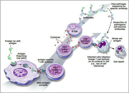
One of the more perplexing questions in biomedical research is—why does the body's protective shield against infections, the immune system, attack its own vital cells, organs, and tissues? The answer to this question is central to understanding an array of autoimmune diseases, such as rheumatoid arthritis, type 1 diabetes, systemic lupus erythematosus, and Sjogren's syndrome. When some of the body's cellular proteins are recognized as "foreign" by immune cells called T lymphocytes, a destructive cascade of inflammation is set in place. Current therapies to combat these cases of cellular mistaken identity dampen the body's immune response and leave patients vulnerable to life-threatening infections. Research on stem cells is now providing new approaches to strategically remove the misguided immune cells and restore normal immune cells to the body. Presented here are some of the basic research investigations that are being guided by adult and embryonic stem cell discoveries. Introduction The body's main line of defense against invasion by infectious organisms is the immune system. To succeed, an immune system must distinguish the many cellular components of its own body (self) from the cells or components of invading organisms (nonself). "Nonself" should be attacked while "self" should not. Therefore, two general types of errors can be made by the immune system. If the immune system fails to quickly detect and destroy an invading organism, an infection will result. However, if the immune system fails to recognize self cells or components and mistakenly attacks them, the result is known as an autoimmune disease. Common autoimmune diseases include rheumatoid arthritis, systemic lupus erythematosis (lupus), type 1 diabetes, multiple sclerosis, Sjogren's syndrome and inflammatory bowel disease. Although each of these diseases has different symptoms, they share the unfortunate reality that, for some reason, the body's immune system has turned against itself (see Box 6.1. Immune System Components: Common Terms and Definitions). How Does the Immune System Normally Keep Us Healthy? The "soldiers" of the immune system are white blood cells, including T and B lymphocytes, which originate in the bone marrow from hematopoietic stem cells. Every day the body comes into contact with many organisms such as bacteria, viruses, and parasites. Unopposed, these organisms have the potential to cause serious infections, such as pneumonia or AIDS. When a healthy individual is infected, the body responds by activating a variety of immune cells. Initially, invading bacteria or viruses are engulfed by an antigen presenting cell (APC), and their component proteins (antigens) are cut into pieces and displayed on the cell's surface. Pieces of the foreign protein (antigen) bind to the major histocompatibility complex (MHC) proteins, also known as human leukocyte antigen (HLA) molecules, on the surface of the APCs (see Figure 6.1 Immune Response to Self or Foreign Antigens). This complex, formed by a foreign protein and an MHC protein, then binds to a T cell receptor on the surface of another type of immune cell, the CD4 helper T cell. They are so named because they "help" immune responses proceed and have a protein called CD4 on their surface. This complex enables these T cells to focus the immune response to a specific invading organism. The antigen-specific CD4 helper T cells divide and multiply while secreting substances called cytokines, which cause inflammation and help activate other immune cells. The particular cytokines secreted by the CD4 helper T cells act on cells known as the CD8 "cytotoxic" T cells (because they can kill the cells that are infected by the invading organism and have the CD8 protein on their surface). The helper T cells can also activate antigen-specific B cells to produce antibodies, which can neutralize and help eliminate bacteria and viruses from the body. Some of the antigen-specific T and B cells that are activated to rid the body of infectious organisms become long-lived "memory" cells. Memory cells have the capacity to act quickly when confronted with the same infectious organism at later times. It is the memory cells that cause us to become "immune" from later reinfections with the same organism. Figure 6.1. Immune Response to Self or Foreign Antigens. (© 2001 Terese Winslow) How Do the Immune Cells of the Body Know What to Attack and What Not To? All immune and blood cells develop from multipotent hematopoietic stem cells that originate in the bone marrow. Upon their departure from the bone marrow, immature T cells undergo a final maturation process in the thymus, a small organ located in the upper chest, before being dispersed to the body with the rest of the immune cells (e.g., B cells). Within the thymus, T cells undergo an important process that "educates" them to distinguish between self (the proteins of their own body) and nonself (the invading organism's) antigens. Here, the T cells are selected for their ability to bind to the particular MHC proteins expressed by the individual. The particular array of MHCs varies slightly between individuals, and this variation is the basis of the immune response when a transplanted organ is rejected. MHCs and other less easily characterized molecules called minor histocompatibility antigens are genetically determined and this is the reason why donor organs from relatives of the recipient are preferred over unrelated donors. In the bone marrow, a highly diverse and random array of T cells is produced. Collectively, these T cells are capable of recognizing an almost unlimited number of antigens. Because the process of generating a T cell's antigen specificity is a random one, many immature T cells have the potential to react with the body's own (self) proteins. To avoid this potential disaster, the thymus provides an environment where T cells that recognize self-antigens (autoreactive or self-reactive T cells) are deleted or inactivated in a process called tolerance induction. Tolerance usually ensures that T cells do not attack the "autoantigens" (self-proteins) of the body. Given the importance of this task, it is not surprising that there are multiple checkpoints for destroying or inactivating T cells that might react to auto-antigens. Autoimmune diseases arise when this intricate system for the induction and maintenance of immune tolerance fails. These diseases result in cell and tissue destruction by antigen-specific CD8 cytotoxic T cells or autoantibodies (antibodies to self-proteins) and the accompanying inflammatory process. These mechanisms can lead to the destruction of the joints in rheumatoid arthritis, the destruction of the insulinproducing beta cells of the pancreas in type 1 diabetes, or damage to the kidneys in lupus. The reasons for the failure to induce or maintain tolerance are enigmatic. However, genetic factors, along with environmental and hormonal influences and certain infections, may contribute to tolerance and the development of autoimmune disease [4, 7]. Hematopoietic Stem Cell Therapy for Autoimmune Diseases The current treatments for many autoimmune diseases include the systemic use of anti-inflammatory drugs and potent immunosuppressive and immunomodulatory agents (i.e., steroids and inhibitor proteins that block the action of inflammatory cytokines). However, despite their profound effect on immune responses, these therapies are unable to induce clinically significant remissions in certain patients. In recent years, researchers have contemplated the use of stem cells to treat autoimmune disorders. Discussed here is some of the rationale for this approach, with a focus on experimental stem cell therapies for lupus, rheumatoid arthritis, and type 1 diabetes. The immune-mediated injury in autoimmune diseases can be organ-specific, such as type 1 diabetes which is the consequence of the destruction of the pancreatic beta islet cells or multiple sclerosis which results from the breakdown of the myelin covering of nerves. These autoimmune diseases are amenable to treatments involving the repair or replacement of damaged or destroyed cells or tissue (see Chapter 7. Stem Cells and Diabetes and Chapter 11. Use of Genetically Modified Stem Cells in Experimental Gene Therapies). In contrast, non-organ-specific autoimmune diseases, such as lupus, are characterized by widespread injury due to immune reactions against many different organs and tissues. One approach is being evaluated in early clinical trials of patients with poorly responsive, life-threatening lupus. This is a severe disease affecting multiple organs in the body including muscles, skin, joints, and kidneys as well as the brain and nerves. Over 239,000 Americans, of which more than 90 percent are women, suffer from lupus. In addition, lupus disproportionately afflicts African-American and Hispanic women [11]. A major obstacle in the treatment of non-organ-specific autoimmune diseases such as lupus is the lack of a single specific target for the application of therapy. The objective of hematopoietic stem cell therapy for lupus is to destroy the mature, long-lived, and auto-reactive immune cells and to generate a new, properly functioning immune system. In most of these trials, the patient's own stem cells have been used in a procedure known as autologous (from "one's self") hematopoietic stem cell transplantation. First, patients receive injections of a growth factor, which coaxes large numbers of hematopoietic stem cells to be released from the bone marrow into the blood stream. These cells are harvested from the blood, purified away from mature immune cells, and stored. After sufficient quantities of these cells are obtained, the patient undergoes a regimen of cytotoxic (cell-killing) drug and/or radiation therapy, which eliminates the mature immune cells. Then, the hematopoietic stem cells are returned to the patient via a blood transfusion into the circulation where they migrate to the bone marrow and begin to differentiate to become mature immune cells. The body's immune system is then restored. Nonetheless, the recovery phase, until the immune system is reconstituted represents a period of dramatically increased susceptibility to bacterial, fungal, and viral infection, making this a high-risk therapy. Recent reports suggest that this replacement therapy may fundamentally alter the patient's immune system. Richard Burt and his colleagues [18] conducted a long-term follow-up (one to three years) of seven lupus patients who underwent this procedure and found that they remained free from active lupus and improved continuously after transplantation, without the need for immunosuppressive medications. One of the hallmarks of lupus is that during the natural progression of disease, the normally diverse repertoire of T cells become limited in the number of different antigens they recognize, suggesting that an increasing proportion of the patient's T cells are autoreactive. Burt and colleagues found that following hematopoietic stem cell transplantation, levels of T cell diversity were restored to those of healthy individuals. This finding provides evidence that stem cell replacement may be beneficial in reestablishing tolerance in T cells, thereby decreasing the likelihood of disease reoccurrence. Development of Hematopoietic Stem Cell Lines for Transplantation The ability to generate and propagate unlimited numbers of hematopoietic stem cells outside the body—whether from adult, umbilical cord blood, fetal, or embryonic sources—would have a major impact on the safety, cost, and availability of stem cells for transplantation. The current approach of isolating hematopoietic stem cells from a patient's own peripheral blood places the patient at risk for a flare-up of their autoimmune disease. This is a potential consequence of repeated administration of the stem cell growth factors needed to mobilize hematopoietic stem cells from the bone marrow to the blood stream in numbers sufficient for transplantation. In addition, contamination of the purified hematopoietic stem cells with the patient's mature autoreactive T and B cells could affect the success of the treatment in some patients. Propagation of pure cell lines in the laboratory would avoid these potential drawbacks and increase the numbers of stem cells available to each patient, thus shortening the at-risk interval before full immune reconstitution. Whether embryonic stem cells will provide advantages over stem cells derived from cord blood or adult bone marrow hematopoietic stem cells remains to be determined. However, hematopoietic stem cells, whether from umbilical cord blood or bone marrow, have a more limited potential for self-renewal than do pluripotent embryonic stem cells. Although new information will be needed to direct the differentiation of embryonic stem cells into hematopoietic stem cells, hematopoietic cells are present in differentiated cultures from human embryonic stem cells [9] and from human fetal-derived embryonic germ stem cells [17]. One potential advantage of using hematopoietic stem cell lines for transplantation in patients with autoimmune diseases is that these cells could be generated from unaffected individuals or, as predisposing genetic factors are defined, from embryonic stem cells lacking these genetic influences. In addition, use of genetically selected or genetically engineered cell types may further limit the possibility of disease progression or reemergence. One risk of using nonself hematopoietic stem cells is of immune rejection of the transplanted cells. Immune rejection is caused by MHC protein differences between the donor and the patient (recipient). In this scenario, the transplanted hematopoietic stem cells and their progeny are rejected by the patient's own T cells, which are originating from the patient's surviving bone marrow hematopoietic stem cells. In this regard, embryonic stem cell-derived hematopoietic stem cells may offer distinct advantages over cord blood and bone marrow hematopoietic stem cell lines in avoiding rejection of the transplant. Theoretically, banks of embryonic stem cells expressing various combinations of the three most critical MHC proteins could be generated to allow close matching to the recipient's MHC composition. Additionally, there is evidence that embryonic stem cells are considerably more receptive to genetic manipulation than are hematopoietic stem cells (see Chapter 11. Use of Genetically Modified Stem Cells in Experimental Gene Therapies). This characteristic means that embryonic stem cells could be useful in strategies that could prevent their recognition by the patient's surviving immune cells. For example, it may be possible to introduce the recipient's MHC proteins into embryonic stem cells through targeted gene transfer. Alternatively, it is theoretically possible to generate a universal donor embryonic stem cell line by genetic alteration or removal of the MHC proteins. Researchers have accomplished this by genetically altering a mouse so that it has little or no surface expression of MHC molecules on any of the cells or tissues. There is no rejection of pancreatic beta islet cells from these genetically altered mice when the cells are transplanted into completely MHC-mismatched mice [13]. Additional research will be needed to determine the feasibility of these alternative strategies for prevention of graft rejection in humans [6]. Jon Odorico and colleagues have shown that expression of MHC proteins on mouse embryonic stem cells and differentiated embryonic stem cell progeny is either absent or greatly decreased compared with MHC expression on adult cells [8]. These preliminary findings raise the intriguing possibility that lines derived from embryonic stem cells may be inherently less susceptible to rejection by the recipient's immune system than lines derived from adult cells. This could have important implications for the transplantation of cells other than hematopoietic stem cells. Another potential advantage of using pure populations of donor hematopoietic stem cells achieved through stem cell technologies would be a lower incidence and severity of graft-versus-host disease, a potentially fatal complication of bone marrow transplantation. Graft-versus-host disease results from the immune-mediated injury to recipient tissues that occurs when mature organ-donor T cells remain within the organ at the time of transplant. Such mature donor alloreactive T cells would be absent from pure populations of multipotent hematopoietic stem cells, and under ideal conditions of immune tolerance induction in the recipient's thymus, the donor-derived mature T cell population would be tolerant to the host. Gene Therapy and Stem Cell Approaches for the Treatment of Autoimmune Diseases Gene therapy is the genetic modification of cells to produce a therapeutic effect (see Chapter 11. Use of Genetically Modified Stem Cells in Experimental Gene Therapies). In most investigational protocols, DNA containing the therapeutic gene is transferred into cultured cells, and these cells are subsequently administered to the animal or patient. DNA can also be injected directly, entering cells at the site of the injection or in the circulation. Under ideal conditions, cells take up the DNA and produce the therapeutic protein encoded by the gene. Currently, there is an extensive amount of gene therapy research being conducted in animal models of autoimmune disease. The goal is to modify the aberrant, inflammatory immune response that is characteristic of autoimmune diseases [15, 19]. Researchers most often use one of two general strategies to modulate the immune system. The first strategy is to block the actions of an inflammatory cytokine (secreted by certain activated immune cells and inflamed tissues) by transferring a gene into cells that encodes a "decoy" receptor for that cytokine. Alternatively, a gene is transferred that encodes an anti-inflammatory cytokine, redirecting the auto-inflammatory immune response to a more "tolerant" state. In many animal studies, promising results have been achieved by using these approaches, and the studies have advanced understanding of the disease processes and the particular inflammatory cytokines involved in disease progression [15, 19]. Serious obstacles to the development of effective gene therapies for humans remain, however. Foremost among these are the difficulty of reliably transferring genetic material into adult and slowly dividing cells (including hematopoietic stem cells) and of producing long-lasting expression of the intended protein at levels that can be tightly controlled in response to disease activity. Importantly, embryonic stem cells are substantially more permissive to gene transfer compared with adult cells, and embryonic cells sustain protein expression during extensive self-renewal. Whether adult-derived stem cells, other than hematopoietic stem cells, are similarly amenable to gene transfer has not yet been determined. Ultimately, stem cell gene therapy should allow the development of novel methods for immune modulation in autoimmune diseases. One example is the genetic modification of hematopoietic stem cells or differentiated tissue cells with a "decoy" receptor for the inflammatory cytokine interferon gamma to treat lupus. For example, in a lupus mouse model, gene transfer of the decoy receptor, via DNA injection, arrested disease progression [12]. Other investigators have used a related but distinct approach in a mouse model of type 1 diabetes. Interleukin-12 (IL-12), an inflammatory cytokine, plays a prominent role in the development of diabetes in these mice. The investigators transferred the gene for a modified form of IL-12, which blocks the activity of the natural IL-12, into pancreatic beta islet cells (the target of autoimmune injury in type 1 diabetes). The islet cell gene therapy prevented the onset of diabetes in these mice [20]. Theoretically, embryonic stem cells or adult stem cells could be genetically modified before or during differentiation into pancreatic beta islet cells to be used for transplantation. The resulting immune-modulating islet cells might diminish the occurrence of ongoing autoimmunity, increase the likelihood of long-term function of the transplanted cells, and eliminate the need for immunosuppressive therapy following transplantation. Researchers are exploring similar genetic approaches to prevent progressive joint destruction and loss of cartilage and to repair damaged joints in animal models of rheumatoid arthritis. Rheumatoid arthritis is a debilitating autoimmune disease characterized by acute and chronic inflammation, in which the immune system primarily attacks the joints of the body. In a recent study, investigators genetically transferred an anti-inflammatory cytokine, interleukin-4 (IL-4), into a specialized, highly efficient antigen-presenting cell called a dendritic cell, and then injected these IL-4-secreting cells into mice that can be induced to develop a form of arthritis similar to rheumatoid arthritis in humans. These IL-4-secreting dendritic cells are presumed to act on the CD4 helper T cells to reintroduce tolerance to self-proteins. Treated mice showed complete suppression of their disease and, in addition to its immune-modulatory properties, IL-4 blocked bone resorption (a serious complication of rheumatoid arthritis), making it a particularly attractive cytokine for this therapy [10]. However, one obstacle to this approach is that human dendritic cells are difficult to isolate in large numbers. Investigators have also directed the differentiation of dendritic cells from mouse embryonic stem cells, indicating that a stem cell-based approach might work in patients with rheumatoid arthritis [5]. Longer-term follow-up and further characterization will be needed in animal models before researchers proceed with the development of such an approach in humans. In similar studies, using other inhibitors of inflammatory cytokines such as a decoy receptor for tumor necrosis factor-α (a prominent inflammatory cytokine in inflamed joints), an inhibitor of nuclear factor-κB (a protein within cells that turns on the production of many inflammatory cytokines), and interleukin-13 (an anti-inflammatory cytokine), researchers have shown promising results in animal models of rheumatoid arthritis [19]. Because of the complexity and redundancy of immune system signaling networks, it is likely that a multifaceted approach involving inhibitors of several different inflammatory cytokines will be successful, whereas approaches targeting single cytokines might fail or produce only short-lived responses. In addition, other cell types may prove to be even better vehicles for the delivery of gene therapy in this disease. Chondrocytes, cells that build cartilage in joints, may provide another avenue for stem cell-based treatment of rheumatoid arthritis. These cells have been derived from human bone marrow stromal stem cells derived from human bone marrow [14]. Little is known about the intermediate cells that ultimately differentiate into chondrocytes. In addition to adult bone marrow as a source for stromal stem cells, human embryonic stem cells can differentiate into precursor cells believed to lead ultimately to the stromal stem cells [16]. However, extensive research is needed to reliably achieve the directed derivation of the stromal stem cells from embryonic stem cells and, subsequently, the differentiation of chondrocytes from these stromal stem cells. The ideal cell for optimum cartilage repair may be a more primitive cell than the chondrocyte, such as the stromal cell, or an intermediate cell in the pathway (e.g., a connective tissue precursor) leading to the chondrocyte. Stromal stem cells can generate new chondrocytes and facilitate cartilage repair in a rabbit model [3]. Such cells may also prove to be ideal targets for the delivery of immune-modulatory gene therapy. Like hematopoietic stem cells, stromal stem cells have been used in animal models for delivery of gene therapy [1]. For example, a recent study demonstrated that genetically engineered chondrocytes, expressing a growth factor, can enhance the function of transplanted chondrocytes [2]. Two obstacles to the use of adult stromal stem cells or chondrocytes are the limited numbers of these cells that can be harvested and the difficulties in propagating them in the laboratory. Embryonic stem cells, genetically modified and expanded before directed differentiation to a connective tissue stem cell, may be an attractive alternative. Collectively, these results illustrate the tremendous potential these cells may offer for the treatment of rheumatoid arthritis and other autoimmune diseases. Conclusion Stem cell-based therapies offer many exciting possibilities for the development of novel treatments, and perhaps even cures, for autoimmune diseases. A challenging research effort remains to fully realize this potential and to address the many remaining questions, which include how best to direct the differentiation of specific cell types and determine which particular type of stem cell will be optimum for each therapeutic approach. Gene therapy with cytokines or their inhibitors is still in its infancy, but stem cells or their progeny may provide one of the better avenues for future delivery of immune-based therapies. Ultimately, the potential to alleviate these devastating chronic diseases with the use of stem cell-based technologies is enormous. References Allay, J.A., Dennis, J.E., Haynesworth, S.E., Majumdar, M.K., Clapp, D.W., Shultz, L.D., Caplan, A.I., and Gerson, S.L. (1997). LacZ and interleukin-3 expression in vivo after retroviral transduction of marrow-derived human osteogenic mesenchymal progenitors. Hum. Gene Ther. 8, 1417–1427. Brower-Toland, B.D., Saxer, R.A., Goodrich, L.R., Mi, Z., Robbins, P.D., Evans, C.H., and Nixon, A.J. (2001). Direct adenovirus-mediated insulin-like growth factor I gene transfer enhances transplant chondrocyte function. Hum. Gene Ther. 12, 117–129. Caplan, A.I., Elyaderani, M., Mochizuki, Y., Wakitani, S., and Goldberg, V.M. (1997). Principles of cartilage repair and regeneration. Clin. Orthop. 342, 254–269. Cooper, G.S., Dooley, M.A., Treadwell, E.L., St Clair, E.W., Parks, C.G., and Gilkeson, G.S. (1998). Hormonal, environmental, and infectious risk factors for developing systemic lupus erythematosus. Arthritis Rheum. 41, 1714–1724. Fairchild, P.J., Brook, F.A., Gardner, R.L., Graca, L., Strong, V., Tone, Y., Tone, M., Nolan, K.F., and Waldmann, H. (2000). Directed differentiation of dendritic cells from mouse embryonic stem cells. Curr. Biol. 10, 1515–1518. Gearhart, J. (1998). New potential for human embryonic stem cells. Science. 282, 1061–1062. Grossman, J.M. and Tsao, B.P. (2000). Genetics and systemic lupus erythematosus. Curr. Rheumatol. Rep. 2, 13–18. Harley, C.B., Gearhart, J., Jaenisch, R., Rossant, J., and Thomson, J. (2001). Keystone Symposia. Pluripotent stem cells: biology and applications. Durango, CO. Itskovitz-Eldor, J., Schuldiner, M., Karsenti, D., Eden, A., Yanuka, O., Amit, M., Soreq, H., and Benvenisty, N. (2000). Differentiation of human embryonic stem cells into embryoid bodies comprising the three embryonic germ layers. Mol. Med. 6, 88–95. Kim, S.H., Kim, S., Evans, C.H., Ghivizzani, S.C., Oligino, T., and Robbins, P.D. (2001). Effective treatment of established murine collagen-induced arthritis by systemic administration of dendritic cells genetically modified to express IL-4. J. Immunol. 166, 3499–3505. Lawrence, R.C., Helmick, C.G., Arnett, F.C., Deyo, R.A., Felson, D.T., Giannini, E.H., Heyse, S.P., Hirsch, R., Hochberg, M.C., Hunder, G.G., Liang, M.H., Pillemer, S.R., Steen, V.D., and Wolfe, F. (1998). Estimates of the prevalence of arthritis and selected musculoskeletal disorders in the United States. Arthritis Rheum. 41, 778–799. Lawson, B.R., Prud'homme, G.J., Chang, Y., Gardner, H.A., Kuan, J., Kono, D.H., and Theofilopoulos, A.N. (2000). Treatment of murine lupus with cDNA encoding IFN-gammaR/Fc. J. Clin. Invest. 106, 207–215. Osorio, R.W., Ascher, N.L., Jaenisch, R., Freise, C.E., Roberts, J.P., and Stock, P.G. (1993). Major histocompatibility complex class I deficiency prolongs islet allograft survival. Diabetes. 42, 1520–1527. Pittenger, M.F., Mackay, A.M., Beck, S.C., Jaiswal, R.K., Douglas, R., Mosca, J.D., Moorman, M.A., Simonetti, D.W., Craig, S., and Marshak, D.R. (1999). Multilineage potential of adult human mesenchymal stem cells. Science. 284, 143–147. Prud'homme, G.J. (2000). Gene therapy of autoimmune diseases with vectors encoding regulatory cytokines or inflammatory cytokine inhibitors. J. Gene. Med. 2, 222–232. Schuldiner, M., Yanuka, O., Itskovitz-Eldor, J., Melton, D., and Benvenisty, N. (2000). Effects of eight growth factors on the differentiation of cells derived from human embryonic stem cells. Proc. Natl. Acad. Sci. U. S. A. 97, 11307–11312. Shamblott, M.J., Axelman, J., Littlefield, J.W., Blumenthal, P.D., Huggins, G.R., Cui, Y., Cheng, L., and Gearhart, J.D. (2000). Human embryonic germ cell derivatives express a broad range of develpmentally distinct markers and proliferate extensively in vitro. Proc. Natl. Acad. Sci. U. S. A. 98, 113–118. Traynor, A.E., Schroeder, J., Rosa, R.M., Cheng, D., Stefka, J., Mujais, S., Baker, S., and Burt, R.K. (2000). Treatment of severe systemic lupus erythematosus with high-dose chemotherapy and haemopoietic stem-cell transplantation: a phase I study. Lancet. 356, 701–707. Tsokos, G.C. and Nepom, G.T. (2000). Gene therapy in the treatment of autoimmune diseases. J. Clin. Invest. 106, 181–183. Yasuda, H., Nagata, M., Arisawa, K., Yoshida, R., Fujihira, K., Okamoto, N., Moriyama, H., Miki, M., Saito, I., Hamada, H., Yokono, K., and Kasuga, M. (1998). Local expression of immunoregulatory IL-12p40 gene prolonged syngeneic islet graft survival in diabetic NOD mice. J. Clin. Invest. 102, 1807–1814. Chapter 5 | Table of Contents | Chapter 7 Historical content: June 17, 2001
Via Krishan Maggon
|
|
Scooped by
Gilbert C FAURE
June 24, 2019 5:07 AM
|
Objectives. Considering the critical role of microRNAs (miRNAs) in regulation of cell activation, we investigated their role in circulating type-2 conventional dendritic cells (cDC2s) of patients with primary Sjögren’s syndrome (pSS) compared to healthy controls (HC).Methods. CD1c-expressing cDC2s were isolated from peripheral blood. A discovery cohort (15 pSS, 6 HC) was used to screen the expression of 758 miRNAs and a replication cohort (15 pSS, 11 HC) was used to confirm differential expression of 18 identified targets. Novel targets for two replicated miRNAs were identified by SILAC in HEK-293T cells and validated in primary cDC2s. Differences in cytokine production between pSS and HC cDC2s were evaluated by intracellular flow-cytometry. cDC2s were cultured in the presence of MSK1-inhibitors to investigate their effect on cytokine production. Results. Expression of miR-130a and miR-708 was significantly decreased in cDC2s from pSS patients compared to HC in both cohorts, and both miRNAs were downregulated upon stimulation via endosomal TLRs. Upstream mediator of cytokine production MSK1 was identified as a novel target of miR-130a and overexpression of miR-130a reduced MSK1 expression in cDC2s. pSS cDC2s showed higher MSK1 expression and an increased fraction of IL-12 and TNF-α-producing cells. MSK1-inhibition reduced cDC2 activation and production of IL-12, TNF-α and IL-6. Conclusions. The decreased expression of miR-130a and miR-708 in pSS cDC2s seems to reflec
|
|
Scooped by
Gilbert C FAURE
May 20, 2019 1:49 PM
|
Journal of Immunology Research is a peer-reviewed, Open Access journal that provides a platform for scientists and clinicians working in different areas of immunology and therapy. The journal publishes research articles, review articles, as well as clinical studies related to classical immunology,...
|
|
Scooped by
Gilbert C FAURE
April 29, 2019 2:29 PM
|
Full text KEYWORDS Antiphospholipid syndrome, antiphospholipid antibodies, thrombosis, pregnancy morbidity, catastrophic antiphospholipid syndrome, treatment INTRODUCTION The antiphospholipid syndrome (APS) is defined by the occurrence of venous and/or arterial thrombotic events and/or pregnancy-related morbidity (≥ 3 unexplained consecutive spontaneous abortions < 10 weeks with exclusion of chromosomal causes, foetal death or severe pre-eclampsia before 34th week of gestation), combined with the presence of circulating antiphospholipid antibodies (aPL) and/or a lupus anticoagulant (LAC), see table 1.1 Formally, LAC is the result of aPL binding to plasma proteins, mainly β2-glycoprotein, that have affinity for the negatively-charged phospholipids; therefore, the pathologic auto-antibodies are not directed against phospholipids. In this paper, the term ‘aPL’ refers to both LAC and anticardiolipin (aCL)/anti-β2-glycoprotein-I (anti-β2GPI) antibodies, following clinical practice and literature. APS is considered a primary autoimmune disease, but is often diagnosed as being secondary to other auto-immune diseases; 30-40% of patients with systemic lupus erythematodes (SLE) also have APS, and APS can be diagnosed secondary to rheumatoid arthritis, systemic sclerosis, dermatomyositis or other autoimmune diseases.2 Large, controlled, randomised clinical trials for interventions in APS are limited, and the quality of current data is not sufficient to develop a structured guideline or assess evidence-based management strategies. However, clinically-relevant questions about diagnostic criteria and treatment of APS patients arise on a daily basis. We aim to provide clinicians with an expert consensus on the management of APS, with a focus on classification, diagnostics, risk stratification, and treatment. METHODS A literature review was performed (KdL and ML) with the following Pubmed search terms: ‘antiphospholipid syndrome’, "Antiphospholipid Syndrome"[Mesh], ‘antiphospholipid antibodies’, "Antibodies, Antiphospholipid"[Mesh], ‘obstetric antiphospholipid syndrome’, ‘catastrophic antiphospholipid syndrome’, ‘laboratory diagnostics’, ‘Clinical Laboratory Techniques"[Mesh] ‘diagnosis’, "Diagnosis"[Mesh], ‘treatment’ and "Therapeutics"[Mesh]; papers focusing on diagnostics and/or treatment for APS were included. Non-English papers and papers published before 2000 were excluded. The literature search was performed on April 3rd, 2018; papers published after that date but with high impact according to the writing committee were included until September 1st, 2018. Statements on APS diagnostics and treatment were extracted from the papers selected, and a first draft of the consensus paper was written (KdL and ML). This draft was circulated amongst all authors and comments were collected. On June 6th, 2018, a first consensus meeting was held in Utrecht, The Netherlands. Based on the outcomes of this meeting, a second draft of the paper was written (JS, ML) and circulated again for comments. On November 5th, 2018, a second consensus meeting was held in Utrecht, The Netherlands. Degree of consensus on statements was assessed by a Delphi procedure and a final report was written afterwards (see table 3). Diagnosis, classification, risk stratification Diagnosis of APS No diagnostic criteria for APS exist. If a patient meets the classification criteria – developed for research purposes – for APS (e.g., a thrombotic event and/or pregnancy morbidity, combined with repeated presence of antiphospholipid antibodies (aPL); see table 1), most likely a clinical diagnosis of APS will be made, although thrombotic and pregnancy complications are not necessarily causally related to circulating aPL. In addition to the thrombotic or pregnancy-related events listed in the classification criteria, APS may be associated with a variety of non-criteria manifestations, such as superficial vein thrombosis, thrombocytopenia, renal microangiopathy, heart valve disease, livedo reticularis or racemosa, migraine, chorea, seizures and myelitis; see table 2. As a result, a clinical diagnosis of APS can be made in patients who do not fulfil the classification criteria.3 An even more complicating factor is the concept of seronegative APS, a term coined to include patients with clinical (criteria and non-criteria) features suggestive of APS, but who are persistently negative for aPL.4 The existence of seronegative APS is a point of international discussion among experts. If the suspicion of seronegative APS arises, we suggest referral to an expert centre. Catastrophic antiphospholipid syndrome (CAPS) is the most extreme APS variant that includes simultaneous multiple organ thrombosis and develops in a short period of time with a high mortality rate. Although strongly associated with the presence of LAC, no other laboratory or clinical determinants are known to be associated with CAPS.2 Risk stratification in APS Several assessment tools to risk-stratify patients have been proposed, mainly focusing on presence and levels of aPL and/or on clinical parameters. For aPL, the presence of LAC is the strongest risk factor for both arterial and venous thrombosis in APS.5,6 For aCL and anti-β2GPI, the association between (levels of) antibodies and thrombosis is less clear. It has been suggested that anti-β2GPIdependent LAC has a strong association with thrombotic risk.7 In several studies, it has been demonstrated that the risk of arterial and venous thrombosis increases with the number of positive tests for aPL, with the highest risks in patients with both LAC, aCL and anti-β2GPI antibodies, so-called ‘triple positive patients’.6,8 In clinical practice, risk stratification does not affect treatment decisions in most situations. The antiphospholipid score (aPL-S) has been developed to predict the risk of APS-related clinical events in patients with APS and other autoimmune diseases (such as SLE, rheumatoid arthritis, and Sjögren’s syndrome) based on the presence of aPL.9 The global APS score (GAPSS) is another clinical score, including both aPL and conventional cardiovascular risk factors, predicting the risk of thrombotic events in patients with SLE.10 The GAPSS has been validated in a cohort of APS patients, and a correlation between higher GAPSS values and recurrence of thrombotic events was observed.11 However, these scores are not sufficient to design treatment strategies for the individual patient. We consider triple positive patients to be at highest risk for recurrent thrombosis. Laboratory diagnostics in APS The classification criteria for APS indicate three different antibody subsets of aPL. For two of these, the antigen is well-defined: aCL antibodies recognize the plasma glycoprotein β2GPI in complex with the anionic phospholipid cardiolipin, and anti-β2GPI antibodies recognize the protein β2GPI in the absence of cardiolipin. Both antibody subtypes can be detected with quantitative solid phase assays in which the antigen is immobilized on a surface. The third aPL subtype, known as LAC, is detected with a functional assay: these antibodies manifest as phospholipid-dependent inhibitors of in vitro coagulation. They are detected with phospholipid sensitive coagulation assays. Although the exact antigen to which LAC are directed is currently unclear, there is ample evidence that antibodies against β2GPI as well as antibodies against prothrombin can induce the LAC phenomenon.12,13 The diagnosis of APS is made in cases of persistent presence of aPL (titre > 99th percentile), assessed in two separate samples taken with an arbitrarily defined interval of at least twelve weeks. This is an important distinction, as several aPL occur transiently in relation to viral and bacterial infections and are of uncertain clinical relevance.14 Moreover, since aPL is prevalent (1-5%) in the general population,15 aPL status should only be tested in patients considered at risk of having APS, such as those < 50 years of age, unprovoked arterial or venous thrombosis, thrombosis at an unusual site, recurrent thrombosis, and thrombotic/pregnancy complications with or without association with a systemic autoimmune disease.16-18 Unfortunately, gold standards for aPL detection are lacking, although aCL assays based on pooled human serum have been in use for over 20 years.19 Reports have been published of new standards based on human(ized) monoclonal antibodies against β2GPI and purified patient-derived polyclonal antibody preparations, but these are not yet available.20 No such standards exist for LAC-positive plasmas. For further harmonization of results between diagnostic laboratories, centres performing these tests participate in diagnostic surveys as part of laboratory accreditation. Measurement of anticardiolipin and anti-β2-glycoprotein I antibodies The classification criteria for APS specify that both immunoglobin G (IgG) and IgM class immunoglobulins against cardiolipin or β2GPI should be measured. Several commercial entities supply kits to measure these antibodies and many laboratories have developed their own solid phase assays. To minimize the effect of the lack of gold standards and the large number of assays in use for detection of aPL on assay standardization, guidance on assay characteristics has been provided by the Scientific Standardization Committee of the International Society on Thrombosis and Haemostasis.16 Detection of aCL and β2GPI antibodies can be performed in both trisodium citrate anticoagulated plasma and in serum. Since aCL antibodies associated with APS are β2GPI-dependent, diagnostic laboratories should use assays in which cardiolipin is saturated with human β2GPI. Samples should be considered positive when the value obtained in these assays exceeds the 99th percentile of the normal population, rather than antibody levels exceeding 40 arbitrary units as indicated in the classification criteria, as this appears to be more specific for APS.21 We recommend that the laboratory report mentions both a cut-off value (< 99th or > 99th percentile, e.g., negative or positive) and a continuous numeric value. Detection of lupus anticoagulant LAC are phospholipid-dependent coagulation inhibitors and are detected with sensitive coagulation assays in trisodium citrate anticoagulated plasma. Plasma samples should be double centrifuged to minimize contamination with platelets, as they are a major source of phospholipid and might therefore interfere with LAC detection.17,18 LAC can be detected with any phospholipid-dependent coagulation assay, however, no gold standard for LAC testing exists. For this reason, it is warranted to perform two tests based on a different assay principle for LAC detection, preferably a dilute Russell’s viper venom time (dRVVT) and a LAC-sensitive activated partial thromboplastin time (APTT).17 In order to be deemed LAC-positive, a sample should have a prolonged clotting time when a reagent with a low phospholipid content is used (screening test), which should correct when a reagent with a high phospholipid content is used (confirmatory test), indicating phospholipid-dependence of the prolongation. The presence of a coagulation inhibitor as the cause of the prolongation should be shown with a mixing test, in which patient plasma is mixed with an equal volume of pooled normal plasma. This will normalise any coagulation factor deficiencies that are present, and any remaining prolongation of the clotting time is therefore caused by an inhibitor. Cut-off values for LAC should be determined locally in each diagnostic laboratory, based on the 99th percentile of the local normal population or alternatively, on the mean + 2 standard deviation (SD) of the clotting time of the normal population. The strength of the LAC should be expressed as a ratio between screen and confirm clotting times, preferably normalized on the mean of the normal population, according to the following equation: Samples are deemed positive for LAC when one or both tests for LAC detection (APTT and dRVVT-based tests) indicate the presence of LAC. The use of anticoagulant drugs interferes with detection of LAC, possibly resulting in false-positive test results. Samples for LAC-detection should therefore be collected before treatment with anticoagulants has started, or sufficiently long after cessation of treatment to minimize confounding effects. However, it should be avoided to use samples obtained in the acute phase after a thrombotic event or during infection, as this is associated with high Factor VIII levels, which might interfere with LAC detection by APTT-based assays. Although LAC can be determined in samples from patients receiving vitamin K antagonists when they are mixed with an equal volume of pooled normal plasma, the outcome of LAC tests in samples with an INR within therapeutic range (INR 2-3) should be interpreted with caution and measurement of LAC in samples with an international normalised ratio (INR) > 3 is not recommended. Mixing antagonists with normal plasma dilutes the titre of LAC and thus reduces the sensitivity of the assay. On the other hand, the mixing test may not completely correct the clotting time for samples in the high INR range and may lead to false-positive interpretation. Temporary discontinuation of vitamin K antagonists (or co-administration of vitamin K and continuing vitamin K antagonists) and bridging with low molecular weight heparin is possible. Unfractionated heparin, however, is incompatible with LAC testing, as is the use of direct oral anticoagulants, even at trough levels of factor Xa or thrombin inhibitors, with factor Xa inhibitors producing false-positive LAC.22 No alternatives exist for LAC detection in samples containing direct thrombin inhibitors. A possible means to detect LAC in samples containing rivaroxaban is the Taipan snake venom time/Ecarin clotting time combination, as these tests are insensitive to factor Xa-inhibitors.23 More studies on the specificity and sensitivity for LAC of this test combination are required before these tests will be widely adopted for LAC detection. Non-criteria aPL There are several reports on the association between various non-criteria aPL subtypes and thrombosis. Amongst these are IgA anti-β2GPI antibodies,24-26 antibodies against the phospholipid phosphatidylethanolamine (PE),27 antibodies against the complex between the phospholipid phosphatidylserine, and the coagulation factor prothrombin (aPS/PT).28 Currently, however, there is insufficient evidence of their clinical relevance to warrant routine detection of these antibodies. Treatment Venous thrombosis in APS A first venous thrombotic event (VTE; amongst others including deep vein thrombosis and pulmonary embolism, abdominal vein thrombosis, cerebral vein thrombosis) should be treated according to the current guidelines for treatment of VTE; no routine testing for aPL is indicated in the general population. In cases of recurrent thrombosis (both provoked and unprovoked), or patients with a pre-existing autoimmune disease (and in particular, SLE), additional testing for aPL should be performed. Low molecular weight heparin (LMWH) in therapeutic doses and subsequent vitamin K antagonists (VKA) are first-line treatments for a first or recurrent APS-related venous thrombotic event (VTE). Treatment with direct oral anticoagulants (DOACs; see separate section below) is not recommended. For APS patients with a first VTE, life-long anticoagulation is recommended. After treatment with LMWH in the acute phase, treatment will be switched to VKA, with an INR target range of 2.0-3.0 for venous events.29,30 High-intensity treatment with an INR ≥ 3.0 after a first VTE is not recommended.31,32 Arterial thrombosis in APS Optimal long-term treatment for arterial thrombosis (other than cerebral arterial thrombosis; see below) is still a matter of debate; either anti-platelet therapy such as aspirin or clopidogrel, VKA with an INR target range of 2.5-3.5 or combined therapy with VKA with an INR target range of 2.0-3.0 and anti-platelet therapy has been recommended.30 As combined therapy, VKA and anti-platelet therapy has not been shown to be superior to anti-platelet therapy alone, and since more major bleeding complications were observed in the combination group,33 we do not recommend up-front combined treatment with both VKA and anti-platelet therapy. Based on expert opinion and in slight contrast with international recommendations, we prefer treatment with either clopidogrel or VKA with an INR target range of 2.0-3.0 in these patients.33,34 In patients with a cerebral ischemic event (transient ischemic attack (TIA) or ischemic stroke) as clinical manifestation, there is no evidence supporting one therapy over the other. A prospective, comparative study in aPL-positive stroke patients showed no benefit of warfarin over aspirin (325 mg/day) on recurrent events; more (minor) haemorrhagic complications in the warfarin group were observed. However, these patients did not necessarily fulfil APS criteria.35 A small (n = 20), randomized, controlled trial in APS patients with ischemic stroke compared VKA and low-dose aspirin with low-dose aspirin alone, and demonstrated less recurrent stroke in the combined-therapy group.36 In APS patients with a first ischemic stroke or TIA without any cause other than APS on work-up, we propose treatment with either VKA with an INR target range of 2.0-3.0 or clopidogrel (since this is superior to aspirin for stroke prevention in a general population suffering from atherosclerotic cerebrovascular disease).37 Position of direct oral anticoagulants in APS DOACs such as dabigatran, rivaroxaban, apixaban, and edoxaban, were shown to be non-inferior to VKAs for treatment and secondary prevention of venous thromboembolic events and prevention of stroke and systemic embolism in patients with nonvalvular atrial fibrillation.38 Furthermore, these drugs have some advantages compared to VKAs as it is a fixed dose, no monitoring is required, interaction is limited, and there is a lower risk of intracranial and other major bleeding. Nonetheless, few studies are published concerning the use of DOACs in known APS. A recent review summarised the available data, including one randomized controlled trial (RAPS trial) and case series.39 From all available case series and case reports, 122 patients were analysed.39 High-risk APS patients with triple positivity or with several clinical criteria for definite APS, developed recurrent thrombosis more frequently while on DOACs in comparison to warfarin. The RAPS trial, comparing 54 APS patients treated with rivaroxaban to 56 APS patients treated with warfarin, demonstrated that the in vitro anticoagulant effect of rivaroxaban may be inferior to that of warfarin.40 A randomized controlled clinical trial in triple positive patients, comparing VKA treatment with rivaroxaban, was terminated early due to more thrombotic events in the rivaroxaban-arm, with particularly more ischemic strokes in the DOAC group.41 At this moment VKAs, compared to DOACs, remain the standard of care in the treatment of APS, especially in high-risk APS patients. DOACs might be an alternativetreatment modality for only those patients with instable INR or poor adherence to INR monitoring, and we anticipate more data from future trials using DOACs in thrombotic APS. Recurrent venous thrombotic events while using anticoagulation therapy Recurrent thrombosis in APS patients, despite adequate anticoagulation therapy (INR target range, 2.0-3.0) is common and occurs in up to one-third of all patients.2 However, recurrent thrombosis is uncommon in APS patients with a higher INR target range of 2.5-3.5 and most patients treated with a VKA with recurrent thrombosis, appear to have an INR target < 3.0.42 Therefore, anticoagulation therapy should be intensified with a INR target range of 2.5-3.5 in case of a recurrent venous event despite adequate INR. When a recurrent venous thrombosis is diagnosed with a suboptimal INR (< 2.0), therapeutic LMWH should be given for a period of two weeks and the INR treatment range should not be changed. Note that the INR value can be underestimated due to interference of thromboplastin and LAC, although this is mostly problematic with higher INR values, i.e. > 4.0.43 If the target INR cannot be reached and/or maintained with VKA, referral to an expert centre is recommended. Based on expert opinion, in patients with recurrent venous thrombosis, despite an adequate INR target range of 2.5-3.5 and after two weeks of therapeutic LMWH, additional long-term use of aspirin can be recommended, or intensifying of VKA therapy with to an INR target range of 3-4. Alternatively, a permanent switch to therapeutic LMWH can be considered. Combining VKA with hydroxychloroquine may also decrease the risk of recurrent venous thrombosis, although randomized studies with hydroxychloroquine in APS are still lacking.44,45 Until now, no clinical support for the use of statins in patients with recurrent venous thrombosis despite anticoagulation exists. However, based on in vitro work and surrogate endpoints, a beneficial role of statins has been suggested.46,47 Recurrent arterial thrombotic events while using anticoagulation therapy No consensus on treatment of recurrent arterial thrombosis was reached at the last meeting of the APS task force.30 Optimal treatment strategies for recurrent arterial thrombotic events have not been studied. In recurrent arterial thrombosis, including TIA or ischemic stroke, despite treatment with clopidogrel, we suggest switching treatment to VKA with an INR target range of 2.0-3.0. If recurrent arterial thrombosis occurs despite adequate treatment with VKA, referral to an expert centre is recommended. We do not recommend to routinely perform brain magnetic resonance imaging (MRI) in APS patients with an ischemic stroke. However, some experts believe that if neurological symptoms, including migraine or cognitive performance, deteriorate, a repeat brain MRI is indicated and new white matter abnormalities may lead to intensifying treatment. The possible benefits of this strategy have not been confirmed in clinical studies, and for patients with deterioration of neurological symptoms, referral to an expert centre is recommended. Catastrophic antiphospholipid syndrome (CAPS) CAPS is a rare complication of APS and occurs in approximately 1% of APS patients. CAPS is a severe condition, including massive (mostly arterial) thrombosis of small vessels, causing multi-organ failure. Mortality in CAPS is high, up to 50%. Adequate and fast treatment initiation slightly improves clinical outcomes (mortality 20-40%) and treatment should be carried out in an expert centre.48,49 A combination of anticoagulant drugs (mostly unfractionated heparin with APTT ratio 2-2.5), intravenous corticosteroids (methylprednisolone 500-1000 mg/day for 3-5 days), therapeutic plasma exchange (TPE) and/or intravenous immunoglobulins (IVIG) (1 g/kg, for a time period of 3 days) is associated with highest survival rates. Combination of heparin-corticosteroids-plasmapheresis with or without additional IVIG results in 69-78% patient survival.50-52 TPE should be started when the clinical suspicion of CAPS arises, within a minimum of 5 days.52 Clinical response dictates the duration of TPE and no single clinical or laboratory parameter is used to determine when to discontinue treatment. For patients with CAPS and underlying SLE, treatment with cyclophosphamide (750 mg/m2 monthly) has been proposed.48,53 Rituximab (anti-CD20) has been administered to patients with refractory CAPS and may be of adjunctive value in a selected population of patients. Eculizumab (anti-complement 5) has been reported in case reports as a last resort, and could be considered in those patients refractory to all other therapies.54,55 Pregnancy and antiphospholipid antibodies Pregnancy complications of APS include recurrent first trimester pregnancy loss, intrauterine growth restriction (IUGR), preeclampsia (PET), premature birth, and intrauterine death (IUD). Early miscarriages are reported in 26-35% and aPL-related PET, premature birth or foetal death are seen in 10–20% of APS pregnancies.2,56 Women with positive aPL do not all carry the same obstetric risk. The following parameters are associated with an increased risk: the presence of LAC, more than one aPL (especially triple positivity), IgG aPL (instead of IgM), previous thrombosis, previous pregnancy complications, associated autoimmune condition, and hypocomplementemia.57 At present, however, risk stratification does not direct different treatment strategies. The current standard treatment, based on low-dose aspirin and LMWH, increases the percentage of a successful pregnancy from 20% to 54-80%.58 In patients with obstetrical APS, aspirin and a prophylactic dose of LMWH should be administered. Depending on the context (concurrent SLE, maternal age, non-criteria clinical features), treatment of patients with fewer than three miscarriages, and therefore, formally not classified as having APS, can be considered. To prevent overtreatment in these cases, confirmation of miscarriage by ultrasound is recommended. During pregnancy, regular clinical pregnancy follow-up is sufficient; only in specified subpopulations (e.g. patients with SLE, chronic kidney disease, morbid obesity, triple positive antibody profile, and patients with a thrombotic event during pregnancy), follow-up (of foetal growth and hypertensive pregnancy complications) should be intensified. Platelets should be checked at least once every 10-14 days after starting LMWH to exclude heparin-induced thrombocytopenia. If pregnancy complications (foetal death or miscarriage) still occur despite combined treatment with low-dose aspirin and prophylactic dose of LMWH, the dose of LMWH can be increased to a therapeutic dose; reverting back to a prophylactic dose of LMWH after 36 weeks of gestation can be considered. Based on expert opinion, patients with APS who already use therapeutic anticoagulation before pregnancy, should be switched to therapeutic dose of LMWH and low-dose aspirin should be added. Anti-factor Xa levels should be periodically monitored (at least once every trimester) in patients receiving therapeutic doses of LMWH, depending on the context. If this strategy fails, further treatment should take place in an expert centre. The recommendations are again based on expert opinion and retrospective cohort studies and include, for example, the addition of hydroxychloroquine or intravenous immunoglobulins. This should however, be confirmed in randomized controlled trials.59-61 In the postpartum period there is an increased risk for thrombosis. Patients with APS with an indication of therapeutic anticoagulation before pregnancy, should be switched to VKA or continue with therapeutic LMWH. Obstetrical APS patients should receive prophylactic or intermediate doses (a randomized clinical trial to investigate optimal dosing is currently underway62) of LMWH during a period of six weeks postpartum.57 Withdrawal of antithrombotic treatment in APS As mentioned above it is recommended to treat thrombotic APS patients lifelong. However, as long-term anticoagulation therapy is associated with haemorrhagic complications, the question arises whether it is possible to withdraw this therapy in selected patients, especially those who have a long-term event free period or who no longer have positive aPL, also called seroconversion. Only a few (uncontrolled and small) studies have addressed this question. Criado-Garcia et al described the effects of anticoagulation withdrawal in six primary APS patients in whom aPL had disappeared.63 None of these patients experienced a thrombotic event during a follow-up of 21 ± 4.9 months. It should be mentioned, however, that these patients were at low risk for recurrence, as they only had a venous thrombosis history. Similar results were found by Coloma Bazan et al, who reported no thrombotic recurrences after anticoagulation withdrawal durign a median follow-up of 20 months in 11 primary APS patients (seven with history of venous thrombosis and four with obstetrical APS).64 However, in a more recent study, 30 APS patients with anticoagulation withdrawal were compared to a thrombotic APS control population without withdrawal. During a median follow-up of 51 months, anticoagulation withdrawal was associated with a higher risk of thrombotic relapse (HR 4.82). Predictive factors were male gender, anti-β2GPI-positivity and triple positivity at onset as well as persistence positivity over time. Predictive factors for low risk of relapse were aspirin prescription and aPL disappearance during follow-up.65 In conclusion, data is insufficient to draw firm conclusions concerning anticoagulation withdrawal in APS patients. The available data suggests that anticoagulation could be withdrawn in APS patients with a single provoked venous thrombotic event in the presence of a known transient precipitating risk factor (such as smoking, disease activity, oral contraceptive use) together with disappearance of aPL. In all other situations, the high risk of thrombotic relapse favours the continuance of anticoagulation treatment. Randomized studies with larger sample sizes are still needed to confirm these statements.66,67 The decision to withdraw anticoagulation in APS patients should be made in an expert centre. Position of rituximab, hydroxychloroquine, intravenous immunoglobulins Several immunomodulatory drugs are suggested for the treatment for patients with APS, although their exact use in the treatment of APS is unclear; these include. rituximab, hydroxychloroquine (HCQ), and IVIG. Most evidence is based on cohort studies, case series or expert opinion. Only a few randomised controlled trials (RCTs) are published. Furthermore, their application is strongly dependent on the different clinical situations. For example, in secondary APS in SLE patients, HCQ is strongly recommended, as it has been shown that HCQ has ‘thrombo-protective’ effects, resulting in fewer venous and arterial thrombotic events.68 In primary APS, limited data is available. One retrospective cohort study demonstrated strong reduction of aPL titres and a decrease in the incidence of arterial thrombosis recurrence by using HCQ.69,70 One RCT randomizing 40 APS patients to VKAs versus VKAs with HCQ showed a protective effect of adding HCQ upon venous thrombotic events.45 At this moment, HCQ is not included in standard care of primary APS, but refractory cases, including refractory obstetrical APS, might benefit from this treatment. Treatment with HCQ (200-400 mg per day) is safe during pregnancy and lactation. Rituximab and IVIG may be used in difficult-totreat APS patients, especially in those with CAPS or recurrent haematological non-criteria manifestations as thrombocytopenia, but not as standard of care of APS.71,72 Prophylaxis in aPL-positive patients without earlier events The question remains whether patients with obstetric APS or individuals with positive aPL without thrombotic events should be treated to prevent thrombosis (primary thromboprophylaxis). At present, there is insufficient evidence to support prophylactic treatment for all of these patients.8,73 However, in patients with more risk factors for thrombosis (such as obesity, smoking, higher age) and/ or high-risk aPL profile (e.g., triple positive), low dose aspirin might be beneficial. In any case, attention should be paid to avoid or to treat any associated cardiovascular risk factors, e.g. using antihypertensives or cholesterollowering agents and avoidance of smoking, etc. Also, the administration of oral contraceptives should be used with caution and with counselling.74 Lastly, prophylaxis of venous thrombosis using LMWH is required for patients in situations associated with increased risk of thrombosis, such as surgical procedures, plaster casts, and those requiring bed rest. CONCLUSION APS is a rare and heterogeneous disease and as a result, well-designed and well-conducted clinical trials are scarce and the development of a formal guideline is difficult. However, by combining data from completed clinical intervention trials together with observational data and data from research in other thrombotic and/ or inflammatory conditions, recommendations for clinical practice can be formulated. Current national and international initiatives – such as the Dutch Arthritis Research and Collaboration Hub (ARCH) and the European Reference Network on Rare and Complex Connective Tissue and Musculoskeletal Diseases (ERN-ReConnet) – are aiming to structure the care for APS patients to offer a unique future opportunity to collect longitudinal clinical data on APS treatment and outcomes. Until a formal guideline has been made, this consensus paper fills the gap between evidence-based medicine and daily clinical practice for the care of APS patients. ACKNOWLEDGEMENTS KdL and ML performed the literature search and wrote the first article draft. All authors reviewed and commented on the first article draft and attended the first consensus meeting. JS and ML wrote the second article draft. All authors reviewed and commented on the second article draft. ML wrote the final report. All authors read the final report and agreed with the text. DISCLOSURES All authors declare no conflicts of interest. FUNDING This work was supported by the Arthritis Research and Collaboration Hub (ARCH) Foundation. REFERENCES Myakis S, Lockshin MD, Atsumi T, et al. International consensus statement on an update of the classification criteria for definite antiphospholipid syndrome (APS). J Thromb Hemost. 2006;4:295-306. Cervera R, Piette JC, Font J et al. Antiphospholipid syndrome: clinical and immunologic manifestations and patterns of disease expression in a cohort of 1,000 patients. Arthritis Rheum. 2002;46:1019-27. Abreu MM, Danowski A, Wahl DG, et al. The relevance of "non-criteria" clinical manifestations of antiphospholipid syndrome: 14th International Congress on Antiphospholipid Antibodies Technical Task Force Report on Antiphospholipid Syndrome Clinical Features. Autoimmun Rev. 2015;14:401-14. Hughes GRV, Khamashta MA. Seronegative antiphospholipid syndrome. Ann Rheum Dis. 2003;62:1127. Galli M, Luciani D, Bertolini G, Barbui T. Lupus anticoagulants are stronger risk factors for thrombosis than anticardiolipin antibodies in the antiphospholipid syndrome: a systematic review of the literature. Blood. 2003;101:1827-32. De Groot PG, Lutters B, Derksen RH, Lisman T, Meijers JC, Rosendaal FR. Lupus anticoagulants and the risk of a first episode of deep venous thrombosis. J Thromb Haemost. 2005;3:1993-7. de Laat HB, Derksen RH, Urbanus RT, Roest M, De Groot PG. Beta2-glycoprotein I-dependent lupus anticoagulant highly correlates with thrombosis in the antiphospholipid syndrome. Blood. 2004;104:3598-602. Pengo V, Ruffatti A, Legnani C, et al. Incidence of a first thromboembolic event in asymptomatic carriers of high-risk antiphospholipid antibody profile: a multicenter prospective study. Blood. 2011;118:4714-8. Otomo K, Atsumi T, Amenqual O, et al. Efficacy of the antiphospholipid score for the diagnosis of antiphospholipid syndrome and its predictive value for thrombotic events. Arthritis Rheum. 2012;64:504-12. Sciascia S, Sanna G, Murru V, Roccatello D, Khamashta MA, Bertolaccini ML. GAPSS: the global anti-phospholipid syndrome score. Rheumatology (Oxford, England). 2013;52:1397-403. Sciascia S, Sanna G, Murru V, Roccatello D, Khamashta MA, Bertolaccini ML. The global anti-phospholipid syndrome score in primary APS. Rheumatology (Oxford, England). 2015;54:134-8. Bevers EM, Galli M, Barbui T, Comfurius P, Zwaal RF. Lupus anticoagulant IgG's (LA) are not directed to phospholipids only, but to a complex of lipid-bound human prothrombin. Thromb Haemost. 1991;66:629-32. Oosting JD, Derksen RH, Entjes HT, Bouma BN, de Groot PG. Lupus anticoagulant activity is frequently dependent on the presence of beta 2-glycoprotein I. Thromb Haemost. 1992;67:499-502. De Groot PG, Urbanus RT. Antiphospholipid Syndrome--Not a Noninflammatory Disease. Semin Thromb Hemost. 2015;41:607-14. Mchrani T, Petri M. Handbook of systemic autoimmune diseases. 2nd edition. Amsterdam: Elsevier; 2009. Chapter 2: Epidemiology of the antiphospholipid syndrome:13-34. Devreese KM, Pierangeli SS, de Laat B, et al. Testing for antiphospholipid antibodies with solid phase assays: guidance from the SSC of the ISTH. J Thromb Haemost. 2014;12:792-5. Pengo V, Tripodi A, Reber G, et al. Update of the guidelines for lupus anticoagulant detection. Subcommittee on Lupus Anticoagulant/ Antiphospholipid Antibody of the Scientific and Standardisation Committee of the International Society on Thrombosis and Haemostasis. J Thromb Haemost. 2009;7:1737-40. Keeling D, Mackie I, Moore GW, Greer IA, Greaves M. British Committee for Standards in Haematology. Guidelines on the investigation and management of antiphospholipid syndrome. Br J Haematol. 2012;157:47-58. Harris EN. Special report. The Second International Anti-cardiolipin Standardization Workshop/the Kingston Anti-Phospholipid Antibody Study (KAPS) group. Am J Clin Pathol. 1990;94:476-84. Willis R, Grossi C, Orietta Borghi M. et al. International standards for IgG and IgM anti-β2glycoprotein antibody measurement. Lupus. 2014;23:1317-9. Ruffatti A, Olivieri S, Tonello M, et al. Influence of different IgG anticardiolipin antibody cut-off values on antiphospholipid syndrome classification. J Thromb Haemost. 2008;6:1693-6. Merriman E, Kaplan Z, Butler J, Malan E, Gan E, Tran H. Rivaroxaban and false positive lupus anticoagulant testing. Thromb Haemost, 2011;105:385-6. Van Os GM, de Laat B, Kamphuisen PW, Meijers JC, de Groot PG. Detection of lupus anticoagulant in the presence of rivaroxaban using Taipan snake venom time. J Thromb Haemost. 2011;9:1657-9. Tortosa C, Cabrera-Marante O, Serrano M, et al. Incidence of thromboembolic events in asymptomatic carriers of IgA anti β2 glycoprotein-I antibodies. PLoS One. 2017 20;12:e0178889. Pericleous C, Ferreira I, Borghi O, et al. Measuring IgA Anti-β2-Glycoprotein I and IgG/IgA Anti-Domain I Antibodies Adds Value to Current Serological Assays for the Antiphospholipid Syndrome. PLoS One. 2016;11:e0156407. Murthy V, Willis R, Romay-Penabad Z, et al. Value of isolated IgA anti-β2 -glycoprotein I positivity in the diagnosis of the antiphospholipid syndrome. Arthritis Rheum. 2013;65:3186-93. Sanmarco M, Gayet S, Alessi MC, et al. Antiphosphatidylethanolamine antibodies are associated with an increased odds ratio for thrombosis. A multicenter study with the participation of the European Forum on antiphospholipid antibodies. Thromb Haemost. 2007;97:949-54. Nojima J, Iwatani Y, Suehisa E, Kuratsune H, Kanakura Y. The presence of anti-phosphatidylserine/prothrombin antibodies as risk factor for both arterial and venous thrombosis in patients with systemic lupus erythematosus. Haematologica. 2006;91:699-702. Bala MM, Celinska-Lowenhoff M, Szot W, et al. Antiplatelet and anticoagulant agents for secondary prevention of stroke and other thromboembolic events in people with antiphospholipid syndrome. Cochrane Database Syst Rev. 2017;10:CD012169. Ruiz-Irastorza G, Cuadrado MJ, Ruiz-Arruza I, et al. Evidence-based recommendations for the prevention and long-term management of thrombosis in antiphospholipid antibody-positive patients: report of a task force at the 13th International Congress on antiphospholipid antibodies. Lupus. 2011;20:206-18. Crowther MA, Ginsberg JS, Julian J, et al. A comparison of two intensities of warfarin for the prevention of recurrent thrombosis in patients with the antiphospholipid antibody syndrome. N Engl J Med. 2003;349:1133-8. Finazzi G, Marchioli R, Brancaccio V. et al. A randomized clinical trial of high-intensity warfarin vs. conventional antithrombotic therapy for the prevention of recurrent thrombosis in patients with the antiphospholipid syndrome (WAPS). J Thromb Haemost. 2005;3:848-53. Cuadrado MJ, Bertolaccini ML, Seed PT, et al. Low-dose aspirin vs low-dose aspirin plus low-intensity warfarin in thromboprophylaxis: a prospective, multicentre, randomized, open, controlled trial in patients positive for antiphospholipid antibodies (ALIWAPAS). Rheumatology (Oxford). 2014;53:275-84. Gerhard-Herman MD, Gornik HL, Barrett C, et al. 2016 AHA/ACC Guideline on the Management of Patients With Lower Extremity Peripheral Artery Disease: A Report of the American College of Cardiology/American Heart Association Task Force on Clinical Practice Guidelines. Circulation. 2017;135:e726-e779. Levine SR, Brey RL, Tilley BC, et al. Antiphospholipid antibodies and subsequent thrombo-occlusive events in patients with ischemic stroke. JAMA. 2004;291:576-84. Okuma H. et al. Comparison between single antiplatelet therapy and combination of antiplatelet and anticoagulation therapy for secondary prevention in ischemic stroke patients with antiphospholipid syndrome. Int J Med Sci. 2009;7:15-8. Xu H, Ping Y, Lin H, He P, Li W, Dai H. Antiplatelet Strategies and Outcomes in Patients with Noncardioembolic Ischemic Stroke from a Real-World Study with a Five-Year Follow-Up. Transl Stroke Res. 2017;8:228-33. Van der Hulle T, Kooiman J, den Exter PL, Dekkers OM, Klok FA, Huisman MV. Effectiveness and safety of novel oral anticoagulants as compared with vitamin K antagonists in the treatment of acute symptomatic venous thromboembolism: a systematic review and meta-analysis. J Thromb Haemost. 2014;12:320-8. Dufrost V, Risse J, Zuily S, Wahl D. DOAC USE in APS: are the drugs an effective and safe alternative to warfarin? Curr Rheum Report. 2016 Dec;18:74. Cohen H, Hunt BJ, Efthymiou M, et al. Rivaroxaban versus warfarin to treat patients with thrombotic antiphospholipid syndrome, with or without systemic lupus erythematosus (RAPS): a randomised, controlled, open-label, phase 2/3, non-inferiority trial. Lancet Haematol. 2016;3:e426-36. Pengo V, Denas G, Zoppellaro G, et al. Rivaroxaban vs warfarin in high-risk patients with antiphospholipid syndrome. Blood. 2018;132:1365-71. Ruiz-Irastorza G, Hunt BJ, Khamashta MA. A systematic review of secondary thromboprophylaxis in patients with antiphospholipid antibodies. Arthritis Rheum. 2007;57:1487-95. Ferrazzi P, Colombo A, Di Micco P, et al. Differences in the INR evaluation of two different thromboplastins in patients with positivity to lupus anticoagulant in ongoing oral anticoagulation. J Blood Med. 2010;1:57-60. Erkan D, Aguiar CL, Andrade D, et al. 14th International Congress on Antiphospholipid Antibodies task force report on antiphospholipid syndrome treatment trends. Autoimmun Rev. 2014;13:685-96. Schmidt-Tanguy A, Voswinkel J, Henrion D, et al. Antithrombotic effects of hydroxychloroquine in primary antiphospholipid syndrome patients. J Thromb Haemost. 2013;11:1927-9. Ruiz-Irastorza G, Crowther M, Branch W, Khamashta MA. Antiphospholipid syndrome. Lancet. 2010;376:1498-509. Jajoria P, Murthy V, Papalardo E, Romay-Penabad Z, Gleason C, Pierangeli SS. Statins for the treatment of antiphospholipid syndrome? Ann NY Acad Sci. 2009;1173:736-45. Rodriguez-Pinto I, Moitinho M1, Santacreu I, et al. Catastrophic antiphospholipid syndrome (CAPS): Descriptive analysis of 500 patients from the International CAPS Registry. Autoimmun Rev. 2016;15:1120-24. Asherson RA, Cervera R, Piette JC, et al. Catastrophic antiphospholipid syndrome: clues to the pathogenesis from a series of 80 patients. Medicine (Baltimore). 2001;80:355-77. Bucciarelli S, Espinosa G, Cervera R, et al. Mortality in the catastrophic antiphospholipid syndrome: causes of death and prognostic factors in a series of 250 patients. Arthritis Rheum. 2006;54:2568-76. Cervera R. 8th International Congress on Autoimmunity: new perspectives for refractory catastrophic antiphospholipid syndrome. Expert Rev Clin Immunol. 2012;8:617-9. Legault K, Schunemann H, Hillis C, et al. McMaster RARE-Bestpractices clinical practice guideline on diagnosis and management of the catastrophic antiphospholipid syndrome. J Thromb Haemost. 2018;16:1656-64. Bayraktar UD, Erkan D, Bucciarelli S, et al. The clinical spectrum of catastrophic antiphospholipid syndrome in the absence and presence of lupus. J Rheumatol. 2007;34:346-52. Kronbichler A, Frank R, Kirschfink M, et al. Efficacy of eculizumab in a patient with immunoadsorption-dependent catastrophic antiphospholipid syndrome: a case report. Medicine (Baltimore). 2014;93:e143. Shapira I, Andrade D, Allen SL, Salmon JE. Brief report: induction of sustained remission in recurrent catastrophic antiphospholipid syndrome via inhibition of terminal complement with eculizumab. Arthritis Rheum. 2012;64:2719-23. Schreiber K, Radin M, Sciascia S. Current insights in obstetric antiphospholipid syndrome. Curr Opin Obstet Gynaecol. 2017;29:397-403. Chighizola CB, Gerosa M, Trespidi L, Di Giacomo A, Rossi F, Acaia B, Meroni PL. Update on the current recommendations and outcomes in pregnant women with antiphospholipid syndrome. Expert Rev Clin Immunol. 2014;10:1505-17. Bates SM, Greer IA, Middeldorp S, et al. VTE, thrombophilia, antithrombotic therapy, and pregnancy: Antithrombotic therapy and prevention of thrombosis, 9th edition: American college of chest physicians evidence-based clinical practice guidelines. Chest. 2012;141:e691S-736S. Alijotas-Reig J. Treatment of refractory obstetric antiphospholipid syndrome: The state of the art and new trends in the therapeutic management. Lupus. 2013;22:6-17. Mekinian A, Lazzaroni MG, Kuzenko A, et al. The efficacy of hydroxychloroquine for obstetrical outcome in anti-phospholipid syndrome: Data from a European multicenter retrospective study. Autoimmun Rev. 2015;14:498-502. Mekininian A, Alijotas-Reig J, Carrat F, et al. Refractory obstetrical APS: features, treatment and outcome in a European multicenter retrospective study. Autoimmun Rev. 2017;16:730-4. Bleker SM, Buchmüller A, Chauleur C, et al. Low-molecular-weight heparin to prevent recurrent venous thromboembolism in pregnancy: Rationale and design of the Highlow study, a randomized trial of two doses. Throm Res. 2016;144;62-8. Criado-Garcia J, Fernández-Puebla RA, JimŽnez LL, Velasco F, Santamaria M, Blanco-Molina A. Anticoagulation treatment withdrawal in primary antiphospholipid syndrome when anticardiolipin antibodies become negative. Rev Clin Esp. 2008;208:135-7. Coloma Bazan E, Donate López C, Moreno Lozano P, Cervera R, Espinosa G. Discontinuation of anticoagulation or antiaggregation treatment may be safe in patients with primary antiphospholipid syndrome when antiphospholipid antibodies became persistently negative. Immunol Res. 2013;56:358-61. Yelnik CM, Urbanski G, Drumez E, et al. Anticoagulation withdrawal in antiphospholipid syndrome: a retrospective matched-control study. Lupus. 2018;27:357-64. Sciascia S, Coloma-Bazán E, Radin M, et al. Can we withdraw anticoagulation in patients with APS after seroconversion? Autoimmun Rev. 2017;16:1109-14. Kearon C, Parpia S, Spencer FA, et al. Antiphospholipid antibodies and recurrent thrombosis after a first unprovoked venous thromboembolism. Blood. 2018;131;2151-60. Jung H, Bobba R, Su J, et al. The protective effect of antimalarial drugs on thrombovascular events in systemic lupus erythematosus. Arthritis Rheum. 2010;62:863-8. Broder A, Putterman C. Hydroxychloroquine use is associated with lower odds of persisently positive antiphospholipid antibodies and/ or lupus anticoagulant in systemic lupus erythematosus. J Rheumatol. 2013;40:30-3. Nuri E, Taraborelli M, Andreoli L, et al. Long-term use of hydroxychloroquine reduces antiphospholipid antibodies levels in patients with primary antiphospholipid syndrome. Immunol Res. 2017;65:17-24. Al Marzooqi A, Leone A, Al Saleh J, Khamashta M. Current status and future prospects for the treatment of antiphospholipid syndrome. Expert Rev Clin Immunol. 2016;12:927-35. Tenti S, Cheleschi S, Guidelli GM, Galeazzi M, Fioravanti A. Intravenous immunoglobulins and antiphospholipid syndrome: How, when and why? A review of the literature. Autoimmun Rev. 2016;15:226-35. Erkan D, Harrison MJ, Levy R, et al. Aspirin for primary thrombosis prevention in the antiphospholipid syndrome: a randomized, double-blind, placebo-controlled trial in asymptomatic antiphospholipid antibody-positive individuals. Arthritis Rheum. 2007;56:2382-91. Legault KJ, Ugarte A, Crowther MA, Ruiz-Irastorza G. Prevention of Recurrent Thrombosis in APS: different from general population. Curr Rheumatol Rep. 2016;18:26.
|
|
Scooped by
Gilbert C FAURE
April 2, 2019 9:33 AM
|
The Research Report on Antinuclear Antibody Test Market provides a detailed overview of the major drivers, restraints, challenges, opportunities, current market trends, and strategies impacting the ANA testing market, along with revenue estimates & forecast and market share analysis. Major Insights Covered in research Study: It provides valuable information on the products, techniques, and diseases in the ANA testing market. Details on regional markets for these segments are also presented in this report. Also, leading players are profiled to study their product offerings and understand the strategies undertaken by them to be competitive in this Antinuclear Antibody Test Market. Download PDF Brochure (Antinuclear Antibody Test Market): https://www.marketsandmarkets.com/pdfdownloadNew.asp?id=218189007 Market Segment by Diseases for Antinuclear Antibody Test Market; Rheumatoid Arthritis Systemic Lupus Erythematosus Sjögren’s Syndrome Scleroderma Other Diseases Market Segment by Techniques for Antinuclear Antibody Test Market; Based on techniques, the Antinuclear Antibody Test Market is segmented into enzyme-linked immunosorbent assay (ELISA), immunofluorescence assay (IFA), and multiplex assay. The ELISA segment is expected to account for the largest share of this market due to the expanding applications of ANA in autoimmune disease testing and therapeutic drug level monitoring. Speak to Our Subject Expert:https://www.marketsandmarkets.com/speaktoanalystNew.asp?id=218189007 Geographical Growth Insights for Antinuclear Antibody Test Market; North America is the largest regional segment in the ANA testing market, followed by Europe, Asia, and the Rest of the World (RoW). Growth in the North American market is primarily driven by the high incidence of autoimmune diseases, growing population and healthcare spending, and growth in the number of individuals covered under medical insurance in the U.S. Major Key Players Impacting the Global Market for Antinuclear Antibody Test; The major players in the antinuclear antibody test market are Alere Inc. (U.S.), Bio-Rad Laboratories, Inc. (U.S.), ERBA Diagnostics, Inc. (U.S.), Trinity Biotech plc (Ireland), Thermo Fisher Scientific, Inc. (U.S.), Antibodies, Inc. (U.S.), EUROIMMUN AG (Germany), Immuno Concepts (U.S.), Inova Diagnostics (U.S.), and Zeus Scientific, Inc. (U.S.). Request Research Sample Pages: https://www.marketsandmarkets.com/requestsampleNew.asp?id=218189007 Thermo Fisher Scientific was the leading player in the global ANA testing market. The company has a strong presence in North America, Europe, Asia, and Latin America. It focuses on expanding its presence in the ANA testing market by strategically investing in research and development activities, which enables it to launch new products in the Antinuclear Antibody Test Market. Tags:Antinuclear Antibody Test · Antinuclear Antibody Test industry · Antinuclear Antibody Test industry Analysis · Antinuclear Antibody Test Market · Antinuclear Antibody Test Market forecast · Antinuclear Antibody Test Market growth · Antinuclear Antibody Test Market key players · Antinuclear Antibody Test Market Research · Antinuclear Antibody Test Market Share · Antinuclear Antibody Test Market size · Antinuclear Antibody Test SALES
|
|
Scooped by
Gilbert C FAURE
March 31, 2019 7:26 AM
|
PeerJ. 2019 Mar 19;7:e6737. doi: 10.7717/peerj.6737. eCollection 2019.
|
|
Scooped by
Gilbert C FAURE
February 18, 2019 8:15 AM
|
Journal of Immunology Research is a peer-reviewed, Open Access journal that provides a platform for scientists and clinicians working in different areas of immunology and therapy. The journal publishes research articles, review articles, as well as clinical studies related to classical immunology,...
|
|
Scooped by
Gilbert C FAURE
January 10, 2019 4:22 AM
|
Abstract Between 50% and 86% of patients with autoimmune hepatitis (AIH) relapse after immunosuppression withdrawal; long‐term immunosuppression is associated with increased risk of neoplasias and infections. Chloroquine diphosphate (CQ) is an immunomodulatory drug that reduces the risk of flares in rheumatologic diseases. Our aims were to investigate the efficacy and safety of CQ for maintenance of biochemical remission of AIH in a double‐blind randomized trial and to define a subgroup that obtained a greater benefit from its use. A total of 61 patients with AIH in histologic remission (90.1% AIH type 1 [AIH‐1]) were randomized to receive CQ 250 mg/day or placebo for 36 months. Of the 61 patients, 31 received CQ and 30 placebo. At baseline, clinical, laboratory, histologic findings, and human leukocyte antigen (HLA) profile were similar between the two groups. Relapse‐free survival was significantly higher in the CQ group compared to the placebo group (59.3% and 19.9%, respectively P = 0.039). For those patients completing 3‐year treatment, relapse rates were 41.6% and 0% after CQ and placebo withdrawal, respectively. Factors associated with a higher risk of relapse in multiple Cox regression were placebo use (hazard ratio, 2.4; 95% confidence interval [CI], 1.055.5; P = 0.039) and anti‐soluble liver antigen/liver‐pancreas (anti‐SLA/LP) seropositivity (hazard ratio, 5.4; 95% CI, 1.91‐15.3; P = 0.002). Although it was not possible to define a subgroup that obtained a greater benefit from CQ according to anti‐SLA/LP reactivity or HLA profile, 100% of patients who were anti‐SLA/LP‐positive (+) relapsed with placebo compared to 50% with CQ (P = 0.055). In the CQ group, 54.8% had side effects and 19.3% interrupted the drug regimen. Conclusion: CQ safely reduced the risk of relapse of AIH, but it was not possible to define a subgroup that obtained a greater benefit with CQ use, probably because of sample size. Abbreviations – negative + positive AIH autoimmune hepatitis AIH‐1/2 AIH type 1/2 ALT alanine aminotransferase anti‐LKM1 anti‐liver/kidney microsomal antibody type 1 anti‐SLA/LP anti‐soluble liver antigen/liver‐pancreas CI confidence interval CQ chloroquine diphosphate HLA human leukocyte antigen HLA‐DQ human leukocyte antigen isotype DQ HLA‐DR human leukocyte antigen isotype DR IAIHG International Autoimmune Hepatitis Group IgG immunoglobulin G The goal of treatment in autoimmune hepatitis (AIH) is to achieve normalization of aminotransferase and immunoglobulin G (IgG) levels with absent or minimal histologic activity to prevent disease progression. A sustained remission of treatment after its withdrawal is the most desirable endpoint. However, most patients relapse within the first year and need reintroduction of treatment, and only approximately 20% achieve this goal.1-3 Patients who present multiple relapses evolve more frequently for liver cirrhosis, death due to liver disease, and the need for liver transplantation than those who remain in sustained remission.4 A significant advance in the treatment of AIH would be the identification of drugs with a good safety profile and few side effects that would allow the maintenance of AIH remission after withdrawal of the immunosuppressive treatment. Antimalarial drugs have been prescribed for almost 400 years and are used in the treatment of systemic lupus erythematosus with benefits in symptomatic control, general and disease‐free survival, and reduction of severity of disease exacerbations.5, 6 Chloroquine diphosphate (CQ) has multiple mechanisms of action in immune response; this could justify its use in the treatment of AIH. CQ 250 mg/day was used for maintenance of disease remission after immunosuppression withdrawal in a preliminary pilot study with 14 patients in histologic remission; in that study, the CQ‐treated group had a 6.49‐fold lower risk of disease relapse.7 In 2 patients who were anti‐soluble liver antigen/liver‐pancreas (anti‐SLA/LP)(+) and who relapsed after CQ withdrawal, its reintroduction as monotherapy (on request) resulted in normalization of aminotransferases and, in one case, histologic remission. The aims of this study were to evaluate whether CQ is safe and effective in preventing relapse of AIH in patients with histologic remission of the disease after immunosuppression withdrawal and whether there is a specific patient profile in which it could be more beneficial. Participants and Methods Trial Design and Participants This was an interventional, single‐center, randomized, double‐blind, controlled, parallel‐group trial of 36 months of CQ versus placebo in patients with AIH in histologic remission. The authors designed the protocol, monitored patients, acquired and maintained the data, and performed statistical analysis. The protocol was approved by the local ethics committees and registered in the ClinicalTrials.gov database (NCT01980745). All subjects signed a written informed consent before enrollment, and the study was performed according to good clinical practice and the Declaration of Helsinki. Patients 18 to 70 years of age with a probable/definite diagnosis of AIH according to International Autoimmune Hepatitis Group (IAIHG) criteria8 who had biochemical and histologic remission after immunosuppressive therapy were eligible for enrollment in this study. Exclusion criteria were overlapping syndromes with primary biliary cholangitis and primary sclerosing cholangitis, decompensated liver cirrhosis (presence of ascites, hepatic encephalopathy, spontaneous bacterial peritonitis, acute variceal bleeding, or Child‐Turcotte‐Pugh B and C classification), enrollment in a previous study with antimalarial drugs, and pregnancy. Interventions All patients received initial treatment with azathioprine and prednisone, most of them in doses of 50 mg/day and 30 mg/day, respectively. There were monthly reductions in prednisone dose and increases in dose of azathioprine if necessary (up to 2 mg/kg/day if tolerated) in order to maintain the levels of aminotransferases within the normal reference range with the lowest dose possible. Doses of prednisone ranged from 5 to 15 mg/day and azathioprine from 50 to 150 mg/day during maintenance treatment. After normalization of hepatic enzymes with stable doses of the drugs, there were no additional adjustments in the doses (except in the cases of adverse effects that justified some modification) until the liver biopsy was performed to evaluate histologic remission, which never occurred before 18 months of normal aminotransferases and 2 years of treatment. Histologic remission was defined as absence or presence of minimal interface hepatitis (hepatitis activity index <4) according to the IAIHG definition.2 The histologic evaluation was performed by experienced liver pathologists according to the Brazilian Society of Pathology.9 Following confirmation of histologic remission, patients were randomized to receive either a fixed dose of CQ (250 mg/day, drug A) or placebo (drug B). CQ was chosen because it was the only formulation available for dispensing by the public health system. In the first month after enrollment, patients received CQ or placebo in addition to the immunosuppressive regimen that induced histologic remission; this was azathioprine and prednisone in all cases. During the second month, azathioprine was discontinued immediately, with weekly weaning of 2.5 mg of the prednisone maintenance dose. Subsequently, the patients remained on drug A or drug B for up to 36 months, disease relapse, significant side effects, pregnancy, or willing withdrawal from the study. Follow‐up was for at least 1 year after the end of the protocol. Primary Outcomes The primary outcome was the maintenance of biochemical response following immunosuppression withdrawal, which was defined as aminotransferase levels within the normal reference range. Relapse of AIH was defined as elevation of aminotransferase levels above twice the normal upper reference value in at least two different measurements taken within a 15‐day interval during which there was no tendency for liver enzymes to fall or stabilize. Routine blood tests and clinical evaluation were performed monthly during the first 6 months of the study, every 2 months or earlier if necessary until the end of the first year, and every 3 months until the end of the study. Secondary Outcomes Secondary outcomes were to determine whether there was a specific group of patients who would benefit most from the ingestion of CQ and to assess the safety profile of the drug. Autoantibody Testing Anti‐nuclear, anti‐smooth muscle, anti‐liver/kidney microsomal antibody type 1 (anti‐LKM1), anti‐liver cytosol type 1, and anti‐mitochondrial antibodies were detected by indirect immunofluorescence; performed on frozen unfixed rodent substrate that included kidney, liver, and stomach; and were considered positive if the titers were greater than or equal to 1:40 according to IAIHG diagnosis criteria.10 The fluorescent patterns of anti‐nuclear and anti‐microfilament antibodies were detected by indirect immunofluorescence on human epithelial type 2 (HEp2) cells and human fibroblasts. Anti‐SLA/LP antibodies were detected by enzyme‐linked immunosorbent assay using the commercial kits Quanta Lite SLA (Inova Diagnostics Inc., San Diego, CA) and/or anti‐SLA/LP IgG (EUROIMMUN, Luebeck, Germany), according to the manufacturer’s instructions, which consider positive values as ≥25 units and ≥1, respectively. Human Leukocyte Antigen Typing Human leukocyte antigen (HLA) typing was performed with DNA samples previously extracted using the dodecyltrimethylammonium bromide (DTAB)/cetyltrimethylammonium bromide (CTAB) technique and stored at −20°C. For the amplification reaction of the HLA isotype DR (HLA‐DR) and HLA isotype DQ (HLA‐DQ) genes, specific primers were used to test the polymorphism in exon 2. The presence of the allele or allele group was based on the presence or absence of the amplified product. The determination of alleles or groups of class II alleles was performed by polymerase chain reaction (primer‐specific sequence) with Micro SSP HLA DNA typing trays (One Lambda). The reaction was performed following the manufacturer’s instructions. Treatment‐Related Side Effects The relationship between the drug and side effects was defined according to the Naranjo algorithm, which determines the probability that an adverse reaction to the drug is actually due to the drug and not the result of other factors.11 The severity of side effects was classified according to the National Cancer Institute Common Terminology Criteria for Adverse Events version 4.03.12 All patients underwent ophthalmologic evaluation before inclusion in the study and at least one annual evaluation according to protocol thereafter for the detection of retinal antimalarial deposition signals. Sample Size Determination Our main hypothesis to be tested was whether there is a difference in AIH relapse rate between patients who use CQ and patients who use placebo. The rates of AIH relapse were estimated in a pilot study, which resulted in relapse of 23.5% in the CQ group and 72.2% in a historical group without medication.7 Assuming an accrual rate of 7 patients per year, a sample size of 31 patients was defined to allow the study to estimate a minimum detectable difference of 37.2% using a 2‐sided Fisher’s exact test with 80% power at a 5% level of significance. Randomization Eligible patients were randomly assigned to receive CQ or placebo after withdrawal of immunosuppression by simple randomization and were allocated to each group in a 1:1 ratio. A medical assistant unaware of the study generated the random allocation sequence by a draw with 62 numbered papers: 31 named drug A and 31 named drug B. One of the trial investigators enrolled and assigned participants to the proposed interventions after randomization. After patients agreed to participate in the study, the principal investigator checked each patient’s list number and determined which drug was destined for that number. The drugs were formulated at the hospital pharmacy as tablets with an identical external appearance. The research assistant and the patients were all blind to the type of medication used. After the study was completed, the pharmacist revealed the composition of the tablets initially named drugs A and B to the researchers. The secrecy of the formulation was maintained until the end of the study. Statistical Analysis For statistical analysis, all patients undergoing randomization (except those who refused medication immediately after enrollment) were included regardless of the time of follow‐up or withdrawal due to side effects or pregnancy. Relapse‐free survival was defined as the time between a patient’s enrollment and the end of treatment (36 months) or the occurrence of side effects leading to discontinuation of the drug and the subject being censored from the study. Time to relapse was defined as the period between the immunosuppression withdrawal and the biochemical relapse of the disease. Relapse‐free survival curves were estimated using the Kaplan‐Meier method and compared by the log‐rank test. The hazard ratios with their respective 95% confidence intervals (CIs) were estimated using the simple Cox regression. A multivariate regression model was fitted using clinically relevant covariates for AIH relapse defined in previous studies. The interactions between the drug and autoantibody reactivity and HLA profile were evaluated by multivariate Cox regression to investigate whether there would be any subgroup of patients with additional benefits with the use of CQ. The proportional hazard assumption was verified using the Schoenfeld residues.13 Baseline characteristics were compared with Fisher’s exact test for categorical variables and t test or Mann‐Whitney test for quantitative variables. The normality and homoscedasticity hypotheses were evaluated by the Anderson‐Darling14 and Levene15 tests, respectively. All hypothesis tests were 2‐sided and considered P < 0.05 as statistically significant. R, version 3.0.2 (R Core Team, 2013), was used for all calculations. Results Patients were enrolled from May 2002 to October 2011, and the follow‐up ended in 2015. All patients except 1 had a definite diagnosis of AIH before treatment according to IAIHG criteria. One patient who could not undergo a liver biopsy due to the presence of ascites had “probable diagnosis” with a score of 13 points. Clinical, laboratory, and histologic data at diagnosis are shown in Table 1. A total of 62 patients were randomized to receive CQ 250 mg/day (n = 31) or placebo (n = 31) for 36 months. Except for 1 patient in the placebo group who refused treatment immediately after randomization, all patients received their assigned treatment (Fig. 1). The mean follow‐up time was 3,526 ± 1,233 days. The median time of drug intake was 329 days (22‐1,275 days).The median age at diagnosis of AIH was 26.86 years (2.7‐63.4 years), and mean treatment duration time before immunosuppression withdrawal was 5.9 ± 4.08 years (median time, 4.56 years). Approximately 90% had AIH‐1; 23.3% of all patients had anti‐SLA/LP seropositivity. Out of 61 patients, 56 had an initial liver biopsy (91.8%), and hepatic cirrhosis was detected in 52.4%. At baseline, demographic, clinical, laboratory, and histologic data were similar between CQ and placebo groups (Table 2). When the HLA profile was analyzed, HLA‐DR3 (26/61 patients) and HLA‐DR13 (26/61 patients) were the most frequent, followed by HLA‐DR4 (17/61 patients) and HLA‐DR7 (13/61 patients). In the CQ group, there was a lower frequency of HLA‐DR3 (35.5% versus 50%), HLA‐DR4 (19.3% versus 36.7%), and HLA‐DR7 (16.1% versus 26.7%) compared to the placebo group and a higher frequency of DR13 (51.6% versus 33.4%). However, these differences did not reach statistical significance (Table 3). There was no specific HLA‐DR profile significantly related to anti‐SLA/LP seropositivity, although 50% harbored DR3. Data at Diagnosis n (%) Presentation of disease at onset Asymptomatic* 6 (9.8) Acute exacerbation of chronic AIH † 44 (72.2) Insidious onset ‡ 11 (18) Extrahepatic autoimmune diseases 8 (13.1) § AST ‖ (mean ± SD) 26.04 ± 20.1 ALT ‖ (mean ± SD) 22.83 ± 21.2 Alkaline phosphatase ‖ (mean ± SD) 1.73 ± 1.05 Gamma‐glutamyltransferase ‖ (mean ± SD) 4.91 ± 4.1 Albumin g/dL (mean ± SD) 3.37 ± 0.68 Total bilirubin mg/dL (mean ± SD) 9.13 ± 9.08 INR (mean ± SD) 1.5 ± 0.41 Gamma globulin ‖ (mean ± SD) 1.97 ± 1.06 Histologic findings ¶ n (%) Rosettes of hepatocytes 33 (58.9) Plasma cells inflammatory infiltrate 23 (41) Interface hepatitis 52 (92.8) Submassive necrosis 4 (7.14) * Diagnosis due to elevation of liver enzymes or ultrasonographic alterations during follow‐up of extrahepatic diseases (rheumatologic and endocrinologic diseases). † Presentation similar to viral hepatitis, with jaundice, coluria, and fecal acolia with or without signs of portal hypertension. ‡ Nonspecific symptoms of fatigue, general ill health, right upper quadrant pain, lethargy, malaise, anorexia, weight loss, nauseas, pruritus, polyarthargia. § Thyroid disease (n = 2), ulcerative colitis (n = 2), systemic erythematosus lupus (n = 2), Sjögren syndrome (n = 1), idiopathic thrombocytopenic purpura (n = 1). ‖ Values are shown as number of times above the upper normal limits. ¶ Percentage calculated considering 56 patients. Of these patients, 5 patients did not have liver biopsy at diagnosis due to presence of ascites and/or coagulopathy. Liver fibrosis staging according to Brazilian Society of Pathology Classification of chronic hepatitis.9 Abbreviations: AST, aspartate aminotransferase; INR, international normalized ratio. CQ (n = 31) Placebo (n = 30) P Value Female sex (%) 26 (83.9) 26 (86.7) 1 Age at diagnosis of AIH (mean ± SD) 29.4 ± 17.9 30.5 ± 17.1 0.98 Age at study inclusion (mean ± SD) 37.7 ± 16.1 39.1 ± 16.9 0.8 AIH classification (%) 0.27 Type 1 29 (93.5) 26 (86.7) Type 2 1 (3.2) 0 (0) Type 3 (isolated anti‐SLAP/LP) 1 (3.2) 1 (3.3) No serologic markers 0 (0) 3 (10) Anti‐SLA/LP seropositivity (%)* 6 (20) 8 (26.7) 0.76 Antimicrofilament seropositivity (%)† 11 (37.9) 16 (55.2) 0.29 Liver cirrhosis at diagnosis (%)‡ 14 (50) 18 (64.3) 0.42 Advanced fibrosis (F3/F4) at study inclusion (%)§ 18 (58) 16 (53.3) 0.8 Arterial hypertension (%) 3 (9.7) 5 (16.7) 0.47 Diabetes mellitus (%) 4 (12.9) 5 (16.7) 0.73 Normal gamma globulin/IgG levels|| at inclusion (%) 20 (64.5) 23 (76.7) 0.4 Immunosuppression at histologic remission (mg/day) (mean ± SD) Azathioprine 90.7 ± 26.6 97.5 ± 22.1 0.47 Prednisone 8.5 ± 2.8 8.9 ± 2.1 0.8 Treatment time before inclusion at study (years, mean ± SD) 6 ± 4.55 5.8 ± 3.53 0.44 *In 1 patient of the CQ group, it was not possible to assess anti‐SLA/LP seropositivity in initial screening. Regarding differences between patients who were anti‐SLA(+) or (–), patients who were anti‐SLA(+) had lower levels of alkaline phosphatase, gamma‐glutamyltranspeptidase, and bilirubin. There were no differences in histologic findings, IgG levels, albumin, INR, and aminotransferases (data not shown). †Information available in 29 patients in each group. ‡Two patients in each group did not have liver biopsy at diagnosis due to presence of ascites and/or coagulopathy. Liver fibrosis staging according to Brazilian Society of Pathology Classification of chronic hepatitis.9 §Liver biopsy was insufficient to stage liver fibrosis in 1 patient without impairing the degree of inflammatory activity. Classification of liver fibrosis according to METAVIR fibrosis score. ||Gamma globulin and/or IgG levels within the normal range (gamma globulin <1.5 g/dL and IgG <1,538 mg/dL). Abbreviation: INR, international normalized ratio. HLA Profile Chloroquine (n = 31) n (%) Placebo (n = 30) n (%) DR1 4 (12.9) 2 (6.7) DR3 11 (35.5) 15 (50)* DR4 6 (19.3) 11 (36.7)† DR7 5 (16.1) 8 (26.7)‡ DR8 0 (0) 1 (3.3) DR9 2 (6.4) 0 (0) DR10 1 (3.2) 0 (0) DR11 4 (12.9) 2 (6.7) DR13 16 (51.6) 10 (33.4)§ DR14 4 (12.9) 1 (3.3) DR15 3 (9.7) 4 (13.3) DR16 3 (9.7) 2 (6.7) DQ02 14 (45.1) 14 (46.7) DQ04 2 (6.4) 5 (16.7) DQ05 10 (32.2) 5 (16.7) DQ06 16 (51.6) 12 (40)|| DQ07 4 (12.9) 3 (10) DQ08 6 (19.3) 5 (16.7) There were no significant statistical differences in HLA‐DR and HLA‐DQ profiles between the CQ and placebo groups (P > 0.05). *P = 0.3 †P = 0.16 ‡P = 0.36 §P = 0.2 ||P = 0.44 Primary Outcomes Patients in the CQ group completed the study more frequently (38.7% versus 13.3%) and took the medication for a longer period compared to those in the placebo group (616 ± 484.8 versus 386 ± 377.4 days, respectively). The mean alanine aminotransferase (ALT) level at relapse was 271.1 ± 301.5 U/L (reference value <31 U/L). Patients who relapsed restarted immunosuppressive therapy without clinical or laboratory signs of liver dysfunction. Factors associated with higher risk of AIH relapse by univariate Cox regression were anti‐SLA/LP seropositivity, placebo randomization, and HLA‐DR3 profile (Fig. 2; Table 4). In the multivariate analysis, factors associated with AIH relapse were anti‐SLA/LP seropositivity and placebo randomization. Relapse‐free survival was significantly lower in the placebo group compared to the CQ group (CQ group, 59.3% [43.1%‐81.6%] versus placebo group, 19.9% [7.9%‐50.2%]; hazard ratio, 2.4; 95% CI, 1.05‐5.5; P = 0.039) and in patients with anti‐SLA/LP seropositivity (hazard ratio, 5.4; 95% CI, 1.91‐15.3; P = 0.002) (Table 5). After CQ/placebo withdrawal at the end of the study at 3 years, 5 of 12 patients (41.6%) in the CQ group relapsed compared to none of 4 patients in the placebo group. Relapse‐free survival 6 months after the study ended was 60% in the CQ group and 100% in the placebo group (P = 0.03) because no patient in the placebo group who maintained biochemical remission during the 3‐year follow‐up relapsed after the study’s end. Variable Hazard Ratio (95% CI) P Value Droga B (placebo) 2.51 (1.19‐5.25) 0.015 Anti‐SLA/LP seropositivity 2.84 (1.34‐5.98) 0.006 HLA‐DR3 2.04 (1‐4.16) 0.049 HLA‐DQ6 0.53 (0.25‐1.1) 0.089 HLA‐DR13 0.63 (0.3‐1.31) 0.21 HLA‐DR7 0.77 (0.32‐1.89) 0.57 HLA‐DR4 2.02 (0.96‐4.27) 0.066 Antimicrofilament seropositivity 1.24 (0.61‐2.54) 0.55 Classification of AIH* AIH‐2 0 (0‐infinity) 0.99 AIH‐3 0.26 (0.04‐1.93) 0.19 Liver cirrhosis at diagnosis 0.71 (0.34‐1.49) 0.36 Advanced fibrosis at study randomization (F3/F4) 1.09 (0.54‐2.24) 0.8 Normal gamma globulin level at randomization 1.14 (0.51‐2.56) 0.74 Diabetes mellitus 0.38 (0.09‐1.58) 0.18 Age at diagnosis 1 (0.98‐1.02) 0.71 *Considering type 1 AIH as the reference group; (AIH‐3 ‐ anti‐SLA/LP as sole marker). Variable Hazard Ratio (95% CI) P Value Placebo intake 2.4 (1.05‐5.5) 0.039 Anti‐SLA/LP seropositivity 5.4 (1.91‐15.3) 0.002 Age at diagnosis of AIH 1 (0.98‐1.03) 0.99 Normal gamma globulin levels at inclusion 0.54 (0.2‐1.45) 0.22 HLA‐DR3 1.84 (0.73‐4.63) 0.19 HLA‐DR13 0.9 (0.29‐2.79) 0.86 Secondary Outcomes We investigated whether a subgroup showed greater benefit with antimalarial use. Of the 14 patients who had anti‐SLA/LP reactivity, 8 were randomized for placebo and 6 for CQ. Of the 6 patients randomized to receive CQ, 3 relapsed compared to all patients in the placebo group (50% versus 100%, respectively; P = 0.055). The anti‐SLA/LP(+) patients had significantly higher levels of aminotransferases at AIH relapse compared to the anti‐SLA/LP‐negative (–) group (460 ± 429 U/L versus 165 ± 120 U/L; P = 0.048; normal reference value up to 31 U/L). The aminotransferase levels in the relapse of patients with anti‐SLA/LP(+) were higher in the placebo group compared to the CQ group (541 ± 475 U/L versus 243.3 ± 183 U/L), but this difference was not statistically significant (P = 0.33). It was not possible to establish an interaction between the drug and anti‐SLA/LP seropositivity (hazard ratio, 2.17; 95% CI, 0.57‐8.23; P = 0.254) by multivariable Cox analysis. It was also not possible to identify a subgroup achieving the greatest benefit according to HLA profile, AIH type, and antimicrofilament seropositivity (Table 6). Variable Hazard Ratio (95% CI) P Value Antimicrofilament seropositivity 3.82 (0.75‐19.38) 0.10 Anti‐SLA/LP 1.41 (0.28‐7.03) 0.67 HLA‐DR3 0.87 (0.2‐3.86) 0.85 HLA‐DR4 0.98 (0.17‐5.76) 0.98 HLA‐DR7 0.49 (0.07‐3.24) 0.46 HLA‐DR13 0.89 (0.19‐4.13) 0.88 Side Effects In the CQ group, 54.8% of the patients had some side effects compared with 16.7% in the placebo group. The discontinuation due to side effects occurred in 19.3% of patients in the CQ group and 10% in the placebo group. The majority of adverse events in the CQ group were classified as possible according to the Naranjo algorithm; none were classified as definite (Table 7). Adverse Event Chloroquine (n = 31) n (%) Placebo (n = 30) n (%) Any AE 17 (54.8) 5 (16.7) Discontinuation due to AE 6 (19.3) 3 (10) Classification according to Naranjo algorithm Definite (0) Definite (0) Probable (4)* Probable (0) Possible (12)† Possible (3) Doubtful (1)‡ Doubtful (2) Grade 3/4 0 0 Grade 2 Neuropathy 2 (6.4) 0 Dermatologic 6 (19.3) 2 (6.6) Seizure 1 (3.2) 0 Arthralgia 0 1 (3.3) Grade 1 Headache 2 (6.4) 1 (3.3) Worsening renal function 1 (3.2) 0 Dyspepsia 3 (9.7) 0 Myalgia 1 (3.2) 0 Dermatologic 3 (9.7) 0 Retinopathy 1 (3.2) 1 (3.3) *Probable causality between drug and adverse reaction: exfoliative dermatitis (1), neuropathy (1), pruritus (1), impairment of renal function (1). †Possible causality between drug and adverse reaction: headache (2), convulsion (1), nausea (1), cutaneous hyperpigmentation (3), pruritus (4), retinopathy (1), cutaneous papules (1), neuropathy (1), inappetence (1). ‡Doubtful causality between drug and adverse reaction: myalgia (1). Abbreviation: AE, adverse event. There were no grade 3 or 4 side effects. Most side effects in the CQ group were dermatologic and included pruritus (5 patients), skin darkening (3 patients), sporadic pruritic papules (1 patient), and skin lesions similar to pellagra (1 patient). Withdrawal of medications was necessary in only one case; the other symptoms were controlled with antihistamines, skin hydration, and vitamin B3 replacement. The most serious side effects were seizures (1 patient), worsening renal function (1 patient), and peripheral neuropathy (2 patients). Despite drug withdrawal improvement, there was no security that these adverse events were drug related. According to the Naranjo algorithm, none of the side effects were classified as definite in relation to the drug. The seizure was classified as possible (although the patient reported irregular use of anticonvulsants), the impaired renal function as probable (the patient had a further diagnosis of Gitelman’s syndrome), and the two cases of neuropathy as likely and possible (1 patient had the diagnosis of carpal tunnel syndrome). Two patients, 1 in the CQ group (after 19 months) and 1 in the placebo group (after 24 months), had suspicion of CQ deposition in the macula during ophthalmologic evaluations. Although none of these patients had visual loss and the suspicion was not confirmed during follow‐up, the drug was discontinued in both cases. Discussion This was a double‐blind randomized study of 61 patients with AIH in biochemical and histologic remission that demonstrated CQ was safe and effective to significantly reduce the risk of relapse 2.5 times in a period up to 3 years after immunosuppression withdrawal. Antimalarial drugs have immunomodulatory effects and have already been used in other autoimmune diseases. In systemic lupus erythematosus, for which they were better studied, they have beneficial effects on general and disease‐free survival, with a reduction in the risk and delay of quiescent disease reactivation, improvement of the metabolic profile, and protection against occurrence of serious infections.5, 6, 16 Their possible mechanisms of action in the pathophysiology of AIH include inhibition of the processing and presentation of antigens, inhibition of cytokine release (interleukins 1, 2, and 6; tumor necrosis factor α; and interferon‐γ), inhibition of the T helper 17 response (inhibition of the release of interleukins 6, 17, and 22), decreased natural killer cell activity, inhibition of polymorphonuclear chemotaxis, and inhibition of the activity of cytotoxic T lymphocytes and clusters of differentiation (CD)4+ T lymphocytes with autoreactivity.17, 18 In the current study, possible biases that could have interfered with the results were avoided. Patients were blindly randomized, the immunosuppressive treatment was maintained along with CQ for 1 month before withdrawal, and all subjects had at least 18 months of aminotransferase normalization and more than 2 years of immunosuppressive treatment before randomization, as recommended by IAIHG in relation to attempted withdrawal of medication. It appears that duration of treatment may influence relapse rates because patients treated continuously for more than 4 years had more sustained remission rates than those treated for less than 4 years.19 In the current study, the mean duration of treatment before immunosuppressive withdrawal was approximately 6 years. Liver biopsies were performed to confirm histologic remission because approximately 55% of the patients with liver enzymes and normal IgG levels still present interface hepatitis in the biopsy and disease relapse in case of treatment discontinuation.20 However, even normal histology does not completely rule out the possibility of disease relapse because it may occur even in patients with histologic remission.21 The option for the maintenance of immunosuppressive treatment that induced histologic remission during the first month of the study was justified because there is a delay in achieving effective plasma concentrations and stable serum concentrations of CQ are only achieved after 4 to 6 weeks.18 Another reason for this choice was to reduce the risk of confusing possible enzymatic alterations related to immunosuppression withdrawal with hepatotoxicity by the antimalarial, at least in the first month of introduction of a new medication.22 The beneficial effect of CQ on protection against disease relapse may be evidenced by several findings in this study. Patients who used the medication were more apt to complete the study and used the medication for a longer period than those who used the placebo. In addition, after cessation of the drug at the end of the study, relapse in the CQ group was significantly higher than in the placebo group, showing that the antimalarial was actually protecting patients against relapse. The relapse‐free survival was 59.3% in the CQ group and 19.9% in the placebo group during the 3 years of treatment, and this difference was also statistically significant. It can be argued that 59.3% is still a low relapse‐free survival when compared to maintenance of lifelong immunosuppressive therapy, with or without corticosteroids.1-3 These options are associated with the countless side effects of corticosteroids and the increased risk of neoplasias and bacterial and fungal infections secondary to thiopurine drugs, which justifies the search for new therapies.23 The use of immunomodulatory drugs without immunosuppressive properties to maintain AIH remission could add options to this scenario. Another aspect to consider is the adopted definition of disease relapse as the elevation of aminotransferase levels above twice the upper normal limit as proposed by the 1999 IAIHG criteria8 rather than 3 times as proposed by other guidelines.1, 2 This concept certainly increased the rates of relapse diagnosis. Another controversial point is the discontinuation of treatment because of side effects. In several cases, the treatment was interrupted because of the occurrence of clinical manifestations that were not clearly related to CQ. The withdrawal rate due to adverse effects was 19.3% in the CQ group, which may have influenced the final results because these patients did not relapse while taking the medication but after antimalarial withdrawal. In addition, this relapse rate occurred over a 3‐year period during which the patients remained without the side effects of corticosteroids and with the possibility of secondary gains in metabolic profile and bone density. With respect to autoantibody reactivity, anti‐SLA/LP and anti‐LKM1 are associated with an increased risk of relapse, and a meta‐analysis suggested never stopping therapy when the former is present.24 In the current study, anti‐SLA/LP(+) patients had higher levels of aminotransferases at disease relapse compared to anti‐SLA/LP(–) patients, and the relapse rates in this subgroup were 100% in the placebo group and 50% in the CQ group. However, when attempting to establish the relationship between anti‐SLA/LP reactivity and the drug used, the difference was not statistically significant; this may be related to sample size and should be confirmed in further studies. A study reporting a 20‐year experience with AIH treatment in childhood25 showed that all anti‐LKM1(+) patients relapsed after treatment discontinuation. In our series, there was one case of AIH‐2 in the CQ group. This patient completed the study without relapse and had biochemical relapse (aspartate aminotransferase, 197 U/L; ALT, 531 U/L [normal value, <31 U/L]) after approximately 90 days of its withdrawal. The HLA profile determines different clinical presentations and different responses to treatment, resulting in an autoimmune response of greater or lesser severity. Caucasian patients with HLA‐DR3 present earlier and more aggressive disease onset with inferior response to corticosteroid treatment compared to patients with HLA‐DR4.2, 26, 27 In Brazil, the susceptibility to AIH‐1 is primarily linked to HLA‐DR13, and there is a secondary association with DR3.28 In the current study, patients with AIH‐1 with HLA‐DR3 or HLA‐DR13 were the most common profiles. A study of AIH‐1 in North America evaluated 26 patients with non‐HLA‐DR3/DR4 and showed that patients in whom the only marker of susceptibility was HLA‐DR13 had lower rates of relapse after treatment withdrawal than those whose sole marker of susceptibility was HLA‐DR3.29 These findings are similar to those found in our study in which the presence of HLA‐DR3 was associated with a higher risk of relapse. Regarding HLA‐DQ, the two serotypes most frequently found in our series were DQ2 and DQ6. This finding is similar to findings in Brazilian and Argentinean patients with AIH‐1 in which susceptibility to the disease with the DR13‐DQ6 haplotype was observed.28, 30 The presence of HLA‐DQ6 resulted in a trend of lower risk of relapse (P = 0.089) with a risk ratio of 0.53, which should be evaluated in studies with a larger sample size. In a Brazilian study of HLA typing, a significant increase in DQ6 was observed in strong linkage disequilibrium with DR13.28 In the present study, 22 of the 28 patients with HLA‐DQ6 had at least one DR13 allele, which was associated with a lower risk of disease relapse compared to HLA‐DR3.29 Therefore, it remains an open question whether this protective effect is actually related to HLA‐DQ6 or to HLA‐DR13. Concerning the safety of antimalarial use, although hydroxychloroquine causes fewer side effects than CQ in patients with rheumatic diseases, CQ is 2‐3 times more potent than hydroxychloroquine.31 This probably influenced the rates of side effects. To minimize the risk of side effects, only patients with cirrhosis with preserved liver function were included because CQ binds moderately (60%) to plasma proteins and undergoes primarily hepatic metabolism by cytochrome P450 enzymes; in addition, impairments in liver function could affect drug metabolism.18, 32 Treatment suspension rates due to side effects were probably overestimated because of the inflexibility adopted in attributing a causal relationship between the symptoms presented during treatment and drug use. In addition, no higher incidence of side effects was observed when compared to published data, and there were no third‐ or fourth‐degree side effects in the current study. The most commonly reported side effects related to the use of CQ, occurring in up to 20% of patients, are dyspeptic and dermatologic symptoms, which were the most frequent in this series. Most of the time, dermatologic findings were reverted in this study with the use of antihistamines and hydration, without impairment of quality of life. The dyspeptic symptoms, which are more common in the first few weeks of treatment, usually improve over time and are minimized by dose reduction or fractionation and postprandial administration18, 33; this was not necessary in any of our patients. Neuromyopathy and seizures are rare events with few reported cases,18, 33, 34 which was also the case in the present study. One of the most feared side effects is retina damage by the antimalarial, which is usually irreversible and dose dependent and time dependent. It is present in less than 1% of patients after 5 years of use but reaches 20% after 20 years. In this series, retinopathy was detected in one case in the CQ group and one in the placebo group, without visual losses. Some risk factors associated with its occurrence are a cumulative total dose of 460 g or daily dose above 250 mg (or >3 mg/kg/day for individuals with low weight), age over 60 years, liver or kidney dysfunction, maculopathy, or retinal preexisting disease.35 In the case of a 50‐year‐old patient in the CQ group, injury suspicion occurred 19 months after starting the medication, the dose by weight was 2.84 mg/kg/day, and there was no renal or hepatic dysfunction. After drug discontinuation, the suspected lesion was completely reversed in less than 6 months of ophthalmologic follow‐up. It is worth remembering that, like antimalarials, long‐term use of corticosteroids is also associated with visual risk, such as glaucoma and cataracts, and a periodic ophthalmologic assessment is also recommended in patients who use long‐term medication. Some criticisms of this study could be made, such as the reduced sample size, the use of CQ instead of hydroxychloroquine, the early withdrawal of antimalarial drugs following the occurrence of some side effects even without a well‐documented causal relationship with the drug, and suspension in pregnancy. Despite the fact that hydroxychloroquine has a better safety profile in rheumatologic diseases, particularly with regard to retinopathy occurrence, a study with 940 patients with several rheumatic disorders revealed that 28% of patients had side effects with CQ compared to 15% with hydroxychloroquine. However, the interruption rate due to side effects was higher with CQ but due to clinical inefficacy was higher with hydroxychloroquine.36 Therefore, in the current study, the use of CQ was probably related to a higher rate of adverse events but maybe with a lower rate of disease relapse. CQ was the only formulation available for dispensing in our Public Health System, but the side effects most frequently reported in our patients were cutaneous and gastrointestinal; these were usually temporary and easy to alleviate. The placebo group also had a high rate of treatment discontinuation, probably reflecting the rigor of investigators in relation to the side effects due to the off‐label use of the drug in the treatment of AIH. On the other hand, the high rates of discontinuation were minimized by the evaluation of relapse‐free survival by the Kaplan‐Meier method because the patient could be observed until its censorship. Although the drug is considered relatively safe during pregnancy and already used in patients with antiphospholipid syndrome, there are no adequate and well‐controlled studies evaluating the safety and efficacy of CQ in pregnancy, justifying its withdrawal in the current study with more decreases in the sample size. The long enrollment period may have added some biases in this study, such as the changes in the AIH criteria for disease remission. The most recent AIH guidelines suggest that biochemical remission should include the normalization of aminotransferases and IgG levels. When the present study was planned and patient recruitment was started, the 1999 IAIHG recommendations, which are still valid, were the reference, and their considerations on complete remission, including histologic remission, were strictly followed. Liver biopsies at the end of the 3‐year CQ administration were not performed because the criteria for AIH relapse after full complete response were based only on biochemical results. However, this long period of time is justified by the low incidence/prevalence of the disease and the fact that the study is unicentric. This in turn also allowed better control of other confounding factors that could influence the results, such as inflammatory activity in liver biopsies before inclusion, standardization of the exams collected, criteria for suspension of the medication, and poststudy follow‐up. In conclusion, CQ safely and significantly reduces the risk of relapsing AIH in remission following discontinuation of immunosuppressive therapy; however, a subgroup with greater benefit from its use has yet to be defined. The side effects presented were mild and more frequently controlled with the use of symptomatic medication without affecting quality of life. The high rates of adverse effects and discontinuation of the drug were a concern in the study but should be interpreted with caution because of the stringent criteria for discontinuation of treatment in the case of side effects resulting from the “off‐label” treatment of AIH with CQ. Possibly the use of hydroxychloroquine instead of CQ could be a safer and better tolerated option to maintain AIH remission, but this should be investigated in further studies. The most outstanding finding of our study is the possibility of using a drug without immunosuppressive effect as part of the treatment of AIH. This creates the expectation that in the long term other drugs with immunomodulatory action can be formulated, allowing an increase of the scarce therapeutic options available for the treatment of the disease. In order to definitely include the drug as a possible therapeutic option, we should consider carrying out multicenter studies, including different countries and populations, to draw more solid conclusions regarding the role of antimalarial drugs in the treatment of AIH. Potential conflict of interest Nothing to report. References
|
|
Scooped by
Gilbert C FAURE
December 7, 2018 2:42 PM
|
Epitope mapping of anti-Ro52 antibodies (Abs) has been extensively studied in patients with Sjögren’s syndrome (SjS) and systemic lupus erythematosus (SLE). Comprehensive epitope mapping in systemic sclerosis (SSc), where anti-Ro52 antibodies are also frequently detected, has not been performed. The aim of the present study was to fully characterize Ro52 epitopes in anti-Ro52-positive SSc using Ro52 fragments spanning the full antigen. Further analysis was made according to anti-Ro60 status. Epitope mapping was performed in 43 anti-Ro52-positive SSc patients. Seventy eight anti-Ro52-positive pathological controls, including 20 patients with SjS, 28 patients with SLE, 15 patients with dermatomyositis (DM) and 15 patients with primary biliary cholangitis (PBC), and 20 anti-Ro52-negative healthy individuals as normal controls were also tested. Five recombinant Ro52 fragments [Ro52-1 (aa 1-127), Ro52-2 (aa 125-268), Ro52-3 (aa 268-475), Ro52-4 (aa 57-180), and Ro52-5 (aa 181-320) were used to test reactivity by line-immunoassay and in house ELISA. Anti-Ro60 reactivity was tested by ELISA. All anti-Ro52 positive sera reacted with Ro52-2; none recognized Ro52-3. Antibodies against Ro52-1 were less frequently found in SSc than in SjS/SLE (11.6% vs 41.7%, p=0.001); and antibodies against Ro52-4 were less frequently found in SSc than in SjS/SLE (27.9% vs 50%, p=0.03). In SSc patients, reactivity against Ro52-1 was more frequent in anti-Ro52+/anti-Ro60+ than i
|
|
Scooped by
Gilbert C FAURE
November 19, 2018 4:41 AM
|
Sjögren’s syndrome (SS) is characterized by extensive lymphocytic infiltration of the salivary and lacrimal gland (LG), resulting in acinar cell destruction and organ dysfunction. The underlying pathogenesis of SS remains largely unknown, and studies historically focus on defining...
|
|
Scooped by
Gilbert C FAURE
November 18, 2018 7:12 AM
|
RMD Open. 2018 Oct 18;4(Suppl 1):e000789. doi: 10.1136/rmdopen-2018-000789. eCollection 2018.
|
|
Scooped by
Gilbert C FAURE
November 12, 2018 1:07 PM
|
Various cytokines, including interferon (IFN)-γ and IL-17, are augmented, and autoreactive T cells and B cells are activated in the immune pathogenesis of Sjögren’s syndrome (SS). In particular, IFNs are involved in both the early stages of innate immunity by high level of type I...
|




 Scoop.it!