Your new post is loading...

|
Scooped by
Dr. Stefan Gruenwald
February 4, 2013 2:19 PM
|
Salk researchers share a how-to secret for biologists: code for Amazon Cloud that significantly reduces the time necessary to process data-intensive microscopic images . The method promises to speed research into the underlying causes of disease by making single-molecule microscopy of practical use for more laboratories. "This is an extremely cost-effective way for labs to process super-resolution images," says Hu Cang, Salk assistant professor in the Waitt Advanced Biophotonics Center and coauthor of the paper. "Depending on the size of the data set, it can save over a week's worth of time." The latest frontier in basic biomedical research is to better understand the "molecular machines" called proteins and enzymes. Determining how they interact is key to discovering cures for diseases. Simply put, finding new therapies is akin to troubleshooting a broken mechanical assembly line-if you know all the steps in the manufacturing process, it's much easier to identify the step where something went wrong. In the case of human cells, some of the parts of the assembly line can be as small as single molecules. According to the Abbe limit, it is impossible to see the difference between any two objects if they are smaller than half the wavelength of the imaging light. Since the shortest wavelength we can see is around 400 nanometers (nm), that means anything 200 nm or below appears as a blurry spot. The challenge for biologists is that the molecules they want to see are often only a few tens of nanometers in size. "You have no idea how many single molecules are distributed within that blurry spot, so essential features and ideas remain obscure to you," says Jennifer Lippincott-Schwartz, a Salk non-resident fellow and coauthor on the paper. In the early 2000s, several techniques were developed to break through the Abbe Limit, launching the new field of super-resolution microscopy. Among them was a method developed by Lippincott-Schwartz and her colleagues called Photoactivated Localization Microscopy, or PALM. PALM, and its sister techniques, work because mathematics can see what the eye cannot: within the blurry spot, there are concentrations of photons that form bright peaks, which represent single molecules. The downside to these approaches is that it can take several hours to several days to crunch all the numbers required just to produce one usable image.
|
|
Scooped by
Dr. Stefan Gruenwald
January 7, 2013 12:13 PM
|
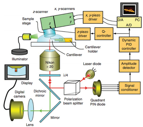
Toshio Ando and co-workers at Kanazawa University have developed and used high-speed atomic force microscopy (HS-AFM) to achieve direct visualization of dynamic structural changes and processes of functioning biological molecules in physiological solution — creating microscopic movies of unprecedented sub-100-ms temporal resolution and submolecular spatial resolution. To produce an image, HS-AFM acquires information on sample height at many points by tapping the sample with the sharp tip of a tiny cantilever and dragging the sharp tip of a tiny cantilever across the sample. Depending on the application, this might involve recording the distance of deflection, the amplitude and phase of oscillations, or the resonant frequency of the cantilever. Ando and co-workers use very small cantilevers that provide 10 to 20 times the sensitivity of larger, conventional cantilevers. Copies of their home-made apparatus are now commercially available through the manufacturer Research Institute of Biomolecule Metrology Co., Ltd in Tsukuba, and record images at least ten times more quickly than their competitors.
|
|
Scooped by
Dr. Stefan Gruenwald
November 30, 2012 11:05 AM
|
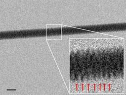
It's the most famous corkscrew in history. Now an electron microscope has captured the famous Watson-Crick double helix in all its glory, by imaging threads of DNA resting on a silicon bed of nails. The technique will let researchers see how proteins, RNA and other biomolecules interact with DNA. The structure of DNA was originally discovered using X-ray crystallography. This involves X-rays scattering off atoms in crystallised arrays of DNA to form a complex pattern of dots on photographic film. Interpreting the images requires complex mathematics to figure out what crystal structure could give rise to the observed patterns. The new images are much more obvious, as they are a direct picture of the DNA strands, albeit seen with electrons rather than X-ray photons. The trick used by Enzo di Fabrizio at the University of Genoa, Italy, and his team was to snag DNA threads out of a dilute solution and lay them on a bed of nanoscopic silicon pillars. The team developed a pattern of pillars that is extremely water-repellent, causing the moisture to evaporate quickly and leave behind strands of DNA stretched out and ready to view. The team also drilled tiny holes in the base of the nanopillar bed, through which they shone beams of electrons to make their high-resolution images. The results reveal the corkscrew thread of the DNA double helix, clearly visible. With this technique, researchers should be able to see how single molecules of DNA interact with other biomolecules.
|
|
Scooped by
Dr. Stefan Gruenwald
October 8, 2012 5:32 PM
|
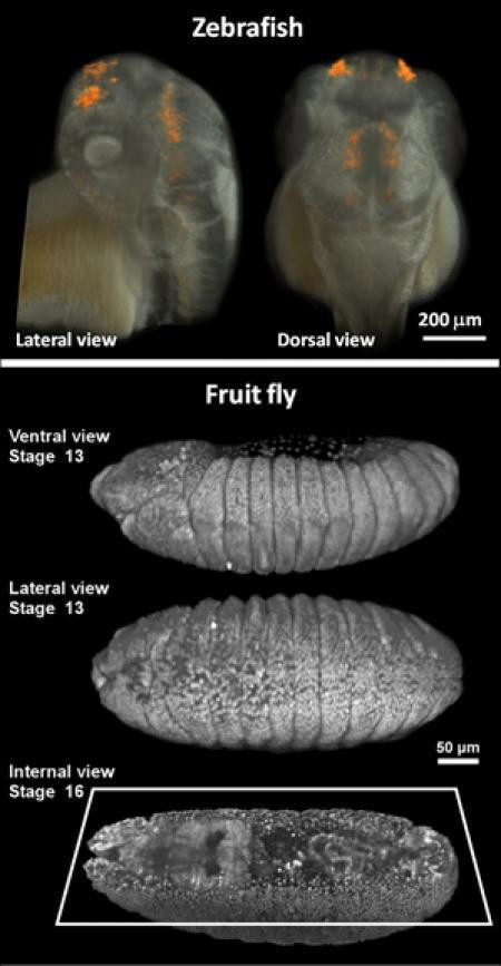
For modern biologists, the ability to capture high-quality, three-dimensional (3D) images of living tissues or organisms over time is necessary to answer problems in areas ranging from genomics to neurobiology and developmental biology. The better the image, the more detailed the information that can be drawn from it. Looking to improve upon current methods of imaging, researchers from the California Institute of Technology (Caltech) have developed a novel approach that could redefine optical imaging of live biological samples by simultaneously achieving high resolution, high penetration depth (for seeing deep inside 3D samples), and high imaging speed.
|
|
Scooped by
Dr. Stefan Gruenwald
September 21, 2012 11:33 PM
|
The growing class of fluorescent proteins useful for detecting events in living cells and animals has almost single-handedly launched and fueled a new era in biology and medicine. It took over thirty years, and the advent of recombinant DNA as well as vastly improved molecular biological approaches to see the pioneering work of Osamu Shimomura developed into a useful tool for live-cell imaging by Doug Prasher and Martin Chalfie. Just in the past decade, however, we have witnessed a truly remarkable expansion in the fluorescent protein palette, largely driven by the innovative studies from Roger Tsien's laboratory. Most of the fluorescent proteins that are commonly used today have been modified through mutagenesis to optimize their expression in biological systems. Continued efforts using directed evolution approaches will no doubt improve the spectral characteristics, photostability, maturation time, brightness, acid resistance, and utility of the fluorescent protein tags for cellular imaging. The current thrust of fluorescent protein development strategies is centered on fine-tuning the current palette of blue to yellow variants from jellyfish, while simultaneously developing monomeric fluorescent proteins emitting in the orange to far-red regions of the visible light spectrum. We now have jellyfish proteins that span an 80-nanometer portion visible spectrum from deep blue to yellow-green, providing a wide choice of genetically encoded markers for studies in cell biology. Fluorescent proteins derived from Anthozoa species (corals and anemones), as well as other sources, span the entire visible spectrum and feature a wide range of useful properties. The unique optical highlighter properties of fluorescent proteins can allow the investigator to change the color or the emission state, providing unique opportunities to track the dynamic behavior of proteins in living cells and animals. Still the "gold standard" in fluorescent protein technology, the enhanced version of GFP features a chromophore based on a para-hydroxybenzylidene substituted imidazolinone. The chromophore of the first reported red fluorescent protein extends conjugation into the polypeptide backbone to generate fluorescence in the longer wavelength regions. The ZsYellow fluorescent protein chromophore features a novel three-ring system and peptide backbone cleavage due to the substitution of lysine for serine as the first amino acid residue in the chromophore tripeptide. The final step in mKO chromophore maturations involves the formation of a novel five-member thiazole ring system when the Cys65 hydroxyl moiety attacks the carbonyl of Phe64 and cyclizes. In a manner similar to mKusabira Orange, mOrange chromophore maturation involves the formation of a novel five-member oxazole (rather than a thiazole) ring system.
http://nic.ucsf.edu/FPvisualization/
|
|
Scooped by
Dr. Stefan Gruenwald
August 29, 2012 7:33 PM
|
Methodology designed to circumnavigate the classical Abbe diffraction barrier in optical microscopy is rapidly advancing using both ensemble and single-molecule techniques. Over the past several decades, fluorescence microscopy has become an essential tool for examining a wide variety of biological molecules, pathways, and dynamics in living cells, tissues, and whole animals. In contrast to other techniques (such as electron microscopy), fluorescence imaging is compatible with cells that are being maintained in culture, which enables minimally invasive optical-based observation of events occurring on a large span of timescales. In terms of spatial resolution, several techniques including positron-emission tomography, magnetic resonance imaging, and optical coherence tomography can generate images of animal and human subjects at resolutions between 10 centimeters and 10 micrometers, whereas electron microscopy and scanning probe techniques feature the highest spatial resolution, often approaching the molecular and atomic levels (see Figure ). Between these two extremes in resolving power lies optical microscopy. Aside from the benefits derived from being able to image living cells, the most significant drawback to all forms of fluorescence microscopy (including widefield, laser scanning, spinning disk, multiphoton, and total internal reflection) are the limits to spatial resolution that were first elucidated and described by Ernst Abbe in the late 1800s. The Abbe diffraction limit (or at least the recognition of this limit) stood for almost a century before inventive microscopists began to examine how their instruments could be improved to circumvent the physical barriers in order to achieve higher resolution. Due to the fact that axial resolution is far lower than lateral resolution (by at least a factor of two), much of the work conducted in the latter part of the twentieth century addressed improvements to performance in the axial dimension. Researchers discovered that laser scanning confocal instruments produced very modest increases in resolution at the cost of signal-to-noise, and that other associated technologies (including multiphoton, structured illumination, and spinning disk) could be used for optical sectioning, but without significant improvement in axial resolution. An important concept to note, and one of the most underappreciated facts associated with optical imaging in biology, is that the achieved microscope resolution often does not reach the physical limit imposed by diffraction. This is due to the fact that optical inhomogeneities in the specimen can distort the phase of the excitation beam, leading to a focal volume that is significantly larger than the diffraction-limited ideal. Furthermore, resolution can also be compromised by improper alignment of the microscope, the use of incompatible immersion oil, coverslips having a thickness outside the optimum range, and improperly adjusted correction collars. The most significant advances in superresolution imaging have been achieved in what is termed far-field microscopy and involve either spatially or temporally modulating the transition between two molecular states of a fluorophore (such as switching between a dark and bright state) or by physically reducing the size of the point-spread function used in the excitation illumination. Among the methods that improve resolution by PSF modification, the most important techniques are referred to by the acronyms STED (stimulated emission depletion; from the Stefan Hell laboratory) and SSIM (saturated structured illumination microscopy; pioneered by Mats Gustafsson). Techniques that rely on the detection and precise localization of single molecules include PALM (photoactivated localization microscopy; introduced by Eric Betzig and Harald Hess) and STORM (stochastic optical reconstruction microscopy; first reported by Xiaowei Zhang). As will be discussed, there are many variations on these techniques, as well as advanced methods that can combine or even improve the performance of existing imaging schemes. Even more importantly, new superresolution techniques are being introduced with almost breathtaking speed (relative to traditional advances in microscopy) and it is not unreasonable to suggest that at some point in the near future, resolution of a single nanometer may well be attainable in commercial instruments.
|
|
Scooped by
Dr. Stefan Gruenwald
August 8, 2012 10:36 AM
|
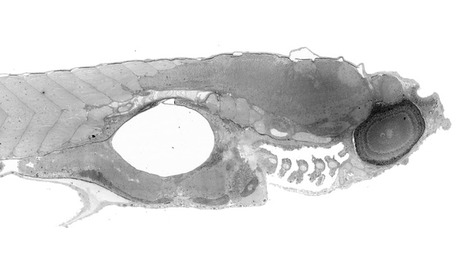
This might just look like a microscope image of some strange, small life-form. But actually its a view of a massive 281-gigapixel image of a zebrafish embryo, which can be zoomed in on to show sub-cellular levels of detail. The image is the product of a new technique called virtual nanoscopy. The process involves stitching together nanometer resolution photographs of what's placed under the microscope, and the result is an image which can be explored a little like a Google Map.
|
|
Scooped by
Dr. Stefan Gruenwald
July 31, 2012 2:06 PM
|
An automated underwater microscope developed by scientists at Woods Hole Oceanographic Institution (WHOI) detected an unexpected bloom of toxic algae in the Gulf of Mexico in February 2008. The fortunate early warning prompted officials to recall shellfish and close down shellfish harvesting, just days before a major regional oyster festival. The instrument, called the Imaging FlowCytobot, was originally designed as a basic research tool to reveal the ebb and flow of a diverse range of microscopic plant and animal life in the ocean, said its developers, Rob Olson and Heidi Sosik. It sits underwater—photographing and counting plankton 24 hours a day for months at a time, and relaying information back to shore via fiber-optic cable.
|
|
Scooped by
Dr. Stefan Gruenwald
April 19, 2012 11:56 AM
|
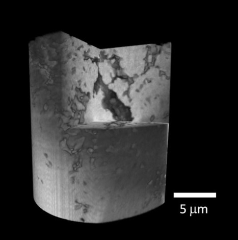
A new x-ray microscope probes the inner intricacies of materials smaller than human cells and creates unparalleled high-resolution 3D images at 25 nm. By integrating unique automatic calibrations, scientists at the U.S. Department of Energy’s Brookhaven National Laboratory are able to capture and combine thousands of images with greater speed and precision than any other microscope. The direct observation of structures spanning 25 nanometers will offer fundamental advances in many fields, including energy research, environmental sciences, biology, and national defense. This innovative full field transmission x-ray microscope (TXM) was developed at Brookhaven Lab’s National Synchrotron Light Source (NSLS), which provides the x-ray source needed to capture images on the nanoscale.
|
|
|
Scooped by
Dr. Stefan Gruenwald
January 11, 2013 1:25 PM
|
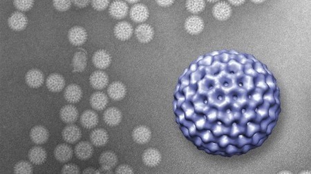
The detailed changes in the structure of a virus as it infects an E. coli bacterium have been observed for the first time. To infect a cell, a virus must be able to first find a suitable cell and then eject its genetic material into its host. This robot-like process has been observed in a virus called T7 and visualized by Ian Molineux, professor of biology in the College of Natural Sciences at The University of Texas at Austin, and colleagues at The University of Texas Health Science Center at Houston (UT Health) Medical School. When searching for its prey, the virus briefly extends — like feelers — one or two of six ultra-thin fibers it normally keeps folded at the base of its head. Once a suitable host has been located, the virus behaves a bit like a planetary rover, extending these fibers to walk randomly across the surface of the cell and find an optimal site for infection — the first experimental evidence for this. At the preferred infection site, the virus goes through a major change in structure in which it ejects some of its proteins through the bacterium’s cell membrane, creating a path for the virus’s genetic material to enter the host. After the viral DNA has been ejected, the protein path collapses and the infected cell membrane reseals. “Although many of these details are specific to T7, the overall process completely changes our understanding of how a virus infects a cell,”.said Molineux. This is also the first time that scientists have made actual images showing how the virus’s tail extends into the host — the very action that allows it to infect a cell with its DNA.
|
|
Rescooped by
Dr. Stefan Gruenwald
from Longevity science
January 3, 2013 11:58 AM
|
Scientists have developed a new technology that allows them to view live viruses in their natural habitat, as opposed to isolated frozen specimens.
Via Ray and Terry's
|
|
Scooped by
Dr. Stefan Gruenwald
November 7, 2012 12:07 PM
|
Traditional microscopy is a powerful tool for imaging small objects, such as cells. However, anybody who has used one will know its limitations: a tiny field of view and a shallow depth of field. That makes it hard to get a sense of the three dimensional shape of objects like cells. One clever way round this is to make a hologram of the sample. The idea here is to split a laser beam in two, use one as a reference beam and bounce the other off the sample to record the pattern of phase shifts that this produces using a digital camera. Recombining the beams produces an interference pattern that can be analysed to retrieve three-dimensional information about the sample in high resolution. This method is clearly powerful. A traditional microscope records only variations in the intensity of light bouncing off a sample. The holographic technique records this and phase information and so obviously stores more information about the subject. With the appropriate image-processing software. it’s possible to change the depth of focus, correct optical aberrations and reconstruct the 3D shape of the sample. There's another advantage too. Digital holographic microscopy has the potential to be incredibly cheap. Various research groups have created devices for as little as $1000 or so. Atsushi Shiraki at the Kisarazu National College of Technology in Japan, and a few pals, show how to cut the cost even further. These guys have built a digital holographic microscope using a web camera, a small solid state laser, an optical pinhole and free open source software. The total cost: just $250 (with the optical pinhole accounting for about half of this!). The entire machine is light and portable, fitting in the palm of the hand (details in the paper below). That's a handy machine that could play a useful role both in field studies for professional researchers and in schools where value for money is a key factor in equipment purchases. It might also be a fun to put together at home. Ref: arxiv.org/abs/1211.0336: Handheld and Low-Cost Digital Holographic Microscopy
|
|
Scooped by
Dr. Stefan Gruenwald
September 26, 2012 1:50 PM
|
Most plankton organisms are too small to be seen with the naked eye. But despite their size, they are vital in marine and freshwater ecosystems, serving as food for larger animals and as oxygen producers in the ocean’s carbon cycle. Microscopy has made it possible to photograph these tiny creatures in the lab, but capturing their behavior in their natural environment has been impossible—until now. Researchers in San Diego are developing the world’s first underwater video microscope, capable of imaging these miniscule organisms in 3D. When plankton are swept into the submersible microscope’s collection chamber, multiple cameras reveal the minute life-forms floating inside. The microscope has been engineered to capture detailed 3D images of plankton moving freely in the salt water chamber. These never-before-seen views of living plankton are a missing piece of the puzzle for scientists who study life in water. Observing activity on a microscopic scale will inform the bigger picture of interactions among creatures of all sizes in oceans, lakes, and rivers around the world.
|
|
Scooped by
Dr. Stefan Gruenwald
September 16, 2012 1:53 PM
|
Use the mouse and scroll down the image by dragging down the grey bar on the right side.

|
Suggested by
Rob Dawson
August 29, 2012 6:23 PM
|
A technology used in projectors can create a high-resolution image 100 times faster than conventional microscopy equipment, which can be too slow to clearly document speedy biological processes.
|
|
Scooped by
Dr. Stefan Gruenwald
August 6, 2012 7:35 PM
|
By blending optical and atomic force microscope technologies, Iowa State University and Ames Laboratory researchers have found a way to complete 3-D measurements of single biological molecules with unprecedented accuracy and precision. Existing technologies allow researchers to measure single molecules on the x and y axes of a 2-D plane. The new technology allows researchers to make height measurements (the z axis) down to the nanometer without custom optics or special surfaces for the samples. Here's how the technology works: Researchers attach a commercial atomic force microscope to a single molecule fluorescence microscope. The tip of the atomic force microscope is positioned over a focused laser beam, creating a standing wave pattern. A molecule that has been treated to emit light is placed within the standing wave. As the tip of the atomic force microscope moves up and down, the fluorescence emitted by the molecule fluctuates in a way that corresponds to its distance from the surface. That distance can be compared to a marker on the surface and measured.
|
|
Rescooped by
Dr. Stefan Gruenwald
from Plant Cell Biology
May 22, 2012 4:45 PM
|
Blood tests convey vital medical information, but the sight of a needle often causes anxiety and results take time. A new device developed by a team of researchers in Israel, however, can reveal much the same information as a traditional blood test in real-time, simply by shining a light through the skin. This optical instrument, no bigger than a breadbox, is able to provide high-resolution images of blood coursing through our veins without the need for harsh and short-lived fluorescent dyes. http://tinyurl.com/bkdn79w
Via Anne Osterrieder
|
|
Rescooped by
Dr. Stefan Gruenwald
from Science News
April 8, 2012 11:01 AM
|
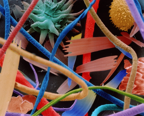
You come into contact with millions of different objects every day. Many videos on scanning and transmission electron microscopy and how to prepare the samples: http://tinyurl.com/chvml2h
Via Sakis Koukouvis
|