Your new post is loading...

|
Scooped by
Dr. Stefan Gruenwald
September 6, 2021 3:23 AM
|
Scientists have long wondered why almost all animals sleep, despite the disadvantages to survival of being unconscious. Now, researchers led by a team from the University of Tsukuba have found new evidence of brain refreshing that takes place during a specific phase of sleep: rapid eye movement (REM) sleep, which is when you tend to dream a lot. Previous studies have measured differences in blood flow in the brain between REM sleep, non-REM sleep, and wakefulness using various methods, with conflicting results. In their latest work, the Tsukuba-led team used a technique to directly visualize the movement of red blood cells in the brain capillaries (where nutrients and waste products are exchanged between brain cells and blood) of mice during awake and asleep states. "We used a dye to make the brain blood vessels visible under fluorescent light, using a technique known as two-photon microscopy," says senior author of the study Professor Yu Hayashi. "In this way, we could directly observe the red blood cells in capillaries of the neocortex in non-anesthetized mice." The researchers also measured electrical activity in the brain to identify REM sleep, non-REM sleep, and wakefulness, and looked for differences in blood flow between these phases. "We were surprised by the results," explains Professor Hayashi. "There was a massive flow of red blood cells through the brain capillaries during REM sleep, but no difference between non-REM sleep and the awake state, showing that REM sleep is a unique state" The research team then disrupted the mice's sleep, resulting in "rebound" REM sleep -- a stronger form of REM sleep to compensate for the earlier disruption. Blood flow in the brain was further increased during rebound REM sleep, suggesting an association between blood flow and REM sleep strength. However, when the researchers repeated the same experiments in mice without adenosine A2a receptors (the receptors whose blockade makes you feel more awake after drinking coffee), there was less of an increase in blood flow during REM sleep, even during rebound REM sleep. "These results suggest that adenosine A2a receptors may be responsible for at least some of the changes in blood flow in the brain during REM sleep," says Professor Hayashi. Given that reduced blood flow in the brain and decreased REM sleep are correlated with the development of Alzheimer's disease, which involves the buildup of waste products in the brain, it may be interesting to address whether increased blood flow in the brain capillaries during REM sleep is important for waste removal from the brain. This study lays preliminary groundwork for future investigations into the role of adenosine A2a receptors in this process, which could ultimately lead to the development of new treatments for conditions such as Alzheimer's disease.
|
|
Scooped by
Dr. Stefan Gruenwald
August 27, 2021 4:54 PM
|
Scientists at Cambridge and Leeds have successfully reversed age-related memory loss in mice and say their discovery could lead to the development of treatments to prevent memory loss in people as they age. In a study recently published in Molecular Psychiatry, the scientists show that changes in the extracellular matrix of the brain -- 'scaffolding' around nerve cells -- lead to loss of memory with aging, but that it is possible to reverse these using genetic treatments. Recent evidence has emerged of the role of perineuronal nets (PNNs) in neuroplasticity -- the ability of the brain to learn and adapt -- and to make memories. PNNs are cartilage-like structures that mostly surround inhibitory neurons in the brain. Their main function is to control the level of plasticity in the brain. They appear at around five years old in humans, and turn off the period of enhanced plasticity during which the connections in the brain are optimized. Then, plasticity is partially turned off, making the brain more efficient but less plastic. PNNs contain compounds known as chondroitin sulphates. Some of these, such as chondroitin 4-sulphate, inhibit the action of the networks, inhibiting neuroplasticity; others, such as chondroitin 6-sulphate, promote neuroplasticity. As we age, the balance of these compounds changes, and as levels of chondroitin 6-sulphate decrease, so our ability to learn and form new memories changes, leading to age-related memory decline. Researchers at the University of Cambridge and University of Leeds investigated whether manipulating the chondroitin sulphate composition of the PNNs might restore neuroplasticity and alleviate age-related memory deficits. To do this, the team looked at 20-month old mice -- considered very old -- and using a suite of tests showed that the mice exhibited deficits in their memory compared to six-month old mice. For example, one test involved seeing whether mice recognized an object. The mouse was placed at the start of a Y-shaped maze and left to explore two identical objects at the end of the two arms. After a short while, the mouse was once again placed in the maze, but this time one arm contained a new object, while the other contained a copy of the repeated object. The researchers measured the amount of the time the mouse spent exploring each object to see whether it had remembered the object from the previous task. The older mice were much less likely to remember the object. The team treated the aging mice using a 'viral vector', a virus capable of reconstituting the amount of 6-sulphate chondroitin sulphates to the PNNs and found that this completely restored memory in the older mice, to a level similar to that seen in the younger mice. Dr Jessica Kwok from the School of Biomedical Sciences at the University of Leeds said: "We saw remarkable results when we treated the aging mice with this treatment. The memory and ability to learn were restored to levels they would not have seen since they were much younger." To explore the role of chondroitin 6-sulphate in memory loss, the researchers bred mice that had been genetically-manipulated such that they were only able to produce low levels of the compound to mimic the changes of aging. Even at 11 weeks, these mice showed signs of premature memory loss. However, increasing levels of chondroitin 6-sulphate using the viral vector restored their memory and plasticity to levels similar to healthy mice. Prof James Fawcett from the John van Geest Centre for Brain Repair at the University of Cambridge said: "What is exciting about this is that although our study was only in mice, the same mechanism should operate in humans -- the molecules and structures in the human brain are the same as those in rodents. This suggests that it may be possible to prevent humans from developing memory loss in old age." The team might have already identified a potential drug, licensed for human use, that can be taken by mouth and inhibits the formation of PNNs. When this compound is given to mice and rats it can restore memory in aging and also improves recovery in spinal cord injury. The researchers are investigating whether it might help alleviate memory loss in animal models of Alzheimer's disease. The approach taken by Professor Fawcett's team -- using viral vectors to deliver the treatment -- is increasingly being used to treat human neurological conditions. A second team at the Centre recently published research showing their use for repairing damage caused by glaucoma and dementia.
|
|
Scooped by
Dr. Stefan Gruenwald
July 17, 2021 8:13 PM
|
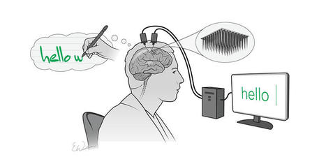
Researchers at UC San Francisco have successfully developed a “speech neuroprosthesis” that has enabled a man with severe paralysis to communicate in sentences, translating signals from his brain to the vocal tract directly into words that appear as text on a screen. The achievement, which was developed in collaboration with the first participant of a clinical research trial, builds on more than a decade of effort by UCSF neurosurgeon Edward Chang, MD, to develop a technology that allows people with paralysis to communicate even if they are unable to speak on their own. The study is published in the New England Journal of Medicine. "To our knowledge, this is the first successful demonstration of direct decoding of full words from the brain activity of someone who is paralyzed and cannot speak," said Chang, the Joan and Sanford Weill Chair of Neurological Surgery at UCSF, Jeanne Robertson Distinguished Professor, and senior author on the study. "It shows strong promise to restore communication by tapping into the brain's natural speech machinery." Each year, thousands of people lose the ability to speak due to stroke, accident, or disease. With further development, the approach described in this study could one day enable these people to fully communicate. Translating Brain Signals into Speech Previously, work in the field of communication neuroprosthetics has focused on restoring communication through spelling-based approaches to type out letters one-by-one in text. Chang's study differs from these efforts in a critical way: his team is translating signals intended to control muscles of the vocal system for speaking words, rather than signals to move the arm or hand to enable typing. Chang said this approach taps into the natural and fluid aspects of speech and promises more rapid and organic communication. "With speech, we normally communicate information at a very high rate, up to 150 or 200 words per minute," he said, noting that spelling-based approaches using typing, writing, and controlling a cursor are considerably slower and more laborious. "Going straight to words, as we're doing here, has great advantages because it's closer to how we normally speak."
|
|
Scooped by
Dr. Stefan Gruenwald
June 28, 2021 5:36 PM
|
Elon Musk's startup company Neuralink has shown off the latest version of its brain-machine interface, which a monkey uses to wirelessly play Pong with its mind. The demonstration is both another step forward for the ambitious company and a fascinating look at the current state of the technology, which, amongst other things, could offer sufferers of paralysis a way to regain control of their limbs. The last time we heard from Neuralink, it had implanted one of its computer chips into the brains of pigs to demonstrate how they can record neural activity as the animals sniff around a pen. These chips consists of arrays of electrodes that record patterns of neuron activity, which can then be decoded and converted into input commands for various devices. Previous research in this field has shown how these brain-machine interfaces can be used to control drones, prosthetic limbs and computer tablets, just by recording and relaying the user's brain activity that signals their intentions. The most impressive feats, however, have required that these brain implants be tethered to computer systems to provide the necessary bandwidth for the signals to be transmitted. The goal for Neuralink, and many researchers in the field, is to develop a completely wireless version, which would allow sufferers of paralysis far greater freedom and quality of life. A consortium of scientists working on this problem, known as BrainGate, earlier this week revealed it had developed the first wireless system to transmit neural signals at a similar bandwidth to wired systems. It uses arrays of 200 electrodes to gather the full spectrum of signals from the brain's motor cortex and pass them along to a connected wireless transmitter on the user's head.
|
|
Scooped by
Dr. Stefan Gruenwald
May 28, 2021 4:49 AM
|
Psychology researchers at UC Santa Cruz have found that playing games in virtual reality creates an effect called "time compression," where time goes by faster than you think. Grayson Mullen, who was a cognitive science undergraduate at the time, worked with Psychology Professor Nicolas Davidenko to design an experiment that tested how virtual reality's effects on a game player's sense of time differ from those of conventional monitors. The results are now published in the journal Timing & Time Perception.
|
|
Scooped by
Dr. Stefan Gruenwald
April 26, 2021 6:27 PM
|
A new study is the first to identify how human brains grow much larger, with three times as many neurons, compared with chimpanzee and gorilla brains. The study, led by researchers at the Medical Research Council (MRC) Laboratory of Molecular Biology in Cambridge, UK, identified a key molecular switch that can make ape brain organoids grow more like human organoids, and vice versa. The study, published in the journal Cell, compared 'brain organoids' -- 3D tissues grown from stem cells which model early brain development -- that were grown from human, gorilla and chimpanzee stem cells. Similar to actual brains, the human brain organoids grew a lot larger than the organoids from other apes. Dr Madeline Lancaster, from the MRC Laboratory of Molecular Biology, who led the study, said: "This provides some of the first insight into what is different about the developing human brain that sets us apart from our closest living relatives, the other great apes. The most striking difference between us and other apes is just how incredibly big our brains are." During the early stages of brain development, neurons are made by stem cells called neural progenitors. These progenitor cells initially have a cylindrical shape that makes it easy for them to split into identical daughter cells with the same shape. The more times the neural progenitor cells multiply at this stage, the more neurons there will be later. As the cells mature and slow their multiplication, they elongate, forming a shape like a stretched ice-cream cone. Previously, research in mice had shown that their neural progenitor cells mature into a conical shape and slow their multiplication within hours. Now, brain organoids have allowed researchers to uncover how this development happens in humans, gorillas and chimpanzees. They found that in gorillas and chimpanzees this transition takes a long time, occurring over approximately five days. Human progenitors were even more delayed in this transition, taking around seven days. The human progenitor cells maintained their cylinder-like shape for longer than other apes and during this time they split more frequently, producing more cells.
|
|
Scooped by
Dr. Stefan Gruenwald
January 9, 2021 2:41 PM
|
Elon Musk today announced a breakthrough in his endeavor to sync the human brain with artificial intelligence. During a live-streamed demonstration involving farm animals and a stage, Musk said that his company Neuralink had built a self-contained neural implant that can wirelessly transmit detailed brain activity without the aid of external hardware. Musk demonstrated the device with live pigs, one of which had the implant in its brain. A screen above the pig streamed the electrical brain activity being registered by the device. “It’s like a Fitbit in your skull with tiny wires,” Musk said in his presentation. “You need an electrical thing to solve an electrical problem.” Musk’s goal is to build a neural implant that can sync up the human brain with AI, enabling humans to control computers, prosthetic limbs, and other machines using only thoughts. When asked during the live Q&A whether the device would ever be used for gaming, Musk answered an emphatic “yes.” Musk’s aspirations for this brain-computer interface (BCI) system are to be able to read and write from millions of neurons in the brain, translating human thought into computer commands, and vice versa. And it would all happen on a small, wireless, battery-powered implant unseen from the outside of the body. His company has been working on the technology for about four years. Teams of researchers globally have been experimenting with surgically implanted BCI systems in humans for over 15 years. The BrainGate consortium and other groups have used BCI to enable people with neurologic diseases and paralysis to operate tablets, type eight words per minute and control prosthetic limbs using only their thoughts.
|
|
Scooped by
Dr. Stefan Gruenwald
November 25, 2020 1:46 PM
|
Even though people tend to remember fewer details about past events as time goes by, the details they do remember are retained with remarkable fidelity, according to a new study. This finding holds true regardless of the age of the person or the amount of time that elapsed since the event took place.
Scientists studying the complex relationship between aging and memory have found that in a controlled experiment, people can remember the details about past events with a surprising 94% accuracy, even accounting for age. These results, published in the journal Psychological Science, suggest that the stories we tell about past events are accurate, although details tend to fade with time.
“These results are surprising to many, given the general pessimism about memory accuracy among scientists and the prevalent idea that memory for one-time events is not to be trusted,” said Nicholas Diamond, the study’s lead researcher, a former graduate student at Baycrest’s Rotman Research Institute (RRI), and currently a postdoctoral researcher at the University of Pennsylvania.
“These results will be helpful for understanding memory in healthy aging.”Brian Levine, Baycrest’s Rotman Research Institute
About 400 academics, including memory scientists, surveyed as part of this study estimated memory accuracy to be around 40% at best, expecting this score to be even lower for older participants or when greater amounts of time had elapsed since the events.
“This study shows us that memory accuracy is actually quite good under normal circumstances, and it remains stable as we age,” said Brian Levine, a senior scientist at RRI and a professor of psychology and neurology at the University of Toronto and co-author on the study. “These results will be helpful for understanding memory in healthy aging.”
|
|
Scooped by
Dr. Stefan Gruenwald
November 12, 2020 12:53 PM
|
A "molecular volume knob" regulating electrical signals in the brain helps with learning and memory, according to a Dartmouth study. The molecular system controls the width of electrical signals that flow across synapses between neurons. The finding of the control mechanism, and the identification of the molecule that regulates it, could help researchers in their search for ways to manage neurological disorders, including Alzheimer's disease, Parkinson's disease and epilepsy. The research, published in Proceedings of the National Academy of Sciences, describes the first study of how the shapes of electrical signals contribute to the functioning of synapses. "The synapses in our brain are highly dynamic and speak in a range of whispers and shouts," said Michael Hoppa, an assistant professor of biological sciences at Dartmouth and the research lead. "This finding puts us on a straighter path toward being able to cure stubborn neurological disorders." The amount of neurotransmitter released changes the numbers and patterns of neurons activated within circuits of the brain. That reshaping of synaptic connection strength is how learning happens and how memories are formed. Two functions support these processes of memory and learning. One, known as facilitation, is a series of increasingly rapid spikes that amplifies the signals that change a synapse's shape. The other, depression, reduces the signals. Together, these two forms of plasticity keep the brain in balance and prevent neurological disorders such as seizures. "As we age, its critical to be able to maintain strengthened synapses. We need a good balance of plasticity in our brain, but also stabilization of synaptic connections," said Hoppa. The research focused on the hippocampus, the center of the brain that is responsible for learning and memory. In the study, the research team found that the electric spikes are delivered as analog signals whose shape impacts the magnitude of chemical neurotransmitter released across the synapses. This mechanism functions similar to a light dimmer with variable settings. Previous research considered the spikes to be delivered as a digital signal, more akin to a light switch that operates only in the "on" and "off" positions. "The finding that these electric spikes are analog unlocks our understanding of how the brain works to form memory and learning," said In Ha Cho, a postdoctoral fellow at Dartmouth and first author of the study. "The use of analog signals provides an easier pathway to modulate the strength of brain circuits."
|
|
Scooped by
Dr. Stefan Gruenwald
November 5, 2020 6:41 PM
|
One in 4,000 people is born without a corpus callosum, a brain structure consisting of neural fibers that are used to transfer information from one hemisphere to the other. A quarter of these individuals do not have any symptoms, while the remainder either have low intelligence quotients or suffer from severe cognitive disorders. In a study published in the journal Cerebral Cortex, neuroscientists from the University of Geneva (UNIGE) discovered that when the neuronal fibers that act as a bridge between the hemispheres are missing, the brain reorganizes itself and creates an impressive number of connections inside each hemisphere. These create more intra-hemispheric connections than in a healthy brain, indicating that plasticity mechanisms are involved. It is thought that these mechanisms enable the brain to compensate for the losses by recreating connections to other brain regions using alternative neural pathways. The corpus callosum develops in utero between the tenth and twentieth week of gestation. Agenesis of the corpus callosum is a congenital brain malformation in which this brain structure fails to develop, resulting in one out of 4,000 babies born without a corpus callosum. When it is missing, nothing replaces this structure measuring about ten centimeters, with the exception of cerebrospinal fluid. This means that the information transmitted from one hemisphere to the other can no longer be conveyed by the neuronal projections from the corpus callosum. «Their role in a healthy brain,» begins Vanessa Siffredi, a researcher in UNIGE’s Faculty of Medicine, «is to ensure the functioning of various cognitive and sensorimotor functions». Surprisingly, 25% of people with this malformation have no visible signs; 50% have average intelligence quotients and learning difficulties; and the remaining 25% suffer from severe cognitive disorders. Mysterious fibers The scientific literature shows that, in the absence of the corpus callosum, certain fibers designed to serve as a bridge between the hemispheres, known as Probst bundles, bypass the absent brain area and curl up inside each hemisphere. «The back-up zones vary from one individual to another. And we don’t understand their functions,» explains the neuroscientist. The UNIGE scientists – working in collaboration with their colleagues at the University of Melbourne – set out to understand this variability and to examine the role of the fibers. Using MRI brain imaging, they studied the anatomical and functional links between different brain regions of approximately 20 Australian children aged 8 to 17 suffering from agenesis of the corpus callosum. This approach first made it possible to observe the physical relationships between the different regions of the brain, i.e. their structural links. In children with corpus callosum agenesis, the neural fibers inside each hemisphere are greater in number and of higher quality than in healthy brains. Furthermore, the UNIGE scientists succeeded in determining the correlations between the activity of different brain regions and their functional links. «If two regions are active together, it means they are communicating with each other,» explains Dr Siffredi. The data shows that intra and inter-hemispheric functional connectivity of brains without the corpus callosum are comparable to those of healthy brains. «Remarkably, communication between the two hemispheres is maintained. We think that plasticity mechanisms, such as the strengthening of structural bonds within each hemisphere, compensated for the lack of neuronal fibers between hemispheres. New connections are created and the signals can be re-routed so that communication is preserved between the two hemispheres.»
|
|
Scooped by
Dr. Stefan Gruenwald
August 29, 2020 8:28 PM
|
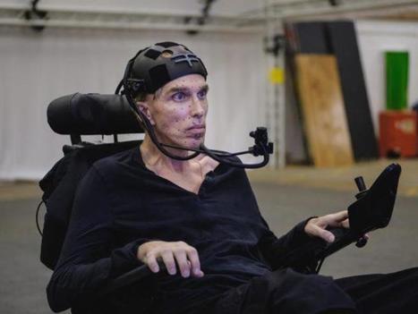
What is it like for a person to live partly inside of the objective function of an AI program? Intel scientist Lama Nachman shares insights from her team’s work with Peter Scott-Morgan, a person willing to transform his body and his life to interact intimately with a machine. Lama Nachman spent years helping the late Stephen Hawking through various upgrades of the computer technology that helped him to work and communicate. Hawking passed away in 2018. Her team at Intel Labs is now working with Peter Scott-Morgan, a roboticist who has undergone several operations to head off the incapacity that comes from ALS, the same affliction as Hawking suffered. Working with a variety of technologies, including GPT-2, OpenAI's generative deep learning model for text, Nachman and team are pushing at the boundaries of how a person can exist in a give and take relationship with AI. Part ethnographer, Nachman shows great sensitivity to the nuances of how humans encounter technology. She explained to ZDNet the differences in temperament between her two very different collaborators, first Hawking, now Scott-Morgan. Hawking was "the best validation engineer ever," says Nachman. He endured tons and tons of trial and error with new technology, and seemed to derive great satisfaction in finding bugs in the software. It was almost like man versus machine, to hear Nachman tell it, John Henry versus the pneumatic drill. Scott-Morgan, in contrast, sees himself as becoming one with the machine, both helping to train it, and at the same time learning a new mode of being from it in a symbiotic fashion. "I think of myself as partly human and partly AI," is how Scott-Morgan views it, in Nachman's telling. "I am willing to be nudged by the AI system," Scott-Morgan has told her. British television is airing a program about Scott-Morgan's transformation, and you can see the trailer here.
|
|
Scooped by
Dr. Stefan Gruenwald
July 15, 2020 10:20 PM
|
The premiere of the movie Scent of Mystery in 1960 marked a singular event in the annals of cinema: the first, and last, motion picture debut "in glorious Smell-O-Vision." Hoping to wow moviegoers with a dynamic olfactory experience alongside the familiar spectacles of sight and sound, select theaters were outfitted with a Rube Goldberg-esque device that piped different scents directly to seats. Audiences and critics quickly concluded that the experience stunk. Fraught with technical issues, Smell-O-Vision was panned and became a running gag that holds a unique place in entertainment history. The flop of Smell-O-Vision, however, failed to deter entrepreneurs from continuing to chase the dream of delivering smells to consumers, particularly in recent years, through digital scent technologies. Such efforts have generated news headlines but scant success, due in part to a limited understanding of how the brain translates odor chemistry into perceptions of smell -- a phenomenon that in many ways remains opaque to scientists. A study by neurobiologists at Harvard Medical School now provides new insights into the mystery of scent. Reporting in Nature on July 1, the researchers describe for the first time how relationships between different odors are encoded in the olfactory cortex, the region of brain responsible for processing smell. By delivering odors with carefully selected molecular structures and analyzing neural activity in awake mice, the team showed that neuronal representations of smell in the cortex reflect chemical similarities between odors, thus enabling scents to be placed into categories by the brain. Moreover, these representations can be rewired by sensory experiences. The findings suggest a neurobiological mechanism that may explain why individuals have common but highly personalized experiences with smell. "All of us share a common frame of reference with smells. You and I both think lemon and lime smell similar and agree that they smell different from pizza, but until now, we didn't know how the brain organizes that kind of information," said senior study author Sandeep Robert Datta, associate professor of neurobiology in the Blavatnik Institute at HMS. The results open new avenues of study to better understand how the brain transforms information about odor chemistry into the perception of smell. "This is the first demonstration of how the olfactory cortex encodes information about the very thing that it's responsible for, which is odor chemistry, the fundamental sensory cues of olfaction," Datta said.
|
|
Scooped by
Dr. Stefan Gruenwald
June 30, 2020 2:47 PM
|
Primates’ neural computations shed new light on the evolution of language. Humans and monkeys may not speak the same lingo, but our ways of thinking are a lot more similar than previously thought, according to new research from UC Berkeley, Harvard University and Carnegie Mellon University. In experiments on 100 study participants across age groups, cultures and species, researchers found that indigenous Tsimane’ people in Bolivia’s Amazon rainforest, American adults and preschoolers and macaque monkeys all show, to varying degrees, a knack for “recursion,” a cognitive process of arranging words, phrases or symbols in a way that helps convey complex commands, sentiments and ideas. The findings, published in the journal Science Advances, shed new light on our understanding of the evolution of language, researchers said. “For the first time, we have strong empirical evidence about patterns of thinking that come naturally to probably all humans and, to a lesser extent, non-human primates,” said study co-author Steven Piantadosi, a UC Berkeley assistant professor of psychology. Indeed, the monkeys were found to perform far better in the tests than the researchers had predicted. “Our data suggest that, with sufficient training, monkeys can learn to represent a recursive process, meaning that this ability may not be as unique to humans as is commonly thought,” said Sam Cheyette, a Ph.D. student in Piantadosi’s lab and co-author of the study.
|
|
|
Scooped by
Dr. Stefan Gruenwald
September 6, 2021 3:18 AM
|
In a discovery published in the journal Nature, an international team of researchers has described a novel molecular device with exceptional computing prowess. Reminiscent of the plasticity of connections in the human brain, the device can be reconfigured on the fly for different computational tasks by simply changing applied voltages. Furthermore, like nerve cells can store memories, the same device can also retain information for future retrieval and processing. "The brain has the remarkable ability to change its wiring around by making and breaking connections between nerve cells. Achieving something comparable in a physical system has been extremely challenging," said Dr. R. Stanley Williams, professor in the Department of Electrical and Computer Engineering at Texas A&M University. "We have now created a molecular device with dramatic reconfigurability, which is achieved not by changing physical connections like in the brain, but by reprogramming its logic." Dr. T. Venkatesan, director of the Center for Quantum Research and Technology (CQRT) at the University of Oklahoma, Scientific Affiliate at National Institute of Standards and Technology, Gaithersburg, and adjunct professor of electrical and computer engineering at the National University of Singapore, added that their molecular device might in the future help design next-generation processing chips with enhanced computational power and speed, but consuming significantly reduced energy. Whether it is the familiar laptop or a sophisticated supercomputer, digital technologies face a common nemesis, the von Neumann bottleneck. This delay in computational processing is a consequence of current computer architectures, wherein the memory, containing data and programs, is physically separated from the processor. As a result, computers spend a significant amount of time shuttling information between the two systems, causing the bottleneck. Also, despite extremely fast processor speeds, these units can be idling for extended amounts of time during periods of information exchange. As an alternative to conventional electronic parts used for designing memory units and processors, devices called memristors offer a way to circumvent the von Neumann bottleneck. Memristors, such as those made of niobium dioxide and vanadium dioxide, transition from being an insulator to a conductor at a set temperature. This property gives these types of memristors the ability to perform computations and store data. However, despite their many advantages, these metal oxide memristors are made of rare-earth elements and can operate only in restrictive temperature regimes. Hence, there has been an ongoing search for promising organic molecules that can perform a comparable memristive function, said Williams. Dr. Sreebrata Goswami, a professor at the Indian Association for the Cultivation of Science, designed the material used in this work. The compound has a central metal atom (iron) bound to three phenyl azo pyridine organic molecules called ligands. "This behaves like an electron sponge that can absorb as many as six electrons reversibly, resulting in seven different redox states," said Sreebrata. "The interconnectivity between these states is the key behind the reconfigurability shown in this work."
|
|
Scooped by
Dr. Stefan Gruenwald
August 27, 2021 4:47 PM
|
A new kind of neural interface system that coordinates the activity of hundreds of tiny brain sensors could one day deepen understanding of the brain and lead to new medical therapies. Brain-computer interfaces (BCIs) are emerging assistive devices that may one day help people with brain or spinal injuries to move or communicate. BCI systems depend on implantable sensors that record electrical signals in the brain and use those signals to drive external devices like computers or robotic prosthetics. Most current BCI systems use one or two sensors to sample up to a few hundred neurons, but neuroscientists are interested in systems that are able to gather data from much larger groups of brain cells. Now, a team of researchers has taken a key step toward a new concept for a future BCI system -- one that employs a coordinated network of independent, wireless microscale neural sensors, each about the size of a grain of salt, to record and stimulate brain activity. The sensors, dubbed "neurograins," independently record the electrical pulses made by firing neurons and send the signals wirelessly to a central hub, which coordinates and processes the signals. In a study published on August 12, 2021 in Nature Electronics, the research team demonstrated the use of nearly 50 such autonomous neurograins to record neural activity in a rodent. The results, the researchers say, are a step toward a system that could one day enable the recording of brain signals in unprecedented detail, leading to new insights into how the brain works and new therapies for people with brain or spinal injuries. "One of the big challenges in the field of brain-computer interfaces is engineering ways of probing as many points in the brain as possible," said Arto Nurmikko, a professor in Brown's School of Engineering and the study's senior author. "Up to now, most BCIs have been monolithic devices -- a bit like little beds of needles. Our team's idea was to break up that monolith into tiny sensors that could be distributed across the cerebral cortex. That's what we've been able to demonstrate here." The team, which includes experts from Brown, Baylor University, University of California at San Diego and Qualcomm, began the work of developing the system about four years ago. The challenge was two-fold, said Nurmikko, who is affiliated with Brown's Carney Institute for Brain Science. The first part required shrinking the complex electronics involved in detecting, amplifying and transmitting neural signals into the tiny silicon neurograin chips. The team first designed and simulated the electronics on a computer, and went through several fabrication iterations to develop operational chips. The second challenge was developing the body-external communications hub that receives signals from those tiny chips. The device is a thin patch, about the size of a thumb print, that attaches to the scalp outside the skull. It works like a miniature cellular phone tower, employing a network protocol to coordinate the signals from the neurograins, each of which has its own network address. The patch also supplies power wirelessly to the neurograins, which are designed to operate using a minimal amount of electricity. "This work was a true multidisciplinary challenge," said Jihun Lee, a postdoctoral researcher at Brown and the study's lead author. "We had to bring together expertise in electromagnetics, radio frequency communication, circuit design, fabrication and neuroscience to design and operate the neurograin system."
|
|
Scooped by
Dr. Stefan Gruenwald
July 2, 2021 6:29 PM
|
Case Western Reserve University researchers studying prions—misfolded proteins that cause lethal incurable diseases—have identified for the first time surface features of human prions responsible for their replication in the brain. The ultimate goal of the research is to help design a strategy to stop prion disease in humans—and, ultimately, to translate new approaches to work on Alzheimer’s and other neurodegenerative diseases. Scientists have yet to discover the exact cause of Alzheimer’s disease, but largely agree that protein issues play a role in its emergence and progression. Alzheimer’s disease afflicts more than 6 million people in the U.S., and the Alzheimer’s Association estimates that their care will cost an estimated $355 billion this year. Research was done at the Safar Laboratory in the Department of Pathology and the Center for Proteomics and Bioinformatics at Case Western Reserve University School of Medicine, and at Case Western Reserve’s Center for Synchrotron Bioscience at Brookhaven Laboratories in New York. Jiri Safar, professor of pathology, neurology and neurosciences at the Case Western Reserve School of Medicine, leads the work. The report, “Structurally distinct external domains drive replication of major human prions,” was published in the June 17 issue of PLOS Pathogens. Prions were first discovered in the late 1980s as a protein-containing biological agent that could replicate itself in living cells without nucleic acid. The public health impact of medically transmitted human prion diseases—and also animal transmissions of bovine spongiform encephalopathy (BSE, “mad cow disease”) prions—dramatically accelerated the development of a new scientific concept of self-replicating protein. Human prions can bind to neighboring normal proteins in the brain, and cause microscopic holes. In essence, they turn brains into sponge-like structures and lead to dementia and death. These discoveries led to the ongoing scientific debate on whether prion-like mechanisms may be involved in the origin and spread of other neurodegenerative disorders in humans. “Human prion diseases are conceivably the most heterogenous neurodegenerative disorders, and a growing body of research indicates that they are caused by distinct strains of human prions,” Safar said. “However, the structural studies of human prions have lagged behind the recent progress in rodent laboratory prions, in part because of their complex molecular characteristics and prohibitive biosafety requirements necessary for investigating disease which is invariably fatal and has no treatment.” The researchers developed a new three-step process to study human prions: - Human brain-derived prions were first exposed to a high-intensity synchrotron X-ray beam. That beam created hydroxyl radical species which, with short bursts of light, selectively and progressively changed the prion’s surface chemical composition. The unique properties of this type of light source include its enormous intensity; it can be millions of times brighter than light from the sun to the Earth.
- The rapid chemical modifications of prions by short bursts of light were monitored with anti-prion antibodies. The antibodies recognize the prion surface features, and mass spectrometry that identifies exact sites of prion-specific, strain-based differences, providing an even more precise description of the prion’s defects.
- Illuminated prions were then allowed to replicate in a test tube. The progressive loss of their replication activity as the synchrotron modifies them helped identify key structural elements responsible for prions’ replication and propagation in the brain.
“This work is a critical first step for identifying sites of structural importance that reflect differences between prions of different diagnosis and aggressiveness,” said Mark Chance, vice dean for research at the School of Medicine and a co-investigator on the work. “Thus, we can now envision designing small molecules to bind to these sites of nucleation and replication and block progression of human prion disease in patients.”
|
|
Scooped by
Dr. Stefan Gruenwald
June 17, 2021 5:57 PM
|
It is the first work to show that sonothermogenetics can control behavior by stimulating a specific target deep in the brain. Neurological disorders such as Parkinson's disease and epilepsy have had some treatment success with deep brain stimulation, but those require surgical device implantation. A multidisciplinary team at Washington University in St. Louis has developed a new brain stimulation technique using focused ultrasound that is able to turn specific types of neurons in the brain on and off and precisely control motor activity without surgical device implantation. The team, led by Hong Chen, assistant professor of biomedical engineering in the McKelvey School of Engineering and of radiation oncology at the School of Medicine, is the first to provide direct evidence showing noninvasive, cell-type-specific activation of neurons in the brain of mammal by combining ultrasound-induced heating effect and genetics, which they have named sonothermogenetics. It is also the first work to show that the ultrasound- genetics combination can robustly control behavior by stimulating a specific target deep in the brain. Results of the three years of research, which was funded in part by the National Institutes of Health's BRAIN Initiative, were published online in Brain Stimulation May 11, 2021. The senior research team included experts from both the McKelvey School of Engineering and the School of Medicine, including Jianmin Cui, professor of biomedical engineering; Joseph P. Culver, professor of radiology, of physics and of biomedical engineering; Mark J. Miller, associate professor of medicine in the Division of Infectious Diseases in the Department of Medicine; and Michael Bruchas, formerly of Washington University, now professor of anesthesiology and pharmacology at the University of Washington. "Our work provided evidence that sonothermogenetics evokes behavioral responses in freely moving mice while targeting a deep brain site," Chen said. "Sonothermogenetics has the potential to transform our approaches for neuroscience research and uncover new methods to understand and treat human brain disorders."
|
|
Scooped by
Dr. Stefan Gruenwald
May 14, 2021 1:43 PM
|
Howard Hughes Medical Institute (HHMI) Investigator Krishna Shenoy and his team have decoded the neural signals associated with writing letters, then displayed typed versions of these letters in real time, an invention that could one day help people with paralysis communicate. Scientists are exploring a number of ways for people with disabilities to communicate with their thoughts. The newest and fastest turns back to a vintage means for expressing oneself: handwriting. For the first time, researchers have deciphered the brain activity associated with trying to write letters by hand. Working with a participant with paralysis who has sensors implanted in his brain, the team used an algorithm to identify letters as he attempted to write them. Then, the system displayed the text on a screen – in real time. This innovation could, with further development, let people with paralysis rapidly type without using their hands, says study coauthor Krishna Shenoy, a Howard Hughes Medical Institute Investigator at Stanford University who jointly supervised the work with Jaimie Henderson, a Stanford neurosurgeon. By attempting handwriting, the study participant typed 90 characters per minute – more than double the previous record for typing with such a “brain-computer interface,” Shenoy and his colleagues report in the journal Nature on May 12, 2021. This technology and others like it have the potential to help people with all sorts of disabilities, says Jose Carmena, a neural engineer at the University of California, Berkeley, who was not involved in the study. Though the findings are preliminary, he says, “it’s a big advancement in the field.” Brain-computer interfaces convert thought into action, Carmena says. “This paper is a perfect example: the interface decodes the thought of writing and produces the action.”
|
|
Scooped by
Dr. Stefan Gruenwald
March 6, 2021 3:07 PM
|
Researchers have succeeded in making an AI understand our subjective notions of what makes faces attractive. The device demonstrated this knowledge by its ability to create new portraits on its own that were tailored to be found personally attractive to individuals. The results can be utilized, for example, in modelling preferences and decision-making as well as potentially identifying unconscious attitudes. "In our previous studies, we designed models that could identify and control simple portrait features, such as hair colour and emotion. However, people largely agree on who is blond and who smiles. Attractiveness is a more challenging subject of study, as it is associated with cultural and psychological factors that likely play unconscious roles in our individual preferences. Indeed, we often find it very hard to explain what it is exactly that makes something, or someone, beautiful: Beauty is in the eye of the beholder," says Senior Researcher and Docent Michiel Spapé from the Department of Psychology and Logopedics, University of Helsinki. The study, which combines computer science and psychology, was published in February in the IEEE Transactions in Affective Computing journal. Preferences exposed by the brain Initially, the researchers gave a generative adversarial neural network (GAN) the task of creating hundreds of artificial portraits. The images were shown, one at a time, to 30 volunteers who were asked to pay attention to faces they found attractive while their brain responses were recorded via electroencephalography (EEG). "It worked a bit like the dating app Tinder: the participants 'swiped right' when coming across an attractive face. Here, however, they did not have to do anything but look at the images. We measured their immediate brain response to the images," Spapé explains. The researchers analysed the EEG data with machine learning techniques, connecting individual EEG data through a brain-computer interface to a generative neural network. "A brain-computer interface such as this is able to interpret users' opinions on the attractiveness of a range of images. By interpreting their views, the AI model interpreting brain responses and the generative neural network modelling the face images can together produce an entirely new face image by combining what a particular person finds attractive," says Academy Research Fellow and Associate Professor Tuukka Ruotsalo, who heads the project. Reference: Michiel Spape, Keith Davis, Lauri Kangassalo, Niklas Ravaja, Zania Sovijarvi-Spape, Tuukka Ruotsalo. Brain-computer interface for generating personally attractive images. IEEE Transactions on Affective Computing, 2021; 1 DOI: 10.1109/TAFFC.2021.3059043
|
|
Scooped by
Dr. Stefan Gruenwald
November 25, 2020 2:04 PM
|
If songbirds could appear on "The Masked Singer" reality TV competition, zebra finches would likely steal the show. That's because they can rapidly memorize the signature sounds of at least 50 different members of their flock, according to new research from the University of California, Berkeley. In recent findings published in the journal Science Advances, these boisterous, red-beaked songbirds, known as zebra finches, have been shown to pick one another out of a crowd (or flock) based on a particular peer's distinct song or contact call. Like humans who can instantly tell which friend or relative is calling by the timbre of the person's voice, zebra finches have a near-human capacity for language mapping. Moreover, they can remember each other's unique vocalizations for months and perhaps longer, the findings suggest. "The amazing auditory memory of zebra finches shows that birds' brains are highly adapted for sophisticated social communication," said study lead author Frederic Theunissen, a UC Berkeley professor of psychology, integrative biology and neuroscience. Theunissen and fellow researchers sought to gauge the scope and magnitude of zebra finches' ability to identify their feathered peers based purely on their unique sounds. As a result, they found that the birds, which mate for life, performed even better than anticipated. "For animals, the ability to recognize the source and meaning of a cohort member's call requires complex mapping skills, and this is something zebra finches have clearly mastered," Theunissen said. A pioneer in the study of bird and human auditory communication for at least two decades, Theunissen acquired a fascination and admiration for the communication skills of zebra finches through his collaboration with UC Berkeley postdoctoral fellow Julie Elie, a neuroethologist who has studied zebra finches in the forests of their native Australia. Their teamwork yielded groundbreaking findings about the communication skills of zebra finches. Zebra finches usually travel around in colonies of 50 to 100 birds, flying apart and then coming back together. Their songs are typically mating calls, while their distance or contact calls are used to identify where they are, or to locate one another. "They have what we call a 'fusion fission' society, where they split up and then come back together," Theunissen said. "They don't want to separate from the flock, and so, if one of them gets lost, they might call out 'Hey, Ted, we're right here.' Or, if one of them is sitting in a nest while the other is foraging, one might call out to ask if it's safe to return to the nest."
|
|
Scooped by
Dr. Stefan Gruenwald
November 12, 2020 7:12 PM
|
Just as our brains evolve early in our lives, AI should evolve as we increasingly apply it in real-world scenarios at scale. Mirroring The Intricacies Of The Human Brain In Early Childhood To continue to drive AI advancement in the decades to come, we need to reimagine deep learning at its core. A promising approach is to mirror how the human brain develops, particularly in early childhood. During infancy, the brain experiences synaptogenesis — an explosion of synapse formation as the brain begins to develop. In early childhood, we have the greatest number of synapses that we will have in our lifetime, with totals increasing until about two years old. Over time, our synapses begin to "train" — strengthening, weakening and evolving as the connections in our brains begin to sparsify. From this stage through our late teenage years, while learning is most prevalent, synapse usage and pruning occurs at more rapid levels. Our brain continuously removes unneeded synapses and cells, which sparsifies the brain even further. The connections themselves learn over time, and the entire structure of our brain is modified to remain lean. This is why the brain of a child has a huge amount of plasticity, while the brain of an adult is thought to lose much of its plasticity. Because of this, a child's brain can continuously reform and learn and may better recover from damage. Replicating Neurological Attributes In Deep Learning To improve and achieve real-world AI deployments, we should reinvent the training process of deep learning models to emulate the "training process" of the human brain. For deep learning, the model training stage is very similar to the initial learning stage of humans. During early stages, the model experiences a mass intake of data, which creates a significant amount of information to mine for each decision and requires significant processing time and power to determine the action or answer. But as training occurs, neural connections become stronger with each learned action and adapt to support continuous learning. As each connection becomes stronger, redundancies are created and overlapping connections can be removed. This is why continuously restructuring and sparsifying deep learning models during training time, and not after training is complete, is necessary. After the training stage, the model has lost most of its plasticity and the connections cannot adapt to take over additional responsibility, so removing connections can result in decreased accuracy. Current methods such as the one unveiled in 2020 by MIT researchers where attempts are made to make the deep learning model smaller post-training phase have reportedly seen some success. However, if you prune in the earlier stages of training when the model is most receptive to restructuring and adapting, you can drastically improve results.
|
|
Scooped by
Dr. Stefan Gruenwald
November 10, 2020 6:39 PM
|
At any given moment in time, our brain is involved in various activities. For example, when typing on a keyboard, our brain not only dictates our finger movements but also how thirsty we feel at that time. As a result, brain signals contain dynamic neural patterns that reflect a combination of these activities simultaneously.
A standing challenge has been isolating those patterns in brain signals that relate to a specific behavior, such as finger movements. Further, developing brain-machine interfaces (BMIs) that help people with neurological and mental disorders requires the translation of brain signals into a specific behavior, a problem called decoding.
This decoding also depends on our ability to isolate neural patterns related to specific behaviors. These neural patterns can be masked by patterns related to other activities and can be missed by standard algorithms.
Led by Maryam Shanechi, Assistant Professor and Viterbi Early Career Chair in Electrical and Computer Engineering at the USC Viterbi School of Engineering, researchers have developed a machine learning algorithm that resolved the above challenge. The algorithm published in Nature Neuroscience uncovered neural patterns missed by other methods and enhanced the decoding of behaviors that originated from signals in the brain. This algorithm is a significant advance in modeling and decoding of complex brain activity which could both enable new neuroscience discoveries and enhance future brain-machine interfaces.
Standard algorithms, says Shanechi, can miss some neural patterns related to a given behavior that are masked by patterns related to other functions happening simultaneously. Shanechi and her PhD student Omid Sani developed a machine learning algorithm to resolve this challenge.
Shanechi, the paper’s lead senior author says, “We have developed an algorithm that, for the first time, can dissociate the dynamic patterns in brain signals that relate to specific behaviors one is interested in. Our algorithm was also much better at decoding these behaviors from the brain signals.”
|
|
Scooped by
Dr. Stefan Gruenwald
August 31, 2020 11:21 AM
|
In ten years artificially intelligent robots will be living and working with us, according to Dr. Mark Sagar, CEO of Soul Machines, an Auckland, New Zealand-based company that develops intelligent, emotionally responsive avatars. Sagar, an AI engineer, is the inventor of a virtual nervous system that powers autonomous animated avatars like Baby X — a virtual infant that learns through experience and can “feel” emotions. “We are creating realistic adult avatars serving as virtual assistants. You can use them to plug into existing systems like IBM Watson or Cortana — putting a face on a chatbot,” said Sagar. Within a decade humans will be interacting with lifelike emotionally-responsive AI robots, very similar to the premise of the the HBO hit series Westworld, said Sagar. But before that scenario becomes a reality robotics will have to catch up to AI technology. “Robotics technology is not really at the level of control that’s required,” he said.
|
|
Scooped by
Dr. Stefan Gruenwald
August 29, 2020 8:26 PM
|
Elon Musk demonstrated a working Neuralink brain-machine interface device implanted on a pig during a live broadcast. He said the purpose of the presentation was to recruit employees that would like to help develop the system. – “We’re not trying to raise money or do anything else, but the main purpose is to convince great people to come work at Neuralink, and help us bring the product to fruition; make it affordable and reliable and such that anyone who wants one can have one,” he said. Neuralink aims to solve brain-related issues with the brain chip called ‘Link’. Musk said the device could help solve memory loss, strokes, addiction, depression, anxiety, even monitor a users’ health to warn if they are about to have a heart attack. The interface could also help return mobility to paralyzed individuals through artificial limbs. The user would be able to move prosthetics with their thoughts via the Link brain-machine interface. Ultimately, Musk's vision for Neuralink is for humans to merge with Artificial Intelligence (AI) – “Such that the future of the world is controlled by the combined will of the people of Earth … I think that that’s obviously gonna’ be the future that we want,” he stated. The Neuralink device is currently in its initial phase of development. The design has changed since it was unveiled in 2019. Before, the device would sit behind a user’s ear and it was visible, now the design looks like a small coin, that will be placed above the skull. It’s “like a Fitbit in your skull with tiny wires,” Musk said a couple of times during the presentation, adding that Neuralink aims to connect humans to the device using Bluetooth to pair with an app on a cell phone. The Link chip features 1,024 tiny electrode threads that are threaded by a surgical robot inside the brain to stimulate neurons. “For the initial device, it’s read/write in every channel with about 1024 channels, all-day battery life that recharges overnight and has quite a long-range, so you can have the range being to your phone,” Musk said. “I should say that’s kind of an important thing, because this would connect to your phone, and so the application would be on your phone, and the Link communicating, by essentially Bluetooth low energy to the device in your head.” The Link device will be capable of inductive charging (wirelessly).
|
|
Scooped by
Dr. Stefan Gruenwald
July 15, 2020 10:18 PM
|
Especially activities in the field of artificial intelligence, like teaching robots to walk or precise automatic image recognition, demand ever more powerful, yet at the same time more economical computer chips. While the optimization of conventional microelectronics is slowly reaching its physical limits, nature offers us a blueprint how information can be processed and stored quickly and efficiently: our own brain. For the very first time, scientists at TU Dresden and the Helmholtz-Zentrum Dresden-Rossendorf (HZDR) have now successfully imitated the functioning of brain neurons using semiconductor materials. They have published their research results in the journal Nature Electronics. Today, enhancing the performance of microelectronics is usually achieved by reducing component size, especially of the individual transistors on the silicon computer chips. "But that can't go on indefinitely -- we need new approaches," Larysa Baraban asserts. The physicist, who has been working at HZDR since the beginning of the year, is one of the three primary authors of the international study, which involved a total of six institutes. One approach is based on the brain, combining data processing with data storage in an artificial neuron. "Our group has extensive experience with biological and chemical electronic sensors," Baraban continues. "So, we simulated the properties of neurons using the principles of biosensors and modified a classical field-effect transistor to create an artificial neurotransistor." The advantage of such an architecture lies in the simultaneous storage and processing of information in a single component. In conventional transistor technology, they are separated, which slows processing time and hence ultimately also limits performance. Silicon wafer + polymer = chip capable of learning Modeling computers on the human brain is no new idea. Scientists made attempts to hook up nerve cells to electronics in Petri dishes decades ago. "But a wet computer chip that has to be fed all the time is of no use to anybody," says Gianaurelio Cuniberti from TU Dresden. The Professor for Materials Science and Nanotechnology is one of the three brains behind the neurotransistor alongside Ronald Tetzlaff, Professor of Fundamentals of Electrical Engineering in Dresden, and Leon Chua from the University of California at Berkeley, who had already postulated similar components in the early 1970s. Now, Cuniberti, Baraban and their team have been able to implement it: "We apply a viscous substance -- called solgel -- to a conventional silicon wafer with circuits. This polymer hardens and becomes a porous ceramic," the materials science professor explains. "Ions move between the holes. They are heavier than electrons and slower to return to their position after excitation. This delay, called hysteresis, is what causes the storage effect." As Cuniberti explains, this is a decisive factor in the functioning of the transistor. "The more an individual transistor is excited, the sooner it will open and let the current flow. This strengthens the connection. The system is learning."
|