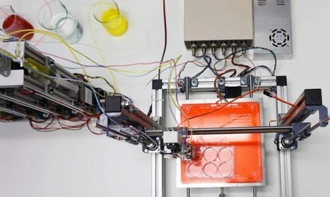
For the past several years, researchers at the University of Illinois at Urbana-Champaign have reverse-engineered native biological tissues and organs — creating tiny walking “bio-bots” powered by muscle cells and controlled with electrical and optical pulses.
Now, in an open-access cover paper in Nature Protocols, the researchers are sharing a protocol with engineering details for their current generation of millimeter-scale soft robotic bio-bots*. Using 3D-printed skeletons, these devices would be coupled to tissue-engineered skeletal muscle actuators to drive locomotion across 2D surfaces, and could one day be used for studies of muscle development and disease, high-throughput drug testing, and dynamic implants, among other applications.
In research that could one day lead to advances against neurodegenerative diseases like Alzheimer's and Parkinson's, University of Michigan engineering researchers have demonstrated a technique for precisely measuring the properties of individual protein molecules floating in a liquid.
Proteins are essential to the function of every cell. Measuring their properties in blood and other body fluids could unlock valuable information, as the molecules are a vital building block in the body. The body manufactures them in a variety of complex shapes that can transmit messages between cells, carry oxygen and perform other important functions.
Sometimes, however, proteins don't form properly. Scientists believe that some types of these misshapen proteins, called amyloids, can clump together into masses in the brain. The sticky tangles block normal cell function, leading to brain cell degeneration and disease.
But the processes of how amyloids form and clump together are not well understood. This is due in part to the fact that there's currently not a good way to study them. Researchers say current methods are expensive, time-consuming and difficult to interpret, and can only provide a broad picture of the overall level of amyloids in a patient's system.
The University of Michigan and University of Fribourg researchers who developed the new technique believe that it could help solve the problem by measuring an individual molecule's shape, volume, electrical charge, rotation speed and propensity for binding to other molecules.
They call this information a "5-D fingerprint" and believe that it could uncover new information that may one day help doctors track the status of patients with neurodegenerative diseases and possibly even develop new treatments. Their work is detailed in a paper published in Nature Nanotechnology.
"Imagine the challenge of identifying a specific person based only on their height and weight," said David Sept, a U-M biomedical engineering professor who worked on the project. "That's essentially the challenge we face with current techniques. Imagine how much easier it would be with additional descriptors like gender, hair color and clothing. That's the kind of new information 5-D fingerprinting provides, making it much easier to identify specific proteins."
Michael Mayer, the lead author on the study and a former U-M researcher who's now a biophysics professor at Switzerland's Adolphe Merkle Institute, says identifying individual proteins could help doctors keep better tabs on the status of a patient's disease, and it could also help researchers gain a better understanding of exactly how amyloid proteins are involved with neurodegenerative disease.
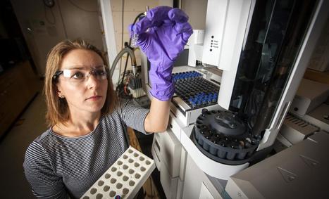
To take the detailed measurements, the research team uses a nanopore 10-30 nanometers wide—so small that only one protein molecule can fit through at a time. The researchers filled the nanopore with a salt solution and passed an electric current through the solution.
As a protein molecule tumbles through the nanopore, its movement causes tiny, measurable fluctuations in the electric current. By carefully measuring this current, the researchers can determine the protein's unique five-dimensional signature and identify it nearly instantaneously.
"Amyloid molecules not only vary widely in size, but they tend to clump together into masses that are even more difficult to study," Mayer said. "Because it can analyze each particle one by one, this new method gives us a much better window to how amyloids behave inside the body."
Ultimately, the team aims to develop a device that doctors and researchers could use to quickly measure proteins in a sample of blood or other body fluid. This goal is likely several years off; in the meantime, they are working to improve the technique's accuracy, honing it in order to get a better approximation of each protein's shape. They believe that in the future, the technology could also be useful for measuring proteins associated with heart disease and in a variety of other applications as well.
"I think the possibilities are pretty vast," Sept said. "Antibodies, larger hormones, perhaps pathogens could all be detected. Synthetic nanoparticles could also be easily characterized to see how uniform they are."
The study is titled "Real-time shape approximation and fingerprinting of single proteins using a nanopore."
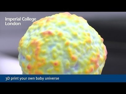
Physicists have created a 3D printed cosmic microwave background - a map of the oldest light in the universe - and provided the files for download. The cosmic microwave background (CMB) is a glow that the universe has in the microwave range that maps the oldest light in the universe. It was imprinted when the universe first became transparent, instead of an opaque fog of plasma and radiation.
The CMB formed when the universe was only 380,000 years old – very early on in its now 13.8 billion-year history. The Planck satellite is making ever-more detailed maps of the CMB, which tells astronomers more about the early universe and the formation of structures within it, such as galaxies. However, more detailed maps are increasingly difficult to view and explore.
To address this issue, Dr Dave Clements from the Department of Physics at Imperial, and two final-year undergraduate students in Physics, have created the plans for 3D printing the CMB. A paper describing the process is published today in the European Journal of Physics.
Dr Clements said: “Presenting the CMB in a truly 3D form, that can be held in the hand and felt rather than viewed, has many potential benefits for teaching and outreach work, and is especially relevant for those with a visual disability.
One of the hurdles to longevity is that bodies seemed designed to break down. It's a part of aging. It might be a part of evolution. Transplants and implants aren’t painless and can be risky. Now, researchers are saying that you won’t have to worry about bodies breaking down when you can just print a new part.
Nature Magazine reports that scientists are using 3D bioprinting technology that can print everything from new knees and bones to eyes and ears.
Penn State biological engineer Ibrahim Ozbolat, who is studying how to use 3D bioprinting to repair cartilage and other tissue types, tells Nature that one day a patient could lay under a machine that would inject cells into a trick knee to repair it.
“In the future, we can have the patient under the bioprinter,” says Ozbolat.
Ozbolat ads that this bioprinter machine could fix any body part, not just knees.
Cornell researchers have developed an interactive prototyping system that prints a wire frame of your design as you design it. You can pause anywhere in the process to test or measure and make needed changes, which will be added to the physical model still in the printer.
In conventional 3-D printing, a nozzle scans across a stage depositing drops of plastic, rising slightly after each pass to build an object in a series of layers. With the On-the-Fly-Print system, the nozzle instead extrudes a rope of quick-hardening plastic to create a wire frame that represents the surface of the solid object described in a computer-aided design (CAD) file and allows the designer to make refinements while printing is in progress.
3-D printing has become a powerful tool for engineers and designers, allowing them to do "rapid prototyping" by creating a physical copy of a proposed design.
But what if you decide to make changes? You may have to go back, change the design and print the whole thing again, perhaps more than once. So Cornell researchers have come up with an interactive prototyping system that prints what you are designing as you design it; the designer can pause anywhere in the process to test, measure and, if necessary, make changes that will be added to the physical model still in the printer.
"We are going from human-computer interaction to human-machine interaction," said graduate student Huaishu Peng, who described the On-the-Fly-Print system in a paper presented at the 2016 ACM Conference for Human Computer Interaction. Co-authors are François Guimbretière, associate professor of information science; Steve Marschner, professor of computer science; and doctoral student Rundong Wu.
Their system uses an improved version of an innovative "WirePrint" printer developed in a collaboration between Guimbretière's lab and the Hasso Platner Institute in Potsdam, Germany.
In conventional 3-D printing, a nozzle scans across a stage depositing drops of plastic, rising slightly after each pass to build an object in a series of layers. With the WirePrint technique the nozzle extrudes a rope of quick-hardening plastic to create a wire frame that represents the surface of the solid object described in a computer-aided design (CAD) file. WirePrint aimed to speed prototyping by creating a model of the shape of an object instead of printing the entire solid. The On-the-Fly-Print system builds on that idea by allowing the designer to make refinements while printing is in progress.
The world's first 3D-printed office building opened this week in Dubai, Reuters reports. The 2,700-square-foot, single-story building was built in just 17 days using a gigantic, 20-foot tall 3D printer and a special mix of concrete, fiber reinforced plastic and glass fiber reinforced gypsum.
Although the "printer" was massive at about two stories tall, 120 feet long and 40 feet wide, it only needed one staffer to make sure it was functioning properly. The rest of the 18-person construction crew consisted of installers, electricians and mechanical engineers who completed the job for a mere $140,000 in construction and labor costs — or about half the price of a comparable structure built with conventional methods. Of course, the building is more than just another gold star in the UAE's ultramodernplayland — it will also serve, appropriately enough, as the temporary headquarters for the Dubai Future Foundation. Next year, the structure is scheduled to become the home of Dubai's Museum of the Future.
"This is the first 3D-printed building in the world, and it's not just a building, it has fully functional offices and staff," the UAE Minister of Cabinet Affairs, Mohamed Al Gergawi said. According to Gergawi, Dubai plans to have 25 percent of the buildings in the emirate built via 3D printing by the year 2030.
Artificial Intelligence is starting to turn invisible from the outside in -- and vice versa. The exact effects and workings of AI technologies are becoming..
In the near future, artificial intelligence will commonly become intangible, indistinguishable and incomprehensible for humans.
Firstly, AI doesn’t necessarily need a tangible embodiment. It can manifest itself through different mediators, such as a graphical user interface or a voice interface. Already we trust Spotify recommendations without a glance or talk to Siri and Alexa like they were summoned spirits, intelligences without a tangible form.
Secondly, AI becomes invisible by passing the Turing test, or its more relevant variants. An intelligent system that manages to simulate human-level communication, and cognitive as well as emotional abilities, can become indistinguishable from humans and, thus, the “artificiality” of its intelligence becomes imperceptible for us.
Thirdly, and most importantly, AI escapes human gaze when the details of its effects and technological dynamics go beyond human perception and understanding. We can be aware of the existence, presence and effects of intelligent systems, but we no longer fully comprehend what these systems do, how they achieve their goals or what are their definite effects. This means that AI technologies will soon go beyond Clarke’s third law, stating that “any sufficiently advanced technology is indistinguishable from magic.” Indeed, we don’t anymore have a chance to figure out the trick — or even realize that any trick occurred in the first place.
Following this, we are able to perceive manifestations and presentations of artificial intelligence, but the intelligence itself becomes unknowable to humans through human senses. Currently there are two distinct traits in this development. First, most algorithmic systems, as well as the latest advancements in AI technologies, are black boxes; inaccessible, unfathomable and uncontrollable to most people.
Therefore, it’s hard to perceive or assess how intelligent systems shape your life online and offline, from your latest song recommendations to your personalized insurance policy, not to mention the algorithmic stock market trading that shapes the global market economy affecting almost every aspect of modern life.
Concretely, when the actions of intelligent systems become more holistically intertwined with personal, social, cultural, political and economical systems, it becomes challenging to distinguish the exact effects or impact of the machine intelligence itself.
Second, AI technologies are becoming so complex that they are hard to understand — even for the experts designing and developing them. In his recent book, The Master Algorithm, machine learning expert Pedro Domingos points out that already back in 1950s scientists created an algorithm that could do something that humans couldn’t fully comprehend.
This development hasn’t changed its course; rather, to the contrary. With the current pace of AI development, even seasoned experts have a hard time keeping up. Today various machine learning systems can already provide unexpected insights in varying fields, from personalization technologies to particle physics, from cooking recipes and outlandish game moves to crime prevention and bioengineering. Concretely, specialized systems can empower scientific discoveries in biology or help you choose the best route to your next meeting.
Northwestern University scientists created a prosthetic ovary using a 3D printer – an implant that allowed mice that had their ovaries surgically removed to bear live young. The results will be presented Saturday, April 2, at the Endocrine Society’s annual meeting, ENDO 2016, in Boston. Researchers hope to use the technology to develop an ovary bioprosthesis that could be implanted in women to restore fertility. One group that could benefit is survivors of childhood cancers, who have an increased risk of infertility as adults. An estimated 1 in 250 adults has survived childhood cancer.
“One of the biggest concerns for patients diagnosed with cancer is how the treatment may affect their fertility and hormone health,” said lead study author Monica M. Laronda, PhD, a postdoctoral research fellow at Northwestern University’s Feinberg School of Medicine. “We are developing new ways to restore their quality of life by engineering ovary bioprosthesis implants.”
The researchers used a 3D printer to create a scaffold to support hormone-producing cells and immature egg cells, called oocytes. The structure was made out of gelatin – a biological material derived from the animal protein collagen. The scientists applied biological principles to manufacture the scaffold, which needed to be rigid enough to be handled during surgery and to provide enough space for oocyte growth, blood vessel formation and ovulation.
Using human cell cultures, the researchers determined the optimal scaffold design should have crisscrossing struts that allowed the cells to anchor at multiple points. The scaffolds were seeded with ovarian follicles – the spherical unit that contains a centralized oocyte with surrounding supportive, hormone-producing cells – to create the bioprosthesis.
To test the implant, researchers removed the ovaries of mice and replaced them with the ovary bioprosthesis. Following the procedure, the mice ovulated, gave birth to healthy pups and were able to nurse.
Implanting the prosthetic ovary in mice also restored the estrous, or female hormone cycle. Researchers theorize a similar implant could help maintain hormone cycling in women who were born with or have undergone disease treatments that have reduced ovarian function. These women often experience decreased production of reproductive hormones that can cause issues with the onset of puberty as well as bone and vascular health problems later in life.
“We developed this implant with downstream human applications in mind, as it is made through a scalable 3D printing method, using a material already used in humans,” Laronda said. “We hope to one day restore fertility and hormone function in women who suffer from the side effects of cancer treatments or who were born with reduced ovarian function.”
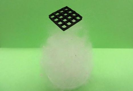
Aerogels are among the world’s lightest materials. Graphene aerogel, a record holder in that category, is so light that a large block of it wouldn’t make a dent on a tiny ball of cotton. Water is about one thousand times more dense. The minimal density of aerogels allows for a number of possible applications, researchers have found, ranging from soaking up oil spills to “invisibility” cloaks.
Now, scientists from State University of New York (SUNY) at Buffalo and Kansas State University report in the journal Small that they have found a way to 3D print graphene aerogel, which has only been used in lab prototypes. This technology will make the material much easier to use, and open it, and hopefully other aerogel materials, up to wider applications.
Graphene is just a single layer of carbon atoms. Since it was isolated for the first time in 2004, it has been touted as a wonder material for its strength, pliability and conductivity. Aerogel is essentially a gel where the liquid is replaced by air. Graphene aerogel is known to be highly compressible (so it can bear pressure without breaking apart) and highly conductive (so it can carry electricity efficiently). The very structure of the material that gives it these properties, however, makes it difficult to manufacture using 3D printing technology.
SUNY Buffalo and Kansas State University researchers came up with a solution. They mixed graphene oxide—graphene with extra oxygen atoms—with water and deposited layers on a surface at -25°C. This instantly froze each layer, and allowed the undisrupted construction of the aerogel, with ice as its support.
3D Printing aerogels containing graphene? This material gets some interesting properties. While it is quite hard to manufacture in a controlled fashion I believe it will open the way for compressible circuits.
A team of biomedical researchers at Wake Forest Institute for Regenerative Medicine has just completed an invention 10 years in the making. It's a 3D printer that can craft relatively simple tissues like cartilage into large complex shapes—like an infant's ear. Using cartridges that are brimming with biodegradable plastic and human cells bound up in gel, this new kind of 3D printer builds complex chunks of growing muscle, cartilage, and even bone. When implanted into animals, these simple fabricated tissues survive and thrive indefinitely.
The scientists led by Anthony Atala surmounted two particularly thorny challenges that have long impeded the futuristic goal of printing living human tissues. First, their new device manufactures large, stable chunks of printed tissue that don't fall apart. Second, it keeps those large structures alive and growing. The new 3D printer is unveiled and outlined today in the journal Nature Biotechnology.
"This is the first bioprinter that can print tissue at the large scales relevant for human implantation," Atala says. "Basically, once we've printed a structure, we can keep it alive for several weeks before we implant it. Now the next step is to test these [printed tissues] for safety so we can implant them in the future in patients."
Atala's new device—named the Integrated Tissue and Organ Printing System, or ITOP—is straightforward. The programmed printer slowly squirts out layer upon layer of a rapidly hardening material in the form of tiny droplets. Like other 3D printers, this layered approach allows ITOP to print highly complex shapes in three dimensions with incredible detail. The materials ITOP uses, and the way it structures the tissues that it builds, are what make this machine revoutionary.
“This work represents an elegant advance in programmable materials assembly, made possible by a multidisciplinary approach,” said Jennifer Lewis, Sc.D., senior author of a new study reported on January 25 in a new in Nature Materials. “We have now gone beyond integrating form and function to create transformable architectures.”
In nature, flowers and plants have tissue compositions and microstructures that result in dynamic morphologies (forms) that change according to their environments. Mimicking the variety of shape changes undergone by plant organs such as tendrils, leaves, and flowers in response to environmental stimuli like humidity and/or temperature, the 4D-printed hydrogel composites developed by Lewis and her team are programmed to contain precise, localized swelling behaviors.
The trick: the hydrogel composites contain cellulose fibrils that are derived from wood and are similar to the microstructures that enable shape changes in plants. By aligning cellulose fibrils during printing, the hydrogel composite ink is encoded with anisotropic swelling and stiffness, which can be patterned to produce intricate shape changes. The anisotropic (irregular) nature of the cellulose fibrils gives rise to varied directional properties that can be predicted and controlled. That’s why wood can be split easier along the grain rather than across it.
Likewise, when immersed in water, the hydrogel-cellulose fibril ink undergoes differential swelling behavior along and orthogonal to the printing path. Combined with a proprietary mathematical model developed by the team that predicts how a 4D object must be printed to achieve prescribed transformable shapes, the new method opens up many new and exciting potential applications for 4D printing technology including smart textiles, soft electronics, biomedical devices, and tissue engineering.
The composite ink that the team uses flows like liquid through the printhead, yet rapidly solidifies once printed. A variety of hydrogel materials can be used interchangeably resulting in different stimuli-responsive behaviors, while the cellulose fibrils can be replaced with other anisotropic fillers of choice, including conductive fillers. The mathematical model prescribes the printing pathways required to achieve the desired shape-transforming response. Specifically, it solves the “inverse problem” — the challenge of being able to predict what the printing toolpath must be to encode swelling behaviors toward achieving a specific desired target shape.
“It is wonderful to be able to design and realize, in an engineered structure, some of nature’s solutions,” said L. Mahadevan, Ph.D., a Wyss Core Faculty member as well as the Lola England de Valpine Professor of Applied Mathematics, Professor of Organismic and Evolutionary Biology, and Professor of Physics at Harvard University and Harvard SEAS, is a co-author on the study. “By solving the inverse problem, we are now able to reverse-engineer the problem and determine how to vary local inhomogeneity, i.e. the spacing between the printed ink filaments, and the anisotropy, i.e. the direction of these filaments, to control the spatiotemporal response of these shapeshifting sheets.”
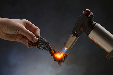
There's a reason they're used in everything from jet engines to Formula 1 race car brakes: Ceramics are tough. They can withstand an absurd amount of heat and pressure without warping or breaking, all while brushing off many of the physical and chemical assaults that would rust metals and wear away plastics.
"The problem is that ceramics are just notoriously difficult to process," says Zak Eckel, an engineer at HRL Laboratories in Malibu, California.
Heat-resistant ceramics require crazy-high temperatures to melt, so it's been a struggle to develop methods to 3D-print them. Today, there are just a few 3D printing techniques on the market that use any ceramics (developed by companies like 3DCERAM and Lithoz), but the approaches are severely limited in the types of ceramics they can print, as well as the end quality of their materials. Eckel and his team have just developed an altogether new way to 3D print practically flawless ceramics—including fantastically heat-resistant varieties that've so far been beyond our reach. Their research is announced today in the journal Science.
A prototype 3D bioprinter that can create totally functional human skin has been developed by scientists from Universidad Carlos III de Madrid (UC3M) and BioDan Group in Spain. The skin has been used to treat burns as well as traumatic and surgical wounds in a large number of patients in Spain, according to the scientists.
The system provides two processes. Autologous skin (from the patient’s own cells to generate human collagen) for therapeutic use, such as in the treatment of severe burns, instead of the animal collagen used in other methods. The researchers have applied for approval by various European regulatory agencies to guarantee that the skin that is produced is adequate for use in transplants on burn patients and on those with other skin problems.
The 3D-printed skin replicates human bilayered skin, using “bioinks” (biological components) containing human plasma as well as primary human fibroblasts and keratinocytes obtained from skin biopsies. These are controlled by a computer, which deposits them on a print bed in an orderly manner to then produce the skin.
The researchers were able to generate 100 cm2 of printed skin in less than 35 minutes (including the 30 min required for fibrin gelation).
Allogeneic skin (from a stock of cells), done on a large scale for industrial processes. This skin can be used to test pharmaceutical products, cosmetics, and consumer chemical products where current regulations require testing that does not use animals.
“This method of bioprinting allows skin to be generated in a standardized, automated way, and the process is less expensive than manual production,” says Alfredo Brisac, CEO of BioDan Group, the Spanish bioengineering firm specializing in regenerative medicine that is collaborating on this research and commercializing this technology. The research was published online in the journal Biofabrication.
L'injection de graisse auto logue est une réalité scientifique et médicale prouvée par des injections de graisse sous la peau du sein pour une patiente donnée en Tunisie, cette technique très avancée à l'époque est devenue aujourd'hui une possibilité grand public pour des patientes qui veulent s'offrir une augmentation mammaire sans risque d'allergie contre les implants mammaires de silicone ou autres types. En Espagne, on a développé un prototype machine 3D BIOPRINTER qui intervient dans la génération de Kollagene pour des usages thérapeutiques de la peau endommagée.
At the Los Angeles Auto Show, automaker Divergent 3D showed off their 3D-printed Blade Supercar. The 635 kilogram (1,400 pound) car is made of a combination of aluminum and carbon fiber; accelerates to 97 kilometers per hour (60 miles per hour) in 2.2 seconds with its 700 hp engine; and can use either gasoline or compressed natural gas as fuel.
The Blade Supercar debuted last year in June, heralding the company’s radical, environmentally-sustainable approach to manufacturing. Divergent calls the manufacturing approach NODE, where they 3D print aluminum nodes joined together by carbon fiber tubing.
The process, which is similar to using Lego blocks, requires less capital and uses up fewer materials. The ease of assembly means that even semi-skilled workers can run the process.As an added bonus, Divergent 3D’s cars are 90 percent lighter and more durable than cars built with traditional techniques.
Over the past few years, we’ve seen 3D printing prove itself an invaluable resource for engineers. It’s helped us build prosthetics, instruments, drones and a mini jet engine that can blast at 33,000 RPM. But in the hands of creatives, the hackable tech has done something else entirely: allowed them to make unbelievable art. For University of Chicago assistant professor Dr. Allan Drummond, that art comes in the form of resurrecting ancient beasts.
A biochemistry and human genetics researcher, Drummond studies everything from how cells adapt, to the multi-million-year evolution of the species we share our planet with. It’s no surprise then, that he took a particular interest in trilobites. The extinct arthropods cruised the world’s oceans for some 270 million years – with over 17,000 known species, they are the most diverse group of animals preserved in the fossil record.
“We find their shells fossilized everywhere,” explains Drummond. “They’re museum staples – but we rarely see what they really looked like, with all of their soft tissues (legs, antennae, gills) intact.”
Determined to print a trilobite in all its glory, Drummond turned to the literature and online forums for guidance. “The first step was to look at as many trilobites as possible and choose one,” he recalls. “I’ve always loved these fossils, but the moment they turned from fossils, into living organisms for me, was when I saw the new generation of preparations displayed at Chicago’s Field Museum. I couldn’t believe what I was seeing. In my mind, trilobites were flat, if beautiful, primitive creatures. Seeing those preparations made it clear how not-flat and not-primitive they were.”
In order to narrow down the options, Drummond eliminated any trilobite groups with delicate spines – which many of them had – that would easily break, as well as any that were too simple to print with extensive detail. He settled on Ceraurus, a genus that roamed the Earth in the middle to upper Ordovician, 470-445 million years ago.
“Ceraurus is ideal,” he says. “They have long yet substantial genal [head segement] and pygidial [tail segment] spines, complex thoracic armor, gorgeous curves, unmistakable trilobite form. Enough detail to warrant 3D printing, enough structural solidity to survive it.”
Scientists fabricated isotropic, near-net-shape, neodymium-iron-boron (NdFeB) bonded magnets at DOE's Manufacturing Demonstration Facility at ORNL using the Big Area Additive Manufacturing (BAAM) machine. The result, published in Scientific Reports, was a product with comparable or better magnetic, mechanical, and microstructural properties than bonded magnets made using traditional injection molding with the same composition.
The additive manufacturing process began with composite pellets consisting of 65 volume percent isotropic NdFeB powder and 35 percent polyamide (Nylon-12) manufactured by Magnet Applications, Inc. The pellets were melted, compounded, and extruded layer-by-layer by BAAM into desired forms.
While conventional sintered magnet manufacturing may result in material waste of as much as 30 to 50 percent, additive manufacturing will simply capture and reuse those materials with nearly zero waste, said Parans Paranthaman, principal investigator and a group leader in ORNL's Chemical Sciences Division.
Using a process that conserves material is especially important in the manufacture of permanent magnets made with neodymium, dysprosium—rare earth elements that are mined and separated outside the United States. NdFeB magnets are the most powerful on earth, and used in everything from computer hard drives and head phones to clean energy technologies such as electric vehicles and wind turbines.
The printing process not only conserves materials but also produces complex shapes, requires no tooling and is faster than traditional injection methods, potentially resulting in a much more economic manufacturing process, Paranthaman said.
"Manufacturing is changing rapidly, and a customer may need 50 different designs for the magnets they want to use," said ORNL researcher and co-author Ling Li. Traditional injection molding would require the expense of creating a new mold and tooling for each, but with additive manufacturing the forms can be crafted simply and quickly using computer-assisted design, she explained.
As the name suggests, these remarkable 3D-printed objects seem to change shape depending on their orientation. "People see it as a circle or as kind of a diamond — in reality, that structure is neither of those things," Sekuler said. "It's a shape that was created so that from a particular perspective you'll see it as one thing or another."
And because the two shapes are so different, it's difficult for people's brains to combine them, she added.
"It makes more sense to say there are two different objects," Sekuler said. "It sounds almost counterintuitive … in this case, the simplicity that's winning out is our familiarity with those shapes."
The researcher, Kokichi Sugihara of Meiji University in Japan, used ambiguous shapes and perspective to craft an illusion that was named a 2015 finalist and one that was a 2010 winner.
Lawrence Livermore National Laboratory scientists have combined biology and 3-D printing to create the first reactor that can continuously produce methanol from methane at room temperature and pressure.
The team removed enzymes from methanotrophs, bacteria that eat methane, and mixed them with polymers that they printed or molded into innovative reactors. The research, which could lead to more efficient conversion of methane to energy production, appears in the June 15 edition of Nature Communications.
"Remarkably, the enzymes retain up to 100 percent activity in the polymer," said Sarah Baker, LLNL chemist and project lead. "The printed enzyme-embedded polymer is highly flexible for future development and should be useful in a wide range of applications, especially those involving gas-liquid reactions."
Advances in oil and gas extraction techniques have made vast new stores of natural gas, composed primarily of methane, available. However, a large volume of methane is leaked, vented or flared during these operations, partly because the gas is difficult to store and transport compared to more-valuable liquid fuels. Methane emissions also contribute about one-third of current net global warming potential, primarily from these and other distributed sources such as agriculture and landfills.
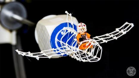
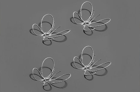
"Flat" and "rigid" are terms typically used to describe electronic devices. But the increasing demand for flexible, wearable electronics, sensors, antennas and biomedical devices has led a team at Harvard's Wyss Institute for Biologically Inspired Engineering and the John A. Paulson School of Engineering and Applied Sciences (SEAS) to innovate an eye-popping new way of printing complex metallic architectures -- as though they are seemingly suspended in midair.
Reported online May 16, 2016 in the Proceedings of the National Academy of Sciences, this laser-assisted direct ink writing method allows microscopic metallic, free-standing 3D structures to be printed in one step without auxiliary support material. The research was led by Wyss Core Faculty member Jennifer Lewis, Sc.D., who is also the Hansjörg Wyss Professor of Biologically Inspired Engineering at SEAS.
"I am truly excited by this latest advance from our lab, which allows one to 3D print and anneal flexible metal electrodes and complex architectures 'on-the-fly,' " said Lewis.
Lewis' team used an ink composed of silver nanoparticles, sending it through a printing nozzle and then annealing it using a precisely programmed laser that applies just the right amount of energy to drive the ink's solidification. The printing nozzle moves along x, y, and z axes and is combined with a rotary print stage to enable freeform curvature. In this way, tiny hemispherical shapes, spiral motifs, even a butterfly made of silver wires less than the width of a hair can be printed in free space within seconds. The printed wires exhibit excellent electrical conductivity, almost matching that of bulk silver.
When compared to conventional 3D printing techniques used to fabricate conductive metallic features, laser-assisted direct ink writing is not only superior in its ability to produce curvilinear, complex wire patterns in one step, but also in the sense that localized laser heating enables electrically conductive silver wires to be printed directly on low-cost plastic substrates.
According to the study's first author, Wyss Institute Postdoctoral Fellow Mark Skylar-Scott, Ph.D., the most challenging aspect of honing the technique was optimizing the nozzle-to-laser separation distance.
"If the laser gets too close to the nozzle during printing, heat is conducted upstream which clogs the nozzle with solidified ink," said Skylar-Scott. "To address this, we devised a heat transfer model to account for temperature distribution along a given silver wire pattern, allowing us to modulate the printing speed and distance between the nozzle and laser to elegantly control the laser annealing process 'on the fly.' "
From visible light to radio waves, most people are familiar with the different sections of the electromagnetic spectrum. But one wavelength is often forgotten, little understood, and, until recently, rarely studied.
"Terahertz is somewhat of a gap between microwaves and infrared," said Northwestern University's Cheng Sun. "People are trying to fill in this gap because this spectrum carries a lot of information."
Sun and his team have used metamaterials and 3-D printing to develop a novel lens that works with terahertz frequencies. Not only does it have better imaging capabilities than common lenses, but it opens the door for more advances in the mysterious realm of the terahertz. Supported by the National Science Foundation, the work was published online on April 22 in the journal Advanced Optical Materials.
"Typical lenses—even fancy ones—have many, many components to counter their intrinsic imperfections," said Sun, associate professor of mechanical engineering at Northwestern's McCormick School of Engineering. "Sometimes modern imaging systems stack several lenses to deliver optimal imaging performance, but this is very expensive and complex."
The focal length of a lens is determined by its curvature and refractive index, which shapes the light as it enters. Without components to counter imperfections, resulting images can be fuzzy or blurred. Sun's lens, on the other hand, employs a gradient index, which is a refractive index that changes over space to create flawless images without requiring additional corrective components.
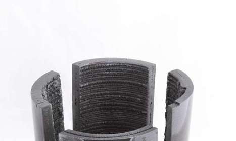
The researchers began with a vat of resin containing silicon, carbon and oxygen. They shone a pattern of ultraviolet light beams onto this resin, causing it to harden where the light shone through it. In 30 to 60 seconds, an item 0.5 to 1 inches (1.27 to 2.54 centimeters) thick can form, with a lattice or honeycomb shape, Schaedler said. The researchers then heat these objects to convert the material into silicon oxycarbide ceramic. This new method is 100 to 1,000 times faster than previous 3D-ceramic-printing techniques, the researchers said. Furthermore, electron microscopy of the end products detected none of the porosity or surface cracks that normally weaken ceramics; indeed, these silicon carbide materials were 10 times stronger than commercially available ceramic foams of similar density, the scientists noted. Since ceramics are notoriously brittle, Schaedler said, "We are working to reinforce our ceramics with fibers." However, it will take some time before these ceramics reach the market, he said. "We are at the discovery phase. It will take at least five years for an application to be commercialized," Schaedler said.
HRL Laboratories, LLC, Malibu, California is a corporate research-and-development laboratory owned by The Boeing Company and General Motors specializing in research into sensors and materials, information and systems sciences, applied electromagnetics, and microelectronics.
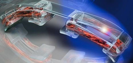
HRL’s Senior Chemical Engineer Zak Eckel and Senior Chemist Dr. Chaoyin Zhou invented a resin formulation that can be 3D printed into parts of virtually any shape and size. The printed resin can then be fired, converting it into a high strength, fully dense ceramic. The resulting material can withstand ultrahigh temperatures in excess of 1700°C and exhibits strength ten times higher than similar materials.
Ceramics are much more difficult to process than polymers or metals because they cannot be cast or machined easily. Traditionally ceramic parts are consolidated from powders by sintering, which introduces porosity and limits both achievable shapes and final strength. "With our new 3D printing process we can take full advantage of the many desirable properties of this silicon oxycarbide ceramic, including high hardness, strength and temperature capability as well as resistance to abrasion and corrosion." says program manager Dr. Tobias Schaedler.
The novel process and material could be used in a wide range of applications from large components in jet engines and hypersonic vehicles to intricate parts in microelectromechanical systems and electronic device packaging.
University of California, San Diego researchers have 3D-printed a tissue that closely mimics the human liver’s sophisticated structure and function. The new model could be used for patient-specific drug screening and disease modeling and could help pharmaceutical companies save time and money when developing new drugs, according to the researchers.
The liver plays a critical role in how the body metabolizes drugs and produces key proteins, so liver models are increasingly being developed in the lab as platforms for drug screening. However, so far, the models lack both the complex micro-architecture and diverse cell makeup of a real liver. For example, the liver receives a dual blood supply with different pressures and chemical constituents.
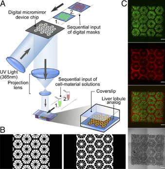
So the team employed a novel bioprinting technology that can rapidly produce complex 3D microstructures that mimic the sophisticated features found in biological tissues.
The team printed a honeycomb pattern of 900-micrometer-sized hexagons, each containing liver cells derived fromhuman induced pluripotent stem cells. An advantage of human induced pluripotent stem cells is that they are patient-specific, which makes them ideal materials for building patient-specific drug screening platforms. And since these cells are derived from a patient’s own skin cells, researchers don’t need to extract any cells from the liver to build liver tissue.
Then, endothelial and mesenchymal supporting cells were printed in the spaces between the stem-cell-containing hexagons.
The entire structure — a 3 × 3 millimeter square, 200 micrometers thick — takes just seconds to print. The researchers say this is a vast improvement over other methods to print liver models, which typically take hours. Their printed model was able to maintain essential functions over a longer time period than other liver models. It also expressed a relatively higher level of a key enzyme that’s considered to be involved in metabolizing many of the drugs administered to patients.
“It typically takes about 12 years and $1.8 billion to produce one FDA-approved drug,” said Shaochen Chen, NanoEngineering professor at the UC San Diego Jacobs School of Engineering. “That’s because over 90 percent of drugs don’t pass animal tests or human clinical trials. We’ve made a tool that pharmaceutical companies could use to do pilot studies on their new drugs, and they won’t have to wait until animal or human trials to test a drug’s safety and efficacy on patients. This would let them focus on the most promising drug candidates earlier on in the process.”
Acquista Online La Prescrizione Di Perdita Di Peso Crediamo che i farmaci a volte possano essere molto urgenti da assumere. Se hai urgente bisogno di farmaci, possiamo anche fornirti una consegna espressa,
Electromashina JSC, a manufacturer of armored vehicles for the Russian army, has revealed that it is using an industrial 3D printer to produce an Armata tank, the standard next-generation armored vehicle platform of the Russian military.
When Russia’s impressive T-14 Armata tank first hit the streets of Moscow last year, very little was known about it. The armored vehicle drove around Red Square during the May 2015 Victory Day Parade, a celebration of the 70th anniversary of Russia’s victory in WWII. Drivers showed off the Armata’s impressive turn radius, its radar-baffling paint, and its thick armor plates. Now, information has been released about how certain Armata tanks, perhaps including the T-14, are being manufactured. Electromashina JSC, an armored vehicle manufacturer and part of the UralVagonZavod corporation, has revealed the important role played by 3D printing technology in the production of its new line of Armata tanks.
Anton Ulrich, Manager of the Rapid Prototyping Lab at Electromashina, explained how 3D printing has been used since 2015 to produce prototype parts. These parts can be created in small numbers, tested, and then redesigned as appropriate until ready for series production. Electromashina has also been using its 3D printers to produce master models, used in the casting of metal and plastic parts. In the near future, the company will start using 3D printers to produce 3D printed titanium parts, several meters in length, for use in its armored vehicles.
“3D printing has been implemented to speed up trial production,” Ulrich explained. “When a designer develops new products, he uses CAD software to produce a 3D model. So, using a 3D printer, we can quickly turn those 3D models into prototype parts. Now there is no need to order a sample component, and then, realizing that it doesn’t fit, have to order a re-run and waste metal. Furthermore, it is possible to produce not just small elements of a part, but the whole assembly, evaluating its mechanical characteristics before production.”
Although 3D printing machine-ready components for use in armored vehicles and other military equipment is a distinct possibility for Electromashina, these items would have to meet certain requirements of the defense industry. “3D printed components can go straight to consumers in certain industries,” said Ulrich. “But in the defense industry, standards are much higher.”
Ulrich, however, sees no reason why 3D printed components cannot eventually meet those strict defense industry requirements. He cites the cases of 3D printed components being used in the aerospace industry and even produced in space on the International Space Station. If 3D printed parts can be approved for use in space, they could certainly be deemed fit for use in Russian tanks.
The Russian army plans to acquire 2,300 T-14 Armata tanks between now and 2020.
Engineers at MIT, Penn State University, and Carnegie Mellon University have devised a way to manipulate cells in three dimensions using sound waves. These “acoustic tweezers” could make possible 3-D printing of cell structures for tissue engineering and other applications, the researchers say.
Designing tissue implants that can be used to treat human disease requires precisely recreating the natural tissue architecture, but so far it has proven difficult to develop a single method that can achieve that while keeping cells viable and functional.
“The results presented in this paper provide a unique pathway to manipulate biological cells accurately and in three dimensions, without the need for any invasive contact, tagging, or biochemical labeling,” says Subra Suresh, president of Carnegie Mellon and former dean of engineering at MIT. “This approach could lead to new possibilities for research and applications in such areas as regenerative medicine, neuroscience, tissue engineering, biomanufacturing, and cancer metastasis.”
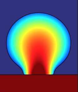
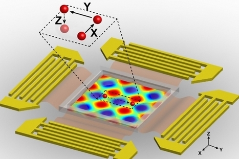
The new acoustic tweezers are based on a microfluidic device that the researchers previously developed to manipulate cells in two dimensions. This device produces two acoustic standing waves, which are waves with a constant height. Where the two waves meet, they create a “pressure node” that can trap single cells. By altering the wavelength and another wave property known as the phase, the researchers can move the node and the cell trapped within it.
The research team previously used a similar approach to separate cancer cells from healthy cells, which could be useful for detecting rare tumor cells in a patient’s bloodstream and predicting whether the tumor will spread.
To get content containing either thought or leadership enter:
To get content containing both thought and leadership enter:
To get content containing the expression thought leadership enter:
You can enter several keywords and you can refine them whenever you want. Our suggestion engine uses more signals but entering a few keywords here will rapidly give you great content to curate.


 Your new post is loading...