 Your new post is loading...
What are some leg-strengthening exercises that will expedite recovery for athletes and physically active individuals who have undergone leg surgery? Post Surgery Leg Strengthening Leg muscles may weaken after hip, knee, ankle, or foot surgery. This happens because those muscles are not used as much during recovery. Gaining back strength and muscle endurance after an injury or surgery is an important step in recovery. Leg exercises can help regain mobility and prevent complications like blood clots and pressure sores after surgery or an injury, which is why engaging in post-surgery leg strengthening is important once the doctor gives the ok. Benefits Post-surgery leg strengthening exercises have several benefits, including - Rebuilds strength and confidence
- Retraining for optimal mobility and flexibility
- Prevents pressure sores
- Prevents blood clots
When the leg muscles are contracted, they move blood like a pump, maintaining proper circulation. Moving in a bed after surgery also helps prevent pressure sores from forming and blood clots. A physical therapy team will determine the right leg exercises for each individual and their injury/condition. This is an important step when moving forward after surgery. (Hoogeboom T. J. et al., 2014) Starting Out The first exercises should target all the major muscles of the leg. (Madara K. C. et al., 2019) Gluteal Sets This isometric exercise means the muscles contract while no motion occurs at the hip joints. To perform: - Lie on your back and tighten your buttock muscles.
- Hold the muscles tight for 5 seconds, then relax.
- Repeat 10 or 15 times.
- Gluteal sets can be done several times per day.
Heel Slide Heel slides can help regain strength in the major muscles of the leg. To perform: - Lie on your back.
- Bend the knee of the surgical leg and slowly slide the heel toward your butt.
- Slide as far as possible and hold for 5 seconds.
- Slowly return to the starting position and repeat.
Short Arc Quad The short arc quad, or SAQ, is a simple way to get the quadricep muscles working. To perform: - Lie on your back with a towel roll, small ball, or something similar under the knee.
- Slowly straighten the knee.
- Tighten the quad muscle on the top of the thigh.
- Hold for 3 seconds, then relax.
- Repeat 10 to 15 times.
Quad Set This exercise helps get the quad muscles working. It also helps control the position of the kneecap. To perform: - Lie on your back.
- Place a small towel roll under the knee.
- Try to press the back of the knee flat against the floor.
- Hold for 10 seconds and release.
- Repeat 10 to 15 times.
Individuals can complete quad sets bilaterally or with both knees simultaneously. This makes the stronger leg help strengthen the weaker side. Straight Leg Raise To perform: - Lie on your back.
- Lift your leg straight off the floor until it is at the height of the opposite bent knee.
- Hold for 10 seconds and slowly lower.
- Repeat 10 to 15 times.
Be sure to keep the knee straight for the entire exercise. Keep the opposite knee bent for comfort. To ensure the knee is straight, individuals can complete a quad set first and then the straight leg raise. The exercise can be more challenging by increasing repetitions or adding a 2- to 3-pound ankle weight on the thigh. For even more challenge, add the ankle weight to the ankle. Hamstring Strengthening Working out the hamstrings after injury or surgery is important. The hamstring muscles bend the knee and extend the hip backward. To perform: - Lie on your stomach.
- Bend one knee to raise the lower limb straight in the air.
- Hold for 5 seconds and lower slowly.
- Repeat 10 to 15 times.
Once the exercise is easy to do, increase the repetitions to 30. Individuals can also add a 2- to 3-pound ankle weight. Physical therapy can help individuals regain mobility after injury or surgery. A therapist may prescribe exercises as part of an at-home exercise program. Over time, progress will go from simple exercises to more challenging ones to improve balance and mobility. (Madara K. C. et al., 2019) Injury Medical Chiropractic & Functional Medicine Clinic Before starting this or any other exercise program, consult a doctor and a physical therapist to find the right exercises for your situation. Injury Medical Chiropractic and Functional Medicine Clinic works with primary healthcare providers and specialists to develop an optimal health and wellness solution. We focus on what works for you to relieve pain, restore function, and prevent injury. Regarding musculoskeletal pain, specialists like chiropractors, acupuncturists, and massage therapists can help mitigate the pain through spinal adjustments that help the body realign itself. They can also work with other medical professionals to integrate a treatment plan to resolve musculoskeletal issues. General Disclaimer * The information herein is not intended to replace a one-on-one relationship with a qualified healthcare professional or licensed physician and is not medical advice. We encourage you to make healthcare decisions based on your research and partnership with a qualified healthcare professional. Our information scope is limited to chiropractic, musculoskeletal, and physical medicines, wellness, sensitive health issues, functional medicine articles, topics, and discussions. We provide and present clinical collaboration with specialists from various disciplines. Each specialist is governed by their professional scope of practice and their jurisdiction of licensure. We use functional health & wellness protocols to treat and support care for the injuries or disorders of the musculoskeletal system. Our videos, posts, topics, subjects, and insights cover clinical matters, issues, and topics that relate to and directly or indirectly support our clinical scope of practice. Our office has reasonably attempted to provide supportive citations and identified the relevant research studies or studies supporting our posts. We provide copies of supporting research studies that are available to regulatory boards and the public upon request. We understand that we cover matters that require an additional explanation of how they may assist in a particular care plan or treatment protocol. To discuss the subject matter above further, please contact Dr. Alex Jimenez or us at 915-50-0900. Dr. Alex Jimenez DC, MSACP, CCST, IFMCP*, CIFM*, ATN* email: coach@elpasofunctionalmedicine.com Licensed in: Texas & New Mexico* References Hoogeboom, T. J., Dronkers, J. J., Hulzebos, E. H., & van Meeteren, N. L. (2014). Merits of exercise therapy before and after major surgery. Current opinion in anaesthesiology, 27(2), 161–166. https://doi.org/10.1097/ACO.0000000000000062 Madara, K. C., Marmon, A., Aljehani, M., Hunter-Giordano, A., Zeni, J., Jr., & Raisis, L. (2019). PROGRESSIVE REHABILITATION AFTER TOTAL HIP ARTHROPLASTY: A PILOT AND FEASIBILITY STUDY. International Journal of Sports Physical Therapy, 14(4), 564–581.
In females, hernia symptoms are often smaller and deeper without a noticeable lump and can mimic gynecological issues, with misdiagnoses being common. Can knowing the risk factors and how female hernias are treated help women get relief? Female Hernia A hernia occurs when an internal structure pushes through a weak spot in the abdominal wall, the muscles, and the tissue covering the front of the torso. The more common include: - Groin hernia, known as an inguinal hernia.
- Upper thigh or femoral hernia.
However, a hernia can develop anywhere from the ribcage to the upper thigh. Hernias are less common in women, have different symptoms than in men, and are often misdiagnosed. Lower abdominal and pelvic hernias present differently in women than men, who typically have a visible bulge. Instead, female hernias tend to be smaller, deeper, and less noticeable. They can also cause chronic pelvic pressure or pain that can be mistaken for gynecological problems. Hernia Symptoms For a Woman Hernias in women tend to be smaller and deeper than male hernias, with no lump showing. Instead, female hernias can cause chronic, deep pelvic pain and occasional sharp, stabbing pain that comes on fast and lingers. (Köckerling F., Koch A., & Lorenz R. 2019) Hernia pain worsens with exercise, laughing, coughing, or straining to evacuate the bowels. The pain is often described as: - Dull
- Aching
- Pinching
- Sharp
- Shooting
- Burning
Inguinal hernia pain is usually felt at or above the groin and may radiate to the hip, lower back, vulva, or thigh. Many women find the pain increases during their menstrual cycle. The pain can also be exacerbated by any activity that generates extra pressure on the pelvic floor, including: - Prolonged sitting or standing.
- Bending
- Getting in or out of bed.
- Getting in or out of a car.
- Sexual intercourse
Emergency Hernias in the pelvic area are at risk of becoming incarcerated hernias. An incarcerated hernia occurs when a portion of the intestine or other abdominal tissue becomes trapped in the hernial sac, making it impossible to push it back into place. If this gets trapped or strangulated, it can cause tissue death. Strangulated hernias are a medical emergency. Symptoms include: - Deep red or purple tissues.
- The hernia bulge does not shrink when you lie down.
Other symptoms that warrant immediate medical attention include: (Johns Hopkins Medicine, 2025) - Worsening pain
- Bloating
- Difficulty with bowel movements
- Nausea
- Fever
- A fast, racing heartbeat.
Contact a healthcare provider or the emergency room if experiencing any of the above symptoms. Types Hernias can occur anywhere on the abdominal wall. They may be caused by: - Internal pressure, such as during pregnancy.
- A sports injury
- Tissue weakness
Hernias in the lower abdomen or groin are typically indirect inguinal hernias. The inguinal canal comprises multiple layers of muscles and fascia that the thin round ligament threads through. Other groin and pelvic hernias include: - A direct inguinal hernia
- A femoral hernia at the top of the inner thigh.
- An obturator hernia in the front upper thigh, although this type is rare.
Other common hernias in women are: - Incisional hernia - at the site of a surgical incision
- Umbilical hernia - around the belly button
- Ventral hernia - abdominal midline
Less common hernias include: - Hiatal hernia - diaphragm
- Perineal hernia - pelvic floor
Risk Factors Risk factors for developing a hernia include: (Johns Hopkins Medicine, 2025) - Obesity
- Frequent constipation
- Abdominal or pelvic surgery.
- Allergies with chronic sneezing.
- A chronic cough.
- Collagen defects or connective tissue disorders.
Pregnancy and repeated pregnancies are linked to an increased risk of hernia. Types that are more common in pregnancy include: - Umbilical hernia
- Ventral hernia
- Inguinal hernia
Umbilical hernias are the most common. However, only a small percentage of pregnant individuals get them. (Kulacoglu H. 2018) Diagnosis A hernia diagnosis is made with a physical examination and, if needed, imaging studies. Patients are asked to describe their symptoms precisely, where the pain is located, and any activities that exacerbate it. To check for a hernia, the healthcare provider will palpate for a hernia while the patient sits, stands, or coughs. Imaging tests can include: - Ultrasound
- CT scan
- Endoscopy - a camera is used to see inside the esophagus and stomach.
Misdiagnoses Female hernia symptoms can be vague, which often points healthcare providers in the wrong direction. Female hernias are commonly misdiagnosed as: (Köckerling F., Koch A., & Lorenz R. 2019) - Cysts in the reproductive organs
- Endometriosis
- Fibroid tumors
Treatment A small hernia that does not cause problems or pain may be treated with a wait-and-evaluate protocol. A hernia often worsens over time and could eventually require surgery. (University of Michigan Health, 2024) Self-care treatments include: Medical treatments usually start with conservative measures, including physical therapy, stretching, exercise, and rest. Physical therapists often use myofascial release techniques to relieve muscle spasms. Surgery may be needed to repair the weak area of the abdominal wall to relieve symptoms. (University of Michigan Health, 2024) Hernia repair surgery is typically performed as a laparoscopic surgery. (Köckerling F., Koch A., & Lorenz R. 2019) Most patients heal quickly from the surgery and can return to regular activities in a week or two. Injury Medical Chiropractic and Functional Medicine Clinic Injury Medical Chiropractic and Functional Medicine Clinic works with primary healthcare providers and specialists to develop an optimal health and wellness solution. We focus on what works for you to relieve pain, restore function, and prevent injury. Regarding musculoskeletal pain, specialists like chiropractors, acupuncturists, and massage therapists can help mitigate the pain through spinal adjustments that help the body realign itself. They can also work with other medical professionals to integrate a treatment plan to resolve musculoskeletal issues. General Disclaimer * The information herein is not intended to replace a one-on-one relationship with a qualified healthcare professional or licensed physician and is not medical advice. We encourage you to make healthcare decisions based on your research and partnership with a qualified healthcare professional. Our information scope is limited to chiropractic, musculoskeletal, physical medicines, wellness, sensitive health issues, functional medicine articles, topics, and discussions. We provide and present clinical collaboration with specialists from various disciplines. Each specialist is governed by their professional scope of practice and their jurisdiction of licensure. We use functional health & wellness protocols to treat and support care for the injuries or disorders of the musculoskeletal system. Our videos, posts, topics, subjects, and insights cover clinical matters, issues, and topics that relate to and directly or indirectly support our clinical scope of practice.* Our office has reasonably attempted to provide supportive citations and identified the relevant research studies or studies supporting our posts. We provide copies of supporting research studies that are available to regulatory boards and the public upon request. We understand that we cover matters that require an additional explanation of how it may assist in a particular care plan or treatment protocol; therefore, to further discuss the subject matter above, please contact Dr. Alex Jimenez or contact us at 915-850-0900. Dr. Alex Jimenez DC, MSACP, CCST, IFMCP*, CIFM*, ATN* email: coach@elpasofunctionalmedicine.com Licensed in: Texas & New Mexico* References Köckerling, F., Koch, A., & Lorenz, R. (2019). Groin Hernias in Women-A Review of the Literature. Frontiers in surgery, 6, 4. https://doi.org/10.3389/fsurg.2019.00004 Johns Hopkins Medicine. (2025). How to tell if you have a hernia. https://www.hopkinsmedicine.org/health/conditions-and-diseases/how-to-tell-if-you-have-a-hernia Kulacoglu H. (2018). Umbilical Hernia Repair and Pregnancy: Before, during, after…. Frontiers in surgery, 5, 1. https://doi.org/10.3389/fsurg.2018.00001 University of Michigan Health. (2024). Inguinal hernia: Should I have surgery now, or should I wait? https://www.uofmhealth.org/health-library/za1162 American Academy of Orthopaedic Surgeons. (2022). Sports hernia. https://orthoinfo.aaos.org/en/diseases--conditions/sports-hernia-athletic-pubalgia/ Northeast Georgia Health System. (2022). Living with a hernia. Northeast Georgia Health System Improving the health of our community in all we do. https://www.nghs.com/2022/02/15/living-with-a-hernia
For individuals dealing with finger injuries, which can occur from various causes, including overuse, jobs, sports, and more, can knowing the cause of finger pain help healthcare providers determine what steps to take for treatment? Finger Injuries Finger injuries are common and can range from minor to serious. (van Veenendaal L. M. et al., 2014) Symptoms can result from an acute injury, including broken fingers and sprains, or chronic conditions like arthritis. Fractures Finger fractures can vary and can be serious and lead to permanent damage, deformity, and loss of function if not treated properly. What is important is that fractures are appropriately diagnosed so the proper treatment plan can be initiated. Most finger fractures can be addressed with simple treatments, while others may require surgery. (Oetgen M. E., and Dodds S. D. 2008) Sprain and Dislocation Sprains and dislocations are common finger injuries. (Prucz R. B. and Friedrich J. B. 2015) Both damage the ligaments that support the finger joints. In more severe injuries, a dislocation can occur, necessitating the finger to be put back into place or reduced. Individuals with a sprain or dislocation often notice finger swelling or stiffness for months after the injury. Ligament Damage Some call this injury skier's or gamekeeper's thumb, which results from a specific type of thumb dislocation. Here, the ulnar collateral ligament of the thumb is damaged. This ligament helps keep the thumb stable and supports grip and hand strength. However, this type of ligament injury often requires surgery. (Christensen T. et al., 2016) Arthritis Arthritis causes damage to normal joint surfaces where two bones come together. Fingers are one of the most common locations where arthritis occurs. (Spies C. K. et al., 2018) Two types of arthritis commonly affect the fingers: osteoarthritis and rheumatoid arthritis. Arthritis of The Thumb Arthritis of the thumb usually occurs at the joint where the thumb meets the wrist. This joint called the carpometacarpal/CMC joint, helps with gripping and pinching. Thumb arthritis is more common in women than men and increases in frequency over 40. (Deveza L. A. et al., 2017) Trigger Finger Trigger finger or stenosing tenosynovitis, is a common injury that causes pain and snapping of the fingers' tendons, resulting in a sensation of locking or catching when bending and straightening the digits. (Makkouk A. H. et al., 2008) Other symptoms include pain and stiffness in the fingers and thumb. Treatments can vary from observation, rest, splinting, injections, and surgery. Tendon Injuries Mallet finger A mallet finger is an injury to the tip of the finger. Usually, it occurs when the end of a straightened finger or thumb is hit, jamming the finger. After the injury, the individual may notice that they cannot fully straighten the tip of the finger. Treatment almost always uses a splint that has to stay on for about six weeks without removal. (Alla, S. R., Deal, N. D., and Dempsey, I. J. 2014) Very rarely is a surgical procedure necessary. Jersey Finger This is an injury to the finger flexor tendon. The flexor tendon pulls the finger into the palm when contracting the forearm flexor muscles. The injury occurs at the tip of the finger; typically, the tendon snaps back to the finger's base or into the palm. Ring Injuries Injuries to the finger while wearing wedding bands or other finger jewelry can lead to serious complications. Even minor injuries can have devastating complications if the severity of the injury is not recognized and addressed. If an injury occurs while wearing the jewelry and there is soft tissue damage, including blood circulation being cut off, immediate medical attention is necessary. Other Injuries Bruises The most common finger injury is caused by direct trauma to the skin and muscles. Symptoms include pain, swelling, tenderness, and discoloration of the skin. Cuts and Scrapes These can range from minor to more serious, such as injuries that cut through blood vessels, nerves, and tendons. Injury Medical Chiropractic and Functional Medicine Clinic After the initial inflammation and swelling have subsided, a doctor will recommend a treatment plan that usually involves physical therapy, self-performed physical rehabilitation, or supervision by a physical therapist or team. At Injury Medical Chiropractic and Functional Medicine Clinic, our areas of practice include Chronic Pain, Personal Injury, Auto Accident Care, Work Injuries, Back Injury, Low Back Pain, Neck Pain, Migraine Headaches, Sports Injuries, Severe Sciatica, Scoliosis, Complex Herniated Discs, Fibromyalgia, Chronic Pain, Complex Injuries, Stress Management, Wellness & Nutrition, Functional Medicine Treatments, and in-scope care protocols. We focus on what works for you to relieve pain and restore function. If other treatment is needed, individuals will be referred to a clinic or physician best suited to their injury, condition, and/or ailment. General Disclaimer * The information herein is not intended to replace a one-on-one relationship with a qualified healthcare professional or licensed physician and is not medical advice. We encourage you to make healthcare decisions based on your research and partnership with a qualified healthcare professional. Our information scope is limited to chiropractic, musculoskeletal, physical medicines, wellness, sensitive health issues, functional medicine articles, topics, and discussions. We provide and present clinical collaboration with specialists from various disciplines. Each specialist is governed by their professional scope of practice and their jurisdiction of licensure. We use functional health & wellness protocols to treat and support care for the injuries or disorders of the musculoskeletal system. Our videos, posts, topics, subjects, and insights cover clinical matters, issues, and topics that relate to and directly or indirectly support our clinical scope of practice.* Our office has reasonably attempted to provide supportive citations and identified the relevant research studies or studies supporting our posts. We provide copies of supporting research studies available to regulatory boards and the public upon request. We understand that we cover matters that require an additional explanation of how it may assist in a particular care plan or treatment protocol; therefore, to further discuss the subject matter above, please contact Dr. Alex Jimenez or contact us at 915-850-0900. Dr. Alex Jimenez DC, MSACP, CCST, IFMCP*, CIFM*, ATN* email: coach@elpasofunctionalmedicine.com Licensed in: Texas & New Mexico* References van Veenendaal, L. M., de Klerk, G., & van der Velde, D. (2014). A painful finger as first sign of a malignancy. Geriatric orthopaedic surgery & rehabilitation, 5(1), 18–20. https://doi.org/10.1177/2151458514522125 Oetgen, M. E., & Dodds, S. D. (2008). Non-operative treatment of common finger injuries. Current reviews in musculoskeletal medicine, 1(2), 97–102. https://doi.org/10.1007/s12178-007-9014-z
Prucz, R. B., & Friedrich, J. B. (2015). Finger joint injuries. Clinics in sports medicine, 34(1), 99–116. https://doi.org/10.1016/j.csm.2014.09.002
Christensen, T., Sarfani, S., Shin, A. Y., & Kakar, S. (2016). Long-Term Outcomes of Primary Repair of Chronic Thumb Ulnar Collateral Ligament Injuries. Hand (New York, N.Y.), 11(3), 303–309. https://doi.org/10.1177/1558944716628482 Spies, C. K., Langer, M., Hahn, P., Müller, L. P., & Unglaub, F. (2018). The Treatment of Primary Arthritis of the Finger and Thumb Joint. Deutsches Arzteblatt international, 115(16), 269–275. https://doi.org/10.3238/arztebl.2018.0269 Deveza, L. A., Hunter, D. J., Wajon, A., Bennell, K. L., Vicenzino, B., Hodges, P., Eyles, J. P., Jongs, R., Riordan, E. A., Duong, V., Min Oo, W., O'Connell, R., & Meneses, S. R. (2017). Efficacy of combined conservative therapies on clinical outcomes in patients with thumb base osteoarthritis: protocol for a randomised, controlled trial (COMBO). BMJ open, 7(1), e014498. https://doi.org/10.1136/bmjopen-2016-014498 Makkouk, A. H., Oetgen, M. E., Swigart, C. R., & Dodds, S. D. (2008). Trigger finger: etiology, evaluation, and treatment. Current reviews in musculoskeletal medicine, 1(2), 92–96. https://doi.org/10.1007/s12178-007-9012-1 Alla, S. R., Deal, N. D., & Dempsey, I. J. (2014). Current concepts: mallet finger. Hand (New York, N.Y.), 9(2), 138–144. https://doi.org/10.1007/s11552-014-9609-y
For athletes and sports enthusiasts, a torn triceps can be a serious injury. Can knowing their symptoms, causes, risk factors, and potential complications help healthcare providers develop an effective treatment plan? Torn Triceps Injury The triceps is the muscle on the back of the upper arm that allows the elbow to straighten. Fortunately, triceps tears are uncommon, but they can be serious. The injury affects men more often than women and usually occurs from trauma, sports, and/or exercise activities. Depending on the extent and severity of the injury, a torn triceps injury can require splinting, physical therapy, and possibly surgery to regain movement and strength. Recovery after a triceps tear typically lasts around six months. (The Ohio State University Wexner Medical Center. 2021) Anatomy The triceps brachii muscle, or triceps, runs along the back of the upper arm. It is named tri- because it has three heads - the long, medial, and lateral head. (Sendic G. 2023) The triceps originates at the shoulder and attaches to the shoulder blade/scapula and upper arm bone/humerus. At the bottom, it attaches to the point of the elbow. This is the bone on the pinky side of the forearm, known as the ulna. The triceps cause movement at the shoulder and the elbow joint. At the shoulder, it performs extension or backward movement of the arm and adduction or moving the arm toward the body. The main function of this muscle is at the elbow, where it performs extension or straightening of the elbow. The triceps work the opposite of the biceps muscle on the front of the upper arm, which conducts flexion or bending of the elbow. Triceps Tear Tears can occur anywhere along the length of a muscle or tendon, which is the structure that attaches the muscle to the bones. Triceps tears commonly occur in the tendon connecting the triceps to the back of the elbow. Muscle and tendon tears are graded from 1 to 3 based on severity. (Alberto Grassi et al., 2016) Grade 1 Mild - These small tears cause pain that worsens with movement.
- There is some swelling, bruising, and minimal loss of function.
Grade 2 Moderate - These tears are larger and have moderate swelling and bruising.
- The fibers are partially torn and stretched.
- Up to 50% loss of function.
Grade 3 Severe - This is the worst type of tear, where the muscle or tendon is completely torn.
- These injuries cause severe pain and disability.
Symptoms Triceps tears cause immediate pain in the back of the elbow and upper arm that worsens when trying to move the elbow. Individuals might also feel and/or hear a popping or tearing sensation. There will be swelling, and the skin will likely be red and/or bruised. With a partial tear, the arm will feel weak. If there is a complete tear, there will be significant weakness when straightening the elbow. Individuals may also notice a lump on the back of their arm where the muscles have contracted and knotted together. Causes Triceps tears usually occur during trauma, when the muscle is contracted and an external force pushes the elbow into a bent position. (Kyle Casadei et al., 2020) One of the most common causes is by falling on an outstretched arm. Triceps tears also occur during sports activities like: - Throwing a baseball
- Blocking in a football game
- Gymnastics
- Boxing
- When a player falls and lands on their arm.
- Tears can also happen when using heavy weights during triceps-targeted exercises, such as the bench press.
- Tears can also occur from direct trauma to the muscle, like a motor vehicle accident, but are less common.
Long-Term Triceps tears can develop over time as a result of tendonitis. This condition usually occurs from repetitive use of the triceps muscle during activities like manual labor or exercise. Triceps tendonitis is sometimes referred to as weightlifter's elbow. (Orthopedic & Spine Center. N.D.) The strain on tendons causes tiny tears that the body typically heals. However, if more strain is placed on the tendon than it can keep up with, the small tears can begin to grow. Risk Factors Risk factors can increase the risk of a triceps tear. Underlying medical conditions can weaken tendons, increasing the risk of injury, and can include: (Tony Mangano et al., 2015) - Diabetes
- Rheumatoid arthritis
- Hyperparathyroidism
- Lupus
- Xanthoma - fatty deposits of cholesterol under the skin.
- Hemangioendothelioma - cancerous or noncancerous tumors caused by abnormal growth of blood vessel cells.
- Chronic kidney failure
- Chronic tendonitis or bursitis in the elbow.
- Individuals who have had cortisone shots in the tendon.
- Individuals using anabolic steroids.
Triceps tears tend to occur more commonly in males between 30 and 50. (Ortho Bullets. 2022) This comes from participating in activities like football, weightlifting, bodybuilding, and manual labor, which also increases the risk of injury. Treatment Treatment depends on which part of the triceps is affected and the extent of the damage. It may only need resting for a few weeks, physical therapy, or require surgery. Nonsurgical Partial tears in the triceps that involve less than 50% of the tendon can often be treated without surgery. (Mehmet Demirhan, Ali Ersen 2016) Initial treatment includes: - Splinting the elbow with a slight bend for four to six weeks allows the injured tissue to heal. (Ortho Bullets. 2022)
- During this time, ice can be applied to the area for 15 to 20 minutes several times daily to help decrease pain and swelling.
- Non-steroidal anti-inflammatory medications/NSAIDs - Aleve, Advil, and Bayer can help reduce inflammation.
- Other over-the-counter medications like Tylenol can help decrease the pain.
- Once the splint is removed, physical therapy will help restore movement and strength in the elbow.
- Full movement is expected to return within 12 weeks, but full strength will not return until six to nine months after the injury. (Mehmet Demirhan, Ali Ersen 2016)
Surgery Triceps tendon tears that involve more than 50% of the tendon require surgery. In some cases, however, surgery may still be recommended for tears smaller than 50% if the individual has a physically demanding job or plans to resume playing sports at a high level. Tears in the muscle belly or area where the muscle and tendon join are typically sewn back together. If the tendon is no longer attached to the bone, it is screwed back on. Recovery and physical therapy after surgery depend on the specific surgeon's protocols. In general, individuals will spend a couple of weeks in a brace. Around four weeks after surgery, individuals will be able to start moving the elbow again. However, they won't be able to start doing heavy lifting for four to six months. (Ortho Bullets. 2022) (Mehmet Demirhan, Ali Ersen 2016) Complications Complications can occur after triceps repair, whether there was surgery or not. For example, individuals may have problems regaining full elbow extension or straightening. They are also at a higher risk of re-rupture if they try to use the arm before it's fully healed. (Mehmet Demirhan, Ali Ersen 2016) General Disclaimer * The information herein is not intended to replace a one-on-one relationship with a qualified healthcare professional or licensed physician and is not medical advice. We encourage you to make healthcare decisions based on your research and partnership with a qualified healthcare professional. Our information scope is limited to chiropractic, musculoskeletal, physical medicines, wellness, sensitive health issues, functional medicine articles, topics, and discussions. We provide and present clinical collaboration with specialists from various disciplines. Each specialist is governed by their professional scope of practice and their jurisdiction of licensure. We use functional health & wellness protocols to treat and support care for the injuries or disorders of the musculoskeletal system. Our videos, posts, topics, subjects, and insights cover clinical matters, issues, and topics that relate to and directly or indirectly support our clinical scope of practice.* Our office has reasonably attempted to provide supportive citations and identified the relevant research studies or studies supporting our posts. We provide copies of supporting research studies that are available to regulatory boards and the public upon request. We understand that we cover matters that require an additional explanation of how it may assist in a particular care plan or treatment protocol; therefore, to further discuss the subject matter above, please contact Dr. Alex Jimenez or contact us at 915-850-0900. Dr. Alex Jimenez DC, MSACP, CCST, IFMCP*, CIFM*, ATN* email: coach@elpasofunctionalmedicine.com Licensed in: Texas & New Mexico* References The Ohio State University Wexner Medical Center. (2021). Distal triceps repair: clinical care guideline. (Medicine, Issue. https://medicine.osu.edu/-/media/files/medicine/departments/sports-medicine/medical-professionals/shoulder-and-elbow/distaltricepsrepair.pdf? Sendic G. Kenhub. (2023). Triceps brachii muscle Kenhub. https://www.kenhub.com/en/library/anatomy/triceps-brachii-muscle Grassi, A., Quaglia, A., Canata, G. L., & Zaffagnini, S. (2016). An update on the grading of muscle injuries: a narrative review from clinical to comprehensive systems. Joints, 4(1), 39–46. https://doi.org/10.11138/jts/2016.4.1.039 Casadei, K., Kiel, J., & Freidl, M. (2020). Triceps Tendon Injuries. Current sports medicine reports, 19(9), 367–372. https://doi.org/10.1249/JSR.0000000000000749 Orthopedic & Spine Center. (N.D.). Triceps tendonitis or weightlifter's elbow. Resource Center. https://www.osc-ortho.com/resources/elbow-pain/triceps-tendonitis-or-weightlifters-elbow/ Mangano, T., Cerruti, P., Repetto, I., Trentini, R., Giovale, M., & Franchin, F. (2015). Chronic Tendonopathy as a Unique Cause of Non Traumatic Triceps Tendon Rupture in a (Risk Factors Free) Bodybuilder: A Case Report. Journal of orthopaedic case reports, 5(1), 58–61. https://doi.org/10.13107/jocr.2250-0685.257 Ortho Bullets. (2022). Triceps rupture https://www.orthobullets.com/shoulder-and-elbow/3071/triceps-rupture Demirhan, M., & Ersen, A. (2017). Distal triceps ruptures. EFORT open reviews, 1(6), 255–259. https://doi.org/10.1302/2058-5241.1.000038
For individuals into sports, fitness enthusiasts, and those that engage in physical activities, musculoskeletal injuries are common. Can using ice tape help during the initial or acute phase of injury decrease inflammation and swelling to expedite recovery and return to activities sooner? Ice Tape After a musculoskeletal injury, individuals are recommended to follow the R.I.C.E. method to help reduce swelling and inflammation. R.I.C.E. is the acronym for Rest, Ice, Compression, and Elevation. (Michigan Medicine. University of Michigan. 2023) The cold helps to decrease pain, lower tissue temperature, and decrease swelling around the site of the injury. By controlling the inflammation with ice and compression early after injury, individuals can maintain the appropriate range of motion and mobility around the injured body part. (Jon E. Block. 2010) There are different ways to apply ice to an injury. - Store-bought ice bags and cold packs.
- Soaking the injured body part in a cold whirlpool or tub.
- Making reusable ice packs.
- A compression bandage can be used together with the ice.
Ice Tape is a compression bandage that provides cold therapy all at once. After an injury, applying it can help decrease the pain and swelling during the acute inflammatory phase of healing. (Matthew J. Kraeutler et al., 2015) How The Tape Works The tape is a flexible bandage that is infused with therapeutic cooling gel. When applied to an injured body part and exposed to air, the gel activates, generating a cold sensation around the area. The therapeutic medicinal effect can last five to six hours. Combined with a flexible bandage, it provides ice therapy and compression. The ice tape can be used straight out of the package but can also be stored in the refrigerator to increase the cold effect. Depending on the maker's instructions, the tape should not be stored in the freezer as this can make it too hard to wrap around the injured area. Advantages The benefits include the following: Easy to Use - The product is easy to use.
- Take out the tape, and start wrapping it around the injured body part.
Fasteners Not Required - The wrap sticks to itself, so the tape stays in place without using clips or fasteners.
Easy to Cut - The standard roll is 48 inches long by 2 inches wide.
- Most injuries require enough to wrap around the injured area.
- Scissors cut the exact amount needed, and store the rest in the resealable bag.
Reusable - After 15 to 20 minutes of application, the product can be easily removed, rolled up, stored in the bag, and used again.
- The tape can be used multiple times.
- The tape begins to lose its cooling quality after several uses.
Portable - The tape does not need to be placed in a cooler when traveling.
- It is easily portable and perfect for a quick ice and compression application immediately after an injury.
- It can decrease pain and inflammation and kept at the workplace.
Disadvantages A few disadvantages include the following: Chemical Odor - The gel on the flexible wrap can have a medicine odor.
- It is not quite as powerful smelling as pain creams, but the chemical odor could bother some individuals.
Might Not Be Cold Enough - The tape works for immediate pain relief and inflammation, but it may not get cold enough for the user when applied right from the package at room temperature.
- However, it can be placed in a refrigerator to increase the coldness and may provide a more therapeutic cooling effect, especially for those dealing with tendinitis or bursitis.
Stickiness Could Be Distracting - The tape could be a bit sticky for some.
- This sticky factor can be a minor annoyance.
- However, it just feels sticky when being applied.
- A couple of flecks of the gel may get left behind when removed.
- The ice tape can also stick to clothing.
For individuals looking for a quick, on-the-go cooling therapy for injured or aching body parts, ice tape may be an option. It could be good to have on hand to provide cooling compression if a minor injury occurs while participating in athletics or physical activities and relief for overuse or repetitive strain injuries. General Disclaimer * The information herein is not intended to replace a one-on-one relationship with a qualified healthcare professional or licensed physician and is not medical advice. We encourage you to make healthcare decisions based on your research and partnership with a qualified healthcare professional. Our information scope is limited to chiropractic, musculoskeletal, physical medicines, wellness, sensitive health issues, functional medicine articles, topics, and discussions. We provide and present clinical collaboration with specialists from various disciplines. Each specialist is governed by their professional scope of practice and their jurisdiction of licensure. We use functional health & wellness protocols to treat and support care for the injuries or disorders of the musculoskeletal system. Our videos, posts, topics, subjects, and insights cover clinical matters, issues, and topics that relate to and directly or indirectly support our clinical scope of practice.* Our office has reasonably attempted to provide supportive citations and identified the relevant research studies or studies supporting our posts. We provide copies of supporting research studies available to regulatory boards and the public upon request. We understand that we cover matters that require an additional explanation of how it may assist in a particular care plan or treatment protocol; therefore, to discuss the subject matter above further, please contact Dr. Alex Jimenez or contact us at 915-850-0900. Dr. Alex Jimenez DC, MSACP, CCST, IFMCP*, CIFM*, ATN* email: coach@elpasofunctionalmedicine.com Licensed in: Texas & New Mexico* References Michigan Medicine. University of Michigan. Rest, Ice, Compression, and Elevation (RICE). Block J. E. (2010). Cold and compression in the management of musculoskeletal injuries and orthopedic operative procedures: a narrative review. Open access journal of sports medicine, 1, 105–113. https://doi.org/10.2147/oajsm.s11102 Kraeutler, M. J., Reynolds, K. A., Long, C., & McCarty, E. C. (2015). Compressive cryotherapy versus ice-a prospective, randomized study on postoperative pain in patients undergoing arthroscopic rotator cuff repair or subacromial decompression. Journal of shoulder and elbow surgery, 24(6), 854–859. https://doi.org/10.1016/j.jse.2015.02.004
Athletes and physically active individuals who participate in activities, exercises, and sports that involve kicking, pivoting, and/or shifting directions can develop pelvis overuse injury of the pubic symphysis/joint at the front of the pelvis known as osteitis pubis. Can recognizing the symptoms and causes help in treatment and prevention? Osteitis Pubis Injury Osteitis pubis is the inflammation of the joint that connects the pelvic bones, called the pelvic symphysis, and the structures around it. The pubic symphysis is a joint in front of and below the bladder. It holds the two sides of the pelvis together in the front. The pubis symphysis has very little motion, but when abnormal or continued stress is placed on the joint, groin and pelvic pain can present. An osteitis pubis injury is a common overuse injury in physically active individuals and athletes but can also occur as the result of physical trauma, pregnancy, and/or childbirth. Symptoms The most common symptom is pain over the front of the pelvis. The pain is most often felt in the center, but one side may be more painful than the other. The pain typically radiates/spreads outward. Other signs and symptoms include: (Patrick Gomella, Patrick Mufarrij. 2017) - Lower abdominal pain in the center of the pelvis
- Limping
- Hip and/or leg weakness
- Difficulty climbing stairs
- Pain when walking, running, and/or shifting directions
- Clicking or popping sounds with movement or when shifting directions
- Pain when lying down on the side
- Pain when sneezing or coughing
Osteitis pubis can be confused with other injuries, including a groin strain/groin pull, a direct inguinal hernia, ilioinguinal neuralgia, or a pelvic stress fracture. Causes An osteitis pubis injury usually occurs when the symphysis joint is exposed to excessive, continued, directional stress and overuse of the hip and leg muscles. Causes include: (Patrick Gomella, Patrick Mufarrij. 2017) - Sports activities
- Exercising
- Pregnancy and childbirth
- Pelvic injury like a severe fall
Diagnosis The injury is diagnosed based on a physical examination and imaging tests. Other tests may be used to rule out other possible causes. - The physical exam will involve manipulation of the hip to place tension on the rectus abdominis trunk muscle and adductor thigh muscle groups.
- Pain during the manipulation is a common sign of the condition.
- Individuals may be asked to walk to look for irregularities in gait patterns or to see if symptoms occur with certain movements.
- X-rays will typically reveal joint irregularities as well as sclerosis/thickening of the pubic symphysis.
- Magnetic resonance imaging - MRI may reveal joint and surrounding bone inflammation.
- Some cases will show no signs of injury on an X-ray or MRI.
Treatment Effective treatment can take several months or longer. Because inflammation is the underlying cause of symptoms, the treatment will often involve: (Tricia Beatty. 2012) Rest - Allows the acute inflammation to subside.
- During recovery, sleeping flat on the back may be recommended to reduce pain.
Ice and Heat Applications - Ice packs help reduce inflammation.
- The heat helps ease pain after the initial swelling has gone down.
Physical Therapy Anti-inflammatory Medication - Over-the-counter nonsteroidal anti-inflammatory medications - NSAIDs like ibuprofen and naproxen can reduce pain and inflammation.
Assistive Walking Devices - If the symptoms are severe, crutches or a cane may be recommended to reduce stress on the pelvis.
Cortisone - There have been attempts to treat the condition with cortisone injections, but the evidence supporting its use is limited and needs further research. (Alessio Giai Via, et al., 2019)
Prognosis Once diagnosed, the prognosis for full recovery is optimal but can take time. It can take some individuals six months or more to return to pre-injury level of function, but most return by around three months. If conservative treatment fails to provide relief after six months, surgery could be recommended. (Michael Dirkx, Christopher Vitale. 2023) General Disclaimer * The information herein is not intended to replace a one-on-one relationship with a qualified healthcare professional or licensed physician and is not medical advice. We encourage you to make healthcare decisions based on your research and partnership with a qualified healthcare professional. Our information scope is limited to chiropractic, musculoskeletal, physical medicines, wellness, sensitive health issues, functional medicine articles, topics, and discussions. We provide and present clinical collaboration with specialists from various disciplines. Each specialist is governed by their professional scope of practice and their jurisdiction of licensure. We use functional health & wellness protocols to treat and support care for the injuries or disorders of the musculoskeletal system. Our videos, posts, topics, subjects, and insights cover clinical matters, issues, and topics that relate to and directly or indirectly support our clinical scope of practice.* Our office has reasonably attempted to provide supportive citations and identified the relevant research studies or studies supporting our posts. We provide copies of supporting research studies available to regulatory boards and the public upon request. We understand that we cover matters that require an additional explanation of how it may assist in a particular care plan or treatment protocol; therefore, to further discuss the subject matter above, don't hesitate to get in touch with Dr. Alex Jimenez or contact us at 915-850-0900. Dr. Alex Jimenez DC, MSACP, CCST, IFMCP*, CIFM*, ATN* email: coach@elpasofunctionalmedicine.com Licensed in: Texas & New Mexico* References Gomella, P., & Mufarrij, P. (2017). Osteitis pubis: A rare cause of suprapubic pain. Reviews in urology, 19(3), 156–163. https://doi.org/10.3909/riu0767 Beatty T. (2012). Osteitis pubis in athletes. Current sports medicine reports, 11(2), 96–98. https://doi.org/10.1249/JSR.0b013e318249c32b Via, A. G., Frizziero, A., Finotti, P., Oliva, F., Randelli, F., & Maffulli, N. (2018). Management of osteitis pubis in athletes: rehabilitation and return to training - a review of the most recent literature. Open access journal of sports medicine, 10, 1–10. https://doi.org/10.2147/OAJSM.S155077 Dirkx M, Vitale C. Osteitis Pubis. [Updated 2022 Dec 11]. In: StatPearls [Internet]. Treasure Island (FL): StatPearls Publishing; 2023 Jan-. Available from: https://www.ncbi.nlm.nih.gov/books/NBK556168/
Individuals that experience nerve pain in the foot could be caused by a number of different conditions, can recognizing the most common causes help in developing an effective treatment plan? Nerve Pain In The Foot These sensations can feel like a burning, shooting, electrical, or stabbing pain and can happen while in motion or at rest. It can occur on the top of the foot or through the arch. The area closest to the nerve may be sensitive to the touch. A number of different conditions can cause nerve pain in the foot, including: - Morton's neuroma
- Pinched nerve
- Tarsal tunnel syndrome
- Diabetic peripheral neuropathy
- Herniated disc
Morton's Neuroma Morton's neuroma involves the nerve that runs between the third and fourth toes, but can sometimes occur between the second and third toes becoming thicker. Typical symptoms include a burning or shooting pain in the area, usually while walking. (Nikolaos Gougoulias, et al., 2019) Another common symptom is the sensation of pressure beneath the toes like the sock is bunched up underneath. Treatments can include: - Arch supports
- Cortisone injections to decrease swelling
- Footwear modifications - can include lifts, orthotics combined with metatarsal pads, and rocker soles, to provide cushion where needed.
Things that increase the risk of developing the condition include: - Regularly wearing high-heels - the condition occurs more frequently in women.
- Shoes that are too tight.
- Participating in high-impact sports like running.
- Having flat feet, high arches, bunions, or hammertoes.
Pinched Nerve A pinched nerve can feel like shooting or burning pain. Nerve entrapment can occur in various regions of the foot or the area on top of the foot may feel sensitive. Causes can be caused by: (Basavaraj Chari, Eugene McNally. 2018) - Trauma that causes swelling.
- Blunt impact.
- Tight shoes.
Treatment can include: - Massage
- Physical therapy
- Rest
- Footwear modifications
- Anti-inflammatories.
Things that increase the risk of developing a pinched nerve in the foot include: - Poor-fitting footwear.
- Repetitive stress injury.
- Trauma to the foot.
- Obesity.
- Rheumatoid arthritis.
Tarsal Tunnel Syndrome Another type of nerve entrapment is tarsal tunnel syndrome. Tarsal tunnel syndrome is "anything that produces compression on the posterior tibial nerve." (American College of Foot and Ankle Surgeons. 2019) The tibial nerve is located near the heel. Symptoms include numbness and foot cramps, burning, tingling, or shooting sensations that often radiate from the instep/arch. Both can worsen while the foot is at rest, like when sitting or sleeping. Treatment can consist of: - Placing padding in the shoe where the foot is being compressed to relieve the pain.
- Custom foot orthotics.
- Cortisone shots or other anti-inflammatory treatments.
- Surgery may be necessary to release the nerve.
Conditions that compress the tibial nerve and can lead to tarsal tunnel syndrome include: - Flat feet
- Fallen arches
- Ankle sprain
- Diabetes
- Arthritis
- Varicose veins
- Bone spurs
Diabetic Peripheral Neuropathy Long-term high blood sugar/glucose associated with diabetes can lead to a form of nerve damage known as peripheral neuropathy. (Centers for Disease Control and Prevention. 2022) Neuropathy pain feels like burning or shooting pain, or the sensation of walking on bubble wrap that usually shows up overnight. The pain can come and go as well as a gradual loss of feeling in the feet that begins in the toes and moves up the foot. It's estimated that around half of individuals with diabetes will eventually develop neuropathy. (Eva L. Feldman, et al., 2019) Treatments can include: - Physical therapy massage to increase circulation.
- Topical treatments with capsaicin.
- Vitamin B.
- Blood sugar management.
- Alpha lipoic acid.
- Medication.
Individuals with diabetes have an increased risk of developing peripheral neuropathy if: - Blood sugar is not well-controlled.
- Diabetes has been present for many years.
- Kidney disease.
- Smoke.
- Overweight or obese.
Herniated Disc Nerve pain in the foot can be caused by spinal issues. A herniated disc in the lower back can irritate and compress the nerves, causing pain that radiates down the leg and foot. Additional symptoms usually include muscle weakness in the legs and/or numbness and tingling. Most herniated discs don't require surgery and get better with conservative treatment. (Wai Weng Yoon, Jonathan Koch. 2021) If symptoms don't improve or worsen, a healthcare provider may recommend surgery. Herniated discs are most common in young and middle-aged adults. Increased chances of developing a herniated disc can come from: - Degenerative changes in the spine from normal age wear and tear.
- Physically demanding job.
- Lifting incorrectly.
- Overweight or obese.
- Genetic predisposition - family history of herniated discs.
Spinal Stenosis Spinal stenosis occurs when the spaces in the spine begin to narrow, creating pressure on the spinal cord and nerve roots. It is usually caused by wear and tear on the spine as the body ages. Stenosis in the lower back can cause burning pain in the buttocks and leg. As it progresses pain can radiate into the feet along with numbness and tingling. Conservative treatment consists of physical therapy exercises and non-steroidal anti-inflammatory medications/NSAIDs. (Jon Lurie, Christy Tomkins-Lane. 2016) Cortisone injections can be beneficial and if the condition worsens, surgery may be an option. Risk factors include: - Age 50 or older.
- A narrow spinal canal.
- Previous injury.
- Previous spinal surgery.
- Osteoarthritis that is affecting the back.
Other Possible Causes Other conditions can result in nerve damage and pain symptoms and sensations. Examples include: (Nathan P. Staff, Anthony J. Windebank. 2014) - Vitamin deficiencies (Nathan P. Staff, Anthony J. Windebank. 2014)
- Physical trauma - after surgery or an automobile or sports accident.
- Certain cancer, antiviral medications, or antibiotics.
- Complex regional pain syndrome.
- Tumors that irritate and/or compress a nerve.
- Liver or kidney disease.
- Infectious diseases - Lyme disease complications or viral infections.
Nerve pain in the foot is definitely a reason to see a healthcare provider. Early diagnosis can help prevent symptom progression and future problems. Once the cause of the pain has been identified, the healthcare team can work together to develop a personalized treatment plan to release compressed nerves and restore mobility and function. See a healthcare provider right away if the pain and symptoms worsen, or if there are difficulties standing or walking. General Disclaimer * The information herein is not intended to replace a one-on-one relationship with a qualified healthcare professional or licensed physician and is not medical advice. We encourage you to make healthcare decisions based on your research and partnership with a qualified healthcare professional. Our information scope is limited to chiropractic, musculoskeletal, physical medicines, wellness, sensitive health issues, functional medicine articles, topics, and discussions. We provide and present clinical collaboration with specialists from various disciplines. Each specialist is governed by their professional scope of practice and their jurisdiction of licensure. We use functional health & wellness protocols to treat and support care for the injuries or disorders of the musculoskeletal system. Our videos, posts, topics, subjects, and insights cover clinical matters, issues, and topics that relate to and directly or indirectly support our clinical scope of practice.* Our office has reasonably attempted to provide supportive citations and identified the relevant research study or studies supporting our posts. We provide copies of supporting research studies available to regulatory boards and the public upon request. We understand that we cover matters that require an additional explanation of how it may assist in a particular care plan or treatment protocol; therefore, to further discuss the subject matter above, please contact Dr. Alex Jimenez or contact us at 915-850-0900. Dr. Alex Jimenez DC, MSACP, CCST, IFMCP*, CIFM*, ATN* email: coach@elpasofunctionalmedicine.com Licensed in: Texas & New Mexico* References Gougoulias, N., Lampridis, V., & Sakellariou, A. (2019). Morton's interdigital neuroma: instructional review. EFORT open reviews, 4(1), 14–24. https://doi.org/10.1302/2058-5241.4.180025 Chari, B., & McNally, E. (2018). Nerve Entrapment in Ankle and Foot: Ultrasound Imaging. Seminars in musculoskeletal radiology, 22(3), 354–363. https://doi.org/10.1055/s-0038-1648252 American College of Foot and Ankle Surgeons. Tarsal tunnel syndrome. Centers for Disease Control and Prevention. Diabetes and nerve damage. Feldman, E. L., Callaghan, B. C., Pop-Busui, R., Zochodne, D. W., Wright, D. E., Bennett, D. L., Bril, V., Russell, J. W., & Viswanathan, V. (2019). Diabetic neuropathy. Nature reviews. Disease primers, 5(1), 42. https://doi.org/10.1038/s41572-019-0097-9 Yoon, W. W., & Koch, J. (2021). Herniated discs: when is surgery necessary?. EFORT open reviews, 6(6), 526–530. https://doi.org/10.1302/2058-5241.6.210020 Lurie, J., & Tomkins-Lane, C. (2016). Management of lumbar spinal stenosis. BMJ (Clinical research ed.), 352, h6234. https://doi.org/10.1136/bmj.h6234 Staff, N. P., & Windebank, A. J. (2014). Peripheral neuropathy due to vitamin deficiency, toxins, and medications. Continuum (Minneapolis, Minn.), 20(5 Peripheral Nervous System Disorders), 1293–1306. https://doi.org/10.1212/01.CON.0000455880.06675.5a
Golfing wrist injuries are common with treatment requiring 1-3 months of rest and immobilization and if tears are present surgery. Can chiropractic treatment help avoid surgery, expedite recovery, and rehabilitation? Golfing Wrist Injuries Golfing Wrist Injuries: According to a study, there are over 30,000 golf-related injuries treated in American emergency rooms every year. (Walsh, B. A. et al, 2017) Nearly a third are related to a strain, sprain, or stress fracture. - One of the most common causes of wrist pain is overuse. (Moon, H. W. et al, 2023)
- Repeated swinging generates added stress on the tendons and muscles, leading to inflammation and pain.
- Improper swing techniques can cause the wrists to twist uncomfortably, resulting in inflammation, soreness, and injuries.
- Golfers who grip the club too tightly can add unnecessary strain on their wrists, leading to pain and weakened grip.
Wrist Tendonitis - The most common wrist injury is an inflammation of the tendons. (Ray, G. et al, 2023)
- This condition is often caused by overuse or repetitive motion.
- It usually develops in the leading hand from bending the wrist forward on the backswing and then extends backward at the finish.
Wrist Sprains - These can occur when the golf club hits an object, like a tree root, and makes the wrist bend and/or twist awkwardly. (Zouzias et al., 2018)
Hamate Bone Fractures - When the club hits the ground abnormally it can compress the handle against the bony hooks at the end of the smaller hamate/carpal bones.
Ulnar Tunnel Syndrome - This can cause inflammation, and numbness, and is usually caused by an improper or loose grip.
- It causes nerve damage to the wrist from repeated bumping of the golf club handle against the palm.
de Quervain's Tenosynovitis - This is a repetitive motion injury below the thumb at the wrist. (Tan, H. K. et al, 2014)
- This causes pain and inflammation and is usually accompanied by a grinding sensation when moving the thumb and wrist.
Chiropractic Treatment Given the nature of these injuries, medical attention should be sought out for image scans to look at any damage and properly immobilize the wrist. Once a fracture has been ruled out or healed, golfing wrist injuries can benefit from chiropractic and physical therapy. (Hulbert, J. R. et al, 2005) A typical treatment may involve a multifaceted approach involving various therapies including: - Active release therapy, myofascial release, athletic taping, corrective exercise, and stretching.
- A chiropractor will examine the wrist and its functioning to determine the nature of the injury.
- A chiropractor may recommend using a splint to immobilize the wrist, particularly in cases of overuse.
- They will relieve pain and swelling first, then focus on strengthening the joint.
- They may recommend a regimen of icing the hand.
- Adjustments and manipulations will relieve pressure on the nerves to reduce swelling and restore mobility.
General Disclaimer * The information herein is not intended to replace a one-on-one relationship with a qualified healthcare professional or licensed physician and is not medical advice. We encourage you to make healthcare decisions based on your research and partnership with a qualified healthcare professional. Our information scope is limited to chiropractic, musculoskeletal, physical medicines, wellness, sensitive health issues, functional medicine articles, topics, and discussions. We provide and present clinical collaboration with specialists from various disciplines. Each specialist is governed by their professional scope of practice and their jurisdiction of licensure. We use functional health & wellness protocols to treat and support care for the injuries or disorders of the musculoskeletal system. Our videos, posts, topics, subjects, and insights cover clinical matters, issues, and topics that relate to and directly or indirectly support our clinical scope of practice.* Our office has reasonably attempted to provide supportive citations and identified the relevant research study or studies supporting our posts. We provide copies of supporting research studies available to regulatory boards and the public upon request. We understand that we cover matters that require an additional explanation of how it may assist in a particular care plan or treatment protocol; therefore, to further discuss the subject matter above, please contact Dr. Alex Jimenez or contact us at 915-850-0900. Dr. Alex Jimenez DC, MSACP, CCST, IFMCP*, CIFM*, ATN* email: coach@elpasofunctionalmedicine.com Licensed in: Texas & New Mexico* References Walsh, B. A., Chounthirath, T., Friedenberg, L., & Smith, G. A. (2017). Golf-related injuries treated in United States emergency departments. The American journal of emergency medicine, 35(11), 1666–1671. https://doi.org/10.1016/j.ajem.2017.05.035 Moon, H. W., & Kim, J. S. (2023). Golf-related sports injuries of the musculoskeletal system. Journal of exercise rehabilitation, 19(2), 134–138. https://doi.org/10.12965/jer.2346128.064 Ray, G., Sandean, D. P., & Tall, M. A. (2023). Tenosynovitis. In StatPearls. StatPearls Publishing. Zouzias, I. C., Hendra, J., Stodelle, J., & Limpisvasti, O. (2018). Golf Injuries: Epidemiology, Pathophysiology, and Treatment. The Journal of the American Academy of Orthopaedic Surgeons, 26(4), 116–123. https://doi.org/10.5435/JAAOS-D-15-00433 Tan, H. K., Chew, N., Chew, K. T., & Peh, W. C. (2014). Clinics in diagnostic imaging (156). Golf-induced hamate hook fracture. Singapore medical journal, 55(10), 517–521. https://doi.org/10.11622/smedj.2014133 Hulbert, J. R., Printon, R., Osterbauer, P., Davis, P. T., & Lamaack, R. (2005). Chiropractic treatment of hand and wrist pain in older people: systematic protocol development. Part 1: informant interviews. Journal of chiropractic medicine, 4(3), 144–151. https://doi.org/10.1016/S0899-3467(07)60123-2
Sports activities will result in aches, pains, and injuries that need to be examined by a doctor or specialist for proper diagnosis and treatment. Finding the right sports injury specialist can be one of the most difficult parts of dealing with an injury. The following may help when deciding if a sports chiropractic specialist can help. Sports Injury Specialist Sports medicine is the study and practice of medical principles related to the science of sports: - Injury prevention
- Injury diagnosis and treatment
- Nutrition
- Psychology
Sports medicine focuses on the medical and therapeutic aspects of sports physical activity. These individuals can be physicians, surgeons, chiropractors, physical therapists, or providers who regularly work with athletes. Athletes often prefer providers with athletic treatment experience. Doctor To See First for a Sports Injury - Individuals that belong to an HMO or PPO may find that their primary care physician is the first doctor to see for injury.
- A family doctor may not be a sports medicine specialist but may have the expertise to deal with the injury.
- Minor musculoskeletal injuries like acute sprains and strains respond well to immediate standard treatments like rest, ice, compression, and elevation.
- Individuals with complicated overuse or training injuries, chronic conditions such as tendonitis, or who require surgery will be referred to a specialist.
Family Doctor Treatment - Nearly all family practice physicians can diagnose and treat various sports-related injuries.
- They will refer the individual to a doctor with additional training in sports medicine or an orthopedic sports surgeon if necessary.
When to See a Surgeon - If the injury will likely require surgery and the insurance allows self-referral, individuals may choose to see an orthopedic surgeon first.
- Primary care or sports medicine physicians can treat most sports injuries and fractures.
- A primary care doctor can recommend an orthopedic surgeon if surgery is required.
Specialists to Consider After diagnosis, other providers may be involved in caring for sports-related injuries. Athletic Trainers - Certified athletic trainers are trained professionals that work exclusively with athletes.
- Many work with high school and college sports teams, but also work in health clubs and medical clinics.
- A certified trainer can help decide which injuries require a specialist and can make the referral.
Physical Therapists - Physical therapists treat injuries based on a doctor's clinical diagnosis.
- Physical therapy integrates training and rehabilitation principles into recovery.
- Therapists often subspecialize in sports medicine and orthopedic injuries.
Chiropractors - Chiropractors perform treatments that relieve pressure on various areas of the body.
- Many athletes prefer chiropractic care first because the treatment is done without prescription medications or surgery.
- Chiropractors often work in conjunction with massage therapists to treat various musculoskeletal conditions.
Podiatrists - A podiatrist is recommended for problems with the foot.
- These clinicians have several years of residency, exclusively studying foot and ankle musculoskeletal problems.
- Podiatrists who focus on sports medicine injuries often work with runners and athletes prone to foot and ankle injuries.
- They also perform biomechanical analysis, assess gait, and make customized foot orthotics.
Holistic Practitioners Holistic healthcare practitioners use non-invasive, non-pharmaceutical techniques and therapies that include: - Acupuncture
- Medical herbalism
- Homeopathy
- Other non-traditional methods to treat conditions and illnesses.
- Some may have specific experience in treating sports-related injuries.
Finding the Right Specialist It is important to find a doctor who can design a treatment plan to heal and rehabilitate the injury properly and get the athlete back to their sport quickly and safely. Medicine is science and art, and injury treatment should be personalized to specific goals of healing and performance. When selecting a healthcare provider to treat injuries or provide advice, personal recommendations from trusted sources are recommended to screen providers, as well as asking other athletes, local teams, gyms, athletic clubs, and healthcare organizations can direct individuals in the right direction. If you can't find a confident recommendation, look for a certified sports medicine physician online or call the clinic. When calling the office, questions to think about include: - What is your treatment specialty?
- What experience do you have treating athletes?
- What special training do you have in sports injury care?
- What degrees and certifications do you have?
General Disclaimer * The information herein is not intended to replace a one-on-one relationship with a qualified healthcare professional or licensed physician and is not medical advice. We encourage you to make healthcare decisions based on your research and partnership with a qualified healthcare professional. Our information scope is limited to chiropractic, musculoskeletal, physical medicines, wellness, sensitive health issues, functional medicine articles, topics, and discussions. We provide and present clinical collaboration with specialists from various disciplines. Each specialist is governed by their professional scope of practice and their jurisdiction of licensure. We use functional health & wellness protocols to treat and support care for the injuries or disorders of the musculoskeletal system. Our videos, posts, topics, subjects, and insights cover clinical matters, issues, and topics that relate to and directly or indirectly support our clinical scope of practice.* Our office has reasonably attempted to provide supportive citations and identified the relevant research study or studies supporting our posts. We provide copies of supporting research studies available to regulatory boards and the public upon request. We understand that we cover matters that require an additional explanation of how it may assist in a particular care plan or treatment protocol; therefore, to further discuss the subject matter above, don't hesitate to get in touch with Dr. Alex Jimenez or contact us at 915-850-0900. Dr. Alex Jimenez DC, MSACP, CCST, IFMCP*, CIFM*, ATN* email: coach@elpasofunctionalmedicine.com Licensed in: Texas & New Mexico* References Bowyer, B L et al. "Sports medicine. 2. Upper extremity injuries." Archives of physical medicine and Rehabilitation vol. 74,5-S (1993): S433-7. Chang, Thomas J. "Sports Medicine." Clinics in podiatric medicine and surgery vol. 40,1 (2023): xiii-xiv. doi:10.1016/j.cpm.2022.10.001 Ellen, M I, and J Smith. "Musculoskeletal rehabilitation and sports medicine. 2. Shoulder and upper extremity injuries." Archives of physical medicine and Rehabilitation vol. 80,5 Suppl 1 (1999): S50-8. doi:10.1016/s0003-9993(99)90103-x Haskell, William L et al. "Physical activity and public health: updated recommendation for adults from the American College of Sports Medicine and the American Heart Association." Medicine and science in sports and exercise vol. 39,8 (2007): 1423-34. doi:10.1249/mss.0b013e3180616b27 Sherman, A L, and J L Young. "Musculoskeletal rehabilitation and sports medicine. 1. Head and spine injuries." Archives of physical medicine and Rehabilitation vol. 80,5 Suppl 1 (1999): S40-9. doi:10.1016/s0003-9993(99)90102-8 Zwolski, Christin, et al. "Resistance Training in Youth: Laying the Foundation for Injury Prevention and Physical Literacy." Sports Health vol. 9,5 (2017): 436-443. doi:10.1177/1941738117704153
|
Finger pulley injuries are unique digital injuries distinct from sprains or dislocations. They occur specifically in rock climbers and occasionally in baseball pitchers. What are the symptoms, diagnoses, and treatments available? Finger Pulley Injury A finger pulley injury, common in activities like climbing, involves damage to the fibrous bands (pulleys) that hold tendons against bones. This causes pain, swelling, and potentially bowstringing of the tendons. - Finger pulleys are structures that hold tendons against the bones of the fingers.
- Injury symptoms include pain, swelling, and a popping sound heard at the time of the injury.
- Finger pulley injuries, or ruptures of the digital pulley, are seen almost exclusively in rock climbers. (Miro P. H. et al., 2021)
This activity stresses the digits when maneuvering along uneven surfaces while supporting the entire body's weight. The injuries result from the mechanics of the finger tendons and joints and the position the fingers hold while rock climbing. Rock climbing has grown in popularity. The only other sport in which this injury has been described is baseball, in pitchers. The forces acting on the finger are very different in these activities, but both place high stress on the finger pulleys. Digital Pulleys Everyone has structures in their fingers called digital pulleys. These pulleys hold the tendons against the bones of the fingers. Each finger has eight pulleys, but only two are considered critical to prevent the finger tendons' bowstringing (when one pulley gives out or ruptures). This can result in various injury outcomes, from a simple strain of the pulley to ruptures of multiple pulleys in a single digit. Pain, stiffness, and an inability to fully flex the finger can occur. (Carruthers K. H., Skie M., & Jain M. 2016) In severe situations, when the tendons are bowstringing, the tendon may lift away from the finger when making a fist. Symptoms Pain and Tenderness - Localized pain and tenderness at the finger's base, particularly when gripping or bending. Pain on the palm side of finger and tenderness with pressure
Swelling - Swelling and bruising around the affected finger joint, especially on the palm side.
Popping Sound Stiffness and Difficulty Bending - Stiffness and pain when bending the fingers or difficulty gripping. Difficulty forming a fist
Bowstringing - Visible displacement of the tendon from its normal position, causing a bulge at the finger's base.
Most commonly, the middle or index digit is the injured finger. The two critical pulleys in the finger are designated the A2 and A4. (Carruthers K. H., Skie M., & Jain M. 2016) Individuals may see swelling, redness, and inflammation at the base of the finger (A2) and/or in the space between the two finger joints closest to the tip of the finger (A4). In rock climbers, either or both of those pulleys may be injured. In baseball pitchers, the injury is typically isolated to the A4 pulley. Causes - Overuse and Repetitive Strain: Frequent or intense gripping or crimping, common in rock climbing and other activities, can cause pulley injuries.
- Dynamic or Sudden Movements: Desperate or dynamic moves or poor technique can lead to injury.
- Excessive Force: Pulleys can rupture when the force exerted on them is too great.
- Mechanism of injury: The A2 pulley is the most commonly injured, followed by the A4 pulley.
Diagnosis Emergency treatment is generally unnecessary. However, it is important to have suspected digital pulley injuries examined by a specialist within several days to a week after the injury. The most important aspect of an evaluation is determining whether the injury has caused the bowstringing of the tendons. Imaging tests may be performed to help with the diagnosis and plan treatment. An ultrasound is recommended as the initial imaging technique. (Miro P. H. et al., 2021) If an ultrasound is inconclusive, an MRI may be advised. Sometimes, an MRI is performed with the finger held straight and then bent to see if the tendons are bowstringing. An X-ray can also help exclude other causes of finger pain, including sprains and fractures. Treatment Conservative Care - Immobilization, physical therapy, and pulley-protective measures, such as splints or taped fingers, are often used.
Surgery - Surgery may be necessary for severe grade IV injuries where conservative care fails.
- Only in situations where there are multiple pulley ruptures or if there is delayed treatment should surgery be necessary.
Rehabilitation - Focuses on regaining flexibility, strength, and grip function through exercises and physical therapy.
If the tendons do not bowstring, treatment usually protects the injured finger until swelling and pain subside. If there is bowstringing of the tendons, more careful management of the injury is needed. Individuals who suspect a pulley injury rest or splint the finger and use nonsteroidal anti-inflammatory drugs as necessary for pain until they can get a medical evaluation. (Carruthers K. H., Skie M., & Jain M. 2016) Physical therapy, along with immobilization, the H-tape method, and a protective pulley splint, are recommended for most injuries. (Miro P. H. et al., 2021) Specialized splints and therapy techniques can allow the pulleys to heal properly. Returning to activity varies significantly with the severity of the injury. With mild pulley strains, full activity can be resumed as soon as swelling and pain have subsided. Treatment for full ruptures that are treated non-surgically typically lasts between one and three months. For individuals requiring surgical reconstruction of a pulley injury, restrictions may apply up to a year after the surgery. Injury Medical Chiropractic & Functional Medicine Clinic To prevent complications, a healthcare provider should evaluate pulley injuries immediately. Treatment most often consists of physical therapy, but surgery may be necessary. Injury Medical Chiropractic and Functional Medicine Clinic works with primary healthcare providers and specialists to develop an optimal health and wellness solution. We focus on what works for you to relieve pain, restore function, and prevent injury. Regarding musculoskeletal pain, specialists like chiropractors, acupuncturists, and massage therapists can help mitigate the pain through spinal adjustments that help the body realign itself. They can also work with other medical professionals to integrate a treatment plan to resolve musculoskeletal issues. General Disclaimer * The information herein is not intended to replace a one-on-one relationship with a qualified healthcare professional or licensed physician and is not medical advice. We encourage you to make healthcare decisions based on your research and partnership with a qualified healthcare professional. Our information scope is limited to chiropractic, musculoskeletal, physical medicines, wellness, sensitive health issues, functional medicine articles, topics, and discussions. We provide and present clinical collaboration with specialists from various disciplines. Each specialist is governed by their professional scope of practice and their jurisdiction of licensure. We use functional health & wellness protocols to treat and support care for the injuries or disorders of the musculoskeletal system. Our videos, posts, topics, subjects, and insights cover clinical matters, issues, and topics that relate to and directly or indirectly support our clinical scope of practice. Our office has reasonably attempted to provide supportive citations and identified the relevant research studies or studies supporting our posts. We provide copies of supporting research studies that are available to regulatory boards and the public upon request. We understand that we cover matters that require an additional explanation of how they may assist in a particular care plan or treatment protocol. To discuss the subject matter above further, please contact Dr. Alex Jimenez or us at 915-50-0900. Dr. Alex Jimenez DC, MSACP, CCST, IFMCP*, CIFM*, ATN* email: coach@elpasofunctionalmedicine.com Licensed in: Texas & New Mexico* References Miro, P. H., vanSonnenberg, E., Sabb, D. M., & Schöffl, V. (2021). Finger Flexor Pulley Injuries in Rock Climbers. Wilderness & environmental medicine, 32(2), 247–258. https://doi.org/10.1016/j.wem.2021.01.011 Carruthers, K. H., Skie, M., & Jain, M. (2016). Jam Injuries of the Finger: Diagnosis and Management of Injuries to the Interphalangeal Joints Across Multiple Sports and Levels of Experience. Sports Health, 8(5), 469–478. https://doi.org/10.1177/1941738116658643
Could incorporating standing lumbar flexion exercise into a daily routine help decrease pain and improve overall spinal mobility for individuals with low back pain? Standing Lower Back Flexion Exercise A chiropractic physical therapy team visit can help determine which exercises are best for an individual's injury or condition and teach them what to stop doing if they have low back pain. Exercise and proper posture can decrease discomfort and improve mobility for individuals with low back pain. (Suh, J. H. et al., 2019) Sometimes, exercises that bend backward are recommended, while other times, flexion or forward bending movements are the best way to manage lower back pain. Many find the standing Williams lumbar flexion exercises maneuver helpful for low back pain. (Amila A, Syapitri H, Sembiring E. 2021) Benefits Individuals with certain diagnoses may benefit from spinal flexion. These diagnoses include: Be sure to speak with a healthcare provider to understand the diagnosis and low back symptoms, and work with a physical therapist to be sure that forward flexion of the spine is the correct exercise for your back. When To Avoid Lumbar Flexion Some should avoid excessive forward bending, which could cause further damage or injury to the spine. Reasons to avoid flexion include: Before starting this or any other exercise program for your spine, check with a healthcare provider or physical therapist. How to Perform Gradually progressing with other gentle lumbar flexion exercises before full-standing lumbar flexion is recommended. These include performing a week or two of lumbar flexion lying down, followed by a couple weeks of lumbar flexion seated. Once these exercises are easy to perform and pain-free, progress with lumbar flexion standing postures.To perform, follow these steps: - Stand with your feet shoulder-width apart.
- Slowly bend forward by sliding your hands down the front of your thighs.
- Reach down as far as possible and let your lower back bend forward.
- Grab your ankles and gently pull into more forward flexion to increase the backstretch.
- Hold the end position for a second or two, then slowly return to the starting position.
As you exercise, be sure to monitor changes in symptoms. Pain worsening in the back or traveling down your leg indicates that you should stop the exercise (Spine-health, 2017). If the pain decreases in your leg or centralizes to your back, continue the exercise. Standing lumbar flexion can be repeated for 10 repetitions a couple of times daily. It can help decrease low back or leg pain symptoms and stretch tight hamstrings and back muscles. (Montefiore Pediatric Orthopedic and Scoliosis Center, 2003) Injury Medical Chiropractic and Functional Medicine Clinic Exercise can also prevent future lower back problems. Standing back flexion, postural correction, regular physical activity, and exercise are tools for keeping the spine healthy. Injury Medical Chiropractic and Functional Medicine Clinic works with primary healthcare providers and specialists to build optimal health and wellness solutions. We focus on what works for you to relieve pain, restore function, prevent injury, and help mitigate issues through adjustments that help the body realign itself. They can also work with other medical professionals to integrate a treatment plan to resolve musculoskeletal problems. General Disclaimer * The information herein is not intended to replace a one-on-one relationship with a qualified healthcare professional or licensed physician and is not medical advice. We encourage you to make healthcare decisions based on your research and partnership with a qualified healthcare professional. Our information scope is limited to chiropractic, musculoskeletal, physical medicines, wellness, sensitive health issues, functional medicine articles, topics, and discussions. We provide and present clinical collaboration with specialists from various disciplines. Each specialist is governed by their professional scope of practice and their jurisdiction of licensure. We use functional health & wellness protocols to treat and support care for the injuries or disorders of the musculoskeletal system. Our videos, posts, topics, subjects, and insights cover clinical matters, issues, and topics that relate to and directly or indirectly support our clinical scope of practice.* Our office has reasonably attempted to provide supportive citations and identified the relevant research studies or studies supporting our posts. We provide copies of supporting research studies available to regulatory boards and the public upon request. We understand that we cover matters that require an additional explanation of how it may assist in a particular care plan or treatment protocol; therefore, to further discuss the subject matter above, please contact Dr. Alex Jimenez or contact us at 915-850-0900. Dr. Alex Jimenez DC, MSACP, CCST, IFMCP*, CIFM*, ATN* email: coach@elpasofunctionalmedicine.com Licensed in: Texas & New Mexico* References Suh, J. H., Kim, H., Jung, G. P., Ko, J. Y., & Ryu, J. S. (2019). The effect of lumbar stabilization and walking exercises on chronic low back pain: A randomized controlled trial. Medicine, 98(26), e16173. https://doi.org/10.1097/MD.0000000000016173 Amila A, Syapitri H, Sembiring E. (2021). The effect of William Flexion Exercise on reducing pain intensity for elderly with low back pain. Int J Nurs Health Serv., 4(1), 28-36. https://doi.org/https://doi.org/10.35654/ijnhs.v4i1.374 Lurie, J., & Tomkins-Lane, C. (2016). Management of lumbar spinal stenosis. BMJ (Clinical research ed.), 352, h6234. https://doi.org/10.1136/bmj.h6234 Sfeir, J. G., Drake, M. T., Sonawane, V. J., & Sinaki, M. (2018). Vertebral compression fractures associated with yoga: a case series. European journal of physical and rehabilitation medicine, 54(6), 947–951. https://doi.org/10.23736/S1973-9087.18.05034-7 Howell E. R. (2012). Conservative management of a 31 year old male with left sided low back and leg pain: a case report. The Journal of the Canadian Chiropractic Association, 56(3), 225–232. Spine-health. (2017). Exercise with lower back pain: Should you work through the pain? Spine-health
Knowledge from Veritas. https://www.spine-health.com/blog/exercising-lower-back-pain-should-you-work-through-pain Montefiore Pediatric Orthopedic and Scoliosis Center. Center, M. P. O. a. S. (2003). Low Back Strain. https://www.cham.org/File%20Library/Global%20Navigation/Expertise%20And%20Programs/Pediatric%20Expertise/Orthopedics/Monte-LOW-BACK-STRAIN-WITH-EXERCISES.pdf
For wrestling athletes or those thinking about getting into the sport, can knowing about common injuries help in rehabilitation and prevention? Wrestling Injuries Wrestling is an intense and demanding sport. Studies have found that football and wrestling are the two high school sports with the highest risk of serious injury to athletes (Center for Injury Research and Policy, 2009). The injury rate for college wrestlers is 9 injuries per 1,000 athlete exposures. (Kroshus, E. et al., 2018) While most wrestling injuries include strains and sprains, there can also be serious traumatic and unusual injuries. Using proper safety gear and learning correct techniques can significantly reduce the risk of injuries. The majority occur during competition. Common The most common wrestling injuries are similar to those in other sports and include: Muscle Soreness - Muscle soreness that is experienced 12 to 48 hours after an intense workout or competition.
- Resting is often all that is needed to recover.
Bruises and Contusions - Sparring, take-downs, and hard landings can result in various bruises and contusions.
Sprains and Strains - Rest, ice, compression, and elevation are recommended to treat sprains and strains immediately.
Ankle Sprains - Ankle sprains occur when surrounding ligaments stretch and tear around the joint.
Wrist Sprains - Typically, it occurs when stretching or tearing the ligaments.
- Falling or landing on the hands is a common cause.
Overtraining Syndrome - Frequently occurs in athletes who train beyond the body's ability to recover.
Dehydration - When trying to make weight, dehydration can be a serious health problem that many wrestlers experience.
Other Injuries Other injuries common in wrestling: - Wrist tendinitis
- Finger fractures
- Iliotibial band syndrome
- Meniscus tears
- Groin pull
- Hamstring pull or tear
- Pulled calf muscle
- Achilles tendonitis
- Achilles tendon rupture
- Clavicle/Collarbone fracture
- Concussion
Serious The forcing of a joint beyond its normal range of motion is the most common cause of serious injuries. The most serious wrestling injuries affect the neck, shoulder, elbow, and knee and include: Neck - The cervical vertebrae are often forced into vulnerable positions during various techniques and movements, which can result in a neck injury. Common types include:
- Neck Strain
- Whiplash
- Cervical Fracture
Shoulder - A combination of leverage and twisting causes most upper body and shoulder injuries during competition. Types of shoulder injuries include:
- Rotator cuff injury
- Shoulder separation
- Shoulder dislocation
Elbow Dislocation - Elbows are under tremendous strain when maneuvering.
- Dislocations of the radial head are often related to the athlete bracing for a fall on an outstretched arm during take-downs.
Knee - Most knee injuries occur to the ligaments of the knee joint.
- These include anterior and posterior cruciate ligament or ACL/PCL injuries.
Safety Wrestling requires flexibility, strength, and proper technique to prevent injury, combined with thorough instruction and coaching and following basic safety precautions. Some tips include. Safety Gear - Wear appropriate headgear and mouthguards during practices, meets, and tournaments.
Improving Joint Flexibility - Wrestlers with a high degree of shoulder flexibility have fewer injuries.
- The flexibility of the lower back, hamstrings, elbows, and cervical spine should also be worked on.
Gain or Lose Weight Safely - Avoid dramatic weight loss and weight-gaining strategies by maintaining healthy nutrition and hydration during the season.
Avoiding Dangerous Holds and Slam Moves - Safe wrestling techniques need to be followed as these can generate severe injuries.
Regardless of how common or seemingly not serious an injury or medical condition is, it's important to rest and recover and tell a coach and health care professional, as some injuries and conditions can become serious if left untreated. Injury Medical Chiropractic and Functional Medicine Clinic focuses on and treats injuries and chronic pain syndromes through personalized care plans that improve ability through flexibility, mobility, and agility programs to relieve pain. Our providers use an integrated approach to create personalized care plans for each patient, including Functional Medicine, Acupuncture, Electro-Acupuncture, and Sports Medicine principles. Our goal is to relieve pain naturally by restoring health and function to the body. If other treatment is needed, Dr. Jimenez has teamed up with top surgeons, clinical specialists, medical researchers, and rehabilitation providers to provide the most effective treatments. General Disclaimer * The information herein is not intended to replace a one-on-one relationship with a qualified healthcare professional or licensed physician and is not medical advice. We encourage you to make healthcare decisions based on your research and partnership with a qualified healthcare professional. Our information scope is limited to chiropractic, musculoskeletal, physical medicines, wellness, sensitive health issues, functional medicine articles, topics, and discussions. We provide and present clinical collaboration with specialists from various disciplines. Each specialist is governed by their professional scope of practice and their jurisdiction of licensure. We use functional health & wellness protocols to treat and support care for the injuries or disorders of the musculoskeletal system. Our videos, posts, topics, subjects, and insights cover clinical matters, issues, and topics that relate to and directly or indirectly support our clinical scope of practice.* Our office has reasonably attempted to provide supportive citations and identified the relevant research studies or studies supporting our posts. We provide copies of supporting research studies available to regulatory boards and the public upon request. We understand that we cover matters that require an additional explanation of how it may assist in a particular care plan or treatment protocol; therefore, to further discuss the subject matter above, please get in touch with Dr. Alex Jimenez or contact us at 915-850-0900. Dr. Alex Jimenez DC, MSACP, CCST, IFMCP*, CIFM*, ATN* email: coach@elpasofunctionalmedicine.com Licensed in: Texas & New Mexico* References Nationwide Children’s Hospital. (2024). Center for Injury Research and Policy. https://www.nationwidechildrens.org/research/areas-of-research/center-for-injury-research-and-policy Kroshus, E., Utter, A. C., Pierpoint, L. A., Currie, D. W., Knowles, S. B., Wasserman, E. B., Dompier, T. P., Marshall, S. W., Comstock, R. D., & Kerr, Z. Y. (2018). The First Decade of Web-Based Sports Injury Surveillance: Descriptive Epidemiology of Injuries in US High School Boys' Wrestling (2005-2006 Through 2013-2014) and National Collegiate Athletic Association Men's Wrestling (2004-2005 Through 2013-2014). Journal of athletic training, 53(12), 1143–1155. https://doi.org/10.4085/1062-6050-154-17
Individuals who participate in physical and sports activities could suffer an Achilles tendon tear. Can understanding the symptoms and risks help in treatment and return the individual back to their sports activity sooner? Achilles Tendon This is a common injury that occurs when the tendon attaching the calf muscle to the heel gets torn. About the Tendon - The Achilles tendon is the largest tendon in the body.
- In sports and physical activities, intense explosive movements like running, sprinting, quickly shifting positions, and jumping are exerted on the Achilles.
- Males are more likely to tear their Achilles and sustain a tendon rupture. (G. Thevendran et al., 2013)
- The injury often occurs without any contact or collision but rather the running, starting, stopping, and pulling actions placed on the feet.
- Certain antibiotics and cortisone shots can increase the likelihood of Achilles tear injuries.
- A specific antibiotic, fluoroquinolones, has been shown to increase the risk of Achilles tendon problems.
- Cortisone shots are also associated with Achilles tears, which is why many healthcare providers don't recommend cortisone for Achilles tendonitis. (Anne L. Stephenson et al., 2013)
Symptoms - A tendon tear or rupture causes sudden pain behind the ankle.
- Individuals may hear a pop or a snap and often report the feeling as being kicked in the calf or heel.
- Individuals have difficulty pointing their toes downward.
- Individuals may have swelling and bruising around the tendon.
- A healthcare provider will examine the ankle for continuity of the tendon.
- Squeezing the calf muscle is supposed to cause the foot to point downwards, but in individuals with a tear, the foot will not move, resulting in positive results on the Thompson test.
- A defect in the tendon can usually be felt after a tear.
- X-rays may be used to rule out other conditions, including ankle fracture or ankle arthritis.
Risk Factors - Achilles tendon ruptures are most seen in men around 30 or 40. (David Pedowitz, Greg Kirwan. 2013)
- Many individuals have symptoms of tendonitis prior to sustaining a tear.
- The majority of individuals have no history of previous Achilles tendon problems.
- The majority of Achilles tendon tears are associated with ball sports. (Youichi Yasui et al., 2017)
Other risk factors include: - Gout
- Cortisone injections into the Achilles tendon
- Fluoroquinolone antibiotic use
Fluoroquinolone antibiotics are commonly used for the treatment of respiratory infections, urinary tract infections, and bacterial infections. These antibiotics are associated with Achilles tendon rupture, but further research is needed to determine how they affect the Achilles tendon. Individuals taking these medications are advised to consider an alternative medication if Achilles tendon problems begin to develop. (Anne L. Stephenson et al., 2013) Treatment Depending on the severity of the injury, treatment can consist of non-surgical techniques or surgery. - The benefit of surgery is there is usually less immobilization.
- Individuals can often return to sports activities sooner, and there is less chance of re-rupturing the tendon.
- Non-surgical treatment avoids the potential surgical risks, and the long-term functional results are similar. (David Pedowitz, Greg Kirwan. 2013)
General Disclaimer * The information herein is not intended to replace a one-on-one relationship with a qualified healthcare professional or licensed physician and is not medical advice. We encourage you to make healthcare decisions based on your research and partnership with a qualified healthcare professional. Our information scope is limited to chiropractic, musculoskeletal, physical medicines, wellness, sensitive health issues, functional medicine articles, topics, and discussions. We provide and present clinical collaboration with specialists from various disciplines. Each specialist is governed by their professional scope of practice and their jurisdiction of licensure. We use functional health & wellness protocols to treat and support care for the injuries or disorders of the musculoskeletal system. Our videos, posts, topics, subjects, and insights cover clinical matters, issues, and topics that relate to and directly or indirectly support our clinical scope of practice.* Our office has reasonably attempted to provide supportive citations and identified the relevant research studies or studies supporting our posts. We provide copies of supporting research studies available to regulatory boards and the public upon request. We understand that we cover matters that require an additional explanation of how it may assist in a particular care plan or treatment protocol; therefore, to discuss the subject matter above further, please contact Dr. Alex Jimenez or contact us at 915-850-0900. Dr. Alex Jimenez DC, MSACP, CCST, IFMCP*, CIFM*, ATN* email: coach@elpasofunctionalmedicine.com Licensed in: Texas & New Mexico* References Thevendran, G., Sarraf, K. M., Patel, N. K., Sadri, A., & Rosenfeld, P. (2013). The ruptured Achilles tendon: a current overview from biology of rupture to treatment. Musculoskeletal surgery, 97(1), 9–20. https://doi.org/10.1007/s12306-013-0251-6
Stephenson, A. L., Wu, W., Cortes, D., & Rochon, P. A. (2013). Tendon Injury and Fluoroquinolone Use: A Systematic Review. Drug safety, 36(9), 709–721. https://doi.org/10.1007/s40264-013-0089-8
Pedowitz, D., & Kirwan, G. (2013). Achilles tendon ruptures. Current reviews in musculoskeletal medicine, 6(4), 285–293. https://doi.org/10.1007/s12178-013-9185-8 Yasui, Y., Tonogai, I., Rosenbaum, A. J., Shimozono, Y., Kawano, H., & Kennedy, J. G. (2017). The Risk of Achilles Tendon Rupture in the Patients with Achilles Tendinopathy: Healthcare Database Analysis in the United States. BioMed research international, 2017, 7021862. https://doi.org/10.1155/2017/7021862
For individuals experiencing a turf toe injury, can knowing the symptoms help athletes and non-athletes with treatment, recovery time, and returning to activities? Turf Toe Injury A turf toe injury affects the soft tissue ligaments and tendons at the base of the big toe under the foot. This condition usually occurs when the toe is hyperextended/forced upward, such as when the ball of the foot is on the ground and the heel is lifted. (American Academy of Orthopaedic Surgeons. 2021) The injury is common among athletes who play sports on artificial turf, which is how the injury got its name. However, it can also affect non-athletes, like individuals working on their feet all day. - Recovery time after turf toe injury depends on the severity and the type of activities the individual plans to return to.
- Returning to high-level sports activities after a severe injury can take six months.
- These injuries vary in severity but usually improve with conservative treatment. In severe cases, surgery could be required.
- Pain is the primary issue that stops physical activities after a grade 1 injury, while grades 2 and 3 can take weeks to months to heal completely.
Meaning A turf toe injury refers to a metatarsophalangeal joint strain. This joint comprises ligaments that connect the bone on the sole of the foot, below the big toe/proximal phalanx, to the bones that connect the toes to the larger bones in the feet/metatarsals. The injury is usually caused by hyperextension that often results from a pushing-off motion, like running or jumping. Grading Turf toe injuries can range from mild to severe and are graded as follows: (American Academy of Orthopaedic Surgeons. 2021) - Grade 1 - The soft tissue is stretched, causing pain and swelling.
- Grade 2 - The soft tissue is partially torn. Pain is more pronounced, with significant swelling and bruising, and it is difficult to move the toe.
- Grade 3 - Soft tissue is completely torn, and symptoms are severe.
Is This What's Causing My Foot Pain? Turf toe can be an: - Overuse injury - caused by repeating the same motion repeatedly for an extended period, that causes symptoms to worsen.
- Acute injury - that occurs suddenly, causing immediate pain.
Symptoms can include the following: (Mass General Brigham. 2023) - Limited range-of-motion.
- Tenderness in the big toe and surrounding area.
- Swelling.
- Pain in the big toe and surrounding area.
- Bruising.
- Loose joints can indicate there is a dislocation.
Diagnosis If experiencing turf toe symptoms, see a healthcare provider for a proper diagnosis so they can develop a personalized treatment plan. They will perform a physical exam to assess pain, swelling, and range of motion. (American Academy of Orthopaedic Surgeons. 2021) If the healthcare provider suspects tissue damage, they may recommend imaging with X-rays and (MRI) to grade the injury and determine the proper course of action. Treatment A healthcare provider will determine the best treatment based on the severity of the injury. All turf toe injuries can benefit from the RICE protocol: (American College of Foot and Ankle Surgeons. Foot Health Facts. 2023) - Rest - Avoid activities that worsen symptoms. This can include using an assistive device like a walking boot or crutches to reduce pressure.
- Ice - Apply ice for 20 minutes, then wait 40 minutes before reapplying.
- Compression - Wrap the toe and foot with an elastic bandage to support and reduce swelling.
- Elevation - Prop the foot above the level of the heart to help decrease swelling.
Grade 1 Grade 1 turf toe is classified by stretched soft tissue, pain, and swelling. Treatments can include: (Ali-Asgar Najefi et al., 2018) - Taping to support the toe.
- Wearing shoes with a rigid sole.
- Orthotic support, like a turf toe plate.
Grades 2 and 3 Grades 2 and 3 come with partial or complete tissue tearing, severe pain, and swelling. Treatments for more severe turf toe can include: (Ali-Asgar Najefi et al., 2018) - Limited weight bearing
- Using assistive devices like crutches, a walking boot, or a cast.
Other Treatment - Less than 2% of these injuries require surgery. It is usually recommended if there is instability in the joint or when conservative treatments are unsuccessful. (Ali-Asgar Najefi et al., 2018) (Zachariah W. Pinter et al., 2020)
- Physical therapy is beneficial for decreasing pain and improving the range of motion and strength after injury. (American Academy of Orthopaedic Surgeons. 2021)
- Physical therapy also includes proprioception and agility training exercises, orthotics, and wearing recommended shoes for specific physical activities. (Lisa Chinn, Jay Hertel. 2010)
- A physical therapist can also help ensure that the individual does not return to physical activities before the injury is fully healed and prevent the risk of re-injury.
Recovery Time Recovery depends on injury severity. (Ali-Asgar Najefi et al., 2018) - Grade 1 - Subjective as it varies depending on the individual's pain tolerance.
- Grade 2 - Four to six weeks of immobilization.
- Grade 3 - Eight weeks minimum of immobilization.
- It can take up to six months to return to normal function.
Returning To Normal Activities After a grade 1 turf toe injury, individuals can return to normal activities once the pain is under control. Grades 2 and 3 take longer to heal. Returning to sports activities after a grade 2 injury can take around two or three months, while grade 3 injuries and cases that require surgery can take up to six months. (Ali-Asgar Najefi et al., 2018) General Disclaimer * The information herein is not intended to replace a one-on-one relationship with a qualified healthcare professional or licensed physician and is not medical advice. We encourage you to make healthcare decisions based on your research and partnership with a qualified healthcare professional. Our information scope is limited to chiropractic, musculoskeletal, physical medicines, wellness, sensitive health issues, functional medicine articles, topics, and discussions. We provide and present clinical collaboration with specialists from various disciplines. Each specialist is governed by their professional scope of practice and their jurisdiction of licensure. We use functional health & wellness protocols to treat and support care for the injuries or disorders of the musculoskeletal system. Our videos, posts, topics, subjects, and insights cover clinical matters, issues, and topics that relate to and directly or indirectly support our clinical scope of practice.* Our office has reasonably attempted to provide supportive citations and identified the relevant research studies or studies supporting our posts. We provide copies of supporting research studies available to regulatory boards and the public upon request. We understand that we cover matters that require an additional explanation of how it may assist in a particular care plan or treatment protocol; therefore, to further discuss the subject matter above, don't hesitate to contact Dr. Alex Jimenez or contact us at 915-850-0900. Dr. Alex Jimenez DC, MSACP, CCST, IFMCP*, CIFM*, ATN* email: coach@elpasofunctionalmedicine.com Licensed in: Texas & New Mexico* References American Academy of Orthopaedic Surgeons. (2021). Turf toe. Mass General Brigham. (2023). Turf toe. American College of Foot and Ankle Surgeons. Foot Health Facts. (2023). RICE protocol. Najefi, A. A., Jeyaseelan, L., & Welck, M. (2018). Turf toe: A clinical update. EFORT open reviews, 3(9), 501–506. https://doi.org/10.1302/2058-5241.3.180012 Pinter, Z. W., Farnell, C. G., Huntley, S., Patel, H. A., Peng, J., McMurtrie, J., Ray, J. L., Naranje, S., & Shah, A. B. (2020). Outcomes of Chronic Turf Toe Repair in Non-athlete Population: A Retrospective Study. Indian journal of orthopaedics, 54(1), 43–48. https://doi.org/10.1007/s43465-019-00010-8 Chinn, L., & Hertel, J. (2010). Rehabilitation of ankle and foot injuries in athletes. Clinics in sports medicine, 29(1), 157–167. https://doi.org/10.1016/j.csm.2009.09.006
Individuals' feet will heat up when walking or running; however, burning feet could be a symptom of medical conditions like athlete's foot or a nerve injury or damage. Can awareness of these symptoms help identify solutions to relieve and heal the underlying condition? Burning Feet Walkers and runners often experience heat in their feet. This is natural from the increased circulation, heart rate, warm or hot sidewalks, and pavement. But the feet could experience an abnormal hot or burning sensation. Usually, the overheating is caused by socks and shoes and fatigue after a long workout. The first self-care steps include trying new or specialized footwear and workout adjustments. If burning feet persist or there are signs of infection, tingling, numbness, or pain, individuals should see their healthcare provider. (Mayo Clinic. 2018) Footwear The shoes and how they are worn may be the cause. - First, look at the material of the shoes. They could be shoes and/or insoles that don't circulate air. They can get hot and sweaty without proper air circulation around the feet.
- When choosing running shoes, consider a mesh material that allows airflow to keep the feet cool.
- Consider getting fitted for shoes that are the right size, as the feet swell when running or walking.
- If the shoes are too small, air can't circulate, creating more friction between the foot and the shoe.
- Shoes that are too large can also contribute to friction as the feet move around too much.
- Insoles could also contribute.
- Some insoles can make the feet hot, even if the shoes are breathable.
- Swap the insoles from another pair of shoes to see if they are contributing, and if so, look into new insoles.
Tips to help prevent hot feet: Topical Ointments - Use an anti-blister/chafing topical cream to lubricate and protect the feet.
- This will reduce friction and prevent blisters.
Lace Properly - Individuals may be lacing the shoes too tight, constricting circulation or irritating the nerves at the top of the foot.
- Individuals should be able to slide one finger under the knot.
- Remember that the feet will swell as walking or running commences
- Individuals may need to loosen their laces after warming up.
- Individuals are recommended to learn lacing techniques that will ensure they are not too tight over the sensitive areas.
Cushioning - Fatigue from long workouts or long days standing/moving can result in burning feet.
- Individuals may need added cushioning in the shoes.
- Look for work and athletic shoes that have added cushioning.
Shoe Allergies Individuals may have an allergic reaction or a sensitivity to the fabric, adhesives, dyes, or other chemicals. (Cleveland Clinic. 2023) The chemicals used in production vary for leather compared to fabric and are different by brand and manufacturer. - A shoe material allergy may also result in burning, itching, and swelling.
- It's recommended to note whether symptoms only happen when wearing a specific pair of shoes.
- Recommendations are to try different kinds and brands of shoes.
Socks The sock fabric could be contributing to hot or burning feet. Steps to take can include: Avoid cotton - Cotton is a natural fiber but is not recommended for walking and running as it holds sweat that can keep the feet wet.
- It is recommended to use socks made of Cool-Max and other artificial fibers that wick sweat away and cool them down.
Wool - Wool socks can also cause itching and burning sensations.
- Consider athletic socks made from itch-free wool.
Mindfulness - Individuals could be sensitive to other fabrics or dyes in socks.
- Take note of which socks cause hot or burning feet symptoms.
- Individuals could also be sensitive to laundry products and are recommended to try a different brand or type.
Medical Conditions In addition to shoes and socks, medical conditions could cause and contribute to symptoms. Athlete's Foot - Athlete's foot is a fungal infection.
- Individuals may feel a burning sensation in the affected area.
- Typically, it is itchy, red, scaling, or cracking.
- Rotate shoes.
- The fungus grows in damp places, therefore, it is recommended to rotate shoes to allow them to dry out between workouts.
- Wash and dry the feet after walking or running.
- Try home and over-the-counter solutions, powders, and remedies to treat athlete's foot.
Peripheral Neuropathy Individuals frequently experiencing burning feet apart from when they have been exercising could be due to nerve damage known as peripheral neuropathy. (National Institute of Neurological Disorders and Stroke. 2023) Peripheral neuropathy symptoms include pins and needles, numbness, tickling, tingling, and/or burning sensations. Examination - Diabetes is one of the most common causes of peripheral neuropathy.
- Diabetes can come on at any age.
- Individuals need to learn how to protect their feet, as exercise is recommended for diabetes.
Other conditions that can produce peripheral neuropathy include: - Vitamin B-12 deficiency
- Alcohol abuse
- Circulatory disorders
- AIDS
- Heavy metal poisoning
Massage and Movement - Massaging the feet also increases circulation.
- Exercise such as walking is recommended for peripheral neuropathy as it improves circulation to the feet.
Other Causes Symptoms could also be caused by other conditions including: (Cleveland Clinic. 2023) Nerve Entrapment - Degenerative changes in the spine or back trauma can cause injury/damage to the nerves that can cause pain, tingling, and numbness in the feet.
Tarsal Tunnel Syndrome - Compression of the posterior tibial nerve in your lower leg can cause tingling and burning in your feet.
Morton’s Neuroma - Morton’s neuroma, which is caused by thickened nerve tissue, can cause pain and burning at the base of the toes.
Autoimmune Diseases - Diseases such as multiple sclerosis or Lupus can also cause burning feet.
Self-Care Adjustments or additions to routines and habits can help. - Don't walk or run in worn-out shoes.
- Protect the feet by using the right socks, foot powder, and ointments, and cover any areas where rubbing and friction occur.
- Immediately change out of shoes and socks after exercise, allowing thorough air drying.
- This will help reduce the risk of the athlete's foot fungus growth.
- Soak the feet in cool water. Do not use ice, as it could damage the skin.
- Soak the feet in Epsom salts to relieve pain and inflammation and dry up blisters.
- Elevate the feet after exercising.
- Rotate the shoes and socks between workout sessions and during the day.
- Try different shoes, socks, and insoles.
- Overtraining can worsen symptoms.
- Try gradually building on distance while monitoring symptoms.
See a doctor or specialist healthcare provider if symptoms continue and are not associated with walking or running exercise. General Disclaimer * The information herein is not intended to replace a one-on-one relationship with a qualified healthcare professional or licensed physician and is not medical advice. We encourage you to make healthcare decisions based on your research and partnership with a qualified healthcare professional. Our information scope is limited to chiropractic, musculoskeletal, physical medicines, wellness, sensitive health issues, functional medicine articles, topics, and discussions. We provide and present clinical collaboration with specialists from various disciplines. Each specialist is governed by their professional scope of practice and their jurisdiction of licensure. We use functional health & wellness protocols to treat and support care for the injuries or disorders of the musculoskeletal system. Our videos, posts, topics, subjects, and insights cover clinical matters, issues, and topics that relate to and directly or indirectly support our clinical scope of practice.* Our office has reasonably attempted to provide supportive citations and identified the relevant research studies or studies supporting our posts. We provide copies of supporting research studies available to regulatory boards and the public upon request. We understand that we cover matters that require an additional explanation of how it may assist in a particular care plan or treatment protocol; therefore, to further discuss the subject matter above, don't hesitate to get in touch with Dr. Alex Jimenez or contact us at 915-850-0900. Dr. Alex Jimenez DC, MSACP, CCST, IFMCP*, CIFM*, ATN* email: coach@elpasofunctionalmedicine.com Licensed in: Texas & New Mexico* References Mayo Clinic. (2018). Burning Feet. National Institute of Neurological Disorders and Stroke. (2023). Peripheral Neuropathy. Cleveland Clinic. (2023) Burning Feet Syndrome.
The Q or quadriceps angle is a measurement of pelvic width that is believed to contribute to the risk of sports injuries in women athletes. Can non-surgical therapies and exercises help rehabilitate injuries? Quadriceps Q - Angle Injuries The Q angle is the angle where the femur/upper leg bone meets the tibia/lower leg bone. It is measured by two intersecting lines: - One from the center of the patella/kneecap to the anterior superior iliac spine of the pelvis.
- The other is from the patella to the tibial tubercle.
- On average the angle is three degrees higher in women than men.
- Average 17 degrees for women and 14 degrees for men. (Ramada R Khasawneh, et al., 2019)
- Sports medicine experts have linked a wider pelvis to a larger Q-angle. (Ramada R Khasawneh, et al., 2019)
Women have biomechanical differences that include a wider pelvis, making it easier to give birth. However, this difference can contribute to knee injuries when playing sports, as an increased Q angle generates more stress on the knee joint, as well as leading to increased foot pronation. Injuries Various factors can increase the risk of injury, but a wider Q angle has been linked to the following conditions. Patellofemoral Pain Syndrome - An increased Q angle can cause the quadriceps to pull on the kneecap, shifting it out of place and causing dysfunctional patellar tracking.
- With time, this can cause knee pain (under and around the kneecap), and muscle imbalance.
- Foot orthotics and arch supports could be recommended.
- Some researchers have found a link, while others have not found the same association. (Wolf Petersen, et al., 2014)
Chondromalacia of the Knee - This is the wearing down of the cartilage on the underside of the kneecap.
- This leads to degeneration of the articular surfaces of the knee. (Enrico Vaienti, et al., 2017)
- The common symptom is pain under and around the kneecap.
ACL Injuries - Women have higher rates of ACL injuries than men. (Yasuhiro Mitani. 2017)
- An increased Q angle can be a factor that increases stress and causes the knee to lose its stability.
- However, this remains controversial, as some studies have found no association between the Q angle and knee injuries.
Chiropractic Treatment Strengthening Exercises - ACL injury prevention programs designed for women have resulted in reduced injuries. (Trent Nessler, et al., 2017)
- The vastus medialis obliquus or VMO is a teardrop-shaped muscle that helps move the knee joint and stabilize the kneecap.
- Strengthening the muscle can increase the stability of the knee joint.
- Strengthening may require a specific focus on muscle contraction timing.
- Closed-chain exercises like wall squats are recommended.
- Glute strengthening will improve stability.
Stretching Exercises - Stretching tight muscles will help relax the injured area, increase circulation, and restore range of motion and function.
- Muscles commonly found to be tight include the quadriceps, hamstrings, iliotibial band, and gastrocnemius.
Foot Orthotics - Custom-made, flexible orthotics decrease the Q angle and reduce pronation, relieving the added stress on the knee.
- A custom orthotic ensures that the foot and leg dynamics are accounted for and corrected.
- Motion-control shoes can also help correct overpronation.
General Disclaimer * The information herein is not intended to replace a one-on-one relationship with a qualified healthcare professional or licensed physician and is not medical advice. We encourage you to make healthcare decisions based on your research and partnership with a qualified healthcare professional. Our information scope is limited to chiropractic, musculoskeletal, physical medicines, wellness, sensitive health issues, functional medicine articles, topics, and discussions. We provide and present clinical collaboration with specialists from various disciplines. Each specialist is governed by their professional scope of practice and their jurisdiction of licensure. We use functional health & wellness protocols to treat and support care for the injuries or disorders of the musculoskeletal system. Our videos, posts, topics, subjects, and insights cover clinical matters, issues, and topics that relate to and directly or indirectly support our clinical scope of practice.* Our office has reasonably attempted to provide supportive citations and identified the relevant research study or studies supporting our posts. We provide copies of supporting research studies available to regulatory boards and the public upon request. We understand that we cover matters that require an additional explanation of how it may assist in a particular care plan or treatment protocol; therefore, to further discuss the subject matter above, please contact Dr. Alex Jimenez or contact us at 915-850-0900. Dr. Alex Jimenez DC, MSACP, CCST, IFMCP*, CIFM*, ATN* email: coach@elpasofunctionalmedicine.com Licensed in: Texas & New Mexico* References Khasawneh, R. R., Allouh, M. Z., & Abu-El-Rub, E. (2019). Measurement of the quadriceps (Q) angle with respect to various body parameters in young Arab population. PloS one, 14(6), e0218387. https://doi.org/10.1371/journal.pone.0218387 Petersen, W., Ellermann, A., Gösele-Koppenburg, A., Best, R., Rembitzki, I. V., Brüggemann, G. P., & Liebau, C. (2014). Patellofemoral pain syndrome. Knee surgery, sports traumatology, arthroscopy: Official journal of the ESSKA, 22(10), 2264–2274. https://doi.org/10.1007/s00167-013-2759-6 Vaienti, E., Scita, G., Ceccarelli, F., & Pogliacomi, F. (2017). Understanding the human knee and its relationship to total knee replacement. Acta bio-medica : Atenei Parmensis, 88(2S), 6–16. https://doi.org/10.23750/abm.v88i2-S.6507 Mitani Y. (2017). Gender-related differences in lower limb alignment, range of joint motion, and the incidence of sports injuries in Japanese university athletes. Journal of Physical Therapy Science, 29(1), 12–15. https://doi.org/10.1589/jpts.29.12 Nessler, T., Denney, L., & Sampley, J. (2017). ACL Injury Prevention: What Does Research Tell Us? Current reviews in musculoskeletal medicine, 10(3), 281–288. https://doi.org/10.1007/s12178-017-9416-5
Massage guns can help relieve aching muscles and prevent soreness when used before and after physical activity, work, school, and exercise. They provide massage therapy benefits by targeting muscles with rapid burst pulses. Massage guns can be percussive or vibration-based. Percussive therapy helps increase blood flow to a targeted area, which reduces inflammation and muscle tension, and breaks up knots/trigger points that may have formed in the tissues from added stress or intense physical activity. One of the benefits is that they come with interchangeable massage gun head attachments that target different muscle groups and provide different types of massage. There are many types of interchangeable massage heads, we go over the most common to give a general idea of how they work. If experiencing joint pain, injury, acute muscle pain, or other musculoskeletal disorders, make sure to get clearance from a doctor before using a massage gun. Massage Gun Head Attachments The variations of attachments/heads are designed and shaped differently to effectively apply the right amount of pressure to rejuvenate the body's pressure points, soothe tissues, and release tight and sore muscles. The different heads are designed with a distinctive purpose based on the muscle groups targeted. This maximizes effectiveness and ensures maximum comfort and safety. Ball Head - The ball attachment is for overall muscle recovery.
- It provides a broad surface area and mimics the hands of a skilled massage therapist, delivering a soothing kneading sensation.
- Made of durable material, the ball massage head can reach deep into the muscles.
- Its round shape makes it more flexible to use anywhere, especially the large muscle groups like the quads and glutes.
U/Fork Shaped Head - A plastic, dual-pronged head also known as a fork head.
- The attachment provides relief to areas like the shoulders, spine, neck, calves, and Achilles tendon.
Bullet Head - The plastic head is named as such because of its pointed shape.
- This is recommended for tightness and discomfort in the joints, deep tissues, trigger points, and/or small muscle areas like the feet and wrist.
Flat Head - The multipurpose flat head is for full-body general massage.
- It helps relieve stiffness and pain for total body muscle relaxation, including muscle groups closer to bone joints.
Shovel-Shaped Head - The shovel-shaped head is for the abdominal muscles and lower back.
- The attachment provides stimulation to release stiff muscles.
Using The Right Head Which head to use depends on the individual's specific needs and preferences. Consider the following factors when selecting a massage gun head: Targeted Areas - Identify the body areas requiring the most attention.
- If muscle tightness or soreness is occurring in larger muscle groups, like the back or legs, the ball attachment is recommended.
- For more precise areas like trigger points, the bullet head is recommended.
- Heads can be used in conjunction - for example, a large surface area head is used to relax and loosen and relax a general area, then a more precise head is used to focus the massage on the actual tight spot or trigger point.
Massage Intensity - Massage intensity levels can be varied from a light massage to full force.
- For a softer touch on sensitive muscles, flat head or forkhead attachments are recommended.
- For deep muscle penetration and consistent pressure, the bullet head or shovel head attachments are recommended.
Specific Conditions - Consider any specific conditions or injuries previous and current.
- For individuals recovering from an injury or with sensitive areas, it's important to choose a massage gun head that provides the necessary relief without causing discomfort or worsening an injury.
Try Out Different Heads and Settings - Experiment with different massage head attachments and speeds to find the one that works best for the intended purpose.
- Explore each to discover personal preferences.
- Start with the lowest setting and gradually increase, based on comfort level.
- Always consult with a qualified healthcare provider regarding any medical concerns before using a massage gun.
General Disclaimer * The information herein is not intended to replace a one-on-one relationship with a qualified healthcare professional or licensed physician and is not medical advice. We encourage you to make healthcare decisions based on your research and partnership with a qualified healthcare professional. Our information scope is limited to chiropractic, musculoskeletal, physical medicines, wellness, sensitive health issues, functional medicine articles, topics, and discussions. We provide and present clinical collaboration with specialists from various disciplines. Each specialist is governed by their professional scope of practice and their jurisdiction of licensure. We use functional health & wellness protocols to treat and support care for the injuries or disorders of the musculoskeletal system. Our videos, posts, topics, subjects, and insights cover clinical matters, issues, and topics that relate to and directly or indirectly support our clinical scope of practice.* Our office has reasonably attempted to provide supportive citations and identified the relevant research study or studies supporting our posts. We provide copies of supporting research studies available to regulatory boards and the public upon request. We understand that we cover matters that require an additional explanation of how it may assist in a particular care plan or treatment protocol; therefore, to further discuss the subject matter above, please contact Dr. Alex Jimenez or contact us at 915-850-0900. Dr. Alex Jimenez DC, MSACP, CCST, IFMCP*, CIFM*, ATN* email: coach@elpasofunctionalmedicine.com Licensed in: Texas & New Mexico* References Bergh, Anna, et al. “A Systematic Review of Complementary and Alternative Veterinary Medicine in Sport and Companion Animals: Soft Tissue Mobilization.” Animals: an open access journal from MDPI vol. 12,11 1440. 2 Jun. 2022, doi:10.3390/ani12111440 Imtiyaz, Shagufta, et al. “To Compare the Effect of Vibration Therapy and Massage in Prevention of Delayed Onset Muscle Soreness (DOMS).” Journal of Clinical and diagnostic research: JCDR vol. 8,1 (2014): 133-6. doi:10.7860/JCDR/2014/7294.3971 Konrad, Andreas, et al. “The Acute Effects of a Percussive Massage Treatment with a Hypervolt Device on Plantar Flexor Muscles' Range of Motion and Performance.” Journal of sports science & medicine vol. 19,4 690-694. 19 Nov. 2020 Leabeater, Alana et al. “Under the Gun: The effect of percussive massage therapy on physical and perceptual recovery in active adults.” Journal of athletic training, 10.4085/1062-6050-0041.23. 26 May. 2023, doi:10.4085/1062-6050-0041.23 Lupowitz, Lewis. “Vibration Therapy - A Clinical Commentary.” International journal of sports physical therapy vol. 17,6 984-987. 1 Aug. 2022, doi:10.26603/001c.36964 Yin, Yikun, et al. “The effect of vibration training on delayed muscle soreness: A meta-analysis.” Medicine vol. 101,42 (2022): e31259. doi:10.1097/MD.0000000000031259
Percussive massage guns have become a standard tool in osteopathy, physical and massage therapy, and chiropractic care. They provide rapid bursts of force into muscle tissues to quickly loosen and relax the muscles to alleviate soreness, stiffness and increase circulation. Percussive massager therapy devices can be a healthy part of an exercise and recovery routine. They allow individuals to give themselves quick, powerful massages anytime and anywhere. But they need to be used correctly to reap the benefits. Percussive Massager These devices can be found in stores and online. Many are on the market, making it tough to separate high-quality massagers from low-quality ones. With a little research and advice from a physical therapist or chiropractic professional, they can help the individual get the right one and training on how to use them to maintain a healthy musculoskeletal system. Percussive Therapy Massage guns utilize percussive therapy or vibration therapy, sometimes simultaneously. Percussion and vibration therapy are slightly different. They are soft tissue manipulation, which reduces muscle soreness and post-physical activity, and workout fatigue. - Vibration therapy uses vibration movements to relax the body, alleviate stress and improve circulation.
- Vibration therapy applies force to targeted areas but with less intensity.
- This type of soft tissue therapy is generally defined as reaching eight to 10 millimeters into the soft tissues.
- Vibration therapy is often recommended for individuals with chronic pain conditions, overly sensitive muscles, or a medical condition that prevents them from using percussive therapy.
- Percussive therapy involves the application of force to muscles and fascia to break up adhesions and increase circulation to sore and sensitive areas.
- Percussive therapy extends deeper into the muscles and reaches deep into soft tissue, estimated to be about 60% deeper.
Using It Properly It's important to know how to use the machine to get the most out of a percussive massager. Using the massager incorrectly can lead to further injury or the development of new injuries. Before Workouts A pre-workout massage session can help warm up the body by increasing circulation and improving the range of motion of the muscles that will be engaged during the workout. Spend one to two minutes massaging each muscle group that will be worked out, plus 30 seconds on supporting muscle groups. For example, here is a pre-workout massage for a leg workout. - Sixty seconds on each quadricep.
- Sixty seconds on each hamstring.
- Thirty seconds on the lower back.
- Thirty seconds on each calf.
Circulation increases in less than five minutes, and the muscles are ready for exercise. However, this does not replace proper warming-up like dynamic stretching and light cardio to increase heart rate. After Workouts After working out, a percussive massage can be part of the cool-down. - Post-workout percussive therapy can help return the body from a heightened state to a resting state.
- Percussive therapy helps reduce inflammation, which helps reduce post-workout muscle soreness is thought to occur due to microscopic tears in muscle fibers and inflammation in the tissues.
- Percussive therapy maintains increased circulation after a workout, providing oxygen and nutrients to tired muscles.
- The massage helps relax the nervous system by reducing soreness and pain signals, similar to a TENS unit.
Sore Muscles The muscles may still be sore a day or two after working out. This is called delayed-onset muscle soreness/DOMS. - A percussive massage can help but may not completely alleviate DOMS but it will provide temporary relief.
- The massager's speed and depth settings should be adjusted to where they don't cause pain.
- Sore muscles tend to remain sensitive, and it is recommended to use the lower settings.
- Once a setting feels good, use the massager for one to two minutes on each sore area.
How Not to Use Individuals are recommended to consult a doctor if not sure of percussive massage therapy and should avoid using a percussive massager on: - Musculoskeletal injuries - sprains and strains.
- Bony areas.
- Areas of severe or unexplained pain.
- Sensitive areas.
- Bruises or open wounds.
- Individuals with arthritis, osteoporosis, fibromyalgia, or other musculoskeletal conditions.
Percussive massage devices are safe to use for muscle soreness and as a tool to improve fitness. Individuals can safely use a percussive massager every day as long as they use proper techniques and don’t exceed the recommended usage time, usually provided with instructions on how long to use the device during a session. And some massagers have an automatic shut-off so the individual doesn't exceed the recommended time. General Disclaimer * The information herein is not intended to replace a one-on-one relationship with a qualified healthcare professional or licensed physician and is not medical advice. We encourage you to make healthcare decisions based on your research and partnership with a qualified healthcare professional. Our information scope is limited to chiropractic, musculoskeletal, physical medicines, wellness, sensitive health issues, functional medicine articles, topics, and discussions. We provide and present clinical collaboration with specialists from various disciplines. Each specialist is governed by their professional scope of practice and their jurisdiction of licensure. We use functional health & wellness protocols to treat and support care for the injuries or disorders of the musculoskeletal system. Our videos, posts, topics, subjects, and insights cover clinical matters, issues, and topics that relate to and directly or indirectly support our clinical scope of practice.* Our office has reasonably attempted to provide supportive citations and identified the relevant research study or studies supporting our posts. We provide copies of supporting research studies available to regulatory boards and the public upon request. We understand that we cover matters that require an additional explanation of how it may assist in a particular care plan or treatment protocol; therefore, to further discuss the subject matter above, don't hesitate to get in touch with Dr. Alex Jimenez or contact us at 915-850-0900. Dr. Alex Jimenez DC, MSACP, CCST, IFMCP*, CIFM*, ATN* email: coach@elpasofunctionalmedicine.com Licensed in: Texas & New Mexico* References Cheatham, Scott W et al. “Mechanical Percussion Devices: A Survey of Practice Patterns Among Healthcare Professionals.” International journal of sports physical therapy vol. 16,3 766-777. 2 Jun. 2021, doi:10.26603/001c.23530 Dupuy, Olivier, et al. “An Evidence-Based Approach for Choosing Post-exercise Recovery Techniques to Reduce Markers of Muscle Damage, Soreness, Fatigue, and Inflammation: A Systematic Review With Meta-Analysis.” Frontiers in physiology vol. 9 403. 26 Apr. 2018, doi:10.3389/fphys.2018.00403 García-Sillero, Manuel et al. “Acute Effects of a Percussive Massage Treatment on Movement Velocity during Resistance Training.” International journal of environmental research and public health vol. 18,15 7726. 21 Jul. 2021, doi:10.3390/ijerph18157726 Hotfiel, Thilo, et al. “Advances in Delayed-Onset Muscle Soreness (DOMS): Part I: Pathogenesis and Diagnostics.” “Delayed Onset Muscle Soreness – Teil I: Pathogenese und Diagnostik.” Sportverletzung Sportschaden : Organ der Gesellschaft fur Orthopadisch-Traumatologische Sportmedizin vol. 32,4 (2018): 243-250. doi:10.1055/a-0753-1884 Imtiyaz, Shagufta, et al. “To Compare the Effect of Vibration Therapy and Massage in Prevention of Delayed Onset Muscle Soreness (DOMS).” Journal of Clinical and diagnostic research: JCDR vol. 8,1 (2014): 133-6. doi:10.7860/JCDR/2014/7294.3971 Konrad, Andreas, et al. “The Acute Effects of a Percussive Massage Treatment with a Hypervolt Device on Plantar Flexor Muscles' Range of Motion and Performance.” Journal of sports science & Medicine vol. 19,4 690-694. 19 Nov. 2020
|


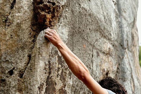
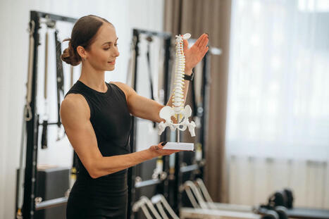
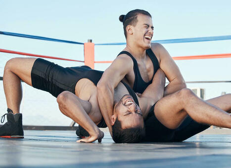

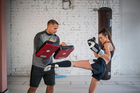
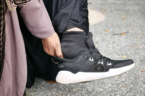
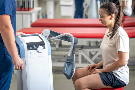


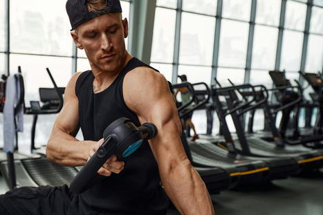


Explore the benefits of post-surgery leg strengthening. Prevent complications and rebuild confidence through leg exercises. For answers to any questions you may have, call Dr. Alexander Jimenez at 915-850-0900 or 915-412-6677