 Your new post is loading...

|
Scooped by
Gilbert C FAURE
November 12, 2025 5:13 AM
|
|
|
Scooped by
Gilbert C FAURE
November 9, 2024 4:33 AM
|
A newly approved drug blocks two cytokines...
|
|
Scooped by
Gilbert C FAURE
December 2, 2023 4:35 AM
|
WHAT IS ALREADY KNOWN ON THIS TOPICPatients with rheumatoid arthritis (RA) can be divided into seropositive and seronegative subgroups. The presence of antibodies against post-translationally modified (PTM) proteins such as citrullinated proteins is nowadays used as a diagnostic and prognostic marker in RA. Antibodies directed against carbamylated proteins have more recently been shown to be present in a subset of the seronegative patients and are associated with bone erosions in that group.WHAT THIS STUDY ADDSIn this study, two different anti-PTM antibodies are investigated: anti-advanced glycation end-product modified protein antibodies (anti-AGE) and anti-malondialdehyde-acetaldehyde adduct modified protein antibodies (anti-MAA). These antibodies can be detected in several forms of inflammatory arthritis. Within seronegative RA (negative for rheumatoid factor, anti-citrullinated protein antibodies and anti-carbamylated protein antibodies), 16.9% of patients are positive for anti-MAA and/or anti-AGE antibodies. This subgroup is characterised by an association with HLA-DRB1*03, increased radiographic joint damage and (for anti-MAA) inflammation.HOW THIS STUDY MIGHT AFFECT RESEARCH, PRACTICE OR POLICYThe presence of anti-PTM antibodies like anti-AGE and anti-MAA in patients with RA and other patients with inflammatory arthritis previously considered to be seronegative, may not only serve as a prognostic marker, but importantly may contribute to understanding the pathogenesis of these conditions, including a subset of RA.IntroductionIn rheumatoid arthritis (RA) around two-thirds of patients are autoantibody positive for rheumatoid factor (RF), anti-citrullinated protein antibodies (ACPA) and/or anti-carbamylated protein (anti-CarP) antibodies.1 The remaining seronegative subgroup of RA is clinically heterogeneous and thus far no reliable biomarkers are available to identify these patients or predict their disease course.1ACPA and anti-CarP are antibodies that recognise proteins that have undergone post-translational modification (PTM), citrullination of arginine and carbamylation of lysine respectively.2 3 However, many other types of PTMs exist.4 Two examples of PTMs that are found to associate with disease are advanced glycation end-products (AGE) and malondialdehyde-acetaldehyde adducts (MAA). AGEs are a result of oxidative stress and tissue damage5 and are, for example, present in patients with diabetes mellitus type 2.6 Interestingly, in these patients also antibodies directed against this PTM were observed.6 MAA modifications are a result of reactive oxygen species that are formed during inflammation and oxidative stress.7 Both MAA-modified proteins and anti-MAA antibodies are found in patients with RA, as well as in other diseases.7 AGE and MAA are both highly immunogenic PTMs.8 9 Therefore, it is plausible that antibodies against AGE and MAA are also present in patients with arthritis.Seronegative RA is associated with HLA-DRB1*03, suggesting a role for immunopathology driven by, for example, B cell immunity.10 Indeed, within the ACPA-negative patients, the presence of anti-CarP was associated with HLA-DRB1*03.11 12 However, it did not yet explain the full HLA-DRB1*03 association, raising the possibility that other anti-PTM responses may be present in ‘seronegative’ RA that are present in the remainder of the HLA-DRB1*03 positive individuals.13 14 On top of this haplotype association, within these patients with ACPA-negative RA, anti-CarP was found to associate with a more severe radiological progression.3 Patients with seronegative RA are a diverse group of patients that in many ways resemble undifferentiated arthritis. Presence of antibodies, like anti-PTM antibodies, might help to better understand and characterise subgroups that possibly belong to this so-called seronegative RA.We therefore investigated whether anti-AGE and anti-MAA antibodies are present in patients with RA and other forms of arthritis, and whether they could potentially close the so-called serological gap1 in seronegative RA.MethodsPatientsOne thousand one hundred eighty-six patients with arthritis of at least one joint and a symptom duration of less than 2 years were included in the Leiden Early Arthritis Clinic (EAC) cohort.15 Data were collected at baseline and follow-up (4, 12 months and yearly thereafter). Patients were being followed as long as the patient remained being seen clinically by the rheumatologist. RA was classified based on the 1987 American College of Rheumatology criteria (n=648).16 Definitive diagnoses other than RA (n=538) were made by the treating physician after 1 year of follow-up and were predominantly psoriatic arthritis (PsA) (n=100), inflammatory osteoarthritis (n=95) and gout (n=93) besides other more rare forms of arthritis. For this manuscript, the following diagnoses were termed autoimmune (AI): RA, PsA, spondyloarthritis, sarcoidosis, systemic lupus erythematosus (SLE) and paraneoplastic arthritis. The diagnoses termed as non-autoimmune (non-AI) were: gout, pseudogout and septic arthritis. Clinical and demographic patient characteristics were collected as described previously.17Genotyping, radiological progression and remissionFrom all patients, HLA genotypes were established as described previously.18 The alleles that were marked as shared epitope-encoding HLA (HLA-SE) positive were: HLA-DRB1*01:01, 01:02, 04:01, 04:04, 04:05, 04:08, 10:01 and 14:02. For the radiological progression analyses, 2853 X-ray sets of the hands and feet of 635 patients with RA were scored as described previously using the Sharp-van der Heijde score (SHS).19 20 Sustained drug-free remission (SDFR) was defined as the absence of clinical synovitis after discontinuation of disease-modifying antirheumatic drug treatment, that persisted for the entire follow-up, being at least 1 year.21Anti-AGE and anti-MAA measurementsAnti-AGE and anti-MAA antibodies were detected using an in-house ELISA based on modified fetal calf serum (FCS) as described previously.22 Briefly, modified and non-modified FCS were coated to a Nunc Maxisorp ELISA plate (430341, Thermofisher). In between each sequential step, plates were washed three times using phosphate buffered saline (PBS)/0.05%Tween (Sigma, P1379). After blocking (PBS/1%bovine serum albumin) for 6 hours at 4°C plates were incubated overnight at 4°C with 1/100 or 1/1000 diluted serum for anti-AGE and anti-MAA, respectively. Each plate contained a standard of anti-PTM positive serum to calculate arbitrary units. After incubation, IgG levels were detected using Rabbit-anti-Human IgG-HRP (Dako, P0214). Plates were developed by incubating with 2,2'-azino-bis(3-ethylbenzothiazoline-6-sulfonic acid (ABTS)/0.015% H2O2 (A1888 and 7722-84-1, both from Merck) and absorbance at 415 nm was measured using a microplate reader (Bio-Rad iMark). The cut-off for positivity was set as the mean arbitrary units plus two times the SD of 80 healthy controls, excluding values higher than 10× the mean.Statistical analysisIndependent samples t-test and Mann-Whitney U tests were used to analyse the baseline characteristics. The association of HLA-DRB1*03 with autoantibodies was assessed with logistic regression, and stratified for anti-cyclic citrullinated peptide 2 (anti-CCP2) and anti-CarP if relevant. Correlations between anti-PTM antibodies and inflammatory markers were calculated using Spearman’s rank correlation. For the radiological progression analyses, a multivariate normal regression model for longitudinal data was used with SHS as response variable. The model controlled for the age, sex and inclusion year of the patients.19 SDFR development until follow-up was calculated using Kaplan-Meier survival analysis and Cox’s regression. All statistical analysis were performed using SPSS statistics V.25 (IBM).ResultsAnti-AGE and anti-MAA in patients with arthritisBaseline characteristics are described in table 1. Anti-PTM antibody levels were measured in RA and non-RA arthritis patients and compared with healthy controls (figure 1A,B and online supplemental table 1). The non-RA arthritis group was divided into subgroups and separately depicted based as AI arthritis (without RA) including PsA, paraneoplastic arthritis, SLE, sarcoidosis and spondyloarthritis and as non-AI arthritis including septic arthritis, gout and pseudogout.Supplemental material[rmdopen-2023-003480supp001.pdf]View inline View popup Table 1 Baseline characteristics of the rheumatoid arthritis (RA), non-RA, autoimmune no RA and non-autoimmune group<img class="highwire-fragment fragment-image" height="440" src="https://rmdopen.bmj.com/content/rmdopen/9/4/e003480/F1.medium.gif"; width="315" alt="Figure 1">Download figure Open in new tab Download powerpoint Figure 1 Anti-AGE and anti-MAA show higher levels in RA and occur in a subgroup of patients with anti-CarP anti-CCP2 negative RA. IgG antibody levels of anti-AGE (A) and anti-MAA (B) in patients with (n=648) and without (n=538) RA. Early patients with arthritis were separately depicted as groups: AI without RA (including psoriatic arthritis, paraneoplastic arthritis, SLE, sarcoidosis and spondyloarthritis) and non-AI (including septic arthritis, gout, pseudogout). (C) Upset plots of groups of patients with RA (n=499*) positive for anti-PTM combinations; anti-AGE, anti-MAA, anti-CarP, anti-CCP2 and RF. *Data for anti-CarP was missing for 149 patients with RA. AGE, advanced glycation end-product; AI, autoimmune; aU/mL, arbitrary units per mL; CarP, carbamylated protein; CCP2, citrullinated cyclic peptide 2; MAA, malondialdehyde acetaldehyde adduct; RA, rheumatoid arthritis; RF, rheumatoid factor.Compared with healthy controls, anti-AGE and anti-MAA were most prevalent in RA (anti-AGE: 7.5% in HC vs 44.6% in RA and anti-MAA: 3.8% in HC vs 46.1% in RA) but were also present in other types of early arthritis. Within patients without RA, anti-AGE and anti-MAA were present in 32.9% and 30.3%, respectively and in non-RA AI arthritis anti-AGE and anti-MAA were found in 38.5% and 41.5%, respectively. These data indicate that the presence of anti-PTM antibodies is not specific for RA. When analysing combinations of autoantibodies, the largest subgroup of patients with RA (n=99) had all four anti-PTM antibodies (anti-AGE, anti-MAA, anti-CarP, anti-CCP2) as well as RF, after which the second largest group (n=63) was characterised by the combination of RF, anti-CCP2 and anti-CarP (figure 1C).Interestingly, 67 (34.0%) and 57 (28.9%) of patients with seronegative (RF negative, ACPA negative and anti-CarP negative) RA were positive for anti-AGE and anti-MAA, respectively. Moreover, 40 (20.3%) of these patients with seronegative RA were positive for both anti-AGE and anti-MAA. These anti-PTM responses may identify a new subgroup in the patients with otherwise seronegative RA.HLA-DRB1*03 associates with anti-AGE and anti-MAA independently of anti-CarP in patients with anti-CCP2-negative RASince HLA class II alleles are known to associate with autoantibody positivity in RA, we sought to investigate the presence of HLA-SE and its association with anti-AGE and anti-MAA antibodies. Of all patients with RA, 63.3% were HLA-SE+ (table 1). Based on the well-known association between HLA-SE and RA, the HLA-SE alleles were assessed and were significantly more prevalent in all RA subgroups compared with healthy controls. In the anti-AGE-positive group, as compared with patients with anti-AGE-negative RA however, the prevalence of HLA-SE alleles was similar (table 2). The same was true for anti-MAA; therefore, both anti-AGE and anti-MAA antibodies were not associated with HLA-SE.View inline View popup Table 2 Association between anti-AGE and anti-MAA antibodies and HLA-SE and HLA-DRB1*03 presence in RA and HLA-DRB1*03 presence in non-RA patients from the Leiden EAC cohortSince HLA-DRB1*03 is associated with seronegative RA and anti-CarP antibodies in this disease subset, we sought to investigate the association of HLA-DRB1*03 with anti-AGE and anti-MAA. In patients with RA, HLA-DRB1*03 was more prevalent in anti-AGE-positive and anti-MAA-positive patients as compared with healthy controls with OR values of 1.34 (95% CI 1.01 to 1.78, p=0.05) and 1.29 (95% CI 0.96 to 1.73, p=0.09), although this did not achieve statistical significance compared with anti-AGE-negative or anti-MAA-negative patients, respectively (table 2, part I). To investigate whether HLA-DRB1*03 is associated with anti-MAA and anti-AGE in anti-CCP2-negative RA, we focused on this subset and stratified the analysis for anti-CarP. Within the patients with anti-CCP2 negative RA, anti-AGE and anti-MAA antibodies were associated with HLA-DRB1*03 compared with healthy controls (OR: 1.98, 95% CI 1.27 to 3.07, p=0.003, and OR: 2.37, 95% CI 1.50 to 3.74, p<0.001, respectively). Anti-MAA was associated with HLA-DRB1*03 in the anti-CCP2 negative stratum independent of anti-CarP (OR: 1.91, 95% CI 1.11 to 3.30, p=0.02) (table 2, part II). In this stratified analysis, anti-AGE showed the same trend for association but did not reach significance (OR: 1.48, 95% CI 0.86 to 2.52, p=0.16). Since anti-AGE and anti-MAA often co-occur, we next stratified the association analysis for these autoantibodies, to dissect whether the observed association to HLA-DRB1*03 could be attributed to one of them in particular. After stratification for anti-AGE or anti-MAA, only patients with double positive RA showed a significant association with HLA-DRB1*03 compared with healthy controls (online supplemental table 2, part I). Since some controversy exists on the association of HLA-DRB1*03 in patients with anti-CCP2 negative RA, we investigated the association between anti-AGE and anti-MAA with HLA-DRB1*03 within patients with HLA-SE negative RA. In both HLA-SE negative and anti-CCP2 negative stratum, we find similar associations with anti-AGE/-MAA and HLA-DR1*03 (table 2, part III).In non-RA arthritis patients, both anti-AGE and anti-MAA showed a similar association with HLA-DRB1*03 with OR values of 2.34 (95% CI 1.58 to 3.47, p<0.001) and 1.94 (95% CI 1.29 to 2.92, p=0.002) compared with healthy controls (table 2, part I). In a comparison within the non-RA arthritis patients, HLA DRB1*03 remained significantly associated with anti-AGE-positive compared with anti-AGE-negative patients (OR: 2.22, 95% CI 1.28 to 3.84, p=0.01), while the association with anti-MAA did not remain significant. To disentangle the effects of anti-AGE and anti-MAA, analyses were again stratified, after which only the presence of anti-AGE in patients with anti-MAA-negative without RA remained significantly associated with HLA-DRB1*03 (online supplemental table 2, part II).Taken together, these data indicate that anti-AGE and anti-MAA associate with HLA-DRB1*03 in RA and non-RA arthritis patients, and that this association (which cannot be ascribed to anti-AGE or anti-MAA in particular) is mainly present in patients with anti-CCP2 negative RA. Similar associations were observed in patients with HLA-SE negative RA.Inflammation markers associate with anti-MAA positivity in RA and non-RA arthritisNext, we sought to investigate whether anti-PTM antibodies correlate with inflammation markers erythrocyte sedimentation rate (ESR) and C reactive protein (CRP) (table 3). Higher inflammation parameters in anti-AGE- and anti-MAA-positive individuals were observed in RA and non-RA arthritis patients, and in both the autoimmune and non-autoimmune subgroups of patients with arthritis. To investigate whether both anti-MAA and anti-AGE were associated with acute phase reactants in RA independently, anti-AGE and anti-MAA were stratified for each other. After this stratification, anti-AGE was no longer associated with either CRP or ESR whereas the association of anti-MAA with these inflammation markers remained significant (online supplemental table 3). These data indicate that anti-PTM responses, especially anti-MAA, is associated with markers of inflammation in early arthritis in both RA and non-RA arthritis patients.View inline View popup Table 3 Association between anti-AGE and anti-MAA antibin RA and non-RA arthritis patientsodies and ESR and CRP levelsAnti-AGE and anti-MAA associate with radiological progression in patients with anti-CCP2-negative RAWe next analysed if the presence of anti-AGE and anti-MAA is associated with radiological progression in RA. Anti-AGE-positive patients displayed more radiographic damage per year than anti-AGE-negative patients (p<0.001) (figure 2A). Data were then stratified for anti-CCP2, which revealed that this association was mainly present in the anti-CCP2-negative subgroup (figure 2B). When anti-CCP2 negative patients were further stratified for anti-CarP, the association between anti-AGE and radiographic progression remained significant (figure 2C). This indicates that in patients with anti-CCP2 negative RA, anti-AGE is associated with radiological progression independent of anti-CarP, suggesting that this anti-PTM antibody could discriminate a different subgroup. Anti-MAA positivity was also associated with radiological progression (p=0.002) (figure 2D). This effect was also observed in the anti-CCP2-negative stratum (figure 2E), although no longer significant after stratifying for anti-CCP2. The latter could be a consequence of power as the effect size (beta) which decreased only slightly to 1.03/year, p=0.16 (figure 2E).<img width="393" alt="Figure 2" height="440" class="highwire-fragment fragment-image" src="https://rmdopen.bmj.com/content/rmdopen/9/4/e003480/F2.medium.gif">Download figure Open in new tab Download powerpoint Figure 2 Anti-AGE and anti-MAA associate with radiological progression in patients with RA (n=600). (A) Radiological progression in anti-AGE positive and negative RA. (B) Data stratified for CCP2. (C) Data stratified for anti-CarP in anti-CCP2-negative stratum. (D) Radiological progression in anti-MAA positive and negative RA. (E) Data stratified for CCP2. Data presented as estimate (95% CI), p value. AGE, advanced glycation end-product; CarP, carbamylated protein; CCP2, citrullinated cyclic peptide 2; MAA, malondialdehyde acetaldehyde adduct.Presence of anti-MAA or anti-AGE is not associated with SDFR in RANext, we sought to investigate whether anti-AGE and anti-MAA were associated with SDFR over time (online supplemental figure 3). Anti-AGE was not associated with SDFR, HR 0.93 (95% CI 0.66 to 1.30; p=0.66) which did not differ after adjusting for CCP2 status (HR 1.14, 95% CI 0.81 to 1.61, p=0.46). Anti-MAA-positive patients were less likely to achieve SDFR, compared with anti-MAA-negative patients, HR 0.72 (95% CI 0.51 to 1.00, p=0.053). After adjusting for CCP2 status, there was no longer an association between anti-MAA and SDFR, HR 1.05 (95% CI 0.74 to 1.50, p=0.80).DiscussionIn this study, we demonstrated that anti-AGE and anti-MAA are present in patients with RA, and interestingly also in a substantial part of patients with otherwise seronegative RA. This is not specific for RA, as anti-AGE and anti-MAA antibodies were also present in other forms of early arthritis. Both anti-AGE and anti-MAA are associated with HLA-DRB1*03 in RA, and anti-AGE is also associated with HLA-DR1*03in non-RA arthritis patients. Anti-AGE and anti-MAA are associated with a distinct clinical phenotype: anti-AGE associates with radiological progression in RA whereas anti-MAA only showed a trend with radiological progression but associated with increased inflammatory parameters in both RA and non-RA arthritis.Associations with particular HLA class II alleles have been described to occur in many seropositive AI diseases.13 14 More specifically, HLA-DRB1*03, initially reported to be associated with anti-CCP2 negative RA, was later associated with the presence of anti-CarP, although not all HLA-DRB1*03-positive patients were anti-CarP-positive.10 12 In this study, we observed that HLA-DRB1*03 was associated with anti-AGE and anti-MAA in patients with anti-CCP2 negative RA which was independent of anti-CarP, thereby identifying another subgroup of anti-CCP2 negative RA that is associated with HLA-DRB1*03. In addition, anti-AGE associated with HLA-DRB1*03 in non-RA confirming the robustness of this finding. Together, these observations provide additional insight into the association of HLA-DBR1*03 with (rheumatoid) arthritis; although these alleles are not associated with the presence of ACPA, they do appear to predispose to the formation of other autoantibodies (anti-CarP, anti-AGE and anti-MAA) in a process in which HLA class II-associated T-cell-dependent immune responses are likely to be involved.Interestingly, in RA, anti-AGE associated with radiological progression independent of anti-CCP2 and anti-CarP suggesting an additive value of anti-AGE in determining disease evolution as it could define a new subgroup of patients with RA. Strikingly, anti-AGE was not associated with SDFR. In RA and non-RA, a subgroup of patients is characterised by more extensive inflammation and the presence of anti-MAA antibodies, while a subgroup of patients with CCP2-negative RA is characterised by radiological progression and presence of anti-AGE antibodies. Based on these results, distinct subgroups within RA and non-RA can be delineated based on their specific clinical phenotype.The presence of AGE-modified proteins and anti-AGE antibodies has been observed in diabetes and hypertension.6 23 Also, in synovial tissue and sera of patients with RA, AGE-modified proteins have been detected.24–26 In addition, MAA-modified proteins have been observed before in RA tissue7 and it is clear that both modifications can be induced by inflammation and oxidative stress in the inflamed joint.5 7 Our study now adds that in a subset of the patients with RA antibodies against these PTMs are present. Additionally, anti-AGE and anti-MAA have been found to be associated with ESR in previous studies in RA and SLE.22 PTMs and anti-PTMs such as anti-AGE and anti-MAA add to the understanding that the combined presence of the antigen and the antibody could trigger effector mechanisms and contribute to the overall process of arthritis and joint damage, in RA and also in non-RA. It would therefore be interesting to investigate whether next to carbamylated proteins27 also the modifications AGE and MAA are present in cartilage and synovium. Additionally, experimental pathogenicity studies on anti-AGE and anti-MAA specifically should be performed to elucidate on the contribution of these anti-PTM antibodies to pathogenesis.There are some limitations to our study. Data on anti-CarP antibody levels were missing for 149 patients with RA; therefore, analysis using stratification including anti-CarP could only be performed in a subgroup of all patients with RA. However, this group still consists of 499 patients with RA and therefore still appears a good representation of the RA population. Radiological progression was assessed in 635 patients with RA included before 2006. Thereafter, radiographs have not been scored since radiographic damage has become rare/nearly non-existent with current treatment strategies. This effectively enabled us to detect differences in the, earlier, informative part of the cohort. When stratifying radiological progression data, groups became small and therefore could suffer from insufficient power implicating that significance could not always be reached. It is therefore important to verify associations using different and/or bigger cohorts to be able to generalise findings to the whole RA population. Additionally, in order to verify the results obtained in this study, a replication cohort is needed. In such a study, IgA and IgM responses could be included to elaborate on the full anti-PTM antibody responses in patients with (rheumatoid) arthritis.28 29 One of the strengths of this study is that the EAC is a well-defined cohort containing RA and non-RA early arthritis patients with extensive information on the HLA haplotype and radiological progression for patients with RA.15 Second, antibody responses have been investigated on the PTM-modified proteins and their control proteins. All PTMs were created on the same antigen backbone and reactivity against FCS itself was subtracted from the results. This results in reliable measurements that capture truly PTM-specific signals and decreases the chance of false observations.30 Additionally, correlation analyses were performed (data not shown) and data were stratified for the other investigated anti-PTM and to verify that anti-AGE and anti-MAA are solely responsible for the observed result and not cross-reactive.In conclusion, anti-AGE and anti-MAA antibodies are both prevalent in patients with RA, and other inflammatory rheumatic conditions, and although not specific for RA they each correlate with specific parameters. Anti-MAA associates with HLA-DRB1*03 in CCP2-negative (RA) patients independent of anti-CarP and associates with inflammation. Anti-AGE associates with HLA-DRB1*03 in patients with CCP2-negative RA and is associated with a worse radiological progression especially in patients with anti-CCP2-negative and anti-CarP-negative RA. With this study, we have now characterised a seropositive subgroup within the heterogeneous group of patients with RA that have been thus far been considered seronegative.Data availability statementData are available upon reasonable request. Requests can be sent to l.a.trouw@lumc.nl.Ethics statementsPatient consent for publicationNot applicable.Ethics approvalThe Leiden Early Arthritis Clinic (EAC) cohort was approved by the Medical Ethics Committee Leiden The Hague Delft under reference number: B19.008. For the current study, measurement of anti-PTM antibodies in serum from the EAC cohort is approved under reference number B15.003. Participants gave informed consent to participate in the study before taking part.AcknowledgmentsWe thank Marloes Verstappen and Bianca M. Boxma-de Klerk for their assistance regarding the remission analysis on EAC cohort data.References↵Trouw LA, Mahler M. Closing the serological gap: promising novel biomarkers for the early diagnosis of rheumatoid arthritis. Autoimmun Rev 2012;12:318–22. doi:10.1016/j.autrev.2012.05.007OpenUrlCrossRefPubMed↵Schellekens GA, de Jong BA, van den Hoogen FH, et al. Citrulline is an essential constituent of antigenic determinants recognized by rheumatoid arthritis-specific autoantibodies. J Clin Invest 1998;101:273–81. doi:10.1172/JCI1316OpenUrlCrossRefPubMedWeb of Science↵Shi J, Knevel R, Suwannalai P, et al. Autoantibodies recognizing carbamylated proteins are present in sera of patients with rheumatoid arthritis and predict joint damage. Proc Natl Acad Sci U S A 2011;108:17372–7. doi:10.1073/pnas.1114465108OpenUrlAbstract/FREE Full Text↵Xu H, Wang Y, Lin S, et al. PTMD: a database of human disease-associated post-translational modifications. Genom Proteom Bioinform 2018;16:244–51. doi:10.1016/j.gpb.2018.06.004OpenUrl↵Schmidt AM, Yan SD, Yan SF, et al. The multiligand receptor RAGE as a progression factor amplifying immune and inflammatory responses. J Clin Invest 2001;108:949–55. doi:10.1172/JCI14002OpenUrlCrossRefPubMedWeb of Science↵Nikolov A, Blazhev A, Tzekova M, et al. Serum levels of antibodies to advanced glycation end products in patients with type 2 diabetes mellitus and hypertension. Folia Med (Plovdiv) 2020;62:295–301. doi:10.3897/folmed.62.e47788OpenUrl↵Thiele GM, Duryee MJ, Anderson DR, et al. Malondialdehyde-acetaldehyde adducts and anti-malondialdehyde-acetaldehyde antibodies in rheumatoid arthritis. Arthritis Rheumatol 2015;67:645–55. doi:10.1002/art.38969OpenUrl↵Buongiorno AM, Morelli S, Sagratella E, et al. Immunogenicity of advanced glycation end products in diabetic patients and in nephropathic non-diabetic patients on hemodialysis or after renal transplantation. J Endocrinol Invest 2008;31:558–62. doi:10.1007/BF03346408OpenUrlPubMed↵Thiele GM, Tuma DJ, Willis MS, et al. Soluble proteins modified with acetaldehyde and malondialdehyde are Immunogenic in the absence of adjuvant. Alcohol Clin Exp Res 1998;22:1731–9.OpenUrlCrossRefPubMed↵Verpoort KN, van Gaalen FA, van der Helm-van Mil AHM, et al. Association of HLA-DR3 with anti-cyclic citrullinated peptide antibody-negative rheumatoid arthritis. Arthritis Rheum 2005;52:3058–62. doi:10.1002/art.21302OpenUrlCrossRefPubMedWeb of Science↵Regueiro C, Rodriguez-Rodriguez L, Triguero-Martinez A, et al. Specific association of HLA-Drb1*03 with anti-carbamylated protein antibodies in patients with rheumatoid arthritis. Arthritis Rheumatol 2019;71:331–9. doi:10.1002/art.40738OpenUrl↵Jiang X, Trouw LA, van Wesemael TJ, et al. Anti-carp antibodies in two large cohorts of patients with rheumatoid arthritis and their relationship to genetic risk factors, cigarette smoking and other autoantibodies. Ann Rheum Dis 2014;73:1761–8. doi:10.1136/annrheumdis-2013-205109OpenUrlAbstract/FREE Full Text↵Kirino Y, Remmers EF. Genetic architectures of seropositive and seronegative rheumatic diseases. Nat Rev Rheumatol 2015;11:401–14. doi:10.1038/nrrheum.2015.41OpenUrlCrossRefPubMed↵Cruz-Tapias P, Pérez-Fernández OM, Rojas-Villarraga A, et al. Shared HLA class II in six autoimmune diseases in Latin America: a meta-analysis. Autoimmune Dis 2012;2012:569728. doi:10.1155/2012/569728↵de Rooy DPC, van der Linden MPM, Knevel R, et al. Predicting arthritis outcomes--what can be learned from the Leiden early arthritis clinic Rheumatology (Oxford) 2011;50:93–100. doi:10.1093/rheumatology/keq230OpenUrlCrossRefPubMedWeb of Science↵Arnett FC, Edworthy SM, Bloch DA, et al. The American rheumatism association 1987 revised criteria for the classification of rheumatoid arthritis. Arthritis Rheum 1988;31:315–24. doi:10.1002/art.1780310302OpenUrlCrossRefPubMedWeb of Science↵van Aken J, van Bilsen JH, Allaart CF, et al. The leiden early arthritis clinic. Clin Exp Rheumatol 2003;21:S100–5.OpenUrlPubMedWeb of Science↵Huizinga TWJ, Amos CI, van der Helm-van Mil AHM, et al. Refining the complex rheumatoid arthritis phenotype based on specificity of the HLA-DRB1 shared EPITOPE for antibodies to citrullinated proteins. Arthritis Rheum 2005;52:3433–8. doi:10.1002/art.21385OpenUrlCrossRefPubMedWeb of Science↵Knevel R, Krabben A, Brouwer E, et al. Genetic variants in Il15 associate with progression of joint destruction in rheumatoid arthritis: a multicohort study. Ann Rheum Dis 2012;71:1651–7. doi:10.1136/annrheumdis-2011-200724OpenUrlAbstract/FREE Full Text↵van der Heijde D. How to read radiographs according to the sharp/Van der heijde method. J Rheumatol 2000;27:261–3.OpenUrlPubMedWeb of Science↵Verstappen M, van Steenbergen HW, de Jong PHP, et al. Unraveling heterogeneity within ACPA-negative rheumatoid arthritis: the subgroup of patients with a strong clinical and serological response to initiation of DMARD treatment favor disease resolution. Arthritis Res Ther 2022;24:4. doi:10.1186/s13075-021-02671-z↵Monahan RC, van den Beukel MD, Borggreven NV, et al. Autoantibodies against specific post-translationally modified proteins are present in patients with lupus and associate with major neuropsychiatric manifestations. RMD Open 2022;8:e002079. doi:10.1136/rmdopen-2021-002079↵Aso Y, Inukai T, Tayama K, et al. Serum concentrations of advanced glycation endproducts are associated with the development of atherosclerosis as well as diabetic microangiopathy in patients with type 2 diabetes. Acta Diabetol 2000;37:87–92. doi:10.1007/s005920070025OpenUrlCrossRefPubMedWeb of Science↵Drinda S, Franke S, Canet CC, et al. Identification of the advanced glycation end products N(Epsilon)-carboxymethyllysine in the synovial tissue of patients with rheumatoid arthritis. Ann Rheum Dis 2002;61:488–92. doi:10.1136/ard.61.6.488OpenUrlAbstract/FREE Full Text↵de Groot L, Hinkema H, Westra J, et al. Advanced glycation endproducts are increased in rheumatoid arthritis patients with controlled disease. Arthritis Res Ther 2011;13:R205. doi:10.1186/ar3538↵Ligier S, Fortin PR, Newkirk MM. A new antibody in rheumatoid arthritis targeting glycated IgG: Igm anti-IgG-AGE. Br J Rheumatol 1998;37:1307–14. doi:10.1093/rheumatology/37.12.1307OpenUrlCrossRefPubMed↵Verheul MK, Janssen GMC, de Ru A, et al. Mass-spectrometric identification of carbamylated proteins present in the joints of rheumatoid arthritis patients and controls. Clin Exp Rheumatol 2021;39:570–7. doi:10.55563/clinexprheumatol/0ms5pkOpenUrlPubMed↵van Delft MAM, van der Woude D, Toes REM, et al. Secretory form of rheumatoid arthritis-associated autoantibodies in serum are mainly of the Igm Isotype, suggesting a continuous reactivation of autoantibody responses at mucosal surfaces. Ann Rheum Dis 2019;78:146–8. doi:10.1136/annrheumdis-2018-213724OpenUrlFREE Full Text↵Mikuls TR, Duryee MJ, England BR, et al. Malondialdehyde-acetaldehyde antibody concentrations in rheumatoid arthritis and other rheumatic conditions. Int Immunopharmacol 2018;56:113–8. doi:10.1016/j.intimp.2018.01.022OpenUrl↵Åhlin E, Elshafie AI, Nur MAM, et al. Anti-citrullinated peptide antibodies in sudanese patients with leishmania donovani infection exhibit reactivity not dependent on citrullination. Scand J Immunol 2015;81:201–8. doi:10.1111/sji.12265OpenUrl
|
|
Scooped by
Gilbert C FAURE
March 1, 2023 6:26 AM
|
In conclusion, screening an axSpA cDNA phage display library for IgA reactivity resulted in the identification of 7 novel UH-axSpA-IgA antigens, of which 2 show promising biomarker potential for the diagnosis of a subset of axSpA patients, in combination with previously identified UH-axSpA-IgG antig...
|
|
Scooped by
Gilbert C FAURE
January 19, 2022 5:03 AM
|
bioRxiv - the preprint server for biology, operated by Cold Spring Harbor Laboratory, a research and educational institution...
|
|
Scooped by
Gilbert C FAURE
July 27, 2021 11:27 AM
|
ObjectiveEvaluate the MRI evidence of active inflammatory and chronic structural damages in radiographic axial spondyloarthritis (r-axSpA) and non-radiographic axial spondyloarthritis (nr-axSpA).MethodsA retrospective review of 253 patients who underwent sacroiliac joint (SIJ) MRI between June 2014 and December 2019 was performed. MRI images including short tau inversion recovery scan and T1-weighted spin echo scans were assessed using the Spondyloarthritis Research Consortium of Canada (SPARCC) score and SPARCC MRI SIJ structural score by two independent readers.ResultsHigher mean score of inflammatory (SPARCC) was seen in r-axSpA patients when compared with nr-axSpA patients (8.08 vs 4.37, P<0.05). Frequencies of MRI structural lesions in r-axSpA patients and nr-axSpA patients were as follows: erosion (65.84 vs 88.23%, P=0.002), backfill (33.17 vs 13.73%, P<0.001), fat metaplasia (79.21 vs 60.78%, P=0.01), and ankylosis (37.13 vs 1.96%, P<0.001). Patients with r-axSpA had a higher mean score for fat metaplasia (8.93 vs 4.06, P=0.0003) and ankylosis (4.49 vs 0.04, P<0.001).ConclusionMore active inflammatory and chronic structural damages except for erosion were seen in r-axSpA patients than nr-axSpA patients, while higher percentage of nr-axSpA patients presented with erosion in MRI.
|
|
Scooped by
Gilbert C FAURE
July 14, 2020 2:44 AM
|
Gut inflammation is strongly associated with spondyloarthritis (SpA), as exemplified by the high prevalence of inflammatory bowel disease (IBD) and the even higher occurrence of subclinical gut inflammation in patients with SpA. The gut–joint axis of inflammation in SpA is further reinforced by similarities in immunopathogenesis at both anatomical sites and by the clinical success of therapies blocking TNF and IL-23 in IBD and in some forms of SpA. Many genetic risk factors are shared between SpA and IBD, and changes in the composition of gut microbiota are seen in both diseases. Current dogma is that inflammation in SpA initiates in the gut and leads to joint inflammation; however, although conceptually attractive, some research does not support this causal relationship. For example, therapies targeting IL-17A are efficacious in the joint but not the gut, and interfering with gut trafficking by targeting molecules such as α4β7 in IBD can lead to onset or flares of SpA. Several important knowledge gaps remain that must be addressed in future studies. Determining the true nature of the gut–joint axis has real-world implications for the treatment of patients with co-incident IBD and SpA and for the repurposing of therapeutics from one disease to the other. In this article, the authors summarize the latest clinical and basic research on gut inflammation in spondyloarthritis and highlight important questions to address in future research.
|
|
Scooped by
Gilbert C FAURE
January 16, 2020 2:17 PM
|
Patients with axial spondyloarthritis have a chronic, immune-mediated inflammatory disease that tends to localize to the sacroiliac joints and spine. Ankylosing spondylitis is, perhaps, the most representative of this group of diseases. Rheumatologists treat patients with axial spondyloarthritis with biologics, such as tumor necrosis factor inhibitors (TNFi’s), which can improve quality of life, activity and... [Read More]
|
|
Scooped by
Gilbert C FAURE
November 21, 2019 10:20 AM
|
The cytokines IL-23 and IL-17 have an important role in the pathogenesis of, and as a therapeutic target in, both animal models of chronic inflammation and some human chronic inflammatory diseases. The traditional view is that a main source of IL-17 is T cells and that IL-17 production is under the control of IL-23. IL-17 inhibition has shown good efficacy in clinical trials for ankylosing spondylitis (AS), a subtype of axial spondyloarthritis (axSpA) characterized by radiographic evidence of sacroiliitis. On the basis of data from animal models, genetic studies and the investigation of tissue and blood samples from patients with AS, IL-23 had also been predicted to be important in the pathogenesis of this disease and was therefore considered a potential therapeutic target for axSpA. However, two placebo-controlled, double-blind clinical trials in axSpA of monoclonal antibodies directed against either the p40 protein or the p19 protein of the IL-23 molecule had clear negative results. These findings indicate that IL-23 and IL-17 are at least partly uncoupled in axSpA. Reasons as to why, when and how such an uncoupling might occur are discussed in this Review, with special reference to the unique microenvironment of the subchondral bone marrow in axSpA. Therapies targeting IL-23 have failed in clinical trials in axial spondyloarthritis (axSpA), despite the importance of the IL-23–IL-17 pathway in axSpA pathogenesis. This Review explores whether IL-17 production might be uncoupled from IL-23 in axSpA.
|
|
Scooped by
Gilbert C FAURE
October 10, 2019 12:46 PM
|
MRI of the sacroiliac joints is increasingly acknowledged as being indispensable in the early diagnosis of axial spondyloarthritis (axSpA) and as having a prominent role in the prognosis and classification of axSpA. Technological advances include improvements in the resolution of structural lesions and in methodologies for the quantification of lesions. Limited access and expertise in interpretation of MRI have led to a resurgence of interest in CT, especially the development of low radiation protocols for assessing the sacroiliac joints. Trials of TNF inhibitors in patients with non-radiographic axSpA have led to greater understanding of the role of MRI in selecting which patients might respond well to this therapy. The role of MRI features as target end points in treat-to-target strategies remains unclear because the effect of such targeting on structural damage parameters has only recently been explored. The relative importance of active and structural lesions for prognostic risk assessment and selection of appropriate treatment is also an area of current research. Given the increased capacity to visualize a broad array of lesions in both the sacroiliac joints and the spine using MRI and CT, these modalities will probably be increasingly employed for assessment of the disease-modifying activity of new therapies. Different imaging modalities, such as radiography, MRI and CT, have different advantages and can help the clinician with different aspects in assessing axial spondyloarthritis (axSpA). This Review covers imaging aspects relating to the diagnosis, classification and management of axSpA.

|
Suggested by
Société Francaise d'Immunologie
June 7, 2019 2:01 AM
|
Ankylosing spondylitis (AS) and the related spondyloarthritis (SpA) have become an extremely productive field in immunogenetic research over the last several years, justifying an update. This timely special issue will thoroughly cover the strong heritability of SpA by tackling the explosion of new...
|
|
Suggested by
Société Francaise d'Immunologie
April 24, 2019 5:22 AM
|
Curr Opin Rheumatol. 2018 Sep;30(5):526-532. doi: 10.1097/BOR.0000000000000529.
|
|
Scooped by
Gilbert C FAURE
February 10, 2019 12:55 PM
|
Spondyloarthritis (SpA) is a chronic inflammatory disease affecting primarily axial and peripheral joints and sometimes also extra-articular organs, such as the gut. Rats transgenic for HLA-B27 and human β2-microglobulin (B27-Tg rat) develop clinical manifestations resembling human disease.
|
|
|
Scooped by
Gilbert C FAURE
January 19, 2025 6:57 AM
|
|
|
Scooped by
Gilbert C FAURE
December 13, 2023 4:40 AM
|
Article Text Article menu PDF Viewpoint Goodbye to the term ‘ankylosing spondylitis’, hello ‘axial spondyloarthritis’: time to embrace the ASAS-defined nomenclature http://orcid.org/0000-0002-5781-158XDésirée van der Heijde1, http://orcid.org/0000-0003-2246-1986Anna Molto2,3,...
|
|
Scooped by
Gilbert C FAURE
June 17, 2023 7:02 AM
|
Audio Rheum Editor’s Picks Dr. Earl D. Silverman, MD, shares his monthly Editor’s Picks and their relevance in current clinical practice: June 2023 Featured articles: Kiltz, et al: Clinimetric Validation of the Assessment of Spondyloarthritis International Society Health Index in Patients With Radiographic Axial Spondyloarthritis in Ixekizumab Trials Alduraibi, et al: Clustering Patients With Gout Based on Comorbidities and Biomarkers: A Cross-Sectional Study Primeau, et al: Responders to Medial Opening Wedge High Tibial Osteotomy for Knee Osteoarthritis Kumthekar, et al: Physical Activity Habits Among Older Adults Living With Rheumatic Disease Wohlfahrt, et al: Pain Mechanisms Associated With Disease Activity in Patients With Rheumatoid Arthritis Treated With Disease-Modifying Antirheumatic Drugs: A Regression Tree Analysis May 2023 Featured articles: Cook, et al: Comparative Effectiveness of BNT162b2 and mRNA-1273 Vaccines Against COVID-19 Infection Among Patients With Systemic Autoimmune Rheumatic Diseases on Immunomodulatory Medications Chen, et al: Validation of the Antineutrophil Cytoplasmic Antibody Renal Risk Score and Modification of the Score in a Chinese Cohort With a Majority of Myeloperoxidase-Positive Patients Gorzewski, et al: Predicting Disease Activity in Rheumatoid Arthritis With the Fibromyalgia Survey Questionnaire: Does the Severity of Fibromyalgia Symptoms Matter? Oguro, et al: Effect of Communicative and Critical Health Literacy on Trust in Physicians Among Patients With Systemic Lupus Erythematosus (SLE): The TRUMP2-SLE Project Barber, et al: Investigating Associations Between Access to Rheumatology Care, Treatment, Continuous Care, and Healthcare Utilization and Costs Among Older Individuals With Rheumatoid Arthritis April 2023 Featured articles: Coates, et al: Sex-Specific Differences in Patients With Psoriatic Arthritis: A Systematic Review Johnson, et al: Evaluating the Threshold Score for Classification of Systemic Lupus Erythematosus Using the EULAR/ACR Criteria Gong, et al: The Association Between Quadriceps Strength and Synovitis in Knee Osteoarthritis: An Exploratory Study From the Osteoarthritis Initiative Jatuworapruk, et al: Prevalence, Risk Factors, and Outcomes of Gout Flare in Patients Hospitalized for PCR-Confirmed COVID-19: A Multicenter Retrospective Cohort Study Bosch, et al: Etanercept Withdrawal and Retreatment in Nonradiographic Axial Spondyloarthritis: Results of RE-EMBARK, an Open-Label Phase IV Trial March 2023 Featured articles: Takanashi, et al: Effects of Aging on Rheumatoid Factor and Anticyclic Citrullinated Peptide Antibody Positivity in Patients With Rheumatoid Arthritis Kiltz, et al: Clinically Relevant Deficits in Performance Tests in Patients With Axial Spondyloarthritis Schletzbaum, et al: Age-Stratified 30-day Rehospitalization and Mortality and Predictors of Rehospitalization Among Patients With Systemic Lupus Erythematosus: A Medicare Cohort Study Berard, et al: Canadian Rheumatology Association Recommendations for the Screening, Monitoring, and Treatment of Juvenile Idiopathic Arthritis-Associated Uveitis Schultz, et al: B Cell Reconstitution is Associated With COVID-19 Booster Vaccine Responsiveness in Patients Previously Seronegative Treated With Rituximab February 2023 Featured articles: Macfarlane, et al: Inflammatory Bowel Disease Risk in Patients With Axial Spondyloarthritis Treated With Biologic Agents Determined Using the BSRBR-AS and a MetaAnalysis Gossec, et al: Women With Psoriatic Arthritis Experience Higher Disease Burden Than Men: Findings From a Real-World Survey in the United States and Europe Davis, et al: The Effect of Psychiatric Comorbidity on Healthcare Utilization for Youth With Newly Diagnosed Systemic Lupus Erythematosus Pyo, et al: The Reclassification of Patients With Previously Diagnosed Eosinophilic Granulomatosis With Polyangiitis Based on the 2022 ACR/EULAR Criteria for Antineutrophil Cytoplasmic Antibody–Associated Vasculitis Weng, et al: Adult-Onset Still Disease After ChAdOx1 nCOV-19 Vaccination Mitchell, et al: How to Provide Sexual and Reproductive Health Care to Patients: Focus Groups With Rheumatologists and Rheumatology Advanced Practice Providers January 2023 Featured articles: Kodishala, et al: Risk Factors for Dementia in Patients With Incident Rheumatoid Arthritis: A Population-Based Cohort Study Beauvais, et al: Development and Validation of a Self-Administered Questionnaire Measuring Essential Knowledge in Patients With Axial Spondyloarthritis Orbai, et al: Impact of Physician-Defined Flares on Quality of Life and Work Impairment: An International Survey of 2238 Patients With Psoriatic Arthritis Smitherman, et al: Patient-Reported Outcomes Among Transition-Age Young Adults With Juvenile Idiopathic Arthritis in the Childhood Arthritis and Rheumatology Research Alliance Registry Stull, et al: Cutaneous Involvement in Systemic Lupus Erythematosus: A Review for the Rheumatologist Masi, et al: Reflections for the 50th Anniversary of The Journal of Rheumatology: The Past, Present, and Future of Rheumatology Santos, et al: A Rare Case of Subcutaneous Sarcoidosis in Patient With Psoriatic Arthritis Garg, et al: Timing and Predictors of Incident Cardiovascular Disease in Systemic Lupus Erythematosus: Risk Occurs Early and Highlights Racial Disparities December 2022 Featured articles: Koo, et al: Relationship Between Inflammation and Radiographic Progression in Patients With Ankylosing Spondylitis Attaining a BASDAI of Less Than 4 During Tumor Necrosis Factor Inhibitor Treatment Sun, et al: Development and Initial Validation of a Systemic Lupus Erythematosus–Specific Measure of the Extent of and Reasons for Medication Nonadherence Patterson, et al: Physical Activity Associates With Lower Systemic Inflammatory Gene Expression in Rheumatoid Arthritis Coleman, et al: Long-Term Follow-up of a Randomized Controlled Trial of Allopurinol Dose Escalation to Achieve Target Serum Urate in People With Gout Smith: Rusty and Wooden Tanomogi, et al: Extravascular Necrotizing Granuloma: A Diagnostic Clue for Eosinophilic Granulomatosis With Polyangiitis Isnardi, et al: Immune Response to SARS-CoV-2 Third Vaccine in Patients With Rheumatoid Arthritis Who Had No Seroconversion After Primary 2-Dose Regimen With Inactivated or Vector-Based Vaccines November 2022 Featured articles: Nelson, et al: Narrative Review of Machine Learning in Rheumatic and Musculoskeletal Diseases for Clinicians and Researchers: Biases, Goals, and Future Directions Hermans, et al: Are All Routine Spondyloarthritis Outpatient Visits Considered Useful by Rheumatologists? An Exploratory Clinical Practice Study Kallas, et al: Trajectory of Damage Accrual in Systemic Lupus Erythematosus Based on Ethnicity and Socioeconomic Factors Nozawa, et al: Early Abnormal Nailfold Capillary Changes Are Predictive of Calcinosis Development in Juvenile Dermatomyositis Chevet, et al: COVID-19 Vaccine Uptake Among Patients With Systemic Lupus Erythematosus in the American Midwest: The Lupus Midwest Network (LUMEN) Remize, et al: Melorheostosis or “Dripping Candle Wax” Bone Disease Bermas: The Unintended Consequence of the Overturn of Roe v Wade: Restrictions on Methotrexate Use October 2022 Featured articles: Hazlewood, et al: Canadian Rheumatology Association Living Guidelines for the Pharmacological Management of Rheumatoid Arthritis With Disease-Modifying Antirheumatic Drugs Schneeberger, et al: Simplified Ankylosing Spondylitis Disease Activity Score (SASDAS) Versus ASDAS: A Post Hoc Analysis of a Randomized Controlled Trial Kasiem, et al: A Practical Guide for Assessment of Skin Burden in Patients With Psoriatic Arthritis Tollisen, et al: Personally Generated Quality of Life Outcomes in Adults With Juvenile Idiopathic Arthritis Glintborg, et al: Long-term Behavioral Changes During the COVID-19 Pandemic and Impact of Vaccination in Patients With Inflammatory Rheumatic Diseases September 2022 Featured articles: Cagnotto, et al: Male Sex Predicts a Favorable Outcome in Early ACPA-Negative Rheumatoid Arthritis: Data From an Observational Study Hazlewood, et al: Frequency of Symptomatic Adverse Events in Rheumatoid Arthritis: An Exploratory Online Survey John M. Davis III: The Patient Experience of Drug Side Effects in Rheumatoid Arthritis: Intriguing Data From an Exploratory Online Survey Venkatachalam, et al: Taking the Long View: Patients Perceive Benefits and Risks of Treatment as Multidimensional Wallace, et al: The Association of Illness-related Uncertainty With Mental Health in Systemic Autoimmune Rheumatic Diseases August 2022 Featured articles: Verweij, et al: Whole-Body Macrophage Positron Emission Tomography Imaging for Disease Activity Assessment in Early Rheumatoid Arthritis Nguyen, et al: Secukinumab in United States Biologic-Naïve Patients With Psoriatic Arthritis: Results From the Randomized, Placebo-Controlled CHOICE Study Le Ralle, et al: Patient Acceptable Symptom State for Burden From Appearance Changes in People With Systemic Sclerosis: A Cross-sectional Survey Baggett, et al: Incidence Rates of Psoriasis in Children With Inflammatory Bowel Disease and Juvenile Arthritis Treated With Tumor Necrosis Factor Inhibitors and Disease-Modifying Antirheumatic Drugs Carluzzo, et al: Patient Empowerment Among Adults With Arthritis: The Case for Emotional Support July 2022 Featured articles: Kelty, et al: Mortality Rates in Patients With Ankylosing Spondylitis With and Without Extraarticular Manifestations and Comorbidities: A Retrospective Cohort Study Mease, et al: Baseline Disease Activity Predicts Achievement of cDAPSA Treatment Targets With Apremilast: Phase III Results in DMARD-naïve Patients With Psoriatic Arthritis Kuwana, et al: Tacrolimus in Patients With Interstitial Pneumonia Associated With Polymyositis or Dermatomyositis: Interim Report of Postmarketing Surveillance in Japan Kiadaliri, et al: Gout and Hospital Admission for Ambulatory Care–Sensitive Conditions: Risks and Trajectories McDermott, et al: Demographic, Lifestyle, and Serologic Risk Factors for Rheumatoid Arthritis (RA)–associated Bronchiectasis: Role of RA-related Autoantibodies June 2022 Featured articles: Darabian, et al: Using FibroScan to Assess for the Development of Liver Fibrosis in Patients With Arthritis on Methotrexate: A Single-center Experience Maguire, et al: Central Obesity in Axial Spondyloarthritis: The Missing Link to Understanding Worse Outcomes in Women? Miloslavsky, et al: The Challenge of Addressing the Rheumatology Workforce Shortage Maheswaranathan, et al: Association of Health Literacy and Numeracy With Lupus Knowledge and the Creation of the Lupus Knowledge Assessment Test Emad, et al: Why Do Patients With Gout Not Take Allopurinol? May 2022 Featured articles: Movahedi, et al: Physician- and Patient-reported Effectiveness Are Similar for Tofacitinib and TNFi in Rheumatoid Arthritis: Data From a Rheumatoid Arthritis Registry El Tal, et al: Consensus Approach to a Treat-to-target Strategy in Juvenile Idiopathic Arthritis Care: Report From the 2020 PR-COIN Consensus Conference Thompson, et al: Modifiable Factors and Incident Gout Across Ethnicity Within a Large Multiethnic Cohort of Older Adults Widdifield, et al: COVID-19 Vaccination Uptake Among Individuals With Immune-mediated Inflammatory Diseases in Ontario, Canada, Between December 2020 and October 2021: A Population-based Analysis Van Praet, et al: Acute Perimyocarditis in a Case of Multisystem Inflammatory Syndrome in Adults Chang, et al: Systemic Lupus Erythematosus Increases the Risk of Gestational Diabetes: Truth or Illusion? McCormick and Choi: Racial Disparities in the Modern Gout Epidemic April 2022 Featured articles: Exarchou, et al: Lifestyle Factors and Disease Activity Over Time in Early Axial Spondyloarthritis: The SPondyloArthritis Caught Early (SPACE) Cohort - https://doi.org/10.3899/jrheum.210046 van Vollenhoven, et al: Efficacy and Safety of Ustekinumab in Patients With Active Systemic Lupus Erythematosus: Results of a Phase II Open-label Extension Study - https://doi.org/10.3899/jrheum.210805 Giancane, et al: Anakinra in Patients With Systemic Juvenile Idiopathic Arthritis: Long-term Safety From the Pharmachild Registry - https://doi.org/10.3899/jrheum.210563 Barber, et al: Best Practices for Virtual Care: A Consensus Statement From the Canadian Rheumatology Association - https://doi.org/10.3899/jrheum.211017 Zickuhr and Mandell: Rheumatology Education Needs a Splash of Color - https://doi.org/10.3899/jrheum.211233 Li, et al: Brain Abscess Due to Nocardia in a Patient With Systemic Lupus Erythematosus- https://doi.org/10.3899/jrheum.210971 March 2022 Featured articles: te Kampe, et al: Outcomes of Care Among Patients With Gout in Europe: A Cross-sectional Survey - https://doi.org/10.3899/jrheum.210009 Stransky, et al: Exploring Family Planning, Parenting, and Sexual and Reproductive Health Care Experiences of Men With Rheumatic Diseases - https://doi.org/10.3899/jrheum.210785 Beckers, et al: Performance of 3 Composite Measures for Disease Activity in Peripheral Spondyloarthritis - https://doi.org/10.3899/jrheum.210075 Sun, et al: Long-term Risk of Heart Failure and Other Adverse Cardiovascular Outcomes in Granulomatosis With Polyangiitis: A Nationwide Cohort Study - https://doi.org/10.3899/jrheum.210677 Curtis, et al: Characteristics, Comorbidities, and Outcomes of SARS-CoV-2 Infection in Patients With Autoimmune Conditions Treated With Systemic Therapies: A Population-based Study - https://doi.org/10.3899/jrheum.210888 Gunasuntharam: Why Should It Be Different From the Other Side? A Parent and Pediatrician’s Perspective of a Child With Kawasaki Disease - https://doi.org/10.3899/jrheum.211159 Lazarou, et al: Adult-onset Acute Calcific Discitis - https://doi.org/10.3899/jrheum.210838 Khanna: Remission in Gout: Concepts From a Patient Perspective - https://doi.org/10.3899/jrheum.211285 Coates and Tillett: How Should We Measure Peripheral Spondyloarthritis? - https://doi.org/10.3899/jrheum.211043 February 2022 Featured articles: Rosenbaum, et al: The Interplay Between COVID-19 and Spondyloarthritis or Its Treatment - https://doi.org/10.3899/jrheum.210742 van der Heijde, et al: Radiographic Progression of Structural Joint Damage Over 5 Years of Baricitinib Treatment in Patients With Rheumatoid Arthritis: Results From RA-BEYOND - https://doi.org/10.3899/jrheum.210346 van der Meer, et al: Extraskeletal Manifestations in Axial Spondyloarthritis Are Associated With Worse Clinical Outcomes Despite the Use of Tumor Necrosis Factor Inhibitor Therapy - https://doi.org/10.3899/jrheum.210308 Haddad, et al: The Association of Psoriatic Arthritis With All-cause Mortality and Leading Causes of Death in Psoriatic Arthritis - https://doi.org/10.3899/jrheum.210159 Yazdanyar, et al: Risk of 30-day Readmission After Knee or Hip Replacement in Rheumatoid Arthritis and Osteoarthritis by Non-Medicare and Medicare Payer Status - https://doi.org/10.3899/jrheum.201370 Perez Acosta et al: Cystic Ganglionosis in a 3-year-old Mimicking Juvenile Idiopathic Arthritis - https://doi.org/10.3899/jrheum.210558 Leung: Is Psoriatic Arthritis Associated With Higher Risk of Mortality? - https://doi.org/10.3899/jrheum.210963 January 2022 Featured articles: Bermas, et al: COVID-19 in Pregnant Women With Rheumatic Disease: Data From the COVID-19 Global Rheumatology Alliance - https://doi.org/10.3899/jrheum.210480 Hazlewood, et al: Heterogeneity in Patient Characteristics and Differences in Treatment Across 4 Canadian Rheumatoid Arthritis Cohorts - https://doi.org/10.3899/jrheum.201688 Panopoulos, et al: Anti-interleukin 6 Therapy Effect for Refractory Joint and Skin Involvement in Systemic Sclerosis: A Real-world, Single-center Experience - https://doi.org/10.3899/jrheum.210273 Lieber, et al: Evaluation of a Patient-reported Frailty Tool in Women With Systemic Lupus Erythematosus - https://doi.org/10.3899/jrheum.201466 Philpott, et al: Synovitis Is Associated With Constant Pain in Knee Osteoarthritis: A Cross-sectional Study of OMERACT Knee Ultrasound Scores - https://doi.org/10.3899/jrheum.210285 Fernandes, et al: Tuberculosis Presenting as an Inflammatory Pseudotumor of the Sciatic Nerve in a Rheumatoid Arthritis Patient Taking Etanercept - https://doi.org/10.3899/jrheum.210540 Sammaritano: The Effect of COVID-19 Illness on Pregnant Patients With Rheumatic Disease: Early Reassuring Data - https://doi.org/10.3899/jrheum.211050 December 2021 Featured articles: Kremer, et al: The Clinical Disease Activity Index and the Routine Assessment of Patient Index Data 3 for Achievement of Treatment Strategies - https://doi.org/10.3899/jrheum.200692 Müskens, et al: Does Etanercept Biosimilar Prescription in a Rheumatology Center Bend the Medication Cost Curve? - https://doi.org/10.3899/jrheum.200565 Gladman, et al: Oligoarticular vs Polyarticular Psoriatic Arthritis: A Longitudinal Study Showing Similar Characteristics - https://doi.org/10.3899/jrheum.210434 Showalter, et al: Esophageal Dilation and Other Clinical Factors Associated With Pulmonary Function Decline in Patients With Systemic Sclerosis - https://doi.org/10.3899/jrheum.210533 Holyer, et al: What Represents Treatment Efficacy in Long-term Studies of Gout Flare Prevention? An Interview Study of People With Gout - https://doi.org/10.3899/jrheum.210476 Tam et al: My Hips Hurt: An Unusual Presentation of Bilateral Groin Pain in an Adolescent Boy - https://doi.org/10.3899/jrheum.210362 Ferreira, et al: Definition of Treatment Targets in Rheumatoid Arthritis: Is It Time for Reappraisal? - https://doi.org/10.3899/jrheum.210050 Jacobs, et al: Unravelling the Cost of Biological Strategies in Rheumatoid Arthritis: A Kaleidoscope of Methodologies, Interpretations, and Interests - https://doi.org/10.3899/jrheum.201510 November 2021 Featured articles: Colantonio, et al: Higher Serum Urate Levels Are Associated With an Increased Risk for Sudden Cardiac Death - https://doi.org/10.3899/jrheum.210139 Ochi, et al: Similarity of Response to Biologics Between Elderly-onset Rheumatoid Arthritis (EORA) and Non-EORA Elderly Patients: From the FIRST Registry - https://doi.org/10.3899/jrheum.201135 Tanaka, et al: Effects of Denosumab in Japanese Patients With Rheumatoid Arthritis Treated With Conventional Antirheumatic Drugs: 36-month Extension of a Phase III Study - https://doi.org/10.3899/jrheum.201376 Ye, et al: Measuring Physical Function in Psoriatic Arthritis: Comparing the Multidimensional Health Assessment Questionnaire to the Health Assessment Questionnaire–Disability Index - https://doi.org/10.3899/jrheum.200927 Concha, et al: Changes in Treatments and Outcomes After Implementation of a National Universal Access Program for Juvenile Idiopathic Arthritis - https://doi.org/10.3899/jrheum.210011 Meara, et al: A Case of Chilblains-like Lesions Post SARS-CoV-2 Vaccine? - https://doi.org/10.3899/jrheum.210226 Liu: Serum Uric Acid: A Murderer or Bystander for Cardiac-related Mortality? - https://doi.org/10.3899/jrheum.210695 Berard and Batthish: Addressing Healthcare Quality in Juvenile Idiopathic Arthritis With a Universal Access Program - https://doi.org/10.3899/jrheum.210658 October 2021 Featured articles: Safy-Khan, et al: Current Smoking Negatively Affects the Response to Methotrexate in Rheumatoid Arthritis in a Dose-responsive Way, Independently of Concomitant Prednisone Use - https://doi.org/10.3899/jrheum.200213 Mease, et al: Comparison of Men and Women With Axial Spondyloarthritis in the US-based Corrona Psoriatic Arthritis/Spondyloarthritis Registry - https://doi.org/10.3899/jrheum.201549 Feher, et al: Impaired Myocardial Flow Reserve on 82Rubidium Positron Emission Tomography/Computed Tomography in Patients With Systemic Sclerosis - https://doi.org/10.3899/jrheum.210040 Mohajer, et al: Metabolic Syndrome and Osteoarthritis Distribution in the Hand Joints: A Propensity Score Matching Analysis From the Osteoarthritis Initiative - https://doi.org/10.3899/jrheum.210189 Fernández-Ávila, et al: Impact of COVID-19 Pandemic on Rheumatology Practice in Latin America - https://doi.org/10.3899/jrheum.201623https://doi.org/10.3899/jrheum.201623 Gilvaz et al: A Case of Disseminated Cutaneous Mycobacterium chelonae Infection During Treatment With Tofacitinib - https://doi.org/10.3899/jrheum.200730 Jansen, et al: Smoking and Methotrexate Inefficacy in Rheumatoid Arthritis: What About Underlying Molecular Mechanisms? - https://doi.org/10.3899/jrheum.210217 Leung: Gender Differences in Disease Activity and Impact in Axial Spondyloarthritis - https://doi.org/10.3899/jrheum.210564 September 2021 Featured articles: Myasoedova, et al: Improved Incidence of Cardiovascular Disease in Patients With Incident Rheumatoid Arthritis in the 2000s: A Population-based Cohort Study - https://doi.org/10.3899/jrheum.200842 Karmacharya, et al: Diagnostic Delay in Psoriatic Arthritis: A Population-based Study - https://doi.org/10.3899/jrheum.201199 Clément, et al: Real-world Risk of Relapse of Giant Cell Arteritis Treated With Tocilizumab: A Retrospective Analysis of 43 Patients - https://doi.org/10.3899/jrheum.200595 Master, et al: Joint Association of Moderate-to-vigorous Intensity Physical Activity and Sedentary Behavior With Incident Functional Limitation: Data From the Osteoarthritis Initiative - https://doi.org/10.3899/jrheum.201250 Sheth et al: Improving Pneumococcal Vaccination Rates in Rheumatology Patients by Using Best Practice Alerts in the Electronic Health Records - https://doi.org/10.3899/jrheum.200806 Bathon: Is the Gap in Incidence of Cardiovascular Events in Rheumatoid Arthritis Really Closing? - https://doi.org/10.3899/jrheum.210366 Villiger: Giant Cell Arteritis: Real-life Experience - https://doi.org/10.3899/jrheum.210334 Gazitt, et al: Spinal Stenosis Caused by Calcinosis in a Patient With Systemic Sclerosis - https://doi.org/10.3899/jrheum.201389 De Boer and Goekoop: Posttraumatic Chylous Knee Effusion - https://doi.org/10.3899/jrheum.191050 August 2021 Featured articles: Vu, et al: Impact of Comorbid Conditions on Healthcare Expenditure and Work-related Outcomes in Patients With Rheumatoid Arthritis - https://doi.org/10.3899/jrheum.200231 Taylor, et al: A Phase III Randomized Study of Apremilast, an Oral Phosphodiesterase 4 Inhibitor, for Active Ankylosing Spondylitis - https://doi.org/10.3899/jrheum.201088 Falasinnu, et al: The Problem of Pain in Systemic Lupus Erythematosus: An Explication of the Role of Biopsychosocial Mechanisms - https://doi.org/10.3899/jrheum.200595 Hazlewood, et al: Canadian Rheumatology Association Recommendation for the Use of COVID-19 Vaccination for Patients With Autoimmune Rheumatic Diseases - https://doi.org/10.3899/jrheum.210288 Aljaberi et al: Maintaining Hepatitis B Protection in Immunocompromised Pediatric Rheumatology and Inflammatory Bowel Disease Patients - https://doi.org/10.3899/jrheum.200283 Bechman, et al: The COVID-19 Vaccine Landscape: What a Rheumatologist Needs to Know - https://doi.org/10.3899/jrheum.210106 Rahman: Why Do Patients With Systemic Lupus Erythematosus Suffer Pain? - https://doi.org/10.3899/jrheum.210057 Awqati, et al: Ulcerative Paraneoplastic Dermatomyositis in the Setting of Positive Transcriptional Intermediary Factor 1-γ Antibody - https://doi.org/10.3899/jrheum.200399 Shinoda, et al: Widespread Mechanic’s Hands in Antisynthetase Syndrome With Anti-OJ Antibody - https://doi.org/10.3899/jrheum.201043 July 2021 Featured articles: Pathi, et al: The Rheumatoid Arthritis Gene Expression Signature Among Women Who Improve or Worsen During Pregnancy: A Pilot Study - https://doi.org/10.3899/jrheum.201128 Stovall, et al: Relation of NSAIDs, DMARDs, and TNF Inhibitors for Ankylosing Spondylitis and Psoriatic Arthritis to Risk of Total Hip and Knee Arthroplasty - https://doi.org/10.3899/jrheum.200453 Mehta, et al: Giant Cell Arteritis and COVID-19: Similarities and Discriminators. A Systematic Literature Review - https://doi.org/10.3899/jrheum.200766 Widdifield, et al: Feminization of the Rheumatology Workforce: A Longitudinal Evaluation of Patient Volumes, Practice Sizes, and Physician Remuneration - https://doi.org/10.3899/jrheum.201166 Bachiller-Corral et al: Risk of Severe COVID-19 Infection in Patients With Inflammatory Rheumatic Diseases - https://doi.org/10.3899/jrheum.200755 Ornetti, et al: Perforating Rheumatoid Nodule Mimicking Malignant Soft-tissue Mass of the Forearm - https://doi.org/10.3899/jrheum.201290 Guillaune-Czitrom, et al: A Recurrent Central Band Keratopathy in a Child - https://doi.org/10.3899/jrheum.200462 June 2021 Featured articles: Xie, et al: Benefits of Methotrexate Use on Cardiovascular Disease Risk Among Rheumatoid Arthritis Patients Initiating Biologic Disease-modifying Antirheumatic Drugs - https://doi.org/10.3899/jrheum.191326 Jørgensen, et al: Relation Between Fatigue and ACR Response in Patients With Psoriatic Arthritis Treated With Tumor Necrosis Factor Inhibitor Therapy: A Population-based Cohort Study - https://doi.org/10.3899/jrheum.191107 Dominguez, et al: Relationship Between Genetic Risk and Age of Diagnosis in Systemic Lupus Erythematosus - https://doi.org/10.3899/jrheum.200002 Coffey, et al: Hospitalization Rates Are Highest in the First 5 Years of Systemic Sclerosis: Results From a Population-based Cohort (1980–2016) - https://doi.org/10.3899/jrheum.200737 Singh and Cleveland: Hospitalized Infections in People With Osteoarthritis: A National US Study - https://doi.org/10.3899/jrheum.191383 May 2021 Featured articles: Ozen, et al: The Risk of Cardiovascular Events Associated With Disease-modifying Antirheumatic Drugs in Rheumatoid Arthritis - doi.org/10.3899/jrheum.200265 Ogdie, et al: Descriptive Comparisons of the Effect of Apremilast and Methotrexate Monotherapy in Oligoarticular Psoriatic Arthritis: The Corrona Psoriatic Arthritis/Spondyloarthritis Registry Results - doi.org/10.3899/jrheum.191209 Kim, et al: Lupus Low Disease Activity State Achievement Is Important for Reducing Adverse Outcomes in Pregnant Patients With Systemic Lupus Erythematosus - doi.org/10.3899/jrheum.200802 Mossel, et al: Clinical Phenotyping of Primary Sjögren Syndrome Patients Using Salivary Gland Ultrasonography: Data From the RESULT Cohort - doi.org/10.3899/jrheum.200482 Panwar et al: Whole-body MRI Quantification for Assessment of Bone Lesions in Chronic Nonbacterial Osteomyelitis Patients Treated With Pamidronate: A Prevalence, Reproducibility, and Responsiveness Study - doi.org/10.3899/jrheum.200329 April 2021 Featured articles: Barber, et al: Evaluating Quality of Care for Rheumatoid Arthritis for the Population of Alberta Using System-level Performance Measures - doi.org/10.3899/jrheum.200420 Liu, et al: Physical Activity and Attitudes Toward Exercise in People With Axial and Peripheral Spondyloarthritis - doi.org/10.3899/jrheum.200354 Harkey, et al: A Decline in Walking Speed Is Associated With Incident Knee Replacement in Adults With and at Risk for Knee Osteoarthritis - doi.org/10.3899/jrheum.200176 Mendel, et al: CanVasc Consensus Recommendations for the Management of Antineutrophil Cytoplasm Antibody-associated Vasculitis: 2020 Update - doi.org/10.3899/jrheum.200721 Gkrouzman, et al: Antiphospholipid Antibody Profile Stability Over Time: Prospective Results From the APS ACTION Clinical Database and Repository - doi.org/10.3899/jrheum.200513 March 2021 Featured articles: Almaghlouth, et al: Propensity Score Methods in Rare Disease: A Demonstration Using Observational Data in Systemic Lupus Erythematosus - doi.org/10.3899/jrheum.200254 Faye, et al: Risk of Adverse Outcomes in Hospitalized Patients With Autoimmune Disease and COVID-19: A Matched Cohort Study From New York City - doi.org/10.3899/jrheum.200989 Grosse, et al: Evaluation of Bone Erosions in Rheumatoid Arthritis: The Ultrasound Score for Erosions Versus the Modified Sharp/van der Heijde Score for Erosions - doi.org/10.3899/jrheum.200286 Liew, et al: Cardiovascular Risk Scores in Axial Spondyloarthritis Versus the General Population: A Cross-sectional Study - doi.org/10.3899/jrheum.200188 van Leeuwen, et al: Association Between Centromere- and Topoisomerase-specific Immune Responses and the Degree of Microangiopathy in Systemic Sclerosis - doi.org/10.3899/jrheum.191331 February 2021 Featured articles: Kiltz, et al: Ixekizumab Improves Functioning and Health in the Treatment of Radiographic Axial Spondyloarthritis: Week 52 Results from 2 Pivotal Studies - doi.org/10.3899/jrheum.200093 Sarabia, et al: The Pattern of Musculoskeletal Complaints in Patients With Suspected Psoriatic Arthritis and Their Correlation With Physical Examination and Ultrasound - doi.org/10.3899/jrheum.190857 te Kampe, et al: Sex Differences in the Clinical Profile Among Patients With Gout: Cross-sectional Analyses of an Observational Study - doi.org/10.3899/jrheum.200113 Walscheid, et al: Enthesitis-related Arthritis: Prevalence and Complications of Associated Uveitis in Children and Adolescents From a Population-based Nationwide Study in Germany - doi.org/10.3899/jrheum.191085 Master, et al: Does the 1-year Decline in Walking Speed Predict Mortality Risk Beyond Current Walking Speed in Adults With Knee Osteoarthritis? - doi.org/10.3899/jrheum.200259 January 2021 Featured articles: Fisher, et al: Tofacitinib Persistence in Patients with Rheumatoid Arthritis: A Retrospective Cohort Study - doi.org/10.3899/jrheum.191252 Thomas, et al: Tumor Necrosis Factor Inhibitor Monotherapy Versus Combination Therapy for the Treatment of Psoriatic Arthritis: Combined Analysis of European Biologics Databases - doi.org/10.3899/jrheum.190815 Blaja, et al: The Challenge of Very Early Systemic Sclerosis: A Combination of Mild and Early Disease? - doi.org/10.3899/jrheum.190976 Zhai, et al: Phenylalanine Is a Novel Marker for Radiographic Knee Osteoarthritis Progression: The MOST Study - doi.org/10.3899/jrheum.200054 Koppikar, et al: Improving Hydroxychloroquine Dosing and Toxicity Screening at a Tertiary Care Ambulatory Center: A Quality Improvement Initiative - doi.org/10.3899/jrheum.191265 December 2020 Featured articles: Foers, et al: Circulating Small Noncoding RNA Biomarkers of Response to Triple Disease-modifying Antirheumatic Drug Therapy in White Women With Early Rheumatoid Arthritis - doi.org/10.3899/jrheum.191012 Wade, et al: Serum MicroRNA Signature as a Diagnostic and Therapeutic Marker in Patients with Psoriatic Arthritis - doi.org/10.3899/jrheum.190602 Schwartz, et al: Utility of the Brief Illness Perception Questionnaire to Monitor Patient Beliefs in Systemic Vasculitis - doi.org/10.3899/jrheum.190828 Correll, et al: Identifying Research Priorities among Patients and Families of Children with Rheumatic Diseases Living in the United States - doi.org/10.3899/jrheum.190934 Bitar, et al: Five-year Evolution Patterns of Physical Activity and Sedentary Behavior in Patients with Lower-limb Osteoarthritis and Their Sociodemographic and Clinical Correlates - doi.org/10.3899/jrheum.190854 November 2020 Featured articles: Cañete, et al: Expert Consensus on a Set of Outcomes to Assess the Effectiveness of Biologic Treatment in Psoriatic Arthritis: The MERECES Study - doi.org/10.3899/jrheum.191056 Aguirre, et al: Using Process Improvement and Systems Redesign to Improve Rheumatology Care Quality in a Safety Net Clinic - doi.org/10.3899/jrheum.190472 Stern, et al: Analysis of Anti-RNA Polymerase III Antibody-positive Systemic Sclerosis and Altered GPATCH2L and CTNND2 Expression in Scleroderma Renal Crisis - doi.org/10.3899/jrheum.190945 Langlois, et al: Rituximab and Cyclophosphamide in Antisynthetase Syndrome–related Interstitial Lung Disease: An Observational Retrospective Study - doi.org/10.3899/jrheum.190505 Moore, et al: Role of Neutrophil Extracellular Traps Regarding Patients at Risk of Increased Disease Activity and Cardiovascular Comorbidity in Systemic Lupus Erythematosus - doi.org/10.3899/jrheum.190875 October 2020 Featured articles: Pappas, et al: Effectiveness of Tocilizumab in Patients with Rheumatoid Arthritis Is Unaffected by Comorbidity Burden or Obesity: Data from a US Registry - doi.org/10.3899/jrheum.190282 van Bentum, et al: The Ankylosing Spondylitis Performance Index: Reliability and Feasibility of an Objective Test for Physical Functioning - doi.org/10.3899/jrheum.191063 Walsh, et al: Measuring Outcomes in Psoriatic Arthritis: Comparing Routine Assessment of Patient Index Data and Psoriatic Arthritis Impact of Disease - doi.org/10.3899/jrheum.190219 Liang, et al: Hemophagocytic Lymphohistiocytosis: Prevalence, Risk Factors, Outcome, and Outcome-related Factors in Adult Idiopathic Inflammatory Myopathies - doi.org/10.3899/jrheum.190542 Kelly, et al: Scope of Outcomes in Trials and Observational Studies of Interventions Targeting Medication Adherence in Rheumatic Conditions: A Systematic Review - doi.org/10.3899/jrheum.190726 September 2020 Featured articles: Kuettel, et al: Pain and Self-reported Swollen Joints Are Main Drivers of Patient-reported Flares in Rheumatoid Arthritis: Results from a 12-month Observational Study - doi.org/10.3899/jrheum.190760 Ben-Shabat, et al: Mortality among Patients with Giant Cell Arteritis: A Large-scale Population-based Cohort Study - doi.org/10.3899/jrheum.190927 Tselios, et al: Advanced Chronic Kidney Disease in Lupus Nephritis: Is Dialysis Inevitable? - doi.org/10.3899/jrheum.191064 Putman, et al: The Quality of Randomized Controlled Trials in High-impact Rheumatology Journals, 1998–2018 - doi.org/10.3899/jrheum.191306 Loef, et al: Health-related Quality of Life in Patients with Hand Osteoarthritis from the General Population and the Outpatient Clinic - doi.org/10.3899/jrheum.190781 July 2020 Featured articles: Harrold, et al: Longterm, Real-world Safety of Adalimumab in Rheumatoid Arthritis: Analysis of a Prospective US-based Registry - doi.org/10.3899/jrheum.190260 Bakewell,et al: Imaging Techniques: Options for the Diagnosis and Monitoring of Treatment of Enthesitis in Psoriatic Arthritis - doi.org/10.3899/jrheum.190512 Hoge, et al: Association of Poverty Income Ratio with Physical Functioning in a Cohort of Patients with Systemic Lupus Erythematosus - doi.org/10.3899/jrheum.190991 Dai, et al: Sleep Quality Is Related to Worsening Knee Pain in Those with Widespread Pain: The Multicenter Osteoarthritis Study - doi.org/10.3899/jrheum.181365 Aydin, etla: The Relationship Between Physical Examination and Ultrasonography of Large Entheses of the Achilles Tendon and Patellar Tendon Origin - doi.org/10.3899/jrheum.190169 back to top June 2020 Featured articles: Sepriano, et al: Adherence to Treat-to-target Management in Rheumatoid Arthritis and Associated Factors: Data from the International RA BIODAM Cohort - doi.org/10.3899/jrheum.190303 Zardin-Moraes, et al: Prevalence of Psoriatic Arthritis Patients Achieving Minimal Disease Activity in Real-world Studies and Randomized Clinical Trials: Systematic Review with Metaanalysis - doi.org/10.3899/jrheum.190677 Desbois, et al: Rituximab-associated Vasculitis Flare: Incidence, Predictors, and Outcome - doi.org/10.3899/jrheum.190076 Bollhalder, et al: Magnetic Resonance Imaging Followup of Temporomandibular Joint Inflammation, Deformation, and Mandibular Growth in Juvenile Idiopathic Arthritis Patients Receiving Systemic Treatment - doi.org/10.3899/jrheum.190168 Colaco, et al: Predictive Utility of Cardiovascular Risk Prediction Algorithms in Inflammatory Rheumatic Diseases: A Systematic Review - doi.org/10.3899/jrheum.190261 May 2020 Featured articles: Skougaard, et al: ELECTOR: eHealth in rheumatology - doi.org/10.3899/jrheum.181362 Lee, et al: Cesarean births in AS - doi.org/10.3899/jrheum.190754 Quinn, et al: Exercise echocardiography in SSc - doi.org/10.3899/jrheum.190226 Gibson, et al: FM assessment screenting tool - doi.org/10.3899/jrheum.190277 Qendro, et al: Immunization in rheumatic diseases - doi.org/10.3899/jrheum.181376 April 2020 Featured articles: Keystone, et al: Primary/secondary nonresponse to anti-TNF - doi.org/10.3899/jrheum.190102 Singh and Cleveland: Insurance, income, and TSA outcomes - doi.org/10.3899/jrheum.190287 Mukwikwi, et al: SLE retinal complications - doi.org/10.3899/jrheum.181102 Rosato, et al: SSc renal parenchymal thickness - doi.org/10.3899/jrheum.190165 Elfishawi, et al: Changes in incident gout - doi.org/10.3899/jrheum.190346 Cron and Chatham: The Rheumatologist’s Role in COVID-19 - doi.org/10.3899/jrheum.200334 Peschken: Possible Consequences of a Shortage of Hydroxychloroquine for Lupus Patients Amid the COVID-19 Pandemic - doi.org/10.3899/jrheum.200395 Putman and Ruderman: Learning from Adversity: Lessons from the COVID-19 Crisis - doi.org/10.3899/jrheum.200411 March 2020 Featured articles: Agca, et al: CV risk in RA - doi.org/10.3899/jrheum.180726 Perruccio, et al: PASDAS and MDA in PsA - doi.org/10.3899/jrheum.181472 Bruschi, et al: NET in SLE/lupus nephritis - doi.org/10.3899/jrheum.181232 Cook, et al: Statins and revision arthroplasty - doi.org/10.3899/jrheum.180574 Singh, et al: Effectiveness of allopurinol in gout - doi.org/10.3899/jrheum.190522 February 2020 Featured articles: Jamal, et al: Adverse events and cancer immunotherapy - doi.org/10.3899/jrheum.190084 Ormseth, et al: Plasma miRNA RA panel - doi.org/10.3899/jrheum.181029 Rostami, et al: AS risk prediction - doi.org/10.3899/jrheum.181209 de Vries-Bouwstra, et al: Recommendation agreement in SSc - doi.org/10.3899/jrheum.181173 Bowes, et al: Automated cartilage segmentation - doi.org/10.3899/jrheum.180541 back to top January 2020 Featured articles: Bechman, et al: Placebo response in RA - doi.org/10.3899/jrheum.190008 Yusuf: Editorial - doi.org/10.3899/jrheum.190900 Walsh, et al: axSpA identification methods - doi.org/10.3899/jrheum.181005 Urowitz, et al: AVE in SLE in decades - doi.org/10.3899/jrheum.180986 Ying, et al: VA SSc stroke risk - doi.org/10.3899/jrheum.181311 Mills, et al: Rheumatic diseases and pregnancy - doi.org/10.3899/jrheum.181067 Author Interviews Q & A: Catherine Bakewell, MD, Sibel Zehra Aydin, MD, Lihi Eder, MD, PhD, and Gurjit S. Kaeley, MBBS, MRCP, RhMSUS Editor-in-Chief Dr. Earl Silverman speaks with Dr. Catherine Bakewell from the Intermountain Healthcare Medical Group Salt Lake Clinic, Dr. Sibel Zehra Aydin from the University of Ottawa, Dr. Lihi Eder at the Women’s College Hospital, and Dr. Gurjit S. Kaeley from the University of Florida about their and their co-authors' review article "Imaging Techniques: Options for the Diagnosis and Monitoring of Treatment of Enthesitis in Psoriatic Arthritis". For the full article: Imaging Techniques: Options for the Diagnosis and Monitoring of Treatment of Enthesitis in Psoriatic Arthritis by Catherine Bakewell, Sibel Zehra Aydin, Veena K. Ranganath, Lihi Eder and Gurjit S. Kaeley. [Read the full transcript.] For the video interview: Viewing Rheum Q & A: Daniel K. White, PT, ScD, MSc Editor-in-Chief Dr. Earl Silverman speaks with Dr. Daniel K. White from the University of Delaware about his and his co-authors' editorial "Walk At Least 10 Minutes a Day for Adults With Knee Osteoarthritis: Recommendation for Minimal Activity During the COVID-19 Pandemic". For the full article: Walk At Least 10 Minutes a Day for Adults With Knee Osteoarthritis: Recommendation for Minimal Activity During the COVID-19 Pandemic by Jason T. Jakiela, Esther J. Waugh, and Daniel K. White. [Read the full transcript.] For the video interview: Viewing Rheum Q & A: Dr. Roberto Caricchio, MD Editor-in-Chief Dr. Earl Silverman speaks with Dr. Roberto Caricchio from Lewis Katz School of Medicine, Temple University about his and his co-author's letter Rheumatologists and Pulmonologists at Temple University Weather the COVID-19 Storm Together. For the full article: Rheumatologists and Pulmonologists at Temple University Weather the COVID-19 Storm Together by Roberto Caricchio and Gerard J. Criner. [Read the full transcript.] For the video interview: Viewing Rheum Q & A: Drs. Michael S. Putman, MD, and Eric M. Ruderman, MD Editor-in-Chief Dr. Earl Silverman speaks with Drs. Michael S. Putman and Eric M. Ruderman from Northwestern University about their editorial Learning from Adversity: Lessons from the COVID-19 Crisis. For the full article: Learning from Adversity: Lessons from the COVID-19 Crisis by Michael S. Putman and Eric M. Ruderman. [Read the full transcript.] For the video interview: Viewing Rheum Q & A: Drs. Rosie Scuccimarri, MD, Evelyn Sutton, MD, and Mary-Ann Fitzcharles, MB, ChB Editor-in-Chief Dr. Earl Silverman speaks with Drs. Rosie Scuccimarri from McGill University, Evelyn Sutton from Dalhousie University, and Mary-Ann Fitzcharles from McGill University about their editorial Hydroxychloroquine: A Potential Ethical Dilemma for Rheumatologists during the COVID-19 Pandemic. For the full article: Hydroxychloroquine: A Potential Ethical Dilemma for Rheumatologists during the COVID-19 Pandemic by Rosie Scuccimarri, Evelyn Sutton, and Mary-Ann Fitzcharles. [Read the full transcript.] For the video interview: Viewing Rheum back to top Audio Abstracts Dr. James T. Rosenbaum For the full article: The Effect of HLA-B27 on Susceptibility and Severity of Covid-19 by James T. Rosenbaum, Hedley Hamilton, Michael H. Weisman, John D. Reveille, Kevin L. Winthrop, and Dongseok Choi. [Read the full transcript.] Dr. Niv Ben-Shabat, BMSc For the full article: Mortality among Patients with Giant Cell Arteritis: A Large-scale Population-based Cohort Study by Niv Ben-Shabat, Shmuel Tiosano, Ora Shovman, Doron Comaneshter, Yehuda Shoenfeld, Arnon D. Cohen and Howard Amital Dr. Gisele Vajgel, MD For the full article: Effect of a Single Apolipoprotein L1 Gene Nephropathy Variant on the Risk of Advanced Lupus Nephritis in Brazilians by Gisele Vajgel, Suelen Cristina Lima, Diego Jeronimo S. Santana, Camila B.L. Oliveira, Denise Maria N. Costa, Pamela J. Hicks, Maria Alina G.M. Cavalcante, Carl D. Langefeld, Lucila Maria Valente, Sergio Crovella, Gianna Mastroianni Kirsztajn, Barry I. Freedman and Paula Sandrin-Garcia Dr. Paula Muilu, MD For the full article: Opioid Use among Patients with Early Inflammatory Arthritides Compared to the General Population by Paula Muilu, Vappu Rantalaiho, Hannu Kautiainen, Lauri Juhani Virta and Kari Puolakka. [Read the full transcript.] Dr. Marie Skougaard, MD For the full article: Patients with Rheumatoid Arthritis Acquire Sustainable Skills for Home Monitoring: A Prospective Dual-country Cohort Study (ELECTOR Clinical Trial I) by Marie Skougaard, Henning Bliddal, Robin Christensen, Karen Ellegaard, Sabrina M. Nielsen, Jakub Zavada, Sabina Oreska, Niels S. Krogh, Christian C. Holm, Merete L. Hetland, Jiri Vencovsky, Henrik Røgind, Peter C. Taylor and Henrik Gudbergsen. [Read the full transcript.] back to top Techniques Dr. Edward C. Keystone, MD, FRCP(C) For the full article: The Dorsal 4-finger Technique: A Novel Method to Examine Metacarpophalangeal Joints in Patients with Rheumatoid Arthritis by Mohammed A. Omair, Pooneh Akhavan, Ali Naraghi, Shikha Mittoo, Juan Xiong, Deborah Weber, Daming Lin, Melissa Weber, and Edward C. Keystone. [Read the full transcript.] back to top Contents Author Interviews Audio Abstracts Techniques Editor's Picks Archives Author Interviews Audio Abstracts Editor's Picks 2019 Editor's Picks 2018
|
|
Scooped by
Gilbert C FAURE
February 16, 2022 9:26 AM
|
Objectives The importance of IL-17A in the pathogenesis of Axial Spondyloarthritis (AxSpA) has been demonstrated by the success of IL-17A blockade. However, the nature of the cell populations produ...
|
|
Scooped by
Gilbert C FAURE
November 6, 2021 11:27 AM
|
Background/Purpose: Sick leave (SL) represents an often poorly studied adverse work outcome especially in early axSpA, with speculation around the potential role of clinical and socioeconomic (SE) factors.
|
|
Scooped by
Gilbert C FAURE
October 12, 2020 1:27 PM
|
Mechanical loading is an important factor in musculoskeletal health and disease. Tendons and ligaments require physiological levels of mechanical loading to develop and maintain their tissue architecture, a process that is achieved at the cellular level through mechanotransduction-mediated fine tuning of the extracellular matrix by tendon and ligament stromal cells. Pathological levels of force represent a biological (mechanical) stress that elicits an immune system-mediated tissue repair pathway in tendons and ligaments. The biomechanics and mechanobiology of tendons and ligaments form the basis for understanding how such tissues sense and respond to mechanical force, and the anatomical extent of several mechanical stress-related disorders in tendons and ligaments overlaps with that of chronic inflammatory arthritis in joints. The role of mechanical stress in ‘overuse’ injuries, such as tendinopathy, has long been known, but mechanical stress is now also emerging as a possible trigger for some forms of chronic inflammatory arthritis, including spondyloarthritis and rheumatoid arthritis. Thus, seemingly diverse diseases of the musculoskeletal system might have similar mechanisms of immunopathogenesis owing to conserved responses to mechanical stress. Mechanical loading is an important factor in the development of tendon and ligament disorders. In this Review, the authors discuss the evidence for the known role of mechanical loading in tendinopathy and its potential role in inflammatory arthritis.
|
|
Scooped by
Gilbert C FAURE
May 20, 2020 5:30 AM
|
Arthritis Rheumatol. Author manuscript; available in PMC 2016 Dec 28. Published in final edited form as: doi: 10.1002/art.39458 PMCID: PMC5195265 NIHMSID: NIHMS835582 PMID: 26473401 Enthesitis New Insights Into Pathogenesis, Diagnostic Modalities, and Treatment The publisher's final edited version of this article is available free at Arthritis Rheumatol See other articles in PMC that cite the published article. Introduction Enthesitis is a central feature of spondyloarthritis (SpA). Although enthesitis has traditionally been considered to be a focal insertional disorder, advanced imaging and pathologic findings suggest that enthesitis is a diffuse process with effects on adjacent bone and soft tissue. As a result of repeated biomechanical stress, it appears that microdamage at the enthesis triggers an inflammatory response in the synovium, leading to synovitis. Along with mechanical stress, exogenous bacteria may play a role in activating the immune response, especially in genetically predisposed individuals whose major histocompatibility locus encodes the class I molecule HLA–B27. Recent studies in animal models suggest that autoimmunity against versican and fibrocartilage proteins, and bone morphogenetic protein (BMP) signaling play roles in enthesitis development. Finally, interleukin-23 (IL-23) has been implicated in enthesitis with inflammatory effects mediated through IL-17 and tumor necrosis factor (TNF), and new bone formation driven by IL-22. Although prior therapeutic choices were limited to nonsteroidal antiinflammatory drugs (NSAIDs) and activity modification, in recent years TNF inhibitors have proven to be useful. Further research on the effects of IL-22 and IL-23 blockade is needed to understand the effects on the treated patient. While enthesitis is underdiagnosed by physical examination alone, the use of ultrasound has proven to be highly sensitive for the detection of enthesitis, with utility in monitoring response to therapy, and will be an invaluable tool for assessing the efficacy of newer treatments. This review summarizes the substantial progress that has been made in addressing the pathophysiology, molecular mechanisms, genetic associations, clinical features, diagnostic modalities, and treatment of enthesitis. Definitions and evolution of the enthesis concept Historic definition Although the adjective “enthetic” derives from the ancient Greek word “enthetikos,” meaning “introduced into the body from without,” in the nineteenth century the adjective was increasingly used to refer to diseases that were “implanted into the body from external sources” (1). It was not until the twentieth century that the term “enthesis” was used as it is today, referring to focal insertional abnormalities at sites of bony attachments to tendons, ligaments, fascia, muscles, or joint capsules (2,3). The first suggestion that the enthesis is centrally affected in SpA was made by Ball in 1971 and was substantiated after a review of pathologic tissues from both patients with rheumatoid arthritis (RA) and patients with ankylosing spondylitis (AS), where he noted the presence of a unique inflammatory enthesopathy that could help to distinguish SpA from RA (2). Broadening the definition of enthesis with the concept of the “enthesis organ” Magnetic resonance imaging (MRI) and ultrasound findings have suggested that enthesopathy encompasses pathologic changes extending to the adjacent bone and soft tissues (4). Likewise, it has been argued that this entity should be considered an “enthesis organ” encompassing not only the enthesis itself, but also the fibrocartilage, bursa, fat pad, adjacent trabecular bone networks, and deeper fascia (5) (Figure 1). Representing areas where hard and soft tissues meet, entheses are sites of concentrated stress with effects not only on the bony attachment interface and the enthesis itself, but also on these neighboring tissues (4–7). Entheses and mechanical stress The concept of an enthesis organ was extended to that of a synovioentheseal complex (8,9), which refers to the relationship between the proinflammatory synovium and the avascular enthesis. In contrast to other skeletal locations, the enthesis is a site of repetitive biomechanical forces. High biomechanical stress at the enthesis triggers an inflammatory cascade with cytokine production by infiltrating monocytes and lymphocytes in the adjacent synovial tissue, resulting in an articular inflammatory response, and clinically leading to synovitis adjacent to attachment sites (8,9). Support for this theory of a dynamic response to biomechanical stress at the enthesis originates from animal models. In one experiment, botulinum toxin A injection delayed fibrocartilage development, suggesting that enthesis development is sensitive to mechanical environmental factors (10). In a mouse model that overexpresses TNF, enthesitis was reduced when the hind legs of the mice were made non–weight-bearing through tail suspension (11). Those authors proposed that triggering of mechanoreceptors via the MAPK pathway stimulates the production of inflammatory mediators. As a result of biomechanical stress, adjacent bone reacts with formation of surface spurs or enthesophytes, observed both radiographically and on histologic examination (12). In early disease, there is destruction of superficial fibrocartilage, with vascular invasion and inflammatory cell infiltration, predominantly with macrophages (13). This leads to another important microanatomical feature, which is the presence of blood vessels at sites where synovium, subchondral bone, and bone marrow are close to each other. In early experiments using labeled phosphorus, Ball identified capillary-like vessels that pass through the enthesis to the marrow (2). Later studies described the presence of vascular channels penetrating cortical bone in the knees of mice adjacent to the cruciate ligaments with associated subclinical changes, including subchondral bone damage and microcyst formation. In the rat adjuvant-induced arthritis model, vascular channels provided a site for inflammatory tissue entry and osteoclast activation (14). Whether enthesitis is a primary central lesion or a secondary process remains a matter of debate. Studies that have implicated enthesitis as the primary process include studies of TNF-transgenic mice, in which the earliest lesion appears to be in the enthesis (11). However, this may be model specific, and a number of reports have challenged the idea of enthesitis as the primary inflammatory lesion (15,16). In one study examining different stages of spontaneous tail spondylitis and peripheral arthritis in HLA–B27/hβ2m–transgenic mice, histologic samples displayed destructive synovitis with neutrophils and multinucleated giant cells rather than by enthesitis or osteitis (16). Among human studies examining biopsy specimens and MRIs of sacroiliac joints, synovitis and subchondral bone marrow changes were more prominent features while enthesitis was not (17,18). In a subsequent study, in patients with early untreated knee or ankle arthritis, analyses revealed a higher synovitis score by MRI in SpA than in RA, whereas there were no differences in the prevalence of enthesitis as assessed by perientheseal focal tissue, entheseal enhancement, and bone marrow edema (15). However, in light of substantial data in animal models highlighting 3 stages of tendon response to injury that have been defined by distinct pathologic changes, determining the initiating event in the enthesis may be confounded by the timing of the analysis (19–21). Contributing cellular and molecular mechanisms Genetic susceptibility It has long been known that AS susceptibility is largely genetically determined. The strongest genetic association is with the major histocompatibility complex (MHC)–encoded class I molecule, HLA–B27, and it is postulated that HLA–B27 contributes to ~40% of the overall risk for SpA (22). Protein misfolding of nascent HLA–B27 in the endoplasmic reticulum has been hypothesized to trigger an unfolded protein response with aberrant recognition by natural killer cell receptors (23). The HLA–B27–induced unfolded protein response in macrophages has been demonstrated in HLA–B27–transgenic rats and is associated with an increase in IL-23 production by these cells (24). Although HLA–B27 remains the dominant risk factor for susceptibility to the AS phenotype, other important influences of the MHC have been observed (25). More recently, Haroon et al (26) found a positive association of B*27:05:02 with enthesitis, dactylitis, and symmetric sacroiliitis in a cohort of psoriatic arthritis (PsA) patients, whereas B*44 haplotypes were associated with a decreased frequency of enthesitis, dactylitis, and joint fusion. Finally, investigators have recently focused on genes outside of the MHC region, such as ERAP1 and ERAP2, which code for aminopeptidases that are involved in MHC class I presentation (25,27). Although additional HLA class I and class II alleles have also been implicated, the scale and scope of gene identification to date have not yet matched the putative total genetic risk for SpA. Microbial factors Microbial infection with virulent organisms remote from affected joints, as well as gastrointestinal dysbiosis without a directly invading pathogen, are known features of certain phenotypes of SpA, and it has long been appreciated that microbial factors can lead to immune activation (28). Clinically, reactive arthritis (ReA) is known to follow infections with Chlamydia, Campylobacter, Shigella, or Yersinia. AS patients consistently have been found to have subclinical gut inflammation and increased gastrointestinal permeability (29,30). In animal models, HLA–B27–transgenic rats raised in germ-free environments do not develop intestinal inflammatory or peripheral joint disease, yet the disease recurs if rats are reconstituted with Bacteroides, supporting the role of gut flora in the development of joint inflammation (31). In a more recent study, colonoscopic biopsies of the terminal ileum of AS patients showed a discrete microbial signature as revealed by sequencing and quantitative polymerase chain reaction analysis of the 16S ribosomal RNA (16S rRNA) gene, exhibiting higher levels of 5 families of bacteria as compared to healthy controls (32). In that study there was no significant difference in the 16S rRNA copy number between patients with AS and controls, indicating that the observed differences were not due to bacterial overgrowth. It has been postulated that the combination of bacterial adjuvants and mechanical factors act synergistically to activate the immune response, particularly in genetically predisposed individuals (5). Fibrocartilage and versican autoimmunity A number of studies have indicated that autoimmunity against fibrocartilage proteins, including aggrecan, may underlie enthesitis and spondylitis (33). A model of SpA induced by immunizing BALB/c mice with the G1 globular domain of versican, leading to spondylitis and enthesitis, suggests that versican autoimmunity may also play a role in enthesitis (34). The inflammatory lesions are characterized by mononuclear cell infiltration at the entheseal insertions to the vertebrae, as is seen with AS, and are associated with angiogenesis which then progresses to cause destructive discitis (35). Role of bone morphogens In the DBA/1 mouse model, where mice develop spontaneously occurring arthritis that culminates in bone formation and joint ankylosis, male mice in crowded conditions developed arthritis in the hind paws that was entheseal, but not synovially based, with new bone formation driven by BMP-7 signaling (36). In that experiment, the incidence of arthritis was increased in mice that were caged together in crowded conditions, yet decreased when the mice were placed in larger cages (37). Thus, in addition to a genetic predisposition for enthesitis, this observation points to the role of environmental factors in the development of arthritis. Finally, immunohistochemical studies in SpA show increased synovial expression of BMP-2 and BMP-6, which is up-regulated by proinflammatory cytokines such as IL-1 and TNF, suggesting that synovial molecules contribute to chronic arthritis and joint ankylosis (36,38). Role of proinflammatory cytokines The role of IL-23 has been addressed as a major driver of cascades that lead to inflammation and bone remodeling in SpA. Alterations in AS susceptibility are related to the existence of single-nucleotide polymorphisms in the IL-23 receptor as demonstrated in genome-wide association studies, and serum levels of the IL-12/23 p40 subunit have been shown to be significantly higher in patients with PsA compared with controls (39,40). IL-23 is produced in the gut, suggesting that the intestinal mucosa is a key site of IL-23 production in SpA. Additionally, Chlamydia trachomatis also leads to induction of IL-23 via CHOP10. Taken together, these findings indicate that IL-23 is a pivotal cytokine and potentially central to the pathogenesis of SpA (41). Increased IL-17 expression by innate immune cells such as mast cells and neutrophils in SpA has been shown to target the facet joints and synovial tissue (42,43). In a subsequent set of investigations, Sherlock et al found that IL-23 could induce SpA by acting on an isolated population of CD3+CD4−CD8− entheseal resident lymphocytes, leading to increased expression of TNF and IL-6 in the enthesis. When IL-23 was overexpressed, mice developed enthesitis with inflammation, which spread into the adjacent synovium (41). Enthesitis was associated with new bone erosion. IL-23 promoted inflammation through IL-17 and TNF, whereas new bone formation was associated with overproduction of IL-22 (41,44) (Figure 2). Additional support for the role of IL-23 comes from the SKG mouse model, in which curdlan (β-1,3-glucan) injections induce enthesitis and dactylitis. Arthritis and spondylitis were IL-23 dependent and were transferable to SCID mouse recipients with CD4+ T cells (45). In this model, disease severity was dependent on the external microbial environment and the host immunogenetic background. More recent work illustrates the differential impact of microbiota on specific pathologic features of SpA; ileitis development, ileal IL-23 expression, and lymph node IL-17A production were microbiota dependent, but arthritis was not (46). In curdlan-treated SKG mice, enthesitis was specifically dependent on IL-17A and IL-22 (47). The role of up-regulation of the IL-23/Th17 pathway in promoting joint inflammation and bone turnover is further supported by recent murine studies, with inhibition of the PsA phenotype after neutralization of IL-17A (48,49). Clinical enthesitis in SpA SpA is by definition a heterogeneous group of clinical entities long recognized as having unique phenotypes that include AS, ReA, PsA, enteropathic arthritis, and what has traditionally been referred to as undifferentiated arthritis. However, with advances in imaging and careful long-term followup observations, it appears that these diseases share common features, including subclinical spinal and peripheral joint inflammation, along with associations with microbes and gene identifications. In attempting to develop a model for an underlying unifying anatomical basis for SpA, an “enthesitis-based model” has been proposed as the basis for the osteitis, periostitis, and new bone formation that are seen in SpA (5). The association between enthesitis and adjacent osteitis has been further supported by imaging and cadaver studies, primarily in patients with PsA (50–52). Regional sites Patients with SpA have a remarkable propensity for inflammation at certain enthesis sites that are ubiquitous and numerous. Clinically, peripheral enthesitis is observed not only in all forms of SpA, but particularly frequently in juvenile-onset SpA. A number of patients with juvenile SpA are classified as having enthesitis-related arthritis (ERA), a heterogeneous subtype that includes some patients who predominantly have enthesitis, enthesitis and arthritis, or juvenile AS. Compared to other subtypes of juvenile idiopathic arthritis, ERA is associated with worse function, worse quality of life, and increased pain (53,54). Enthesitis can be seen in 33–58% of patients with ReA and may be the only clinical manifestation in some whose disease has been triggered by an enteric infection (55). In SpA, the entheses of the lower extremities are more frequently involved than those of the upper limbs, and the heel is the most frequent site (55). In addition to the Achilles and plantar fascia insertions, identified sites of enthesitis include muscle attachments to the greater and lesser trochanters, the insertion of the quadriceps tendon at the upper patellar pole, the insertions of the patellar ligament at the lower patellar pole and the tibial tubercle, acromial and clavicular insertions of the deltoid muscle, and the insertions of the flexor and extensor tendons at the phalanges (55–57). It is unknown why there is a predilection for the entheses at the lower parts of the lower limbs, although it has been hypothesized that this may be due to the length, anatomy, and higher mechanical load at these sites. Given the presumed role of repetitive biomechanical forces discussed above, it is not surprising that in patients with longstanding AS, those with occupational activities that required more bending, twisting, and stretching had more functional limitations and radiographic damage than those whose jobs required little or no dynamic flexibility (58). A recently published computer-based method that fully quantified syndesmophyte heights and volumes on computed tomography scans has revealed that syndesmophytes grow at different rates over time in AS patients, suggesting that mechanical factors local to the disc space may influence syndesmophyte formation (59). Clearly, there are sites that are not associated with SpA despite being sites of significant biomechanical stress, and perhaps it is the compressive and shear force nature of the stress as well as the putative role of antigen expression adjacent to the enthesis that may underlie this apparent discrepancy (5). Additionally, it cannot be discounted that the increased detection of enthesitis at the lower limbs is explained by the accessibility of these sites to ultrasound. Diagnostic criteria and outcome measures Enthesitis is often underdiagnosed in the clinic; clinical assessment and quantification of peripheral enthesitis in daily practice lacks sensitivity and specificity (56,60,61). Although both the Amor criteria (62) and the European Spondylarthropathy Study Group criteria (63) for SpA include peripheral enthesitis, there are limitations to these criteria with regard to the exact quantification of enthesitis. Two clinical methods have been designed and often implemented for evaluating enthesopathy in AS: Mander’s Entheseal Index and the Maastricht Ankylosing Spondylitis Enthesitis Score (MASES) (64,65). Both rely on pain elicited by local pressure of entheseal points. The intraarticular and deep location of entheseal insertions, however, makes quantification of enthesitis by physical examination alone difficult, and not surprisingly, these scoring systems have only moderate sensitivity and specificity for predicting positive sonogram results, depending on the entheseal site (66). Imaging of the enthesis Because of the clinical limitations described above and the poor sensitivity of markers of inflammation, it is necessary to rely on typical abnormalities seen on various imaging techniques to diagnose SpA. Plain radiographs are limited by their inability to show inflammation or soft tissue changes, although late chronic bony changes such as enthesophyte formation or occasional erosions can be seen at the attachment of the Achilles tendon or plantar aponeurosis. More sensitive methods such as ultrasound and MRI, which are useful in their ability to detect both inflammatory and chronic changes in enthesitis at both early and late stages, can be used. MRI MRI has changed the way we approach both the diagnosis and classification of SpA; it is particularly useful in detecting spinal disease in early AS when conventional radiographs are still normal (67). The use of fat-suppressed, fat-saturated, and water-sensitive MRI sequences has demonstrated that the extracapsular inflammation of joints quite often represents enthesitis with variable degrees of soft tissue and bone marrow edema (68,69) (Figures 3–5). The typical appearance of enthesitis on MRI includes soft tissue inflammatory changes outside the joint capsule and perientheseal bone marrow edema (70). Recent studies have examined the utility of whole-body MRI, which has shown promise in the detection of subclinical axial and peripheral enthesitis (71). Of course, MRI has limitations; structures that make up entheses have a low signal on conventional MRI, with low water accumulation in the areas where fibroblasts are tightly cross-linked. MRI is further limited by its cost and availability, and therefore ultrasound remains the preferred modality for the detection of enthesitis both in the clinical setting as well as in research. Ultrasound Ultrasound has indeed proven to be a highly useful and sensitive tool in the evaluation of enthesitis and improves the ability of the clinical examination to detect enthesopathy. In one study of 92 patients with PsA, ultrasound was useful in detecting subclinical entheseal involvement, independent of clinical examination and symptoms (72). In another study of 600 lower limb entheses, at least 1 ultrasound sign of enthesopathy was detected in 60% of clinically asymptomatic cases of enthesitis, thus demonstrating a higher sensitivity than physical examination (73). Ultrasound may be most useful in the early diagnosis of SpA, and likewise, entheseal abnormalities can be detected prior to overt clinical disease. Nevertheless, in an older cross-sectional single-center study of 51 SpA patients and 24 controls, neither MRI nor power Doppler ultrasound (PDUS) discriminated between SpA and controls (74). In a prospective single-center cohort study of 118 patients with symptoms suggestive of SpA conducted by D’Agostino and colleagues (57), vascularization at cortical bone detected by PDUS of at least one enthesis provided good predictive value for diagnosing SpA with a sensitivity of 76.5% and a specificity of 81.3%. Indeed, PDUS is a sensitive and reliable technique used to detect increased blood flow in the enthesis revealing neovascularity and subclinical active inflammation (56,75) (Figure 6). Recent studies have indicated that ultrasound may accurately predict which patients will go on to develop SpA (51,57,75). In one investigation, ultrasound examination of Achilles erosions correlated with objective activity-based measurements of SpA outcomes, and was sensitive to change (76). In the study by D’Agostino and colleagues described above, vascularized enthesis as detected by PDUS combined with Amor’s criteria proved to be the only independent contributors to a diagnosis of SpA (57). Finally, ultrasound may be used to monitor response to therapeutic interventions. A few studies have illustrated improvement in enthesitis shown on ultrasound after the use of TNF antagonists (77,78). In one investigation of 327 patients with active SpA who were treated with anti-TNF therapy for 6 months, cumulative entheseal morphologic abnormalities, intraenthesis and perienthesis, and bursitis were all significantly decreased on PDUS after 6 months of treatment (77). In another study, D’Agostino et al monitored regression of enthesitis using PDUS after treatment with infliximab (78), providing confirmatory evidence for the utility of ultrasound in a clinical research setting. Treatment of enthesitis Historically, treatment of clinical enthesitis had been limited to NSAIDs. Continuous use of NSAIDs not only controls symptoms of disease, but may also slow progression of bony changes in AS (79,80). Therefore, Assessment of SpondyloArthritis international Society/European League Against Rheumatism guidelines place optimal NSAID therapy as a cornerstone of the management plan for AS (81). Treatment with TNF inhibitors is indicated in patients that do not respond to NSAID therapy. TNF inhibition with adalimumab, etanercept, infliximab, and golimumab has been shown to be efficacious in the treatment of enthesitis (82–87). Olivieri et al (88) have reported that adalimumab and etanercept are effective treatments of MRI-documented refractory heel enthesitis, with progressive improvement of bone edema in a 6-month period (88). Agents that block IL-23 have the potential to inhibit both inflammation and altered bone remodeling, although further analysis of the effect of IL-22 and IL-23 blockade on bone pathologies in animal models and patients with PsA are needed to address this important therapeutic issue (89). Entheseal inflammation in a passive-transfer model of collagen antibody-induced arthritis was reduced by an antibody to the p19 subunit of IL-23, which was also associated with the down-regulation of several inflammatory mediators, such as IL-6 and IL-1β, and genes such as Rankl, Ctsk, and matrix metalloproteinases known to be involved in bone erosion (41). Both ustekinumab, a monoclonal antibody directed against the common p40 subunit of IL-12 and IL-23, and secukinumab, a human anti–IL-17A monoclonal antibody, have already demonstrated promise in PsA, with significant improvements in enthesitis (90,91). Apremilast, an oral inhibitor of phosphodiesterase 4, which increases cAMP and thus modulates multiple proinflammatory mediators, has demonstrated efficacy in PsA, with significant improvements in the severity of both enthesitis and dactylitis evidenced by reductions in MASES over a 52-week period (92). Finally, bisphosphonates may also have a role in peripheral enthesitis felt to be refractory to NSAID therapy. In a 6-month randomized controlled comparison of intravenous pamidronate treatment of NSAID-refractory AS, patients treated with pamidronate showed symptomatic improvement with significant reductions in Bath Ankylosing Spondylitis Functional Index and Bath Ankylosing Spondylitis Disease Activity Index measurements together with regression of periarticular osteitis documented by MRI with gadolinium (93). Treatment of patients with SpA enthesitis with currently available agents has not had universal success. In placebo-controlled trials of methotrexate and leflunomide in PsA, enthesitis measures were not assessed (94,95). In a randomized controlled trial, sulfasalazine was not effective for enthesitis (96). Other agents that have not demonstrated clinical efficacy in AS include tocilizumab, and lymphocyte-targeted therapies such as abatacept (97,98). Rituximab only showed modest therapeutic efficacy in SpA (99,100). Conclusions In summary, investigations and clinical observations uniformly point out with increasing clarity that the enthesis is much more than a simple attachment site. A number of studies have shown that it functions as a unit comprising adjacent tissues, including bone and fibro-cartilage linked to synovium, and serves as a way of dissipating stress over a wide area. Inflammation at the enthesis manifests in the adjacent synovium presumably via immunity to common antigens or via release of proinflammatory cytokines at the enthesis. Although work by Benjamin and McGonagle (9) suggests that the enthesis is the primary SpA lesion, the precise role of the enthesis in early stages of disease, especially regarding issues of cause or effect, remains an area of continued debate and discovery. Improved imaging modalities may in the future be able to detect enthesitis at different stages of disease. However, this will require a clinically diverse and large sample size to help address this question. Inflammation at the enthesis is likely modulated by multiple factors. A more complete role for genetic predisposition will require additional advances in gene sequencing and discovery. Repeated biomechanical stress with the resultant inflammatory response regulated by IL-17, IL-22, and IL-23 now provide clues as to why certain areas of the body are affected, and perhaps why others are not. The spine itself (the clinical hallmark of the disease) remains inaccessible to traditional enthesitis-focused research methodologies thus far. However, newer imaging techniques are on the horizon. Further examination into the role of the inflammatory mediators, including IL-17, IL-22, and IL-23 as well as potentially others, in driving enthesitis and bone formation will be important to direct our attention toward future therapeutic targeted pathways in patients with SpA. Acknowledgments Supported in part by the NIH (National Institute of Arthritis and Musculoskeletal and Skin Diseases grant P01-AR-052915 and National Center for Advancing Translational Sciences grant UL1-TR-000124). The authors wish to thank Joseph Robinson, MD (Cedars-Sinai Medical Center Department of Radiology) for assistance with MRI acquisition and interpretation. Footnotes AUTHOR CONTRIBUTIONS All authors were involved in drafting the article or revising it critically for important intellectual content, and all authors approved the final version to be published. References 1. Lampman JH. Origin of enthesopathy. J Rheumatol. 1985;12:1030–1. [PubMed] [Google Scholar] 2. Ball J. Enthesopathy of rheumatoid and ankylosing spondylitis. Ann Rheum Dis. 1971;30:213–23. [PMC free article] [PubMed] [Google Scholar] 3. Francois RJ, Braun J, Khan MA. Entheses and enthesitis: a histopathologic review and relevance to spondyloarthritides. Curr Opin Rheumatol. 2001;13:255–64. [PubMed] [Google Scholar] 4. Benjamin M, Moriggl B, Brenner E, Emery P, McGonagle D, Redman S. The “enthesis organ” concept: why enthesopathies may not present as focal insertional disorders. Arthritis Rheum. 2004;50:3306–13. [PubMed] [Google Scholar] 5. McGonagle D, Stockwin L, Isaacs J, Emery P. An enthesitis based model for the pathogenesis of spondyloarthropathy: additive effects of microbial adjuvant and biomechanical factors at disease sites. J Rheumatol. 2001;28:2155–9. [PubMed] [Google Scholar] 6. Benjamin M, McGonagle D. The anatomical basis for disease localisation in seronegative spondyloarthropathy at entheses and related sites. J Anat. 2001;199:503–26. [PMC free article] [PubMed] [Google Scholar] 7. McGonagle D. Enthesitis: an autoinflammatory lesion linking nail and joint involvement in psoriatic disease. J Eur Acad Dermatol Venereol. 2009;23(Suppl 1):9–13. [PubMed] [Google Scholar] 8. McGonagle D, Lories RJ, Tan AL, Benjamin M. The concept of a “synovio-entheseal complex” and its implications for understanding joint inflammation and damage in psoriatic arthritis and beyond. Arthritis Rheum. 2007;56:2482–91. [PubMed] [Google Scholar] 9. Benjamin M, McGonagle D. The enthesis organ concept and its relevance to the spondyloarthropathies. Adv Exp Med Biol. 2009;649:57–70. [PubMed] [Google Scholar] 10. Thomopoulos S, Kim HM, Rothermich SY, Biederstadt C, Das R, Galatz LM. Decreased muscle loading delays maturation of the tendon enthesis during postnatal development [published erratum appears in J Orthop Res 2009;27:141] J Orthop Res. 2007;25:1154–63. [PubMed] [Google Scholar] 11. Jacques P, Lambrecht S, Verheugen E, Pauwels E, Kollias G, Armaka M, et al. Proof of concept: enthesitis and new bone formation in spondyloarthritis are driven by mechanical strain and stromal cells. Ann Rheum Dis. 2014;73:437–45. [PubMed] [Google Scholar] 12. Benjamin M, Rufai A, Ralphs JR. The mechanism of formation of bony spurs (enthesophytes) in the Achilles tendon. Arthritis Rheum. 2000;43:576–83. [PubMed] [Google Scholar] 13. McGonagle D, Marzo-Ortega H, O’Connor P, Gibbon W, Hawkey P, Henshaw K, et al. Histological assessment of the early enthesitis lesion in spondyloarthropathy. Ann Rheum Dis. 2002;61:534–7. [PMC free article] [PubMed] [Google Scholar] 14. Binks D, Matzelle M, Bergin D, Hodgson RJ, Tan AL, Gravallese EM, et al. The frequency of bone marrow oedema adjacent to the cruciate ligament peri-entheseal vascular channels in inflammatory and degenerative arthritis [abstract] Arthritis Rheum. 2013;65(Suppl):S25–6. [Google Scholar] 15. Paramarta JE, van der Leij C, Gofita I, Yeremenko N, van de Sande MG, de Hair MJ, et al. Peripheral joint inflammation in early onset spondyloarthritis is not specifically related to enthesitis. Ann Rheum Dis. 2014;73:735–40. [PubMed] [Google Scholar] 16. Van Duivenvoorde LM, Dorris ML, Satumtira N, van Tok MN, Redlich K, Tak PP, et al. Relationship between inflammation, bone destruction, and osteoproliferation in the HLA–B27/human β2-microglobulin–transgenic rat model of spondylarthritis. Arthritis Rheum. 2012;64:3210–9. [PMC free article] [PubMed] [Google Scholar] 17. Francois RJ, Gardner DL, Degrave EJ, Bywaters EG. Histopathologic evidence that sacroiliitis in ankylosing spondylitis is not merely enthesitis: systematic study of specimens from patients and control subjects. Arthritis Rheum. 2000;43:2011–24. [PubMed] [Google Scholar] 18. Muche B, Bollow M, Francois RJ, Sieper J, Hamm B, Braun J. Anatomic structures involved in early- and late-stage sacroiliitis in spondylarthritis: a detailed analysis by contrast-enhanced magnetic resonance imaging. Arthritis Rheum. 2003;48:1374–84. [PubMed] [Google Scholar] 19. Cook JL, Purdam CR. Is tendon pathology a continuum? A pathology model to explain the clinical presentation of load-induced tendinopathy. Br J Sports Med. 2009;43:409–16. [PubMed] [Google Scholar] 20. Aspenberg P. Stimulation of tendon repair: mechanical loading, GDFs and platelets: a mini-review. Int Orthop. 2007;31:783–9. [PMC free article] [PubMed] [Google Scholar] 21. Apostolakos J, Durant TJ, Dwyer CR, Russell RP, Weinreb JH, Alaee F, et al. The enthesis: a review of the tendon-to-bone insertion. Muscles Ligaments Tendons J. 2014;4:333–42. [PMC free article] [PubMed] [Google Scholar] 22. Reveille JD. The genetic basis of spondyloarthritis. Ann Rheum Dis. 2011;70(Suppl 1):i44–50. [PubMed] [Google Scholar] 23. Colbert RA, Tran TM, Layh-Schmitt G. HLA-B27 misfolding and ankylosing spondylitis. Mol Immunol. 2014;57:44–51. [PMC free article] [PubMed] [Google Scholar] 24. DeLay ML, Turner MJ, Klenk EI, Smith JA, Sowders DP, Colbert RA. HLA–B27 misfolding and the unfolded protein response augment interleukin-23 production and are associated with Th17 activation in transgenic rats. Arthritis Rheum. 2009;60:2633–43. [PMC free article] [PubMed] [Google Scholar] 25. Cortes A, Pulit SL, Leo PJ, Pointon JJ, Robinson PC, Weisman MH, et al. Major histocompatibility complex associations of ankylosing spondylitis are complex and involve further epistasis with ERAP1. Nat Commun. 2015;6:7146. [PMC free article] [PubMed] [Google Scholar] 26. Haroon M, Winchester R, Giles JT, Heffernan E, FitzGerald O. Certain class I HLA alleles and haplotypes implicated in susceptibility play a role in determining specific features of the psoriatic arthritis phenotype. Ann Rheum Dis. 2014 E-pub ahead of print. [PubMed] [Google Scholar] 27. Breban M, Costantino F, Andre C, Chiocchia G, Garchon HJ. Revisiting MHC genes in spondyloarthritis. Current Rheumatology Reports. 2015;17:516. [PubMed] [Google Scholar] 28. Hacker G, Redecke V, Hacker H. Activation of the immune system by bacterial CpG-DNA. Immunology. 2002;105:245–51. [PMC free article] [PubMed] [Google Scholar] 29. Jacques P, Van Praet L, Carron P, Van den Bosch F, Elewaut D. Pathophysiology and role of the gastrointestinal system in spondyloarthritides. Rheum Dis Clin North Am. 2012;38:569–82. [PubMed] [Google Scholar] 30. Matzkies FG, Targan SR, Berel D, Landers CJ, Reveille JD, McGovern DP, et al. Markers of intestinal inflammation in patients with ankylosing spondylitis: a pilot study. Arthritis Res Ther. 2012;14:R261. [PMC free article] [PubMed] [Google Scholar] 31. Taurog JD, Richardson JA, Croft JT, Simmons WA, Zhou M, Fernandez-Sueiro JL, et al. The germfree state prevents development of gut and joint inflammatory disease in HLA-B27 transgenic rats. J Exp Med. 1994;180:2359–64. [PMC free article] [PubMed] [Google Scholar] 32. Costello ME, Ciccia F, Willner D, Warrington N, Robinson PC, Gardiner B, et al. Intestinal dysbiosis in ankylosing spondylitis. Arthritis Rheumatol. 2015;67:686–91. [PubMed] [Google Scholar] 33. Guerassimov A, Zhang Y, Banerjee S, Cartman A, Webber C, Esdaile J, et al. Autoimmunity to cartilage link protein in patients with rheumatoid arthritis and ankylosing spondylitis. J Rheumatol. 1998;25:1480–4. [PubMed] [Google Scholar] 34. Shi SL, Ciurli C, Cartman A, Pidoux I, Poole AR, Zhang Y. Experimental immunity to the G1 domain of the proteoglycan versican induces spondylitis and sacroiliitis, of a kind seen in human spondylarthropathies. Arthritis Rheum. 2003;48:2903–15. [PubMed] [Google Scholar] 35. Zhang YP, Guerassimov A, Leroux JY, Cartman A, Webber C, Lalic R, et al. Arthritis induced by proteoglycan aggrecan G1 domain in BALB/c mice. Evidence for T cell involvement and the immunosuppressive influence of keratan sulfate on recognition of T and B cell epitopes. J Clin Invest. 1998;101:1678–86. [PMC free article] [PubMed] [Google Scholar] 36. Lories RJ, Derese I, Luyten FP. Modulation of bone morphogenetic protein signaling inhibits the onset and progression of ankylosing enthesitis. J Clin Invest. 2005;115:1571–9. [PMC free article] [PubMed] [Google Scholar] 37. Braem K, Carter S, Lories RJ. Spontaneous arthritis and ankylosis in male DBA/1 mice: further evidence for a role of behavioral factors in “stress-induced arthritis” Biol Proced Online. 2012;14:10. [PMC free article] [PubMed] [Google Scholar] 38. Lories RJ, Derese I, Ceuppens JL, Luyten FP. Bone morphogenetic proteins 2 and 6, expressed in arthritic synovium, are regulated by proinflammatory cytokines and differentially modulate fibroblast-like synoviocyte apoptosis. Arthritis Rheum. 2003;48:2807–18. [PubMed] [Google Scholar] 39. Wellcome Trust Case Control Consortium, Australo-Anglo-American Spondylitis Consortium (TASC) Burton PR, Clayton DG, Cardon LR, Craddock N, et al. Association scan of 14,500 nonsynonymous SNPs in four diseases identifies autoimmunity variants. Nat Genet. 2007;39:1329–37. [PMC free article] [PubMed] [Google Scholar] 40. Reveille JD. Genetics of spondyloarthritis-beyond the MHC. Nat Rev Rheumatol. 2012;8:296–304. [PubMed] [Google Scholar] 41. Sherlock JP, Joyce-Shaikh B, Turner SP, Chao CC, Sathe M, Grein J, et al. IL-23 induces spondyloarthropathy by acting on ROR-γt+ CD3+CD4−CD8− entheseal resident T cells. Nat Med. 2012;18:1069–76. [PubMed] [Google Scholar] 42. Noordenbos T, Yeremenko N, Gofita I, van de Sande M, Tak PP, Canete JD, et al. Interleukin-17–positive mast cells contribute to synovial inflammation in spondylarthritis. Arthritis Rheum. 2012;64:99–109. [PubMed] [Google Scholar] 43. Appel H, Maier R, Wu P, Scheer R, Hempfing A, Kayser R, et al. Analysis of IL-17+ cells in facet joints of patients with spondyloarthritis suggests that the innate immune pathway might be of greater relevance than the Th17-mediated adaptive immune response. Arthritis Res Ther. 2011;13:R95. [PMC free article] [PubMed] [Google Scholar] 44. Lories RJ, McInnes I. Primed for inflammation: enthesis resident cells. Nat Med. 2012;18:1018–9. [PubMed] [Google Scholar] 45. Ruutu M, Thomas G, Steck R, Degli-Esposti MA, Zinkernagel MS, Alexander K, et al. β-glucan triggers spondylarthritis and Crohn’s disease–like ileitis in SKG mice. Arthritis Rheum. 2012;64:2211–22. [PubMed] [Google Scholar] 46. Rehaume LM, Mondot S, Aguirre de Carcer D, Velasco J, Benham H, Hasnain SZ, et al. ZAP-70 genotype disrupts the relationship between microbiota and host, leading to spondyloarthritis and ileitis in SKG mice. Arthritis Rheumatol. 2014;66:2780–92. [PubMed] [Google Scholar] 47. Benham H, Rehaume LM, Hasnain SZ, Velasco J, Baillet AC, Ruutu M, et al. Interleukin-23 mediates the intestinal response to microbial β-1,3-glucan and the development of spondyloarthritis pathology in SKG mice. Arthritis Rheumatol. 2014;66:1755–67. [PubMed] [Google Scholar] 48. Yamamoto M, Nakajima K, Takaishi M, Kitaba S, Magata Y, Kataoka S, et al. Psoriatic inflammation facilitates the onset of arthritis in a mouse model. J Invest Dermatol. 2015;135:445–53. [PubMed] [Google Scholar] 49. Khmaladze I, Kelkka T, Guerard S, Wing K, Pizzolla A, Saxena A, et al. Mannan induces ROS-regulated, IL-17A-dependent psoriasis arthritis-like disease in mice. Proc Natl Acad Sci U S A. 2014;111:E3669–78. [PMC free article] [PubMed] [Google Scholar] 50. Tan AL, Benjamin M, Toumi H, Grainger AJ, Tanner SF, Emery P, et al. The relationship between the extensor tendon enthesis and the nail in distal interphalangeal joint disease in psoriatic arthritis—a high-resolution MRI and histological study. Rheumatology (Oxford) 2007;46:253–6. [PubMed] [Google Scholar] 51. Naredo E, Moller I, de Miguel E, Batlle-Gualda E, Acebes C, Brito E, et al. High prevalence of ultrasonographic synovitis and enthesopathy in patients with psoriasis without psoriatic arthritis: a prospective case-control study. Rheumatology (Oxford) 2011;50:1838–48. [PubMed] [Google Scholar] 52. Yasser R, Yasser E, Hanan D, Rasker JJ. Enthesitis in seronegative spondyloarthropathies with special attention to the knee joint by MRI: a step forward toward understanding disease pathogenesis. Clin Rheumatol. 2011;30:313–22. [PubMed] [Google Scholar] 53. Weiss P, Beukelman T, Schanberg LE, Kimura Y, Colbert RA CARRAnet Investigators. Enthesitis is a significant predictor of decreased quality of life, function, and arthritis-specific pain across juvenile idiopathic arthritis (JIA) categories: preliminary analyses from the CARRAnet registry. Arthritis Rheum. 2011;63(Suppl):S105. [Google Scholar] 54. Weiss PF. Evaluation and treatment of enthesitis-related arthritis. Curr Med Lit Rheumatol. 2013;32:33–41. [PMC free article] [PubMed] [Google Scholar] 55. D’Agostino MA, Olivieri I. Enthesitis. Best Pract Res Clin Rheumatol. 2006;20:473–86. [PubMed] [Google Scholar] 56. D’Agostino MA, Said-Nahal R, Hacquard-Bouder C, Brasseur JL, Dougados M, Breban M. Assessment of peripheral enthesitis in the spondylarthropathies by ultrasonography combined with power Doppler: a cross-sectional study. Arthritis Rheum. 2003;48:523–33. [PubMed] [Google Scholar] 57. D’Agostino MA, Aegerter P, Bechara K, Salliot C, Judet O, Chimenti MS, et al. How to diagnose spondyloarthritis early? Accuracy of peripheral enthesitis detection by power Doppler ultrasonography. Ann Rheum Dis. 2011;70:1433–40. [PubMed] [Google Scholar] 58. Ward MM, Reveille JD, Learch TJ, Davis JC, Jr, Weisman MH. Occupational physical activities and long-term functional and radiographic outcomes in patients with ankylosing spondylitis. Arthritis Rheum. 2008;59:822–32. [PMC free article] [PubMed] [Google Scholar] 59. Tan S, Yao J, Flynn JA, Yao L, Ward MM. Quantitative syndesmophyte measurement in ankylosing spondylitis using CT: longitudinal validity and sensitivity to change over 2 years. Ann Rheum Dis. 2015;74:437–43. [PMC free article] [PubMed] [Google Scholar] 60. Wiell C, Norregaard J, Szkudlarek M, Hasselquist M, Moller JM, Terslev L, et al. Ultrasonography of finger joints, tendons and entheses in patients with spondyloarthropathy: a comparison with clinical examination and MRI. Ann Rheum Dis. 2005;64(Suppl III):327. [Google Scholar] 61. Wiell C, Norregaard J, Szkudlarek M, Hasselquist M, Moller JM, Terslev L, et al. Ultrasonography of lower extremity tendons and entheses in patients with spondyloarthropathy: a comparison with clinical examination and MRI. Ann Rheum Dis. 2005;64(Suppl III):378. [Google Scholar] 62. Amor B, Dougados M, Mijiyawa M. Criteria for the classification of spondylarthropathies. Rev Rhum Mal Osteoartic. 1990;57:85–9. In French. [PubMed] [Google Scholar] 63. Dougados M, van der Linden S, Juhlin R, Huitfeldt B, Amor B, Calin A, et al. the European Spondylarthropathy Study Group. The European Spondylarthropathy Study Group preliminary criteria for the classification of spondylarthropathy. Arthritis Rheum. 1991;34:1218–27. [PubMed] [Google Scholar] 64. Mander M, Simpson JM, McLellan A, Walker D, Goodacre JA, Dick WC. Studies with an enthesis index as a method of clinical-assessment in ankylosing spondylitis. Ann Rheum Dis. 1987;46:197–202. [PMC free article] [PubMed] [Google Scholar] 65. Heuft-Dorenbosch L, Spoorenberg A, van Tubergen A, Landewe R, van ver Tempel H, Mielants H, et al. Assessment of enthesitis in ankylosing spondylitis. Ann Rheum Dis. 2003;62:127–32. [PMC free article] [PubMed] [Google Scholar] 66. Klauser AS, Wipfler E, Dejaco C, Moriggl B, Duftner C, Schirmer M. Diagnostic values of history and clinical examination to predict ultrasound signs of chronic and acute enthesitis. Clin Exp Rheumatol. 2008;26:548–53. [PubMed] [Google Scholar] 67. Braun J, Bollow M, Eggens U, Konig H, Distler A, Sieper J. Use of dynamic magnetic resonance imaging with fast imaging in the detection of early and advanced sacroiliitis in spondylarthropathy patients. Arthritis Rheum. 1994;37:1039–45. [PubMed] [Google Scholar] 68. McGonagle D, Marzo-Ortega H, O’Connor P, Gibbon W, Pease C, Reece R, et al. The role of biomechanical factors and HLA–B27 in magnetic resonance imaging–determined bone changes in plantar fascia enthesopathy. Arthritis Rheum. 2002;46:489–93. [PubMed] [Google Scholar] 69. Marzo-Ortega H, McGonagle D, O’Connor P, Emery P. Efficacy of etanercept in the treatment of the entheseal pathology in resistant spondylarthropathy: a clinical and magnetic resonance imaging study. Arthritis Rheum. 2001;44:2112–7. [PubMed] [Google Scholar] 70. Aquino MR, Tse SML, Gupta S, Rachlis AC, Stimec J. Whole-body MRI of juvenile spondyloarthritis: protocols and pictorial review of characteristic patterns. Pediatr Radiol. 2015;45:754–62. [PubMed] [Google Scholar] 71. Poggenborg RP, Eshed I, Ostergaard M, Sorensen IJ, Moller JM, Madsen OR, et al. Enthesitis in patients with psoriatic arthritis, axial spondyloarthritis and healthy subjects assessed by ‘head-to-toe’ whole-body MRI and clinical examination. Ann Rheum Dis. 2014;74:823–9. [PubMed] [Google Scholar] 72. Bandinelli F, Prignano F, Bonciani D, Bartoli F, Collaku L, Candelieri A, et al. Ultrasound detects occult entheseal involvement in early psoriatic arthritis independently of clinical features and psoriasis severity. Clin Exp Rheumatol. 2013;31:219–24. [PubMed] [Google Scholar] 73. Ruta S, Gutierrez M, Pena C, Garcia M, Arturi A, Filippucci E, et al. Prevalence of subclinical enthesopathy in patients with spondyloarthropathy: an ultrasound study. J Clin Rheumatol. 2011;17:18–22. [PubMed] [Google Scholar] 74. Feydy A, Lavie-Brion MC, Gossec L, Lavie F, Guerini H, Nguyen C, et al. Comparative study of MRI and power Doppler ultrasonography of the heel in patients with spondyloarthritis with and without heel pain and in controls. Ann Rheum Dis. 2012;71:498–503. [PubMed] [Google Scholar] 75. De Miguel E, Munoz-Fernandez S, Castillo C, Cobo-Ibanez T, Martin-Mola E. Diagnostic accuracy of enthesis ultrasound in the diagnosis of early spondyloarthritis. Ann Rheum Dis. 2011;70:434–9. [PubMed] [Google Scholar] 76. De Miguel E, Falcao S, Castillo C, Plasencia C, Garcia M, Branco JC, et al. Enthesis erosion in spondyloarthritis is not a persistent structural lesion. Ann Rheum Dis. 2011;70:2008–10. [PubMed] [Google Scholar] 77. Naredo E, Batlle-Gualda E, Garcia-Vivar ML, Garcia-Aparicio AM, Fernandez-Sueiro JL, Fernandez-Prada M, et al. Power Doppler ultrasonography assessment of entheses in spondyloarthropathies: response to therapy of entheseal abnormalities. J Rheumatol. 2010;37:2110–7. [PubMed] [Google Scholar] 78. D’Agostino MA, Breban M, Said-Nahal R, Dougados M. Refractory inflammatory heel pain in spondylarthropathy: a significant response to infliximab documented by ultrasound [letter] Arthritis Rheum. 2002;46:840–1. [PubMed] [Google Scholar] 79. Poddubnyy D, Rudwaleit M, Haibel H, Listing J, Marker-Hermann E, Zeidler H, et al. Effect of non-steroidal anti-inflammatory drugs on radiographic spinal progression in patients with axial spondyloarthritis: results from the German Spondyloarthritis Inception Cohort. Ann Rheum Dis. 2012;71:1616–22. [PubMed] [Google Scholar] 80. Kroon F, Landewe R, Dougados M, van der Heijde D. Continuous NSAID use reverts the effects of inflammation on radiographic progression in patients with ankylosing spondylitis. Ann Rheum Dis. 2012;71:1623–9. [PubMed] [Google Scholar] 81. Braun J, van den Berg R, Baraliakos X, Boehm H, Burgos-Vargas R, Collantes-Estevez E, et al. 2010 update of the ASAS/EULAR recommendations for the management of ankylosing spondylitis. Ann Rheum Dis. 2011;70:896–904. [PMC free article] [PubMed] [Google Scholar] 82. Marzo-Ortega H, McGonagle D, Jarrett S, Haugeberg G, Hensor E, O’Connor P, et al. Infliximab in combination with methotrexate in active ankylosing spondylitis: a clinical and imaging study. Ann Rheum Dis. 2005;64:1568–75. [PMC free article] [PubMed] [Google Scholar] 83. Baraliakos X, Davis J, Tsuji W, Braun J. Magnetic resonance imaging examinations of the spine in patients with ankylosing spondylitis before and after therapy with the tumor necrosis factor α receptor fusion protein etanercept. Arthritis Rheum. 2005;52:1216–23. [PubMed] [Google Scholar] 84. Antoni C, Krueger GG, de Vlam K, Birbara C, Beutler A, Guzzo C, et al. Infliximab improves signs and symptoms of psoriatic arthritis: results of the IMPACT 2 trial. Ann Rheum Dis. 2005;64:1150–7. [PMC free article] [PubMed] [Google Scholar] 85. Genovese MC, Mease PJ, Thomson GT, Kivitz AJ, Perdok RJ, Weinberg MA M02-570 Study Group. Safety and efficacy of adalimumab in treatment of patients with psoriatic arthritis who had failed disease modifying antirheumatic drug therapy [published erratum appears in J Rheumatol 2007;34:1439] J Rheumatol. 2007;34:1040–50. [PubMed] [Google Scholar] 86. Kavanaugh A, McInnes I, Mease P, Krueger GG, Gladman D, Gomez-Reino J, et al. Golimumab, a new human tumor necrosis factor α antibody, administered every four weeks as a subcutaneous injection in psoriatic arthritis: twenty-four–week efficacy and safety results of a randomized, placebo-controlled study [published erratum appears in Arthritis Rheum 2010;62:2555] Arthritis Rheum. 2009;60:976–86. [PubMed] [Google Scholar] 87. Van der Heijde D, Schiff MH, Sieper J, Kivitz AJ, Wong RL, Kupper H, et al. Adalimumab effectiveness for the treatment of ankylosing spondylitis is maintained for up to 2 years: long-term results from the ATLAS trial. Ann Rheum Dis. 2009;68:922–9. [PMC free article] [PubMed] [Google Scholar] 88. Olivieri I, Giasi V, Scarano E, Gigliotti P, D’Angelo S, Padula A. A brief course of anti-TNF-α therapy can cure recurrent episodes of HLA-B27-associated severe and refractory heel enthesitis [letter] Clin Exp Rheumatol. 2009;27:1057. [PubMed] [Google Scholar] 89. Hreggvidsdottir HS, Noordenbos T, Baeten DL. Inflammatory pathways in spondyloarthritis. Mol Immunol. 2014;57:28–37. [PubMed] [Google Scholar] 90. McInnes IB, Kavanaugh A, Gottlieb AB, Puig L, Rahman P, Ritchlin C, et al. Efficacy and safety of ustekinumab in patients with active psoriatic arthritis: 1 year results of the phase 3, multicentre, double-blind, placebo-controlled PSUMMIT 1 trial. Lancet. 2013;382:780–9. [PubMed] [Google Scholar] 91. McInnes IB, Mease PJ, Kirkham B, Kavanaugh A, Ritchlin CT, Rahman P, et al. Secukinumab, a human anti-interleukin-17A monoclonal antibody, improves active psoriatic arthritis: 24-week efficacy and safety data from a phase 3 randomized, multicenter, double-blind, placebo-controlled study using subcutaneous dosing [abstract] Arthritis Rheumatol. 2014;66:3529. [Google Scholar] 92. Kavanaugh A, Mease PJ, Gomez-Reino JJ, Adebajo AO, Wollenhaupt J, Gladman DD, et al. Longterm (52-week) results of a phase III randomized, controlled trial of apremilast in patients with psoriatic arthritis. J Rheumatol. 2015;42:479–88. [PubMed] [Google Scholar] 93. Maksymowych WP, Lambert R, Jhangri GS, Leclercq S, Chiu P, Wong B, et al. Clinical and radiological amelioration of refractory peripheral spondyloarthritis by pulse intravenous pamidronate therapy. J Rheumatol. 2001;28:144–55. [PubMed] [Google Scholar] 94. Kingsley GH, Kowalczyk A, Taylor H, Ibrahim F, Packham JC, McHugh NJ, et al. A randomized placebo-controlled trial of methotrexate in psoriatic arthritis. Rheumatology (Oxford) 2012;51:1368–77. [PMC free article] [PubMed] [Google Scholar] 95. Kaltwasser JP, Nash P, Gladman D, Rosen CF, Behrens F, Jones P, et al. for the Treatment of Psoriatic Arthritis Study Group. Efficacy and safety of leflunomide in the treatment of psoriatic arthritis and psoriasis: a multinational, double-blind, randomized, placebo-controlled clinical trial. Arthritis Rheum. 2004;50:1939–50. [PubMed] [Google Scholar] 96. Clegg DO, Reda DJ, Mejias E, Cannon GW, Weisman MH, Taylor T, et al. Comparison of sulfasalazine and placebo in the treatment of psoriatic arthritis: a Department of Veterans Affairs Cooperative Study. Arthritis Rheum. 1996;39:2013–20. [PubMed] [Google Scholar] 97. Song IH, Heldmann F, Rudwaleit M, Haibel H, Weiss A, Braun J, et al. Treatment of active ankylosing spondylitis with abatacept: an open-label, 24-week pilot study. Ann Rheum Dis. 2011;70:1108–10. [PubMed] [Google Scholar] 98. Lekpa FK, Poulain C, Wendling D, Soubrier M, De Bandt M, Berthelot JM, et al. Is IL-6 an appropriate target to treat spondyloarthritis patients refractory to anti-TNF therapy? A multicentre retrospective observational study. Arthritis Res Ther. 2012;14:R53. [PMC free article] [PubMed] [Google Scholar] 99. Song IH, Heldmann F, Rudwaleit M, Listing J, Appel H, Haug-Rost I, et al. One-year follow-up of ankylosing spondylitis patients responding to rituximab treatment and re-treated in case of a flare. Ann Rheum Dis. 2013;72:305–6. [PubMed] [Google Scholar] 100. Wendling D, Dougados M, Berenbaum F, Brocq O, Schaeverbeke T, Mazieres B, et al. on behalf of the French Society of Rheumatology and the Club Rhumatismes et Inflammation. Rituximab treatment for spondyloarthritis. A nationwide series: data from the AIR registry of the French Society of Rheumatology. J Rheumatol. 2012;39:2327–31. [PubMed] [Google Scholar]
|
|
Scooped by
Gilbert C FAURE
December 27, 2019 3:13 PM
|
Spondyloarthritis (SpA) is a group of diseases characterized by inflammatory back
pain resulting from arthritis and enthesitis of the sacroiliac joints and spine. The disorders included in SpA share common clinical, radiological, genetic, and therapeutic characteristics and are often associated...
The Wnt signaling pathway plays a key role in several biological processes, such as cellular proliferation and tissue regeneration, and its dysregulation is involved in the pathogenesis of many autoimmune diseases.
Via Krishan Maggon
|
|
Scooped by
Gilbert C FAURE
September 26, 2019 2:52 PM
|
The cytokines IL-23 and IL-17 have an important role in the pathogenesis of, and as a therapeutic target in, both animal models of chronic inflammation and some human chronic inflammatory diseases. The traditional view is that a main source of IL-17 is T cells and that IL-17 production is under the control of IL-23. IL-17 inhibition has shown good efficacy in clinical trials for ankylosing spondylitis (AS), a subtype of axial spondyloarthritis (axSpA) characterized by radiographic evidence of sacroiliitis. On the basis of data from animal models, genetic studies and the investigation of tissue and blood samples from patients with AS, IL-23 had also been predicted to be important in the pathogenesis of this disease and was therefore considered a potential therapeutic target for axSpA. However, two placebo-controlled, double-blind clinical trials in axSpA of monoclonal antibodies directed against either the p40 protein or the p19 protein of the IL-23 molecule had clear negative results. These findings indicate that IL-23 and IL-17 are at least partly uncoupled in axSpA. Reasons as to why, when and how such an uncoupling might occur are discussed in this Review, with special reference to the unique microenvironment of the subchondral bone marrow in axSpA. Therapies targeting IL-23 have failed in clinical trials in axial spondyloarthritis (axSpA), despite the importance of the IL-23–IL-17 pathway in axSpA pathogenesis. This Review explores whether IL-17 production might be uncoupled from IL-23 in axSpA.
|
|
Suggested by
Société Francaise d'Immunologie
May 5, 2019 5:28 AM
|
Abstract Objective To develop treatment recommendations for children with juvenile idiopathic arthritis manifesting as non‐systemic polyarthritis, sacroiliitis, or enthesitis. Methods The Patient/Population, Intervention, Comparison, and Outcomes (PICO) questions were developed and refined by members of the guideline development teams. A systematic review was conducted to compile evidence for the benefits and harms associated with treatments for these conditions. GRADE (Grading of Recommendations Assessment, Development and Evaluation) methodology was used to rate the quality of evidence. A group consensus process was conducted among the Voting Panel to generate the final recommendations and grade their strength. A Parent and Patient Panel used a similar consensus approach to provide patient/caregiver preferences for key questions. Results Thirty‐nine recommendations were developed (8 strong and 31 conditional). The quality of supporting evidence was very low or low for 90% of the recommendations. Recommendations are provided for the use of nonsteroidal antiinflammatory drugs, disease‐modifying antirheumatic drugs, biologics, and intraarticular and oral glucocorticoids. Recommendations for the use of physical and occupational therapy are also provided. Specific recommendations for polyarthritis address general medication use, initial and subsequent treatment, and adjunctive therapies. Good disease control, with therapeutic escalation to achieve low disease activity, was recommended. The sacroiliitis and enthesitis recommendations primarily address initial therapy and adjunctive therapies. Conclusion This guideline provides direction for clinicians, caregivers, and patients making treatment decisions. Clinicians, caregivers, and patients should use a shared decision‐making process that accounts for patients’ values, preferences, and comorbidities. These recommendations should not be used to limit or deny access to therapies. Guidelines and recommendations developed and/or endorsed by the American College of Rheumatology (ACR) are intended to provide guidance for patterns of practice and not to dictate the care of a particular patient. The ACR considers adherence to the recommendations within this guideline to be voluntary, with the ultimate determination regarding their application to be made by the health care provider in light of each patient's individual circumstances. Guidelines and recommendations are intended to promote beneficial or desirable outcomes, but cannot guarantee any specific outcome. Guidelines and recommendations developed and endorsed by the ACR are subject to periodic revision, as warranted by the evolution of medical knowledge, technology, and practice. ACR recommendations are not intended to dictate payment or insurance decisions. These recommendations cannot adequately convey all uncertainties and nuances of patient care. The American College of Rheumatology is an independent, professional, medical and scientific society that does not guarantee, warrant, or endorse any commercial product or service. Introduction Juvenile arthritis is one of the most common chronic diseases of childhood, with an estimated prevalence of 1 per 1,000 children 1-3. The term juvenile idiopathic arthritis (JIA) defines a heterogeneous collection of inflammatory arthritides of unknown etiology with onset prior to age 16 years and a minimum duration of 6 weeks, following the exclusion of other known causes of synovitis 4. Current International League of Associations for Rheumatology (ILAR) classification criteria divide JIA into 7 mutually exclusive categories defined by the number of joints involved, presence or absence of extraarticular manifestations, and presence or absence of additional markers including rheumatoid factor (RF) and HLA–B27 4. All forms of JIA are associated with decreased health‐related quality of life and risk of permanent joint damage, and the disease may persist into adulthood, causing ongoing significant morbidity and impaired quality of life 5-13. A number of treatments are available, including nonsteroidal antiinflammatory drugs (NSAIDs), systemic and intraarticular glucocorticoids, and nonbiologic and biologic disease‐modifying antirheumatic drugs (DMARDs). Prompt initiation of appropriate therapy is of critical importance in preventing permanent damage and improving outcomes. While earlier disease recognition and expanded treatment options have made good disease control a possibility for many patients, they have also made the decision‐making regarding treatments more complex for physicians, caregivers, and patients. The American College of Rheumatology (ACR) published initial recommendations for JIA in 2011 that provided guidance for the treatment of JIA and for the monitoring of select medical therapies, and an update in 2013 focused on the treatment of systemic arthritis 14, 15. The ACR has subsequently transitioned from the RAND/UCLA Appropriateness Method used to generate these prior recommendations to the Grading of Recommendations Assessment, Development and Evaluation (GRADE) methodology, which has the advantages of a more transparent decision‐making process and well‐defined criteria for moving from evidence to recommendation, including balancing benefits and harms and consideration of patient values and preferences while maintaining methodologic rigor 16. The goal of this guideline project was to provide updated recommendations for juvenile non‐systemic polyarthritis, sacroiliitis, and enthesitis, incorporating recently published data and utilizing the GRADE methodology. Recommendations for the treatment of chronic and acute JIA‐associated uveitis were developed concomitantly and are presented separately 17. Methods Methodology overview This guideline followed the ACR guideline development process (http://www.rheumatology.org/Practice-Quality/Clinical-Support/Clinical-Practice-Guidelines). This process includes using the GRADE methodology (www.gradeworkinggroup.org) to rate the quality of the available evidence and to develop the recommendations 18-20. ACR policy guided disclosures and the management of conflicts of interest (participant disclosures are available at https://www.rheumatology.org/Portals/0/Files/JIA-Guideline-Disclosures.pdf). Supplementary Appendix 1, available on the Arthritis & Rheumatology web site at http://onlinelibrary.wiley.com/doi/10.1002/art.40884/abstract, describes the methods in detail. Guideline development teams This work involved 5 teams: 1) a Core Leadership Team, consisting of 4 pediatric rheumatologists, who supervised and coordinated the project and assisted with developing the scope of the project and initial Patient/Population, Intervention, Comparison, and Outcomes (PICO) questions and drafting the manuscript; 2) a Literature Review Team, led by an experienced literature review consultant, which completed the literature search and data abstraction and rated the quality of evidence; 3) an Expert Panel, composed of 9 pediatric rheumatologists, who assisted with developing the scope of the project and drafting and refining the PICO questions; 4) a Voting Panel, consisting of 15 pediatric rheumatologists and 2 adult patients with JIA, who assisted with developing the scope of the project and refining the PICO questions and voted on the recommendations; and 5) a Parent and Patient Panel, consisting of 9 adult patients with JIA and 2 parents of children with JIA, who reviewed the collated evidence and provided input on their values and preferences within the context of a separate meeting. Supplementary Appendix 2 (available on the Arthritis & Rheumatology web site at http://onlinelibrary.wiley.com/doi/10.1002/art.40884/abstract) presents rosters of the team and panel members. In accordance with ACR policy, the principal investigators and the literature review consultant were free of potential conflicts of interest, and in all teams, >50% of members were free of potential conflicts of interest. PICO question development and importance of outcomes The Core Leadership Team drafted the initial project scope, key principles, and examples of relevant PICO questions. The following topics were proposed to the guideline development groups for consideration: acute and chronic anterior uveitis, oligoarthritis, polyarthritis, systemic arthritis, sacroiliitis, enthesitis, and temporomandibular joint arthritis. PICO questions for each topic were developed and discussed at a face‐to‐face meeting during which the topics were refined. The project scope was subsequently limited to patients with non‐systemic polyarthritis, sacroiliitis, and enthesitis, because these were deemed to be the most high‐impact areas. The PICO questions for these topics were subsequently reviewed and further refined by the Expert and Voting Panels via e‐mail. Populations (Table 1) Term Definition Polyarthritis population Children with JIA and non‐systemic polyarthritis (≥5 joints ever involved); may include children from ILAR JIA categories of polyarticular (rheumatoid factor positive or negative), extended oligoarticular, enthesitis‐related arthritis, psoriatic arthritis, and undifferentiated arthritis. Risk factors One or more of the following: positive rheumatoid factor, positive anti–cyclic citrullinated peptide antibodies, joint damage. Moderate/high disease activity Clinical Juvenile Disease Activity Score based on the cJADAS‐10 >2.5. Low disease activity Clinical JADAS‐10 ≤2.5 and ≥1 active joint. Sacroiliitis population Patients with active sacroiliitis who will most likely be classified within the ILAR categories of enthesitis‐related arthritis, psoriatic arthritis, and undifferentiated arthritis, but may include patients in any of the ILAR JIA categories. Active sacroiliitis Prior or current magnetic resonance imaging findings consistent with sacroiliitis along with clinical examination findings consistent with sacroiliitis (e.g., pain with direct palpation of the sacroiliac joints) and/or patient‐reported symptoms of inflammatory back pain. Enthesitis population Patients with enthesitis (inflammation at tendon‐to‐bone insertion sites) who will most likely be from the ILAR categories of enthesitis‐related arthritis, psoriatic arthritis, and undifferentiated arthritis, but may include patients from any of the ILAR JIA categories. Active enthesitis Tenderness and/or swelling of the entheses determined to require medical treatment per the treatment provider. a Disease activity (moderate/high and low) as defined by the clinical Juvenile Disease Activity Score based on 10 joints (cJADAS‐10) is provided as a general parameter and should be interpreted within the clinical context. JIA = juvenile idiopathic arthritis. ILAR = International League of Associations for Rheumatology. While the current ILAR classification criteria have been useful for identifying homogeneous groups of patients for research, more recent data suggest that these categories may not entirely reflect the underlying genetic and clinical heterogeneity of the disease or be relevant for guiding treatment decisions 20-22. For this reason, it was decided to base the current guideline on broad clinical phenotypes rather than ILAR categories, similar to the approach used for development of the 2011 guideline 15. The patient populations addressed in this guideline are defined below. The current recommendations are intended to address typical patients with the phenotype and may not be applicable to patients with uncommon features or highly refractory disease. Polyarthritis This group includes children with JIA and polyarthritis (≥5 joints ever involved) and may include children from different ILAR JIA categories but excludes children with systemic arthritis or sacroiliitis. These guidelines are not intended to be applicable to children with associated extraarticular manifestations (e.g., psoriasis, uveitis, inflammatory bowel disease) that may also influence treatment decisions. Given the heterogeneity of patients with JIA and polyarthritis, the Expert and Voting Panels initially categorized patients into treatment groups, using combinations of the following categories: 1) presence or absence of risk factors for disease severity and potentially a more refractory disease course, and 2) low disease activity versus moderate/high disease activity. Risk factors were defined as the presence of one or more of the following: positive anti–cyclic citrullinated peptide antibodies, or joint damage. The Juvenile Arthritis Disease Activity Score (JADAS) was proposed as a means of categorizing disease activity, with the acknowledgment that a number of versions exist, and that validation is not fully complete and cutoff scores may change 23-27. A joint with inactive disease was defined using the ACR definition: presence of swelling (not due to currently inactive synovitis or to bony enlargement) or, if swelling is not present, limitation of motion accompanied by pain, tenderness, or both 28, 29. The Voting Panel used the clinical JADAS based on 10 joints (cJADAS‐10) and a cutoff of ≤2.5 versus >2.5 to define low versus high/moderate disease activity. Low disease activity was further defined as a cJADAS‐10 of ≤2.5 and ≥1 joint with active disease to ensure that active arthritis was also present. Moderate and high disease activity were considered together, because it was thought that treatment approaches would be similar 30. The cJADAS‐10 is a sum of the total active joint count (to a maximum of 10), physician's global assessment of disease activity (0–10 scale), and parent/patient's global assessment of well‐being (0–10 scale). It was also acknowledged that one of the limitations of the JADAS is the lack of standardization of the physician's and parent's global assessments. It is therefore recommended that the JADAS be interpreted within the context of the clinical presentation rather than considered an absolute determinant of disease activity. Because ultimately few data were available to support different treatment approaches based on the risk factors and disease activity categories, patients were often grouped together to provide a recommendation. The few instances in which the Voting Panel made differing recommendations based on disease activity or risk factors are explicitly noted. Sacroiliitis This group includes patients with active sacroiliitis who will most likely be classified within the ILAR categories of enthesitis‐related arthritis, psoriatic arthritis, and undifferentiated arthritis but may include patients in any of the ILAR JIA categories. In addition to active sacroiliitis, patients may or may not have active peripheral joint disease and/or enthesitis to be included in this population. It is anticipated that patients with peripheral spondyloarthritis and no sacroiliitis would be treated according to the polyarthritis recommendations included in this update or existing JIA oligoarthritis treatment recommendations from the 2011 ACR JIA guideline 15, depending on the numbers of joints involved. For the purposes of this guideline, patients were considered to have active sacroiliitis if they had prior or current magnetic resonance imaging findings consistent with sacroiliitis along with clinical examination findings consistent with sacroiliitis (e.g., pain with direct palpation of the sacroiliac joints) and/or patient‐reported symptoms of inflammatory back pain. Enthesitis This group is intended to include patients with enthesitis (inflammation at tendon‐to‐bone insertion sites) who will also most likely be from the ILAR categories of enthesitis‐related arthritis, psoriatic arthritis, and undifferentiated arthritis but may include patients from any of the ILAR JIA categories. Patients may or may not have concomitant active peripheral arthritis or sacroiliitis to be included in this guideline, but the recommendations for enthesitis are intended to apply to patients with isolated enthesitis or with active enthesitis despite adequate control of their other disease manifestations. For the purposes of this guideline, active enthesitis is tenderness and/or swelling of the entheses determined to require medical treatment per the treating provider. Interventions The pharmacologic and nonpharmacologic therapies considered are listed in Table 2. Both intraarticular and oral glucocorticoids were considered. For the purposes of the recommendations, a bridging course of oral glucocorticoids was defined as a short course (<3 months) of oral glucocorticoids intended to control disease activity quickly during escalation of DMARD or biologic therapy, using the shortest possible duration and the lowest dose needed to control symptoms. The duration of bridging therapy would likely be primarily determined by the anticipated timing of onset of action of the other DMARD or biologic treatment(s). An optimal trial of methotrexate was considered to be 3 months; however, if no or minimal response was observed after 6–8 weeks, it was agreed that changing or adding therapy may be appropriate. Intervention Name/type NSAIDs Any DMARDs Leflunomide, methotrexate, sulfasalazine, triple non–biologic DMARD (methotrexate, sulfasalazine, hydroxychloroquine) Biologics TNFi Adalimumab, etanercept, infliximab, golimumabb Non‐TNFic Abatacept (CTLA‐4Ig), tocilizumab (anti–interleukin‐6 receptor), rituximab (anti‐CD20) Glucocorticoids Oral Any Intraarticular Triamcinolone acetonide, triamcinolone hexacetonide, methylprednisolone acetate Other interventions Physical therapy Occupational therapy a NSAIDs = nonsteroidal antiinflammatory drugs; DMARDs = disease‐modifying antirheumatic drugs; TNFi = tumor necrosis factor inhibitor. b Certolizumab was not included in the recommendations because no pediatric data were yet available. c Evaluated for polyarthritis only. Outcomes Outcomes were selected during the initial face‐to‐face scoping meeting and subsequently refined by online vote (Supplementary Appendix 3, available on the Arthritis & Rheumatology web site at http://onlinelibrary.wiley.com/doi/10.1002/art.40884/abstract). Critical outcomes included disease activity, quality of life, joint damage, and serious adverse events. Pain was selected as an important outcome. While each of these outcomes was thought to be important in decision‐making by the guideline development teams, they were not routinely reported across studies. Disease activity and serious adverse events were the most consistently reported outcomes. Literature searches, data abstraction, and rating the quality of evidence Systematic searches of the published English‐language literature included Ovid Medline, PubMed, Embase, and the Cochrane Library (including Cochrane Database of Systematic Reviews, Database of Abstracts of Reviews of Effects, Cochrane Central Register of Controlled Trials, and Health Technology Assessments) from the beginning of each database through June 12, 2017 (see Supplementary Appendix 4, available on the Arthritis & Rheumatology web site at http://onlinelibrary.wiley.com/doi/10.1002/art.40884/abstract); updated searches were conducted on October 13, 2017. DistillerSR software (https://distillercer.com/products/distillersr-systematic-reviewsoftware/) facilitated duplicate screening of literature search results. Supplementary Appendix 5, available on the Arthritis & Rheumatology web site at http://onlinelibrary.wiley.com/doi/10.1002/art.40884/abstract, shows the citation flow chart. Reviewers entered extracted data into RevMan software (http://tech.cochrane.org/revman) and evaluated the risk of bias of primary studies using the Cochrane risk of bias tool (http://handbook.cochrane.org/). RevMan files were exported into GRADEpro software to formulate a table showing the GRADE summary of findings (Supplementary Appendix 6, available on the Arthritis & Rheumatology web site at http://onlinelibrary.wiley.com/doi/10.1002/art.40884/abstract) for each PICO question 31. When available, the evidence summaries included the benefits and harms for outcomes of interest across studies, the relative effect (95% confidence interval), the number of participants, and the absolute effects. GRADE criteria provided the framework for judging the overall quality of evidence 16. The Literature Review Team rated the quality of evidence for each critical and important outcome as high, moderate, low, or very low quality, taking into account limitations of study design, risk of bias, inconsistency, indirectness, imprecision, and other considerations. The overall quality listed in this report for a body of evidence (i.e., either an individual article or a group of articles) is not a statement about the methodologic quality of the study (or studies). Rather, the intention was to rate the article(s) in relation to the PICO question under consideration. As a result, a very well‐conducted study might be rated lower in the evidence report. For example, if the population or intervention being studied does not completely match the population or intervention being examined by the PICO question, the evidence is downgraded for indirectness. The overall quality of evidence may also be downgraded due to imprecision in the effect estimate (e.g., wide confidence intervals or a low number of patients or events). During the Voting Panel meeting, the panel also considered relevant data from adult studies, but these studies were not systematically searched or compiled in the evidence report. The Voting Panel was provided with a copy of the evidence report from the ACR/Spondylitis Association of America/Spondyloarthritis Research and Treatment Network 2015 Recommendations for the Treatment of Ankylosing Spondylitis and Nonradiographic Axial Spondyloarthritis as a reference 32 (https://www.rheumatology.org/Portals/0/Files/Axial-SpA-Guideline-Supplement-E.pdf). Moving from evidence to recommendations Each recommendation was made based on a consideration of the balance of relative benefits and harms of the treatment options under consideration, the quality of the evidence (e.g., confidence in the effect estimates), and patients’ values and preferences, as per GRADE methodology. Discussion points and voting results from the Parent and Patient Panel meeting were presented during the Voting Panel meeting, as relevant. When the literature did not clearly guide recommendations, recommendations were based on the experience of the Voting Panel members (including physicians and the 2 patients present) as well as the results from the Parent and Patient Panel. Financial costs were not formally considered during the voting process. Consensus building The Voting Panel voted on the direction and strength of the recommendation related to each PICO question. Recommendations required a 70% level of agreement; if 70% agreement was not achieved during an initial vote, the panel members held additional discussions before re‐voting, including rewording of recommendations if needed, until consensus was attained 33. Discussion and iterative voting occurred until consensus was achieved. An additional round of voting was conducted online after the Voting Panel meeting to address questions that arose during preparation of the final recommendations. For each recommendation, a written explanation is provided, describing the reasons for this decision and conditions under which the alternative choice may be preferable, when relevant. Moving from recommendations to practice These recommendations are designed to help health care providers, caregivers, and patients engage in shared decision‐making regarding treatment choices. Health care providers, caregivers, and patients must take into consideration not only clinical phenotype and level of disease activity but also comorbidities, response to and tolerance of prior therapies, patient's values and preferences, and patient's functional status and functional goals when choosing the optimal therapy for an individual patient at the given point in treatment. Results/Recommendations How to interpret the recommendations 18-20 A strong recommendation means that the Voting Panel was confident that the desirable effects of following the recommendation outweigh the undesirable effects (or vice versa), so the course of action would apply to all or almost all patients, and only a small proportion would not want to follow the recommendation. In some cases, strong recommendations were made even in the absence of moderate‐ or high‐quality evidence based on Voting Panel experience and data from adult studies. A conditional recommendation means that the Voting Panel believed that the desirable effects of following the recommendation probably outweigh the undesirable effects, so the course of action would apply to the majority of the patients, but some may not want to follow the recommendation. Because of this, conditional recommendations are particularly preference‐sensitive and warrant a shared decision‐making approach. Conditional recommendations were generally based on low‐ to very low–quality evidence. Most recommendations in this guideline are conditional. For each recommendation, Supplementary Appendix 6 (available on the Arthritis & Rheumatology web site at http://onlinelibrary.wiley.com/doi/10.1002/art.40884/abstract) provides details regarding the PICO questions and the GRADE evidence tables. PICO questions were combined when possible to create simplified recommendations. General recommendations for patients with JIA and polyarthritis (Table 3) Recommendation Level of evidence Each recommendation is preceded by the phrase: “ In children and adolescents with JIA and active polyarthritis …” NSAIDs NSAIDs are conditionally recommended as adjunct therapy (PICO A.1). Very low DMARDs Using methotrexate is conditionally recommended over leflunomide or sulfasalazine (PICO A.2, A.3). Moderate (leflunomide); very low (sulfasalazine) Using subcutaneous methotrexate is conditionally recommended over oral methotrexate (PICO A.4). Very low Glucocorticoids Intraarticular glucocorticoids are conditionally recommended as adjunct therapy (PICO A.5). Very low Triamcinolone hexacetonide is strongly recommended over triamcinolone acetonide for intraarticular glucocorticoid injections (PICO A.6). Moderate Bridging therapy with a limited course of oral glucocorticoids (<3 months) during initiation or escalation of therapy in patients with high or moderate disease activity is conditionally recommended (PICO A.7).bBridging therapy may be of most utility in the setting of limited mobility and/or significant symptoms. Very low Conditionally recommend against bridging therapy with a limited course of oral glucocorticoids (<3 months) in patients with low disease activity (PICO A.8). Very low Strongly recommend against adding chronic low‐dose glucocorticoid, irrespective of risk factors or disease activity (PICO A.9). Very low Biologic DMARDs In children and adolescents with JIA and polyarthritis initiating treatment with a biologic (etanercept, adalimumab, golimumab, abatacept, or tocilizumab) combination therapy with a DMARD is conditionally recommended over biologic monotherapy (PICO A.10, A.11, A.12, A.13, A.14). Very low (etanercept, golimumab); low (abatacept, tocilizumab); moderate (adalimumab) Combination therapy with a DMARD is strongly recommended for infliximab (PICO A.15). Low Physical therapy and occupational therapy In children and adolescents with JIA and polyarthritis who have or are at risk of functional limitations, using physical therapy and/or occupational therapy is conditionally recommended (PICO A.16, PICO A.17). Low (physical therapy); very low (occupational therapy) a JIA = juvenile idiopathic arthritis; NSAIDs = nonsteroidal antiinflammatory drugs; PICO = Patient/Population, Intervention, Comparison, and Outcomes; DMARDs = disease‐modifying antirheumatic drugs. b A bridging course of oral glucocorticoids was defined as a short course (<3 months) of oral glucocorticoids intended to control disease activity quickly during the initiation or escalation of therapy. An adequate trial of methotrexate was considered to be 3 months. If no or minimal response is observed after 6–8 weeks, it was agreed that changing or adding therapy may be appropriate. For this population, an initial set of general recommendations was made regarding NSAID, DMARD, intraarticular glucocorticoid, and biologic use. These general recommendations are intended to apply to the subsequent specific polyarthritis recommendations addressing these medications, because the recommendations were not anticipated to differ based on initial versus subsequent therapy, level of disease activity, or presence or absence of risk factors. For example, in PICO A.2 and A.3, methotrexate is conditionally recommended over leflunomide and sulfasalazine. It is intended that this recommendation apply to subsequent recommendations referring to DMARD therapy. Each recommendation in this section is prefaced with the statement “In children and adolescents with JIA and active polyarthritis…” PICO A.1. NSAIDs are conditionally recommended as adjunct therapy This recommendation is conditional based on the very low quality of evidence and incorporation of patient and caregiver preferences, particularly concerns regarding medication adverse effects. In general, NSAIDs were thought to be appropriate for symptom management, particularly during initiation or escalation of therapy with DMARDs or biologics 34-36. It was acknowledged that NSAIDs are not appropriate as monotherapy for chronic, persistent synovitis. PICO A.2–A.3. Using methotrexate is conditionally recommended over leflunomide or sulfasalazine Leflunomide. The quality of supporting evidence for this recommendation was moderate. The recommendation to favor methotrexate over leflunomide is due to the greater volume of data supporting the effectiveness of methotrexate. The Voting Panel also specifically noted the lack of data for the dosing, safety, and effectiveness of leflunomide in children younger than age 3 years and lack of long‐term safety data for leflunomide in general for children with polyarthritis 37, 38. In addition, there currently is no liquid form of this medication, which may make administration difficult, particularly in younger children. Sulfasalazine. The recommendation for methotrexate over sulfasalazine is conditional, because the supporting evidence is of very low quality, there are no head‐to‐head comparison studies, and there are more data supporting the effectiveness of methotrexate. There were also concerns raised by the Voting Panel regarding the safety of sulfasalazine as compared to methotrexate, specifically the risk of Stevens‐Johnson syndrome and bone marrow suppression 35, 36. Although methotrexate is conditionally recommended over each of these therapies, it is important to note that the Parent and Patient Voting Panel participants stated that they would want to be made aware of available alternatives to methotrexate, because the adverse effects of methotrexate, particularly gastrointestinal intolerance, are very limiting for some children. PICO A.4. Using subcutaneous methotrexate is conditionally recommended over oral methotrexate This recommendation is conditional, because the supporting evidence is of very low quality, and patient preferences may guide the choice of route of administration 39-46. The strength of the recommendation also reflects Voting Panel experience, lack of certainty regarding differences in adverse event rates between the 2 routes of administration, consideration of data suggesting variable bioavailability of oral methotrexate (particularly at higher doses), and the goal of optimizing methotrexate effectiveness prior to escalating therapy 47, 48. PICO A.5. Intraarticular glucocorticoids are conditionally recommended as adjunct therapy This recommendation is conditional given that the supporting evidence is of very low quality and was primarily generated in children with oligoarthritis, and given the variable parent and patient experiences and preferences regarding a procedure that may require sedation or be painful 49. In addition, intraarticular glucocorticoid injections may not be an appropriate treatment approach for large numbers of joints or joints that have been injected multiple times; escalation of systemic therapy may be preferred in these situations. The Voting Panel also suggested that intraarticular glucocorticoid injections be more strongly considered when arthritis is preventing ambulation or otherwise interfering with important daily activities and more prompt disease control is needed. PICO A.6. Triamcinolone hexacetonide is strongly recommended over triamcinolone acetonide for intraarticular glucocorticoid injections The quality of supporting evidence for this recommendation was moderate 50. This recommendation was further supported by observational studies showing improved outcomes with triamcinolone hexacetonide in oligoarticular JIA and rheumatoid arthritis (RA) 51, 52. Voting panel members specifically noted their consistent and repeated observation of more complete and longer duration of clinical response without increased adverse effects with triamcinolone hexacetonide versus triamcinolone acetonide. The Parent and Patient Voting Panel also supported this judgment on the strength of the recommendation. PICO A.7. Bridging therapy with a limited course of oral glucocorticoids (<3 months) during initiation or escalation of therapy in patients with high or moderate disease activity is conditionally recommended This recommendation is conditional based on very low quality of supporting evidence and the known risks associated with systemic glucocorticoid treatment. Parents and patients agreed that bridging therapy was acceptable in this setting. Bridging therapy with glucocorticoids may have most utility in the setting of high disease activity, limited mobility, and/or significant symptoms. PICO A.8. Conditionally recommend against bridging therapy with a limited course of oral glucocorticoids (<3 months) in patients with low disease activity The quality of evidence for this recommendation was very low. Intraarticular glucocorticoid injection was considered preferable in this setting. PICO A.9. Strongly recommend against adding chronic low‐dose glucocorticoids, irrespective of risk factors or disease activity The quality of supporting evidence for this recommendation was very low. The recommendation was strong, however, given the known adverse effects of long‐term systemic glucocorticoid treatment in children, particularly growth suppression, weight gain, osteopenia and cataracts, and availability of other treatment options. The Voting Panel agreed that in the setting of low disease activity, targeted joint injections may be more appropriate (PICO A.5). In the setting of moderate or high disease activity, escalating DMARD or biologic therapy is likely more appropriate. PICO A.10–A.14. In children and adolescents with JIA and polyarthritis initiating treatment with a biologic (etanercept, adaliumab, golimumab, abatacept, or tocilizumab), combination therapy with a DMARD is conditionally recommended over biologic monotherapy This recommendation is intended for patients initiating biologics for additional disease control and not intended for patients who may be tapering therapy due to inactive disease, for whom tapering or removal of the DMARD while continuing biologic therapy may be an appropriate strategy. The available evidence addresses combination therapy with methotrexate, with no evidence identified for other DMARDs. There was variability in the quality of supporting evidence for the medications, ranging from very low (etanercept, golimumab) to low (abatacept or tocilizumab) and moderate (adalimumab) 39, 53-67. The potential benefit of methotrexate treatment for prevention of antidrug antibodies to adalimumab was included in the discussion for concomitant DMARD use with that medication 61. The overall recommendation is conditional based on the quality of supporting evidence and variable parent and patient preferences given the burden of taking multiple medications and concerns regarding methotrexate intolerance. The Voting Panel recognized that there may be situations in which biologic monotherapy is also acceptable, particularly in the setting of adequate disease control or methotrexate intolerance. PICO A.15. Combination therapy with a DMARD is strongly recommended for infliximab Using infliximab in combination with a DMARD was a strong recommendation despite the low quality of evidence, primarily given more extensive experience with the need for combination therapy to reduce the risk of antidrug antibody formation 68, 69. PICO A.16, A.17. In children and adolescents with JIA and polyarthritis who have or are at risk for functional limitations, using physical therapy (PT) and/or occupational therapy (OT) is conditionally recommended This recommendation is conditional based on the low quality of supporting evidence for PT, very low level of evidence for OT, and Voting Panel experience 70, 71. Recommendations for the initial and subsequent treatment of JIA and polyarthritis (Table 4 and Figure 1) Recommendationb Level of evidence Each recommendation is preceded by the phrase: “ In children and adolescents with JIA and active polyarthritis …” Initial therapy All patients Initial therapy with a DMARD is strongly recommended over NSAID monotherapy (PICO B.1). Moderate Using methotrexate monotherapy as initial therapy is conditionally recommended over triple DMARD therapy (PICO B.2). Low Patients without risk factors:b Initial therapy with a DMARD is conditionally recommended over a biologic (PICO B.3). Low Patients with risk factors: Initial therapy with a DMARD is conditionally recommended over a biologic, recognizing that there are situations where initial therapy that includes a biologic may be preferred (PICO B.4).Initial biologic therapy may be considered for patients with risk factors and involvement of high‐risk joints (e.g., cervical spine, wrist, or hip), high disease activity, and/or those judged by their physician to be at high risk of disabling joint damage. Low Subsequent therapy: Low disease activity (cJADAS‐10 ≤2.5 and ≥1 active joint)For children receiving a DMARD and/or biologic: Escalating therapy is conditionally recommended over no escalation of therapy (PICO B.5, B.6).Escalation of therapy may include: Intraarticular glucocorticoid injection(s), optimization of DMARD dose, trial of methotrexate if not done, and adding or changing biologic. Very low Subsequent therapy: Moderate/high disease activity (cJADAS‐10 >2.5) If patient is receiving DMARD monotherapy: Adding a biologic to original DMARD is conditionally recommended over changing to a second DMARD (PICO B.7). Low Adding a biologic is conditionally recommended over changing to triple DMARD therapy (PICO B.8). Low If patient is receiving first TNFi (± DMARD): Switching to a non‐TNFi biologic (tocilizumab or abatacept) is conditionally recommended over switching to a second TNFi (PICO B.9).A second TNFi may be appropriate for patients with good initial response to their first TNFi (i.e., secondary failure). Very low If patient is receiving second biologic: Using TNFi, abatacept, or tocilizumab (depending on prior biologics received) is conditionally recommended over rituximab (PICO B.10). Very low a Disease activity (moderate/high and low) as defined by the clinical Juvenile Disease Activity Score based on 10 joints (cJADAS‐10) is provided as a general parameter and should be interpreted within the clinical context. TNFi = tumor necrosis factor (etanercept, adalimumab, infliximab, golimumab) (see Table 3 for other definitions). b Risk factors include the presence of any of the following: positive anti–cyclic citrullinated peptide antibodies, positive rheumatoid factor, or presence of joint damage. An adequate trial of methotrexate was considered to be 3 months. If no or minimal response is observed after 6–8 weeks, it was agreed that changing or adding therapy may be appropriate. For the purposes of these recommendations, triple DMARD therapy is methotrexate, sulfasalazine, and hydroxychloroquine. The term biologic refers to TNFi, abatacept, or tocilizumab for each of the recommendations, with the exception of PICO B.10, which includes rituximab. Shared decision‐making between the physician, parents, and patient, including discussion of recommended treatments and potential alternatives, is recommended when initiating or escalating treatment. While the initial set of PICO questions for polyarthritis also included comparisons between specific biologics, these questions were discarded at the in‐person Voting Panel meeting due to lack of evidence to guide decision‐making in those scenarios. Although there is most experience with tumor necrosis factor inhibitors (TNFi) as the initial biologic, the class of initial biologic is not specified in the recommendations, again due to lack of comparative data, and the consideration that non‐TNFi biologics may be preferred in certain scenarios based on patient‐level factors (e.g., family history of demyelinating disease) and preferences. In the recommendations (see below), biologic therapy refers to TNFi, abatacept, or tocilizumab, with the exception of PICO B.9, in which rituximab is also addressed. Each recommendation in this section is prefaced with the statement “In children and adolescents with JIA and active polyarthritis…” Initial therapy PICO B.1 Initial therapy with a DMARD is strongly recommended over NSAID monotherapy This recommendation is strong based on moderate quality of supporting evidence, known risk of permanent joint damage associated with ongoing, active disease, and Voting Panel experience 35, 36, 41. PICO B.2. Using methotrexate monotherapy as initial therapy is conditionally recommended over triple DMARD therapy The quality of evidence for this recommendation was low, based on the available trial being relatively small and not blinded 72. Parents and patients also expressed concerns about the burden of taking the 3 different medications, but they did state a preference to be informed about this treatment option. PICO B.3. For patients without risk factors, initial therapy with a DMARD is conditionally recommended over a biologic This recommendation is conditional based on low quality of supporting evidence and parent and patient differing preferences regarding the risks and benefits of DMARDs and biologics. While initial treatment with a biologic has been studied in TREAT‐JIA (Trial of Early Aggressive Therapy in Polyarticular JIA) and ACUTE‐JIA (Aggressive Combination Drug Therapy in Early Polyarticular JIA), the results were not thought to be conclusive enough to support biologics as initial therapy for low‐risk patients 40, 72. Of note, the majority of the Parent and Patient Panel voted against DMARDs as initial therapy, because a number of the participants had considerable adverse effects with methotrexate and had experienced better outcomes with biologics. PICO B.4. For patients with risk factors, initial therapy with a DMARD is conditionally recommended over a biologic, recognizing that there are situations where initial therapy that includes a biologic may be preferred This recommendation is conditional based on the low quality of supporting evidence and parent and patient differing preferences regarding the risks and benefits of DMARDs and biologics. While initial treatment with a biologic has been studied in TREAT‐JIA and ACUTE‐JIA, the results were not thought to be conclusive enough to support biologics as initial therapy 40, 72. However, the Voting Panel acknowledged that biologics may be appropriate initial therapy for some patients with risk factors and involvement of high‐risk joints (e.g., cervical spine, hip, and wrist), high disease activity, and/or for those judged by their physician to be at high risk of disabling joint damage. Of note, the majority of the Parent and Patient group voted against DMARDs as initial therapy, because a number of the participants had considerable adverse effects with methotrexate and had experienced better outcomes with biologics. Subsequent therapy in patients with low disease activity (cJADAS‐10 ≤2.5 and at least 1 active joint) PICO B.5, B.6. In patients with JIA and polyarthritis and low disease activity (cJADAS‐10 ≤2.5 and at least 1 active joint) despite a DMARD or biologic, escalating therapy is conditionally recommended over no escalation of therapy This recommendation is conditional based on the very low quality of supporting evidence and parent and patient preferences regarding the risks and benefits of the treatment options. For this recommendation, escalating therapy is defined as any of the following: intraarticular glucocorticoid injection, increasing the DMARD or biologic dose (if not at optimal dosage), or changing biologic. Changing to an alternate DMARD (methotrexate) was suggested primarily for patients who had not yet received methotrexate and had not yet escalated to treatment with a biologic. Additional considerations included a degree of improvement while receiving current therapy and the specific joint that was active. Synovitis preventing ambulation or otherwise interfering with important daily activities was identified as a factor that would guide more aggressive intervention (e.g., intraarticular glucocorticoid injection or adding/changing biologic). Subsequent therapy in patients with moderate or high disease activity (cJADAS‐10 >2.5) PICO B.7. In patients with JIA and polyarthritis and moderate or high disease activity despite DMARD monotherapy, adding a biologic to the original DMARD is conditionally recommended over changing to a second DMARD This recommendation was conditional based on the low quality of supporting evidence and parent and patient preferences regarding the risks and benefits of DMARDs and biologic medications 29, 58, 61, 64, 65, 67, 72-82. PICO B.8. In patients with JIA and polyarthritis and moderate or high disease activity receiving DMARD monotherapy, adding a biologic is conditionally recommended over changing to triple DMARD therapy The quality of evidence for this recommendation was low, based on the published pediatric trial being relatively small and not blinded 72. Although studies in RA have suggested that triple therapy is non‐inferior to biologic therapy, the pediatric study showed improved outcomes for biologic therapy over triple DMARD therapy 83, 84. This recommendation was also supported by Voting Panel concerns about adherence to the regimen and tolerability and parent and patient concerns about the burden of taking the 3 different medications. PICO B.9. In patients with JIA and polyarthritis and moderate or high disease activity receiving a first TNFi (with or without DMARD), switching to a non‐TNFi biologic (tocilizumab or abatacept) is conditionally recommended over switching to a second TNFi This recommendation was conditional based on very low quality of supporting evidence and parent and patient preferences regarding the risks and benefits of biologics with different mechanisms of action 79, 85. In making this recommendation, the Voting Panel also considered data from RA that have suggested better outcomes with switching to a non‐TNFi biologic 86-88. The Voting Panel agreed that a second TNFi may be appropriate for patients who had a good initial response to their first TNFi (i.e., secondary failure), particularly failure due to the presence of suspected or measured antidrug antibodies to the first TNFi. PICO B.10. In patients with JIA and polyarthritis and moderate or high disease activity despite a second biologic, using a TNFi, abatacept, or tocilizumab (depending upon prior biologics received) is conditionally recommended over rituximab This recommendation was conditional based on very low quality of supporting evidence, Voting Panel member experience, and parent and patient preferences regarding the risks and benefits of biologics with different mechanisms of action. This recommendation was also supported by the availability of data from randomized clinical trials of tocilizumab and abatacept establishing their efficacy in JIA, which is lacking for rituximab 64, 65, 67. In addition, the article identified for the evidence report showed a higher rate of serious adverse events for rituximab compared to other biologics 78. The Voting Panel did discuss that rituximab may be considered earlier for RF‐positive children based on data from RA, although the other 3 classes of biologics would still be primarily recommended 89. Recommendations for the treatment of JIA and sacroiliitis (Table 5) Recommendation Level of evidence In children and adolescents with active sacroiliitis, treatment with an NSAID is strongly recommended over no treatment with an NSAID (PICO C.1). Very low In children and adolescents with active sacroiliitis despite treatment with NSAIDs: Adding TNFi is strongly recommended over continued NSAID monotherapy (PICO C.2). Low Using sulfasalazine for patients who have contraindications to TNFi or have failed more than one TNFi is conditionally recommended (PICO C.3). Low Strongly recommend against using methotrexate monotherapy (PICO C.4). Very low Glucocorticoids In children and adolescents with active sacroiliitis despite treatment with NSAIDs: Bridging therapy with a limited course of oral glucocorticoids (<3 months) during initiation or escalation of therapy is conditionally recommended (PICO C.5).bBridging therapy may be of most utility in the setting of high disease activity, limited mobility, and/or significant symptoms. Very low Intraarticular glucocorticoid injection of the sacroiliac joints as adjunct therapy is conditionally recommended (PICO C.6). Very low Physical therapy In children and adolescents with sacroiliitis who have or are at risk for functional limitations, using physical therapy is conditionally recommended (PICO C.7). Very low a TNFi = tumor necrosis factor inhibitor (etanercept, adalimumab, infliximab, golimumab) (see Table 3 for other definitions). b A bridging course of oral glucocorticoids was defined as a short course (<3 months) of oral glucocorticoids intended to control disease activity quickly during the initiation or escalation of therapy. PICO C.1. In children and adolescents with JIA and active sacroiliitis, treatment with an NSAID is strongly recommended over no treatment with an NSAID This recommendation was strong despite very low quality of supporting evidence in children, given the established utility of NSAIDs in adult spondyloarthritis and the analgesic effects of NSAIDs in children with other forms of arthritis. This recommendation is in alignment with the treatment recommendations for ankylosing spondylitis co‐developed by the American College of Rheumatology/Spondylitis Association of America Spondyloarthritis Research and Treatment Network 32. PICO C.2. In children and adolescents with active sacroiliitis despite NSAIDs, adding a TNFi is strongly recommended over continued NSAID monotherapy Although the quality of supporting evidence in pediatrics for this recommendation was low, this recommendation is based on evaluation of both pediatric data and data from adult spondyloarthritis that include randomized controlled trials showing benefit 80, 90-99. PICO C.3. In children and adolescents with active sacroiliitis despite NSAIDs, using sulfasalazine for patients who have contraindications to TNFi or have failed more than one TNFi is conditionally recommended This recommendation was conditional based on low quality of supporting evidence, particularly the relatively limited efficacy of sulfasalazine demonstrated in a randomized controlled trial in juvenile spondyloarthritis 100. However, it was considered as a potential option for patients with contraindications to TNFi and based on parent and patient considerations of the risks and benefits of biologics versus sulfasalazine. Sulfasalazine was also considered as an option for patients with adverse events associated with their initial TNFi that would be considered a class effect and who would therefore not be able to receive additional TNFi. Non‐TNFi biologics (e.g., interleukin‐17 [IL‐17] inhibitors) were not considered by the Voting Panel, because there are no published pediatric studies. PICO C.4. In children and adolescents with active sacroiliitis despite NSAIDs, strongly recommend against using methotrexate monotherapy Although the quality of supporting evidence for this recommendation was very low, this recommendation is based on data from adult spondyloarthritis suggesting lack of effectiveness 92, 101-103. While we recommend against using methotrexate monotherapy as a treatment for sacroiliitis, methotrexate may have utility as adjunct therapy in patients with concomitant peripheral polyarthritis or to prevent the development of antidrug antibodies against monoclonal TNFi. PICO C.5. In children and adolescents with active sacroiliitis despite treatment with NSAIDs, bridging therapy with a limited course of oral glucocorticoids (<3 months) during initiation or escalation of therapy is conditionally recommended This recommendation is conditional based on very low quality of supporting evidence and known risks of glucocorticoid treatment. Bridging therapy with oral glucocorticoids may have the most utility in the setting of high disease activity, limited mobility, and/or significant symptoms. PICO C.6. In children and adolescents with active sacroiliitis despite treatment with NSAIDs, intraarticular glucocorticoid injection of the sacroiliac joints as adjunct therapy is conditionally recommended This recommendation was conditional based on very low quality of evidence and on varying parent and patient preferences regarding the procedure. PICO C.7. In children and adolescents with sacroiliitis who have or are at risk for functional limitations, using PT is conditionally recommended This recommendation was conditional based on the very low quality of supporting evidence and Voting Panel experience. It was also discussed that there may be a role for PT and activity modification in specifically identifying and reducing mechanical factors contributing to microtrauma and repetitive stress that could potentially contribute to disease activity in these patients 104. Recommendations for the treatment of JIA and enthesitis (Table 6) Recommendation Level of evidence In children and adolescents with active enthesitis, NSAID treatment is strongly recommended over no treatment with an NSAID (PICO D.1). Very low In children and adolescents with active enthesitis despite treatment with NSAIDs: Using a TNFi is conditionally recommended over methotrexate or sulfasalazine (PICO D.2, D.3). Low Bridging therapy with a limited course of oral glucocorticoids (<3 months) during initiation or escalation of therapy is conditionally recommended (PICO D.4).bBridging therapy may be of most utility in the setting of high disease activity, limited mobility, and/or significant symptoms. Very low Physical therapy In children and adolescents with enthesitis who have or are at risk for functional limitations, using physical therapy is conditionally recommended (PICO D.5). Very low a TNFi = tumor necrosis factor inhibitor (etanercept, adalimumab, infliximab, golimumab) (see Table 3 for other definitions). b A bridging course of oral glucocorticoids was defined a short course (<3 months) of oral glucocorticoids intended to control disease activity quickly during the initiation or escalation of therapy. PICO D.1. In children and adolescents with JIA and active enthesitis, NSAID treatment is strongly recommended over no treatment with an NSAID This recommendation is strong despite the very low quality of supporting evidence based on Voting Panel experience, established analgesic effects, and data from adult disease showing benefit 105. PICO D.2, D.3. In children and adolescents with JIA and active enthesitis despite treatment with NSAIDs, using a TNFi is conditionally recommended over methotrexate or sulfasalazine This recommendation is conditional based on the low quality of supporting evidence. While TNFi is preferred, the Voting Panel discussed that a trial of methotrexate or sulfasalazine may be warranted for patients with contraindications to TNFi, patients with mild enthesitis, and patients with concomitant active peripheral polyarthritis 80, 90-95, 100. PICO D.4. In children and adolescents with JIA and chronic active enthesitis despite treatment with NSAIDs, bridging therapy with a limited course of oral glucocorticoids (<3 months) during initiation or escalation of therapy is conditionally recommended This recommendation is conditional based on very low quality of supporting evidence and known risks of glucocorticoid treatment in the pediatric population. Bridging therapy with glucocorticoids may have most utility in the setting of high disease activity, limited mobility, and/or significant symptoms. PICO D.5. In children and adolescents with JIA and enthesitis who have or are at risk for functional limitations, using PT is conditionally recommended This recommendation was conditional based on very low quality of evidence and Voting Panel experience. Discussion This guideline includes 39 recommendations for the treatment of children
|
|
Suggested by
Société Francaise d'Immunologie
April 2, 2019 1:43 PM
|
Objective It remains unclear if and how inflammation and new bone formation in spondyloarthritis (SpA) are coupled. We undertook this study to assess the hypothesis that interleukin‐17A (IL‐17A) is a pivotal driver of both processes. Methods The effect of tumor necrosis factor (TNF) and IL‐17A on osteogenesis was tested in an osteoblastic differentiation assay using SpA fibroblast‐like synoviocytes (FLS) differentiated with dexamethasone, β‐glycophosphatase, and ascorbic acid. IL‐17A blockade was performed in HLA–B27/human β2‐microglobulin (hβ2m)–transgenic rats, which served as a model for SpA in both prophylactic and therapeutic settings. Inflammation and new bone formation were evaluated by micro–computed tomography imaging, histologic analysis, and gene expression profiling. Results TNF and IL‐17A significantly increased in vitro osteoblastic differentiation. In vivo, prophylactic blockade of IL‐17A significantly delayed spondylitis and arthritis development and decreased arthritis severity. Anti–IL‐17A treatment was also associated with prevention of bone loss and periosteal new bone formation. Therapeutic targeting of IL‐17A after the initial inflammatory insult also significantly reduced axial and peripheral joint inflammation. This treatment was again associated with a marked reduction in spinal and peripheral structural damage, including new bone formation. RNA sequencing of target tissue confirmed that IL‐17A is a key driver of the molecular signature of disease in this model and that therapeutic anti–IL‐17A treatment reversed the inflammatory signature and the selected gene expression related to bone damage. Conclusion Both prophylactic and therapeutic inhibition of IL‐17A diminished inflammation and new bone formation in HLA‐B27/hβ2m–transgenic rats. Taken together with the ability of IL‐17A to promote osteoblastic differentiation of human SpA FLS, these data suggest a direct link between IL‐17A–driven inflammation and pathologic new bone formation in SpA.
|


 Scoop.it!




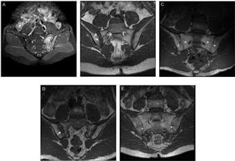
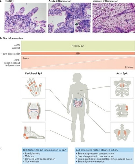

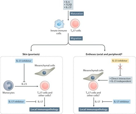




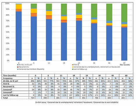
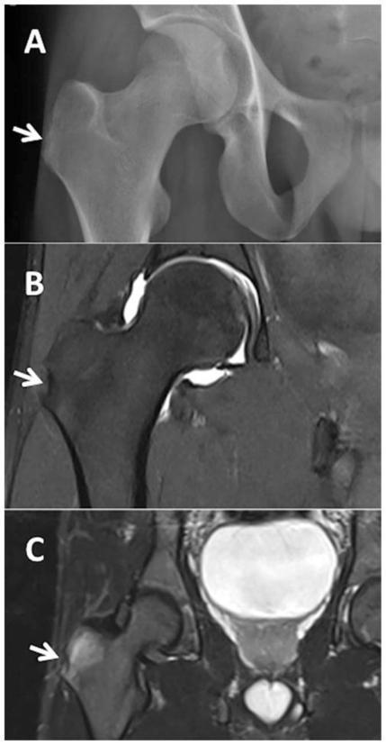
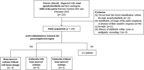
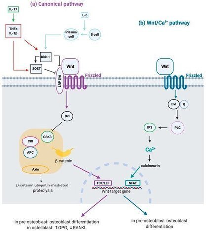







selected information
https://www.scoop.it/topic/rheumatology-rhumatologie?q=spondyloarthritis