Your new post is loading...

|
Scooped by
I2BC Paris-Saclay
January 9, 5:24 AM
|
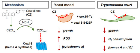
The heme A synthase Cox15, as a target of redox-active 3-benzylmenadiones with antiparasitic activity
We identified a new target for antiparasitic compounds and it is in the mitochondria. Chagas disease, caused by Trypanosoma cruzi, is a neglected parasitic infection. The very limited arsenal of anti-T. cruzi treatments calls for the development of new drugs. Recently, a library of 3-benzylmenadione derivatives was synthesized with cruzidione being the most efficient and specific compound against the parasite. To decipher its mode of action, we used the yeast Saccharomyces cerevisiae as model. Evidence pinpointed at the heme A synthase Cox15 as a primary target of cruzidione: 1) a mutation in Cox15 (i.e., S429F) renders the yeast cells highly sensitive to the drug, 2) treatment with cruzidione led to the loss of cytochrome c oxidase, an enzyme that relies on heme A as an essential cofactor and 3) replacement of the yeast Cox15 by T. cruzi enzyme resulted in a high sensitivity to cruzidione. We then investigated the effect of cruzidione in T. cruzi and observed a significant reduction of heme contents, most likely involving the inhibition of the heme A synthase. This, in turn, led to a decrease in O2 consumption by the parasite. Finally, using the yeast model, we showed that, similarly to what we previously found for the antimalarial benzylmenadione plasmodione, NADH-dehydrogenase plays a key role in cruzidione bioactivation. We proposed that the reduced benzoylmenadione metabolites produced by the reaction with NADH-dehydrogenase, act as Cox15 inhibitors. This study, through the identification of the mode of action of cruzidione, highlighted Cox15 as a novel target for antiparasitic drugs. More information: https://journals.asm.org/doi/10.1128/aac.01161-25 Contact: Brigitte Meunier, brigitte.meunier@i2bc.paris-saclay.fr
|
|
Scooped by
I2BC Paris-Saclay
November 26, 2024 3:23 AM
|
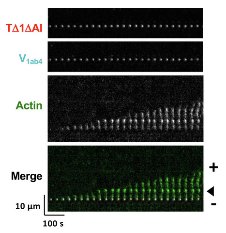
Talin and vinculin combine their activities totrigger actin assembly
In cells, focal adhesions (FAs) strengthen their connection with the actin cytoskeleton to resist force. By combining biochemistry and cell biology, the authors show that the proteins talin and vinculin control actin assembly, thus reinforcing the anchoring of actin to FAs. Focal adhesions (FAs) strengthen their link with the actin cytoskeleton to resistforce. Talin-vinculin association could reinforce actin anchoring to FAs bycontrolling actin polymerization. However, the actin polymerization activity ofthe talin-vinculin complex is not known because it requires the reconstitutionof the mechanical and biochemical activation steps that control the associa-tion of talin and vinculin. By combining kinetic and binding assays with singleactinfilament observations in TIRF microscopy, we show that the associationof talin and vinculin mutants, mimicking mechanically stretched talin andactivated vinculin, triggers a sequential mechanism in whichfilaments arenucleated, capped and released to elongate. In agreement with these obser-vations, FRAP experiments in cells co-expressing the same constitutivemutants of talin and vinculin revealed accelerated growth of stressfibers. Ourfindings suggest a versatile mechanism for the regulation of actin assembly inFAs subjected to various combinations of biochemical and mechanical cues. More information: https://www.nature.com/articles/s41467-024-53859-1 Contact: Christophe LE CLAINCHE <christophe.leclainche@i2bc.paris-saclay.fr>
|
|
Scooped by
I2BC Paris-Saclay
October 17, 2024 5:54 AM
|
The acetyltransferase SCO0988 controls positively specialized metabolism and morphological differentiation in the model strains Streptomyces coelicolor and Streptomyces lividans.
Acetylation of some proteins plays a positive role in the regulation of the production of specialized metabolites and in the morphological differentiation of Streptomyces. Streptomyces species possess in their genomes numerous silent biosynthetic pathways likely to direct the biosynthesis of novel bioactive specialized metabolites. Enhancing the expression of these cryptic pathways might lead to the discovery of most needed novel antibiotics to limit the spreading of pathogenic bacteria resistant to most antibiotics in current use.
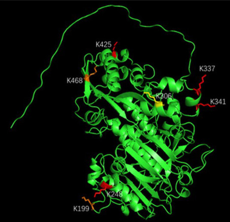
We characterized, an acetyl transferase (SCO0988) whose over-expression led to enhanced antibiotic production and accelerated sporulation of a low antibiotic producing strain, Streptomyces lividans, while the deletion of this gene had opposite effects in a strong antibiotic producer, Streptomyces coelicolor. Comparative analysis of the acetylome of the S. lividans strain overexpressing SCO0988 with that of the original strain revealed that 92 peptides, belonging to various proteins involved in different cellular processes, were exclusively acetylated in the strain over-expressing SCO0988. This led to the definition of a consensus SCO0988 acetylation site (v/q-P-l/g-D-p/e-KR-v/l-h/f-y/a-t/m). Among the acetylated proteins, BldKB was found to be over-acetylated on four lysine residues including Lys425 in the strain overexpressing SCO0988. BldKB is the solute binding protein of an ABC transporter involved in the up-take of an oligopeptide that plays a role in the triggering of a quorum sensing regulatory cascade controlling positively specialized metabolism, aerial mycelium formation and sporulation in S. coelicolor. A bldKB mutant has a reduced antibiotic production and does not develop aerial mycelium. The bldKB mutant phenotypes can be complemented by the native bldKB gene but not by a variant of this gene encoding a protein in which Lys425 was replaced by Arg (that cannot be acetylated) or by Glu (a putative structural analogue of acetylated lysine). Our study thus demonstrated that Lys425 is critical for BldKB function but could not clearly establish the impact of Lys425 acetylation on BldKB function. More information: https://www.ncbi.nlm.nih.gov/pmc/articles/PMC11303876/ Contact: Marie-Joëlle VIROLLE marie-joelle.virolle@i2bc.paris-saclay.fr
|
|
Scooped by
I2BC Paris-Saclay
October 1, 2024 6:19 AM
|
Lipid Metabolism in Relation to Carbohydrate Metabolism
This chapter reviews in the insect world the integration of the carbohydrate-lipid metabolic axis at the entire body level Carbohydrates and lipids integrate into a complex metabolic network that is essential to maintain homeostasis. In insects, as in most metazoans, dietary carbohydrates are taken up as monosaccharides whose excess is toxic, even at relatively low concentrations. To cope with this toxicity, monosaccharides are stored either as glycogen or neutral lipids, the latter constituting a quasi-unlimited energy store. Breakdown of these stores in response to energy demand depends on insect species and on several physiological parameters. In this chapter, we review the multiple metabolic pathways and strategies linking carbohydrates and lipids that insects utilize to respond to nutrient availability, food scarcity or physiological activities. more information: https://www.ncbi.nlm.nih.gov/pubmed/39192070 Contact: jacques.montagne@i2bc.paris-saclay.fr
|
|
Scooped by
I2BC Paris-Saclay
July 26, 2024 8:14 AM
|
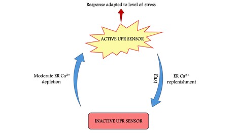
Gradual ER calcium depletion induces a progressive and reversible UPR signaling
The UPR sensors show high plasticity in sensing physiological alterations by closely reporting the variations in ER Ca2+ levels. Through this mechanisms cells could rapidly adapt stress response signaling pathways to variations in ER homeostasis. The unfolded protein response (UPR) is a widespread signal transduction pathway triggered by endoplasmic reticulum (ER) stress. Because calcium (Ca2+) is a key factor in the maintenance of ER homeostasis, massive Ca2+ depletion of the ER is a potent inducer of ER stress. Although moderate changes in ER Ca2+ drive the ubiquitous Ca2+ signaling pathways, a possible incremental relationship between UPR activation and Ca2+ changes has yet to be described. Here, we determine the sensitivity and time-dependency of activation of the three ER stress sensors, IRE1α, PERK and ATF6α in response to controlled changes in the concentration of ER Ca2+ in human cultured cells. Combining Ca2+ imaging, FRAP experiments, biochemical analyses and mathematical modeling, we uncover a non-linear rate of activation of the IRE1α branch of UPR, as compared to the PERK and ATF6α branches that become activated gradually with time and are sensitive to more important ER Ca2+ depletions. However, the three arms are all activated within a 1h timescale. The model predicted the deactivation of PERK and IRE1α upon refilling the ER with Ca2+. Accordingly, we showed that ER Ca2+ replenishment leads to the complete reversion of IRE1α and PERK phosphorylation in less than 15 min, thus revealing the highly plastic character of the activation of the upstream UPR sensors. In conclusion, our results reveal a dynamic and dose-sensitive Ca2+-dependent activation/deactivation cycle of UPR induction, which could tightly control cell fate upon acute and/or chronic stress. More information: https://doi.org/10.1093/pnasnexus/pgae229 Contact: Ilaria Pontisso (ilaria.pontisso@i2bc.paris-saclay.fr)
|
|
Scooped by
I2BC Paris-Saclay
July 1, 2024 8:59 AM
|
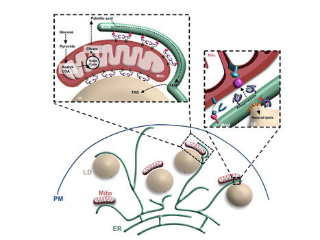
“Ménage à trois”: emerging function of the mitochondria-ER-lipid droplet connection in coordinating lipid transfer, metabolism and storage in cells
“Ménage à trois”: coordination of lipid transfer, metabolism and storage at the new mitochondria-ER-lipid droplet junction. Over the past two decades, we have witnessed a growing appreciation for the importance of membrane contact sites (CS) in facilitating direct communication between organelles. CS are tiny regions where the membranes of two organelles meet but do not fuse and allow the transfer of metabolites between organelles, playing crucial roles in the coordination of cellular metabolic activities. The significant advancements in imaging techniques, and molecular and cell biology research has revealed that CS are more complex than what originally thought, as they are extremely dynamics, they can remodel their shape, composition and functions in accord to metabolic and environmental changes, and can occur between more than two organelles. In this review, for the Special Issue “Lipid droplets, metabolic hubs in health and disease“ in FEBS Letters, Vera F. Monteiro-Cardoso and Francesca Giordano describe how recent studies led to the identification of a three-way mitochondria-ER-lipid droplet CS and discuss the emerging functions of these contacts in maintaining lipid storage, homeostasis and balance. They also summarize properties and functions of key protein components localized at the mitochondria-ER-lipid droplet interface, with a special focus on lipid transfer proteins. Understanding tripartite CS is essential for unraveling the complexities of inter-organelle communication and cooperation within cells. more information: https://febs.onlinelibrary.wiley.com/doi/10.1002/1873-3468.14893 contact: Francesca GIORDANO <francesca.giordano@i2bc.paris-saclay.fr>
|
|
Scooped by
I2BC Paris-Saclay
April 15, 2024 8:53 AM
|
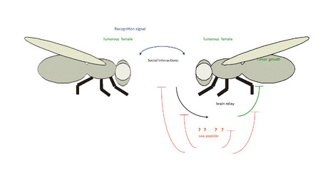
Male manipulation impinges on social-dependent tumor suppression in Drosophila melanogaster females
This study shows that tumorous Drosophila females perceive a cognitive-like signal from their social environment that acts on tumor growth, while this effect is lost in mated females because of peptides transmitted through the male ejaculate. Physiological status can influence social behavior, which reciprocally can impinge on physiology and health. Previously, we reported that tumor growth in Drosophila virgin females depends on the social context, but did not investigate the underlying physiological mechanisms. Here, we sought to characterize the signal perceived between tumorous flies, ultimately discovering that the tumor suppressive effect varies depending on reproductive status. Firstly, we show that the tumor suppressive effect is neither dependent on remnant pheromone-like products nor on the microbiota. Transcriptome analysis of the heads of these tumorous flies reveals social-dependent gene-expression changes related to nervous-system activity, suggesting that a cognitive-like relay might mediate the tumor suppressive effect. The transcriptome also reveals changes in the expression of genes related to mating behavior. Surprisingly, we observed that this social-dependent tumor-suppressive effect is lost in fertilized females. After mating, Drosophila females change their behavior ─favoring offspring survival─ in response to peptides transferred via the male ejaculate, a pehnomenon called “male manipulation”. Remarkably, the social-dependent tumor suppressive effect is restored in females mated by sex-peptide deficient males. Since male manipulation has likely been selected to favor male gene transmission, our findings indicate that this evolutionary trait impedes social-dependent tumor growth slowdown. more information: https://doi.org/10.1038/s41598-024-57003-3 contact: Jacques MONTAGNE <jacques.montagne@i2bc.paris-saclay.fr>
|
|
Scooped by
I2BC Paris-Saclay
March 25, 2024 6:18 AM
|
JAK-STAT-dependent contact between follicle cells and the oocyte controls Drosophila anterior-posterior polarity and germline development
The authors identified a population of somatic cells in Drosophila follicles that elaborate filopodia penetrating the oocyte, thereby maintaining the contact between the two tissues. This is essential to control polarity and germline development of the future embryo. The number of embryonic primordial germ cells is determined, in Drosophila, by the quantity of germ plasm, whose assembly starts in the posterior region of the oocyte during oogenesis. Here, we report that extending JAK-STAT activity in the posterior somatic follicular epithelium leads to an excess of primordial germ cells in the future embryo. We show that JAK-STAT signaling is necessary for the differentiation of approximately 20 specialized follicle cells maintaining tight contact with the oocyte. These cells define, in the underlying posterior oocyte cortex, the anchoring of the germ cell determinant oskar mRNA. We reveal that the apical surface of these posterior anchoring cells extends long filopodia penetrating the oocyte. We identify two JAK-STAT targets in these cells that are each sufficient to extend the zone of contact with the oocyte, thereby leading to production of extra primordial germ cells. JAK-STAT signaling thus determines a fixed number of posterior anchoring cells required for anterior-posterior oocyte polarity and for the development of the future germline. More information: https://doi-org.insb.bib.cnrs.fr/10.1038/s41467-024-45963-z Contact: Marianne MALARTRE <marianne.malartre@i2bc.paris-saclay.fr>
|
|
Scooped by
I2BC Paris-Saclay
January 30, 2024 9:19 AM
|
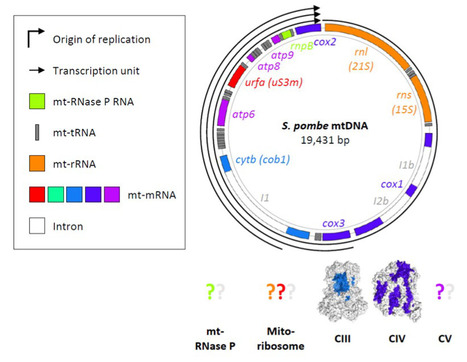
Schizosaccharomyces pombe as a fundamental model for research on mitochondrial gene expression: Progress, achievements and outlooks
This is a comprehensive and critical review on mitochondrial gene expression in fission yeast, presenting up-to-date knowledge, and emphasising numerous contributions of the unicellular model to both fundamental and biomedical research. Schizosaccharomyces pombe (fission yeast) is an attractive model for mitochondrial research. The organism resembles human cells in terms of mitochondrial inheritance, mitochondrial transport, sugar metabolism, mitogenome structure, and dependence of viability on the mitogenome (the petite-negative phenotype). Transcriptions of these genomes produce only a few polycistronic transcripts, which then undergo processing as per the tRNA punctuation model. In general, the machinery for mitochondrial gene expression is structurally and functionally conserved between fission yeast and humans. Furthermore, molecular research on S. pombe is supported by a considerable number of experimental techniques and database resources. Owing to these advantages, fission yeast has significantly contributed to biomedical and fundamental research. Here, we review the current state of knowledge regarding S. pombe mitochondrial gene expression, and emphasise the pertinence of fission yeast as both a model and tool, especially for studies on mitochondrial translation. More information: https://doi.org/10.1002/iub.2801 Contact: Nathalie BONNEFOY <nathalie.bonnefoy@i2bc.paris-saclay.fr>
|
|
Scooped by
I2BC Paris-Saclay
December 27, 2023 7:59 AM
|
Straining the root on and off triggers local calcium signaling
Combining microfluidics and imaging, reveal that a pressure exerted on the root induces an elastic deformation and a calcium signal both at the onset and at the offset of the stimulation. How plant roots perceive mechanical cues encountered in the soil remains elusive. Combining microfluidics and imaging, we have shown that a pressure exerted on the root induces an elastic deformation and a calcium signal both at the onset and at the offset of the stimulation. Although the intensity of the calcium response increased with the pressure applied, successive stimuli led to a remarkable attenuation of the calcium signal. The calcium elevation was restricted to the tissue under pressure without propagation. This study published in Proceedings of the Royal Society B (https://doi.org/10.1098/rspb.2023.1462) contributes to elucidate the mechanisms of root adaptation to the mechanical cues generated by the soil.
This work was supported by the Région Ile de France through the DIM ELICIT program and by Saclay Plant Sciences through the DYNANO project. More information: doi: 10.1128/msphere.00401-23 Contact: Jean-Marie Frachisse – Sébastien Thomine <jean-marie.frachisse@i2bc.paris-saclay.fr> <sebastien.thomine@i2bc.paris-saclay.fr>
|
|
Scooped by
I2BC Paris-Saclay
March 17, 2023 12:20 PM
|
An image-based high-content screening for compounds targeting Toxoplasma gondii repurposed inhibitors effective against the malaria parasite Plasmodium falciparum
Apicomplexan parasites contain specific secretory organelles called rhoptries and micronemes, whose biogenesis depends on the unique receptor sortilin TgSORT. In the present study, we took advantage of the subcellular polarity of the parasite to engineer a clonal transgenic Toxoplasma line that expresses simultaneously the green fluorescent protein TgSORT-GFP in the post-Golgi-endosome-like compartment and the red fluorescent protein rhoptry ROP1-mCherry near the apical end. We utilized this fluorescent transgenic T. gondii to develop a miniaturized image-based phenotype assay coupled to an automated image analysis. By applying this methodology to 1,120 compounds, we identified 12 that are capable of disrupting the T. gondii morphology and inhibiting intracellular replication. Analysis of the selected compounds confirmed that all 12 are kinase inhibitors and intramembrane pumps, with some exhibiting potent activity against Plasmodium falciparum. Our findings highlight the advantage of comparative and targeted phenotypic analysis involving two related parasite species as a means of identifying molecules with a conserved mode of action. More: https://doi.org/10.3389/fcimb.2023.1102551 Contact: Stanislas Tomavo <stanislas.tomavo@i2bc.paris-saclay.fr>
|
|
Scooped by
I2BC Paris-Saclay
March 17, 2023 11:10 AM
|
Biolistic Transformation of Chlamydomonas reinhardtii and Saccharomyces cerevisiae Mitochondria
This review summarizes principles, historical and practical aspects as well as achievements of microalga and budding yeast mitochondrial DNA manipulation using the biolistic technique, and includes a video protocol. Chlamydomonas reinhardtii and Saccharomyces cerevisiae are currently the two micro-organisms in which genetic transformation of mitochondria is routinely performed. The generation of a large variety of defined alterations as well as the insertion of ectopic genes in the mitochondrial genome (mtDNA) are possible, especially in yeast. Biolistic transformation of mitochondria is achieved through the bombardment of microprojectiles coated with DNA, which can be incorporated into mtDNA thanks to the highly efficient homologous recombination machinery present in S. cerevisiae and C. reinhardtii organelles. Despite a low frequency of transformation, the isolation of transformants in yeast is relatively quick and easy, since several natural or artificial selectable markers are available, while the selection in C. reinhardtii remains long and awaits new markers. Here, we describe the materials and techniques used to perform biolistic transformation, in order to mutagenize endogenous mitochondrial genes or insert novel markers into mtDNA. Although alternative strategies to edit mtDNA are being set up, so far, insertion of ectopic genes relies on the biolistic transformation techniques. This reviews is Chapter 24 of a 2023 volume from Springer Protocols devoted to mitochondrial DNA. More: Methods Mol Biol 2023;2615:345-364. Contcat: Nathalie Bonnefoy <nathalie.bonnefoy@i2bc.paris-saclay.fr>
|
|
Scooped by
I2BC Paris-Saclay
October 24, 2022 5:20 AM
|
A functional protein complex, associated to ciliopathies, dictates the future position of distal appendages
Using BioID and expansion microscopy, the study uncover a conserved functional complex required for determining the future position of centriolar distal appendages and their assembly, allowing the basal body anchoring process during ciliogenesis. Cilia/flagella play fundamental and diverse biological functions in a wide variety of eukaryotes. The cilia assembly process, also called ciliogenesis, is a multistep process involving 4 major events: basal body (BB) duplication, migration to the cell surface, membrane anchoring of the BB via the distal appendages, and ciliary growth. The conservation of this sequence of events in most phyla is paralleled by an important conservation of the proteins involved. The BB anchoring step requires the tethering of the distal appendages to a membrane. The interaction of the BB with the membrane leads to the formation of the transition zone (TZ), recognized to act as a diffusion barrier between the intracellular space and the cilium. Mutations in numerous genes involved in BB docking and TZ assembly are associated with the most severe ciliopathies highlighting the importance of these events in ciliogenesis.
To discover novel molecular actors involved in the BB anchoring process, the Tassin’s team used BioID technology in human cells. They focused their attention on a protein, CEP90, together with its bait (FOPNL) and another protein (OFD1) involved in ciliopathies and known to interact with FOPNL. These 3 proteins are conserved from Protist to mammals despite absent from some phyla. They unveiled using ultrastructure expansion microscopy that the 3 proteins localize at the distal end of both centrioles/BB in Paramecium and mammalian cells. Functional analysis performed both in Paramecium and mammalian cells demonstrate that the 3 proteins form a functional complex required for distal appendage assembly and BB docking. Intriguingly, these proteins are recruited early during centriole duplication on the external surface of the procentriole. This early recruitment on procentriole led us to propose that this functional complex dictates the future distal appendage location in mammalian cells one or two cell cycle before their assembly.
|
|
|
Scooped by
I2BC Paris-Saclay
January 9, 5:05 AM
|
Gain and loss of gene function shaped the nickel hyperaccumulation trait in Noccaea caerulescens
Plants that hyperaccumulate nickel open the possibility to mine this metal from soils in an environmetally friendly manner. In this study, sequencing of a nickel hyperaccumulating plant together with genomic and transcriptomic comparisons reveal the molecular mechanisms underlying this extreme trait. Nickel hyperaccumulation is an extreme adaptation to ultramafic soils observed in more than 500 plant species. However, our understanding of the molecular mechanisms underlying the evolution of this trait remains limited. To shed light on these mechanisms, we have generated a high-quality genome assembly of the metal hyperaccumulator Noccaea caerulescens. We then used this genome as reference to conduct comparative intraspecific and interspecific transcriptomic analyses using various accessions of N. caerulescens and the non-accumulating relative Microthlaspi perfoliatum to identify genes associated with nickel hyperaccumulation.
Our results suggest a correlation between nickel hyperaccumulation and a decrease in the expression of genes involved in defense responses and the regulation of membrane trafficking. Surprisingly, these analyses did not reveal a significant enrichment of genes involved in the regulation of metal homeostasis. However, we found that the expression levels of selected metal transporter genes,
namely, NcHMA3, NcHMA4, and NcIREG2, are consistently elevated in N. caerulescens accessions hyperaccumulating nickel.
Furthermore, our analyses identified frameshift mutations in NcIRT1 associated with the loss of nickel hyperaccumulation in a few accessions. We further showed that the expression of a functional NcIRT1 in the roots of the La Calamine accession increases nickel accumulation in shoots. Our results demonstrate that NcIRT1 participates in nickel hyperaccumulation in N. caerulescens. They also
suggest that nickel hyperaccumulation is an ancient trait in N. caerulescens that has evolved from the high and constitutive expression of several metal transporters, including NcIREG2, and that the trait was subsequently lost in a few accessions due to mutations in NcIRT1. More information: https://doi.org/10.1093/plcell/koaf281 Contacts: Sylvain Merlot, sylvain.merlot@univ-tlse3.fr Sébastien Thomine, sebastien.thomine@i2bc.paris-saclay.fr
|
|
Scooped by
I2BC Paris-Saclay
October 17, 2024 6:08 AM
|
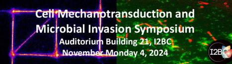
Cell Mechanotransduction and Microbial Invasion Symposium
Learn with great scientists about cell mechanotransduction and microbial invasion processes Morning session: Cell Mechanotransduction 9H00-9H30 : Christophe Leclainche, I2BC, Gif-sur-Yvette.
Understanding mechanotransduction through in vitro reconstitution of actomyosin-dependent protein machineries
9H30-10H00 : Benoit Ladoux, MPZPM, Erlangen, Germany.
Mechanical imprints of cell extrusion from epithelial tissues.
10H00-10H30 : Patricia Bassereau, Institut Curie, Paris.
Cell spreading on fluid membranes requires strong integrin ligands and microtubules
10H30-10H50 : Coffee break
10H50-11H20: Gregory Giannone, Université de Bordeaux.
Unraveling the nanoscale spatial and mechanical regulation of actin binding proteins.
11H20-12H00 : Keynote lecture - Michael Sheetz, UTMB, Texas, USA.
Mechanical Oscillations Kill Cancer Cells and Reverse Aging
Afternoon session « Microbial Invasion» 14H00-14H30 : Sven Hammerschmidt, Greifswald university, Germany
TBA
14H30-15H00 : Sandrine Bourdoulous, Institut Cochin, Paris.
Angptl4 prevents endothelial dysfunction during bacterial sepsis
15H00-15H40 : Keynote lecture - Linda Kenney, UTMB, Texas, USA.
Imaging Salmonella Lifestyles: From Virulence to Biofilms to Tumors
15H40-16H00 : Coffee break
16H00-16H30 : Jost Enninga, Institut Pasteur, Paris.
Endomembrane breakage by enteroinvasive bacterial pathogens- new insights by in cellulo structural biology
16H30-17H00 : Marc Prudhomme, Université Paul Sabatier, Toulouse
Bacterial communication and antibiotic tolerance in Pneumooccus
17H00-17H30 : Guy Tran Van Nhieu, I2BC, Gif-sur-Yvette.
Characterization of pneumococcal secreted factors associated with bacterial meningitis
|
|
Scooped by
I2BC Paris-Saclay
October 1, 2024 6:22 AM
|
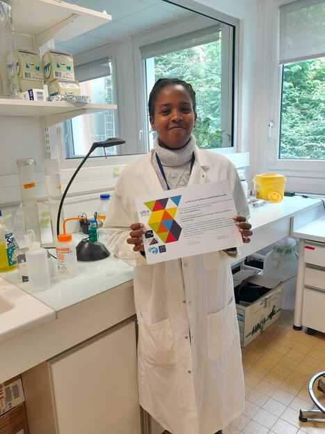
Award for the best conference presentation.
PhD student Audrey Ntadambanya from the Department of Cell Biology, won the prize for best presentation at the Photosciences 2024 conference. PhD student Audrey Ntadambanya, supervised by Marcelina Cardoso Dos Santos, won the prize for best presentation at the Photosciences 2024 conference. She was rewarded for her presentation, which was judged to be of high scientific quality and delivered with great enthusiasm and skill. Audrey's thesis work focuses on the development of new nanosensors based on quantum dot nanoparticles to resolve biomolecular interactions at the sub-nanometre scale. The prize was a scientific book selected by the winner. Furthermore, Audrey was able to attend the conference due to financial support from OI BioProbe for participation in international conferences. more information: https://new.societechimiquedefrance.fr/divisions/photochimie-photophysique-et-photosciences/important-dates/ Contact: marcelina.cardoso-dos-santos@i2bc.paris-saclay.fr
|
|
Scooped by
I2BC Paris-Saclay
September 23, 2024 10:20 AM
|
Sulfur starvation-induced autophagy in Saccharomyces cerevisiae involvesSAM-dependent signaling and transcription activator Met4
Unveiling a new property of bacteriophage-host interaction in the Autophagy has a key protective role under nutrient stresses. The authors unveil in yeast an original mechanism of induction of autophagy under sulfur deprivation that requires the transcription activator Met4 and involves S-adenosylmethionine as signaling molecule. Autophagy is a key lysosomal degradative mechanism allowing a prosurvival response to stresses, especially nutrient starvation. Here we investigate the mechanism of autophagy induction in response to sulfur starvation in Saccharomyces cerevisiae. We found that sulfur deprivation leads to rapid and widespread transcriptional induction of autophagy-related (ATG) genes in ways not seen under nitrogen starvation. This distinctive response depends mainly on the transcription activator of sulfur metabolism Met4. Consistently, Met4 is essential for autophagy under sulfur starvation. Depletion of either cysteine, methionine or SAM induces autophagy flux. However, only SAM depletion can trigger strong transcriptional induction of ATG genes and a fully functional autophagic response. Furthermore, combined inactivation of Met4 and Atg1 causes a dramatic decrease in cell survival under sulfur starvation, highlighting the interplay between sulfur metabolism and autophagy to maintain cell viability. Thus, we describe a pathway of sulfur starvation induced autophagy depending on Met4 and involving SAM as signaling sulfur metabolite. More information: https://doi.org/10.1038/s41467-024-51309-6 Contact: Laurent KURAS laurent.kuras@i2bc.paris-saclay.fr
|
|
Scooped by
I2BC Paris-Saclay
July 2, 2024 10:28 AM
|
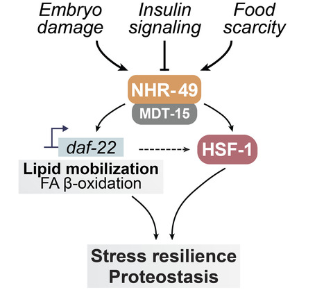
Nuclear receptor signaling via NHR-49/MDT-15 regulates stress resilience and proteostasis in response to reproductive and metabolic cues
The production of unhealthy eggs leads to improved maternal stress resilience and proteostasis in C. elegans. This study identifies the nuclear hormone receptor NHR-49 as a critical regulator that is activated by elevated fat stores and acts by potentiating the heat shock response. The ability to sense and respond to proteotoxic insults declines with age, leaving cells vulnerable to chronic and acute stressors. Reproductive cues modulate this decline in cellular proteostasis to influence organismal stress resilience in Caenorhabditis elegans We previously uncovered a pathway that links the integrity of developing embryos to somatic health in reproductive adults. Here, we show that the nuclear receptor NHR-49, an ortholog of mammalian peroxisome proliferator-activated receptor α (PPARα), regulates stress resilience and proteostasis downstream from embryo integrity and other pathways that influence lipid homeostasis and upstream of HSF-1. Disruption of the vitelline layer of the embryo envelope, which activates a proteostasis-enhancing intertissue pathway in somatic cells, triggers changes in lipid catabolism gene expression that are accompanied by an increase in fat stores. NHR-49, together with its coactivator, MDT-15, contributes to this remodeling of lipid metabolism and is also important for the elevated stress resilience mediated by inhibition of the embryonic vitelline layer. Our findings indicate that NHR-49 also contributes to stress resilience in other pathways known to change lipid homeostasis, including reduced insulin-like signaling and fasting, and that increased NHR-49 activity is sufficient to improve proteostasis and stress resilience in an HSF-1-dependent manner. Together, our results establish NHR-49 as a key regulator that links lipid homeostasis and cellular resilience to proteotoxic stress. More information: https://genesdev.cshlp.org/content/early/2024/05/30/gad.351829.124 Contact: Ambre Sala ambre.sala@i2bc.paris-saaclay.fr
|
|
Scooped by
I2BC Paris-Saclay
April 15, 2024 9:18 AM
|
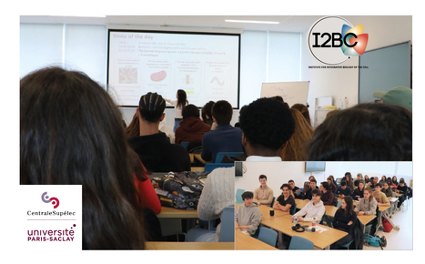
Students from CentraleSupélec visit I2BC groups of the Cellular Biology department
80 students of the Bachelor of Global Engineering, a joint novel program of CentraleSupéléc and McGilles University visited the research groups Kühl, Sala and Siudeja at the Cellular Biology department at I2BC in March. During two afternoons in March three groups from the Cellular Biology department, Kühl, Sala and Siudeja, explained their research questions and model organisms to a total of 80 students from the Bachelor of Global Engineering (BoGe), a joint novel program of CentraleSupéléc, Paris-Saclay and McGilles University, Canada (https://www.centralesupelec.fr/en/bachelor-global-engineering) . The visit was part of the BoGe Biology course taught by Sébastien Bloyer (Genome dep.) and Inge Kühl (BioCell dep.). We thank all the participants for their help, and the I2BC for the support.
|
|
Scooped by
I2BC Paris-Saclay
April 15, 2024 8:35 AM
|
A Fatty Acid Anabolic Pathway in Specialized-Cells Sustains a Remote Signal that Controls Egg Activation in Drosophila
While egg activation, i.e. oocyte to embryo transition, was not known to depend on physiological non-genital signal, this study shows that in Drosophila it depends on a very-long-chain-fatty-acid produced in the oenocytes (lipid metabolism specialized cells). Egg activation, representing the critical oocyte-to-embryo transition, provokes meiosis completion, modification of the vitelline membrane to prevent polyspermy, and translation of maternally provided mRNAs. This transition is triggered by a calcium signal induced by spermatozoon fertilization in most animal species, but not in insects. In Drosophila melanogaster, mature oocytes remain arrested at metaphase-I of meiosis and the calcium-dependent activation occurs while the oocyte moves through the genital tract. Here, we discovered that the oenocytes of fruitfly females are required for egg activation. Oenocytes, cells specialized in lipid-metabolism, are located beneath the abdominal cuticle. In adult flies, they synthesize the fatty acids (FAs) that are the precursors of cuticular hydrocarbons (CHCs), including pheromones. The oenocyte-targeted knockdown of a set of FA-anabolic enzymes, involved in very-long-chain fatty acid (VLCFA) synthesis, leads to a defect in egg activation. Given that some but not all of the identified enzymes are required for CHC/pheromone biogenesis, this putative VLCFA-dependent remote control may rely on an as-yet unidentified CHC or may function in parallel to CHC biogenesis. Additionally, we discovered that the most posterior ventral oenocyte cluster is in close proximity to the uterus. Since oocytes dissected from females deficient in this FA-anabolic pathway can be activated in vitro, this regulatory loop likely operates upstream of the calcium trigger. To our knowledge, our findings provide the first evidence that a physiological extra-genital signal remotely controls egg activation. Moreover, our study highlights a potential metabolic link between pheromone-mediated partner recognition and egg activation. More information: https://doi.org/10.1371/journal.pgen.1011186 Contact: Jacques Montagne jacques.montagne@i2bc.paris-saclay.fr
|
|
Scooped by
I2BC Paris-Saclay
January 30, 2024 9:23 AM
|
Toxoplasma membrane inositol phospholipid binding protein TgREMIND is essential for secretory organelle function and host infection
Apicomplexan parasites possess specialized secretory organelles called rhoptries, micronemes, and dense granules that play a vital role in host infection. In this study, we demonstrate that TgREMIND, a protein found in Toxoplasma gondii, is necessary for the biogenesis of rhoptries and dense granules. TgREMIND contains a Fes-CIP4 homology-Bin/Amphiphysin/Rvs (F-BAR) domain, which binds to membrane phospholipids, as well as a novel uncharacterized domain that we have named REMIND (regulator of membrane-interacting domain). Both the F-BAR domain and the REMIND are crucial for TgREMIND functions. When TgREMIND is depleted, there is a significant decrease in the abundance of dense granules and abnormal transparency of rhoptries, leading to a reduction in protein secretion from these organelles. The absence of TgREMIND inhibits host invasion and parasite dissemination, demonstrating that TgREMIND is essential for the proper function of critical secretory organelles required for successful infection by Toxoplasma. More information: doi: 10.1016/j.celrep.2023.113601 Contact: Stanislas TOMAVO <stanislas.tomavo@i2bc.paris-saclay.fr>
|
|
Scooped by
I2BC Paris-Saclay
December 27, 2023 9:21 AM
|
Visualization and Quantification of Endogenous Intra-Organelle Protein Interactions at ER-Mitochondria Contact Sites by Proximity Ligation Assays
|
|
Scooped by
I2BC Paris-Saclay
July 19, 2023 10:26 AM
|
LGG-1/GABARAP lipidation is not required for autophagy and development in Caenorhabditis elegans
Using a CRISPR approach in C. elegans, this study shows that the ubiquitin-like ATG8 homolog LGG-1 possesses lipidation-independent function in autophagy, and provides a novel insight into the role of ATG family during animal development. The ubiquitin-like proteins Atg8/LC3/GABARAP are required for multiple steps of autophagy, such as initiation, cargo recognition and engulfment, vesicle closure and degradation. Most of LC3/GABARAP functions are considered dependent on their post-translational modifications and their association with the autophagosome membrane through a conjugation to a lipid, the phosphatidyl-ethanolamine. Contrarily to mammals, C. elegans possesses single homologs of LC3 and GABARAP families, named LGG-2 and LGG-1. Using site-directed mutagenesis, we inhibited the conjugation of LGG-1 to the autophagosome membrane and generated mutants that express only cytosolic forms, either the precursor or the cleaved protein. LGG-1 is an essential gene for autophagy and development in C. elegans, but we discovered that its functions could be fully achieved independently of its localization to the membrane. This study reveals an essential role for the cleaved form of LGG-1 in autophagy but also in an autophagy-independent embryonic function. Our data question the use of lipidated GABARAP/LC3 as the main marker of autophagic flux and highlight the high plasticity of autophagy. More information: https://elifesciences.org/articles/85748 Contact: Renaud Legouis <renaud.legouis@i2bc.paris-saclay.fr>
|
|
Scooped by
I2BC Paris-Saclay
March 17, 2023 11:54 AM
|
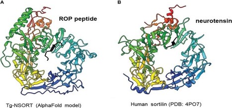
The luminal domain of Toxoplasma gondii sortilin adopts a ring-shaped structure exhibiting motifs specific to apicomplexan parasites
Rhoptries and micronemes are essential for host cell invasion and survival of all apicomplexan parasites, which are composed of protozoan pathogens including Plasmodium falciparum (malaria) and Toxoplasma gondii (toxoplasmosis) that infect humans and animals causing severe diseases. We identifiedToxoplasma gondii TgSORT as an essential cargo receptor, which drives the transport of rhoptry and microneme proteins to ensure the biogenesis of these secretory organelles. Here, we present an optimized protocol for expression of the entire luminal N-terminus of TgSORT (Tg-NSORT) in the yeast Pichia pastoris. Optimization of its coding sequence, cloning and transformation of the yeast P. pastoris allowed the secretion of Tg-NSORT. The protein was purified and further analyzed by negative staining electron microscopy. In addition, molecular modeling using AlphaFold identified key differences between the human and theT gondii sortilin. The structural features that are only present inT. gondii and other apicomplexan parasites were highlighted. Elucidating the roles of these specific structural features may be useful for designing new therapeutic agents against apicomplexan parasites. More: https://doi.org/10.3389/fpara.2023.1103772 Contact: Stanislas Tomavo <stanislas.tomavo@i2bc.paris-saclay.fr>
|
|
Scooped by
I2BC Paris-Saclay
December 2, 2022 10:58 AM
|
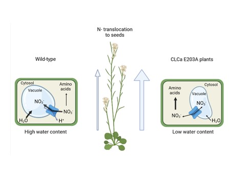
Proton exchange by the vacuolar nitrate transporter CLCa is required for plant growth and nitrogen use efficiency
CLCa exchange mechanism limits nitrogen use efficicency in plants. Nitrogen (N) is quantitatively the most important inorganic nutrient for plants. Roots mainly take it up in the form of nitrate (NO3-). Once inside the cells, nitrate can be assimilated to amino acids or stored in the vacuole, where it regulates cell water content to sustain plant growth. CLCa is the main transporter mediating the entry of NO3- into vacuoles. It works as an exchanger removing one proton from the vacuole to store two NO3- anions. CLCa belongs to a conserved family composed of both anions/protons exchangers and anions channels, with closely similar structures. Most of the exchangers share a conserved glutamate residue (E203 in CLCa). In this publication, we analyzed if the NO3-/H+ exchanger mechanism of CLCa is required for plants to stabilize water and nitrate status and what are the physiological consequences of the conversion of CLCa from an exchanger to a nitrate channel. We generated Arabidopsis lines that express a mutated form of CLCa in the amino acid E203, turning this protein into a NO3- channel. This modification decreases nitrate accumulation in the vacuole and increases amino acids and protein synthesis, thereby leading to high N content in the seeds. Although these plants present a higher nitrogen use efficiency (NUE), their growth is reduced, in association to a diminution of water content. This finding reveals the importance of the CLCa exchanger mechanism in allowing plant cells to maintain cell turgor irrespective of fluctuating nitrogen concentrations in the soil. More information: here Contact: Sophie Filleur <sophie.filleur@i2bc.paris-saclay.fr>
|