Your new post is loading...
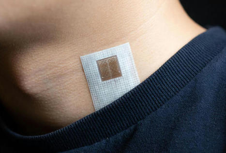
A new ultrasound-based sensor has been unveiled, offering real-time, on-the-go cardiac imaging. Designed by engineers from the University of California San Diego, the wearable device is roughly the size of a postage stamp and can be worn for up to 24 hours. Plus, it even functions during a workout. The engineers say the goal is to make ultrasound capabilities more accessible. Currently, echocardiograms – ultrasound examinations for the heart – can only be done by trained professionals and requires bulky equipment. “The technology enables anybody to use ultrasound imaging on the go,” said Sheng Xu, study lead. The new device uses AI algorithms to measure the activity and structure of the wearer’s heart, monitoring blood flow and flagging any abnormal functions. Using ultrasound waves, the device generates a constant stream of images of the four chambers of the heart, enabling continuous, real-time data capture. “The increasing risk of heart diseases calls for more advanced and inclusive monitoring procedures,” said Xu. “By providing patients and doctors with more thorough details, continuous and real-time cardiac image monitoring is poised to fundamentally optimize and reshape the paradigm of cardiac diagnoses.” In its current iteration, the patch can be connected through cables to a computer, which can download the data automatically while the patch is still on, though plans for a wireless design are underway. The patch is set for commercialization through Softsonics, a UC San Diego spinoff.
Researchers at Duke University are developing an artificial intelligence tool for toilets that would help providers improve care management for patients with gastrointestinal issues through remote patient monitoring. The tool, which can be installed in the pipes of a toilet and analyzes stool samples, has the potential to improve treatment of chronic gastrointestinal issues like inflammatory bowel disease or irritable bowel syndrome, according to a press release. When a patient flushes the toilet, the mHealth platform photographs the stool as it moves through the pipes. That data is sent to a gastroenterologist, who can analyze the data for evidence of chronic issues. A study conducted by Duke University researchers found that the platform had an 85.1 percent accuracy rate on stool form classification and a 76.3 percent accuracy rate on detection of gross blood. “Typically, gastroenterologists have to rely on patient self-reported information about their stool to help determine the cause of their gastrointestinal health issues, which can be very unreliable,” Deborah Fisher, MD, an associate professor of medicine at Duke and one of the study’s lead authors, said in the press release. “Patients often can’t remember what their stool looks like or how often they have a bowel movement, which is part of the standard monitoring process,” she said. “The smart toilet technology will allow us to gather the long-term information needed to make a more accurate and timely diagnosis of chronic gastrointestinal problems.” This remote patient monitoring platform has the potential to benefit both patients and healthcare providers. Patients won’t need to do anything out of the ordinary, and gastroenterologists won’t have to try and diagnose based on a patient’s description or recollection. “An IBD flare-up could be diagnosed using the smart toilet and the patient’s response to treatment could be monitored with the technology,” Sonia Grego, PhD, a lead researcher on the study and founding director of the Duke Smart Toilet Lab, said in the release. “This could be especially useful for patients who live in long-term care facilities who may not be able to report their conditions and could help improve initial diagnosis of acute conditions,” The program makes use of something that nearly every American has at home. It’s not the first study to target the toilet. Back in 2019, researchers at the Rochester Institute of Technology developed a sensor-embedded toilet seat. The toilet seat was designed to allow care providers to remotely monitor a patient’s weight, heart rate, blood pressure, blood oxygenation levels, and stroke volume. Similar to the AI tool for toilet pipes, RIT’s smart toilet seat presents the ability to catch patients’ symptoms early enough before they could lead to something more severe. With gastroenterology appearing on Doximity’s list of specialties that are least engaged in telehealth, this artificial intelligence software, once available to the public, could change minds. Gastroenterology usually requires in-person and hands-on interaction but using remote monitoring in this way might prove to be even more beneficial.
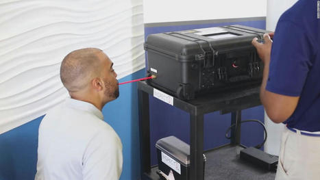
The US Food and Drug Administration has granted emergency use authorization to the first Covid-19 test that spots chemical compounds associated with the coronavirus in breath, the agency said Thursday. The FDA said the InspectIR Covid-19 Breathalyzer, which is about the size of a piece of carry-on luggage, can be used in medical offices and mobile testing sites. It can give results in less than three minutes. The system separates and identifies chemical mixtures to detect five compounds associated with SARS-CoV-2 infection. A study of the InspectIR Breathalyzer found that it accurately identified more than 91% of positive samples and nearly 100% of negative samples. Similar sensitivity was found in another study that focused on the Omicron coronavirus variant. However, a positive result should be confirmed with a PCR test, the FDA said. "It's another tool, and the FDA announcement suggests it's reasonably accurate and a relatively user-friendly tool," Dr. Emily Volk, president of the College of American Pathologists, a board-certified anatomic and clinical pathologist, said Friday. "It waits to be seen how widely this will be adopted," Volk said. "That could rely on how expensive it is." In an email to CNN on Friday, InspectIR Systems said it is not releasing the price of the machine or when it will be available. The agency's authorization "is yet another example of the rapid innovation occurring with diagnostic tests for COVID-19," Dr. Jeff Shuren, director of the FDA's Center for Devices and Radiological Health, said in a statement. "The FDA continues to support the development of novel COVID-19 tests with the goal of advancing technologies that can help address the current pandemic and better position the U.S. for the next public health emergency." FDA Press Release (April 14, 2022): https://www.fda.gov/news-events/press-announcements/coronavirus-covid-19-update-fda-authorizes-first-covid-19-diagnostic-test-using-breath-samples FDA Letter of Authorization (April 14, 2022): https://www.fda.gov/media/157720/download?utm_medium=email&utm_source=govdelivery
Via Juan Lama
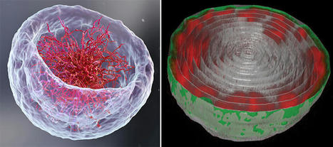
If you open a biology textbook and run through the images depicting how DNA is organized in the cell's nucleus, chances are you'll start feeling hungry; the chains of DNA would seem like a bowl of ramen: long strings floating in liquid. However, according to two new studies—one experimental and the other theoretical—that are the outcome of the collaboration between the groups of Prof. Talila Volk of the Molecular Genetics Department and Prof. Sam Safran of the Chemical and Biological Physics Department at the Weizmann Institute of Science, this image should be reconsidered. Clarifying it is essential since DNA's spatial arrangement in the nucleus can affect the expression of genes contained within the DNA molecule, and hence the proteins found in the cell. This story began when Volk was studying how mechanical forces influence cell nuclei in the muscle and found evidence that muscle contractions had an immediate effect on gene expression patterns. "We couldn't explore this further because existing methods relied on imaging of chemically preserved cells, so they failed to capture what happens in the cell nuclei of an actual working muscle," she says. To address this issue, Dr. Dana Lorber, a research associate in Volk's group, led the design of a device that makes it possible to study muscle nuclei in live fruit fly larvae. The device holds the tiny, translucent larva within a groove that allows it to contract and relax its muscles but keeps its movement constrained so that it can be scanned by a fluorescence microscope. Using the device, the researchers obtained images of the internal, linearly-organized complexes of DNA and its proteins (known as chromatin), surrounded by the membrane of the muscle nuclei. Expecting a bowl full of ramen, Lorber and Dr. Daria Amiad-Pavlov, a postdoctoral fellow in Volk's group, were in for a surprise. Rather than filling up the entire volume of the nucleus, the "noodles," or long chromatin molecules, were organized as a relatively thin layer, attached to its inner walls. Similar to the outcome of the interaction between oil and water, what is known as "phase separation," the chromatin separated itself from the bulk of the liquid inside of the nucleus and found its place at its outskirts, while most of the fluid medium remained at the center. The researchers realized that they were on their way to addressing a fundamental biological question, that is—how is chromatin, and hence DNA, organized in the nucleus in a living organism. "But the findings were so unexpected, we had to make sure no error had crept in and that this organization was universal," Lorber says.
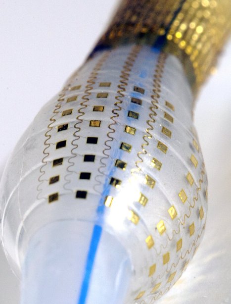
Researchers at the University of California San Diego created an ultrasound patch that can measure blood flow in vessels as deep as 14 cm within the body. The stretchy patch can be applied to the skin and may help clinicians to monitor and diagnose various conditions, including blockages that could cause an infarct. The patch contains an array of ultrasound transducers that can measure blood flow in vessels directly beneath it and the ultrasound beam can also be steered to assess vessels that are nearby, but not directly below. Monitoring blood flow in specific vessels can help clinicians to diagnose various cardiovascular diseases. For instance, measuring changes in blood flow in the carotid artery could show that someone is at a risk of stroke, and help to initiate treatment before a stroke occurs. However, current technology to accurately measure blood flow can be inconvenient, invasive, requiring a qualified technician to use an ultrasound probe to investigate the target. The flexible device contains a 12 x 12 grid of tiny ultrasound transducers embedded in a stretchy polymer. The transducers can be controlled to activate together, allowing the researchers to investigate vessels that are as deep as 14 cm beneath the skin. In another mode, the transducers will activate at different times, causing the ultrasound beam to be steered, which allows the user to investigate vessels that do not lie directly beneath the patch. “With the phased array technology, we can manipulate the ultrasound beam in the way that we want,” said Muyang Lin, another researcher involved in the study. “This gives our device multiple capabilities: monitoring central organs as well as blood flow, with high resolution. This would not be possible using just one transducer.”
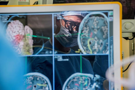
It is the first work to show that sonothermogenetics can control behavior by stimulating a specific target deep in the brain. Neurological disorders such as Parkinson's disease and epilepsy have had some treatment success with deep brain stimulation, but those require surgical device implantation. A multidisciplinary team at Washington University in St. Louis has developed a new brain stimulation technique using focused ultrasound that is able to turn specific types of neurons in the brain on and off and precisely control motor activity without surgical device implantation. The team, led by Hong Chen, assistant professor of biomedical engineering in the McKelvey School of Engineering and of radiation oncology at the School of Medicine, is the first to provide direct evidence showing noninvasive, cell-type-specific activation of neurons in the brain of mammal by combining ultrasound-induced heating effect and genetics, which they have named sonothermogenetics. It is also the first work to show that the ultrasound- genetics combination can robustly control behavior by stimulating a specific target deep in the brain. Results of the three years of research, which was funded in part by the National Institutes of Health's BRAIN Initiative, were published online in Brain Stimulation May 11, 2021. The senior research team included experts from both the McKelvey School of Engineering and the School of Medicine, including Jianmin Cui, professor of biomedical engineering; Joseph P. Culver, professor of radiology, of physics and of biomedical engineering; Mark J. Miller, associate professor of medicine in the Division of Infectious Diseases in the Department of Medicine; and Michael Bruchas, formerly of Washington University, now professor of anesthesiology and pharmacology at the University of Washington. "Our work provided evidence that sonothermogenetics evokes behavioral responses in freely moving mice while targeting a deep brain site," Chen said. "Sonothermogenetics has the potential to transform our approaches for neuroscience research and uncover new methods to understand and treat human brain disorders."
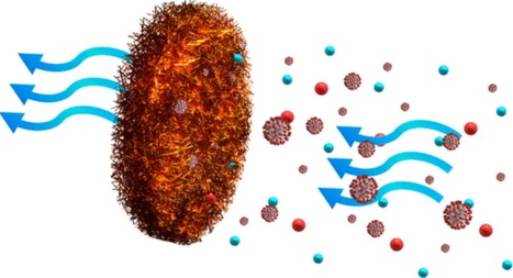
Researchers reporting in ACS’ Nano Letters have transformed copper nanowires into metal foams that could be used in facemasks and air filtration systems. The foams filter efficiently, decontaminate easily for reuse and are recyclable. The ongoing COVID-19 pandemic highlights the severe health risks posed by deep submicrometer-sized airborne viruses and particulates in the spread of infectious diseases. There is an urgent need for the development of efficient, durable, and reusable filters for this size range. Scientists now report the realization of efficient particulate filters using nanowire-based low-density metal foams which combine extremely large surface areas with excellent mechanical properties. The metal foams exhibit outstanding filtration efficiencies (>96.6%) in the PM0.3 regime, with the potential for further improvement. Their mechanical stability, light weight, chemical and radiation resistance, ease of cleaning and reuse, and recyclability further make such metal foams promising filters for combating COVID-19 and other types of airborne particulates.
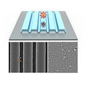
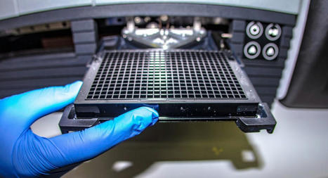
A fast, low-cost technique to see and count viruses or proteins from a sample in real time, without any chemicals or dyes, could underpin a new class of devices for rapid diagnostics and viral load monitoring, including HIV and the virus that causes COVID-19. Researchers at the University of Illinois Urbana-Champaign described the technique, called Photonic Resonator Interferometric Scattering Microscopy, or PRISM, in the journal Nature Communications. “We have developed a new form of microscopy that amplifies the interaction between light and biological materials. We can use it for very rapid and sensitive forms of diagnostic testing, and also as a very powerful tool for understanding biological processes at the scale of individual items, like counting individual proteins or recording individual protein interactions,” said study leader Brian Cunningham, the Intel Alumni Endowed Chair of electrical and computer engineering at Illinois. In optical microscopes, light bounces off any molecules or viruses it encounters on a slide, creating a signal. Instead of a regular glass slide, the PRISM technique uses a photonic crystal — a nanostructured glass surface that brilliantly reflects only one specific wavelength of light. Cunningham’s group designed and fabricated a photonic crystal that reflects red light, so that the light from a red laser would be amplified. “The molecules we are looking at – in this study, viruses and small proteins – are extremely small. They cannot scatter enough light to create a signal that can be detected by a conventional optical microscope,” said graduate student Nantao Li, the first author of the paper. “The benefit of using the photonic crystal is that it amplifies the light’s intensity so it’s easier to detect those signals and enables us to study these proteins and viruses without any chemical labels or dyes that might modify their natural state or hinder their activity – we can just use the intrinsic scattering signal as the gauge for determining if those molecules are present.” The researchers verified their technique by detecting the virus that causes COVID-19. PRISM detected individual coronaviruses as they traveled across the slide’s surface. The researchers also used PRISM to detect individual proteins such as ferritin and fibrinogen. The technique could allow researchers to study such biological targets in their natural states – watching as proteins interact, for example – or researchers could seed the surface of the photonic crystal slide with antibodies or other molecules to capture the targeted items and hold them in place. “It takes 10 seconds to get a measurement, and in that time we can count the number of viruses captured on the sensor,” Cunningham said. “It’s a single-step detection method that works at room temperature. It is also fast, very sensitive and low cost. It’s very different from the standard way we do viral testing now, which involves breaking open the viruses, extracting their genetic material and putting it through a chemical amplification process so we can detect it. That method, called PCR, is accurate and sensitive, but it requires time, specialized equipment and trained technicians.”
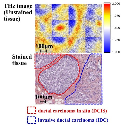
A team of researchers at Osaka University, in collaboration with the University of Bordeaux and the Bergonié Institute in France, has succeeded in terahertz imaging of early-stage breast cancer less than 0.5 mm without staining, which is difficult to identify even by pathological diagnosis. Their work provides a breakthrough towards rapid and precise on-site diagnosis of various types of cancer and accelerates the development of innovative terahertz diagnostic devices. Breast cancer is roughly divided into two types: invasive and non-invasive. The former, invasive ductal carcinoma (IDC), begins in the cells of a breast duct, growing through the duct walls and into the surrounding breast tissue, potentially spreading to other parts of the body. The latter, ductal carcinoma in situ (DCIS), is an early-stage small breast cancer confined to the breast duct, but it can lead to invasive cancer. Therefore, early detection of DCIS is crucial. For pathological diagnosis of cancer, the tissue sample is chemically stained, and a pathologist makes a diagnosis using an image of the stained tissue. However, the staining process takes time, and it is difficult to distinguish DCIS from malignant IDC as they look nearly identical. Terahertz imaging can distinguish cancer tissue from normal tissue without staining and radiation exposure. However, it was still difficult to identify an individual DCIS lesion (which typically range from 50 to 500 µm) by terahertz imaging due to its diffraction-limited spatial resolution of just several millimeters. “To overcome this drawback, we developed a unique imaging technique in which terahertz light sources that are locally generated at irradiation spots of laser beams in a nonlinear optical crystal directly interact with a breast cancer tissue sample. Consequently, we succeeded in clearly visualizing a DCIS lesion of less than 0.5 mm,” explains lead author Kosuke Okada. The accuracy of this technique is approximately 1000 times higher than that of conventional techniques using terahertz waves.
Vanderbilt University engineers have determined that their back-assist exosuit, a clothing-like device that supports human movement and posture, can reduce fatigue by an average of 29-47 percent in lower back muscles. The exosuit's functionality presents a promising new development for individuals who work in physically demanding fields and are at risk for back pain, including medical professionals and frontline workers. The article describing the experiment and findings, "Low-Profile Elastic Exosuit Reduces Back Muscle Fatigue," was published in the Nature journal Scientific Reports on September 29, 2020. The research, led by Assistant Professor of Mechanical Engineering Karl Zelik and recent Ph.D. graduate and primary author Erik Lamers, used surface electromyography techniques to measure changes in low back muscle fatigue in male and female participants, who were given physical tasks to perform both with and without the exosuit. The wearable technology developed by Zelik's team may conjure images of Iron Man's suit, but it does not rely on motors or batteries. Instead, the low-profile, elastic exosuit applies assistive forces that cooperate with the low back extensor muscles, to relieve strain on the muscles and spine, and to help reduce injury risks. This study showed that wearing the exosuit made holding a 35-pound weight (the average weight of a 4-year-old child) less tiring on the back than holding a 24-pound weight (the average weight of an 18-month-old baby) without the exosuit. "These findings show how exosuits could provide valuable back relief to frontline and essential workers who have been taking a physical toll and supporting all of us throughout this pandemic. What we learned has the potential to shape the biomechanical and industrial standards of future wearable technologies," said Zelik, who holds secondary appointments in biomedical engineering and in physical medicine and rehabilitation. The work also demonstrated a sharpened understanding of how the latissimus dorsi muscles (the "lats") -- those that adduct, extend and medially rotate the shoulder joint -- affect low back mechanics. While previous research has shown that the lats generally do not contribute much to supporting the low back, the investigators discovered that, as people get tired, they may suddenly recruit the lats to offload the main back extensor muscles to a significant degree. "The lats act sort of like an exosuit. When a person's low back muscles become over-strained and fatigued, they summon extra assistance from their lats to relieve this back strain and fatigue. The elastic bands in our exosuit work the same way to help sustain endurance and strength," said Lamers, a National Science Foundation Graduate Research Fellow who worked in Vanderbilt's Center for Rehabilitation Engineering and Assistive Technology. Zelik, a former collegiate athlete who competed in the long jump and triple jump, knows firsthand how intensive physical activity can fatigue the body. He also understands the importance of ensuring that the exosuit and its utility are built with inclusive design practices. "Wearables are going to change the way we work and live, and we want to improve safety, health and well-being for everyone. One of the critical challenges moving forward will be to ensure that all wearable technology is developed to serve and protect both women and men. We are thrilled that this research helped lead to the first commercial exosuit or exoskeleton designed with both male- and female-fits," Zelik said.
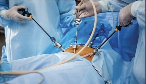
Researchers developed a new class of medical instruments equipped with an advanced soft electronics system that could dramatically improve the diagnoses and treatments of a number of cardiac diseases and conditions. Detailed in a new paper published in the journal Nature Biomedical Engineering, the researchers, led by engineers at the George Washington University and Northwestern University, applied stretchable and flexible matrices of electrode sensors and actuators, along with temperature and pressure sensors, to a balloon catheter system, often used in minimally invasive surgeries or ablations to treat conditions such as heart arrhythmias. The new system, which conforms better to the body's soft tissue than current devices, can perform a variety of functions, including: simultaneous in vivo measurements of temperature, force of contact and electrophysiological parameters; the ability to customize diagnostic and therapeutic functions; and real-time feedback. The new system can also dramatically reduce the length of invasive ablation procedures and exposure of patients and doctors to X-ray radiation. Many minimally invasive surgeries rely on catheters inserted into the body through small incisions to conduct diagnostic measurements and therapeutic interventions. Physicians, for example, use this catheter-based approach to map and treat irregular heartbeats, or arrhythmias, often by locating and killing or ablating cardiac tissue area which is causing the arrhythmias. Though widely used in surgery, the current catheter-based approach has a number of drawbacks. The rigidity of today's catheter devices means they do not conform well to soft, biological tissues, impacting high fidelity mapping of an organ's electrophysiological signals. Current devices make contact with only a small part of an organ at a time, making it necessary to constantly move a probe around, lengthening medical procedures. Current catheter systems are also limited in the number of functions they can perform, requiring physicians to use multiple catheters in a single ablation procedure. Additionally, long procedures—for example, to locate and ablate tissues causing arrhythmias—risk exposing both patient and physician to potentially damaging X-rays, as physicians rely on X-ray images during the course of the surgery to guide their catheters. The benefit The new class of instruments the researchers developed will allow physicians to acquire a rich set of electrophysiological information and to complete surgeries in shorter times with a single instrumented catheter system.
An Indiana University Kelley School of Business study of users with coronavirus symptoms shows that chatbots can be an effective screening tool, which could ease the burden for medical providers. COVID-19 has placed tremendous pressure on health care systems, not only for critical care but also from an anxious public looking for answers. Research from the Indiana University Kelley School of Business found that chatbots -- software applications that conduct online chats via text or text-to-speech -- working for reputable organizations can ease the burden on medical providers and offer trusted guidance to those with symptoms. Researchers conducted an online experiment with 371 participants who viewed a COVID-19 screening session between a hotline agent -- chatbot or human -- and a user with mild or severe symptoms. They studied whether chatbots were seen as being persuasive, providing satisfying information that likely would be followed. Their results showed a slight negative bias against chatbots' ability, perhaps due to recent press reports. When the perceived ability is the same, however, participants reported that they viewed chatbots more positively than human agents, which is good news for health care organizations struggling to meet user demand for screening services. "The primary factor driving user response to screening hotlines -- human or chatbot -- is perceptions of the agent's ability," said Alan Dennis, the John T. Chambers Chair of Internet Systems at Kelley and corresponding author of the paper, "User reactions to COVID-19 screening chatbots from reputable providers." "When ability is the same, users view chatbots no differently or more positively than human agents." Other authors on the paper, forthcoming in the Journal of the American Medical Informatics Association, are Antino Kim, assistant professor of operations and decision technologies at Kelley; and Sezgin Ayabakan, assistant professor of management information systems, and doctoral candidate Mohammad Rahimi, both at Temple University's Fox School of Business. Even before the pandemic, chatbots were identified as a technology that could speed up how people interact with researchers and find medical information online. "Chatbots are scalable, so they can meet an unexpected surge in demand when there is a shortage of qualified human agents," Dennis, Kim and their co-authors wrote, adding that chatbots "can provide round-the-clock service at a low operational cost.
Rice chemist Han Xiao and his colleagues found that replacing a single oxygen atom with a sulfur atom in a common fluorophore turns it into a photosensitizing molecule. When exposed to light, the molecule generated reactive oxygen species (ROS) that destroyed breast cancer cells in the lab.
The study led by co-lead authors Juan Tang and Lushun Wang, both Rice postdoctoral researchers, appears in the Royal Society of Chemistry flagship journal Chemical Science.
This method of photodynamic therapy is already in use, as light-triggered molecules are known to generate cytotoxic ROS. Most current photosensitizers require the incorporation of heavy atoms, but they are difficult and costly to synthesize and remain toxic in the dark, potentially damaging healthy cells, Xiao said.
The Rice lab's one-step compounds have no heavy atoms, yield a high ratio of ROS when triggered and shut off when the light is turned off. The lab's various thio-based fluorophores absorb light in visible to near-infrared wavelengths that penetrate up to 5 millimeters into tissues.
"This work comes through our previous study to make better fluorogenic dyes," Xiao said. "That was a totally new discovery, but once we went deeper into the mechanism, we found that our thio-based fluorophores can lead to a dramatic generation of singlet oxygen when excited with light. This is the real mediator."
For testing, the researchers combined their photosensitizers with trastuzumab, an antibody used to target and treat early and advanced breast cancer. The combination showed "robust cytotoxicity" against HER2-positive (cancerous) cell lines but almost no activity against HER2-negative cells.
Xiao said the experiments showed their photosensitizers targeted both monolayer cancer cells and multicellular tumor spheroids. "We think a big application for this photosensitizer will be skin cancers," he said. "It should be easy for light to penetrate basal cell carcinomas on the surface."
|
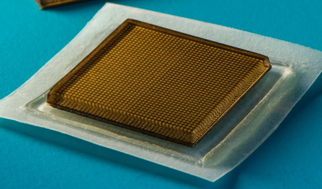
Ultrasound is a convenient, noninvasive tool for doctors to look inside the human body and check out a person’s liver, heart and other internal structures, as well as the developing fetus of a pregnant patient. But today’s ultrasound imaging technology is large and technical, so it’s only available in healthcare facilities and must be operated by highly trained technicians. Plus, patients, who take time out of their schedules to go to an appointment, have to be covered in a sticky gel. Now, researchers say they’ve developed an innovative solution to some of these challenges. Engineers at the Massachusetts Institute of Technology have unveiled a new adhesive ultrasound patch that’s about the size of a postage stamp and can provide continuous imaging of the body’s inner workings for up to 48 hours. The scientists shared their new technology in a paper published last week in the journal Science. “We believe we’ve opened a new era of wearable imaging,” says Xuanhe Zhao, a mechanical engineer at MIT and one of the study’s authors, in a statement. “With a few patches on your body, you could see your internal organs.” In the past, engineers developing wearable ultrasound technologies have run into issues with image quality and flexibility, but the new MIT stickers seem to have struck the right balance. To create the small devices, which are about three millimeters thick and two square centimeters in size, engineers combined rigid transducers with a stretchy, sticky layer that encapsulates a layer of water-based hydrogel.
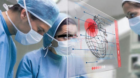
Medical schools are using augmented reality in their curricula to give students hands-on learning opportunities. Medical students can use augmented reality to see and apply theories throughout their training. Augmented reality (AR) and virtual reality (VR) in the healthcare sector are giving a new perspective to the world. Virtual reality (VR) and other innovative technologies such as augmented reality are already being used by many healthcare organizations (AR). Technologists employ virtual reality in various settings, including treating patients, medical teaching, and hospital management. Augmented Surgery Surgeons can utilize AR to learn more about their patients’ anatomy. They may feed their MRI and CT scan results into an AR headset. Then, just before surgery, place-specific patient physiology over their body. Doctors can examine muscles, bones, and essential organs with this technology. Previously, surgery had a significant mortality rate. Procedures can now be a lot safer because of AR. When physicians and experts try to save patients’ lives, AR can help by providing people with all the information they need. Physicians could be more aware of organ placement, vein meshes, and diagnostic reports as they operate since they are all there in front of their eyes. Augmented Diagnosis Some patients, you may have seen, have trouble accurately articulating actual symptoms to doctors. Because of AR, patients will finally be able to communicate their symptoms. As a result of this procedure, doctors can more readily examine their patients’ complaints and diagnose them more accurately. Using augmented reality, the nurse can now swiftly find veins. How? AccuVein, an AR firm that uses a portable scanner, allows nurses to determine the location of veins. Augmented Practice Medical schools use augmented reality in their curricula to give students hands-on learning opportunities. It would be simple to replicate patients and surgical interactions for students using AR in education. Medical students can use augmented reality to see and apply theories throughout their training. Virtual Collaboration Between Physicians With AR What if your surgeon is thousands of miles away? AR may be able to help you. If the surgeon is unavailable and a specialized on hand possesses AR tools, the specialist can assist and follow orders. Doctors may employ AR for collaborative operations and AR video conferencing for successful meetings on any health topic. With doctors from outside the clinic assisting, it might be a life-saving situation.
A completely paralyzed patient can now convey full sentences thanks to electrodes implanted in his brain. Medical research surgically placed brain-computer interfaces (BCIs) into a patient’s brain. The implants tracked electrical activity in the brain and translated the signals to commands to another device. According to a paper published in Nature detailing the procedure, the subject was able to message requests for beer, soup and massages from his nurses. He also asked to watch movies with his son. The participant suffers from Amyotrophic lateral sclerosis, or ALS, and is completely ‘locked-in,’ meaning he can’t use his limbs or make eye movements. In the past, BCIs have helped patients convey ‘yes’ or ‘no’ via thoughts or allowed partially paralyzed patients to move prosthetic limbs. “It’s really remarkable to be able to reestablish communication with someone in a completely locked-in state. To me, that’s a tremendous breakthrough and obviously quite meaningful for the research participant,” said Jaimie Henderson, M.D., a Stanford University neurosurgeon who wasn’t involved in the project. The individual received the innovative treatment through ALS Voice GmbH. A nonprofit that works with noncommunicative participants to incorporate BCIs and other technologies to improve their quality of life. The patient responded well to neurofeedback, which shows brain activity in real time so a patient can control their responses. When brain electrical activity alters, a computer would respond with ascending or descending tones. “Within two days, [the patient] was able to increase and decrease the frequency of a sound tone,” said Ujwal Chaudhary of ALS Voice gGmbH. His team developed software based on a computer system the patient’s family created. One of the patient’s first sentences was, “boys, it works so effortlessly.” In the future, Chaudhary wants to develop software with a word list that has an autocomplete function in the software. “There are many ways in which we could make it faster,” he said.
In a discovery published in the journal Nature, an international team of researchers has described a novel molecular device with exceptional computing prowess. Reminiscent of the plasticity of connections in the human brain, the device can be reconfigured on the fly for different computational tasks by simply changing applied voltages. Furthermore, like nerve cells can store memories, the same device can also retain information for future retrieval and processing. "The brain has the remarkable ability to change its wiring around by making and breaking connections between nerve cells. Achieving something comparable in a physical system has been extremely challenging," said Dr. R. Stanley Williams, professor in the Department of Electrical and Computer Engineering at Texas A&M University. "We have now created a molecular device with dramatic reconfigurability, which is achieved not by changing physical connections like in the brain, but by reprogramming its logic." Dr. T. Venkatesan, director of the Center for Quantum Research and Technology (CQRT) at the University of Oklahoma, Scientific Affiliate at National Institute of Standards and Technology, Gaithersburg, and adjunct professor of electrical and computer engineering at the National University of Singapore, added that their molecular device might in the future help design next-generation processing chips with enhanced computational power and speed, but consuming significantly reduced energy. Whether it is the familiar laptop or a sophisticated supercomputer, digital technologies face a common nemesis, the von Neumann bottleneck. This delay in computational processing is a consequence of current computer architectures, wherein the memory, containing data and programs, is physically separated from the processor. As a result, computers spend a significant amount of time shuttling information between the two systems, causing the bottleneck. Also, despite extremely fast processor speeds, these units can be idling for extended amounts of time during periods of information exchange. As an alternative to conventional electronic parts used for designing memory units and processors, devices called memristors offer a way to circumvent the von Neumann bottleneck. Memristors, such as those made of niobium dioxide and vanadium dioxide, transition from being an insulator to a conductor at a set temperature. This property gives these types of memristors the ability to perform computations and store data. However, despite their many advantages, these metal oxide memristors are made of rare-earth elements and can operate only in restrictive temperature regimes. Hence, there has been an ongoing search for promising organic molecules that can perform a comparable memristive function, said Williams. Dr. Sreebrata Goswami, a professor at the Indian Association for the Cultivation of Science, designed the material used in this work. The compound has a central metal atom (iron) bound to three phenyl azo pyridine organic molecules called ligands. "This behaves like an electron sponge that can absorb as many as six electrons reversibly, resulting in seven different redox states," said Sreebrata. "The interconnectivity between these states is the key behind the reconfigurability shown in this work."
Researchers at UC San Francisco have successfully developed a “speech neuroprosthesis” that has enabled a man with severe paralysis to communicate in sentences, translating signals from his brain to the vocal tract directly into words that appear as text on a screen. The achievement, which was developed in collaboration with the first participant of a clinical research trial, builds on more than a decade of effort by UCSF neurosurgeon Edward Chang, MD, to develop a technology that allows people with paralysis to communicate even if they are unable to speak on their own. The study is published in the New England Journal of Medicine. "To our knowledge, this is the first successful demonstration of direct decoding of full words from the brain activity of someone who is paralyzed and cannot speak," said Chang, the Joan and Sanford Weill Chair of Neurological Surgery at UCSF, Jeanne Robertson Distinguished Professor, and senior author on the study. "It shows strong promise to restore communication by tapping into the brain's natural speech machinery." Each year, thousands of people lose the ability to speak due to stroke, accident, or disease. With further development, the approach described in this study could one day enable these people to fully communicate. Translating Brain Signals into Speech Previously, work in the field of communication neuroprosthetics has focused on restoring communication through spelling-based approaches to type out letters one-by-one in text. Chang's study differs from these efforts in a critical way: his team is translating signals intended to control muscles of the vocal system for speaking words, rather than signals to move the arm or hand to enable typing. Chang said this approach taps into the natural and fluid aspects of speech and promises more rapid and organic communication. "With speech, we normally communicate information at a very high rate, up to 150 or 200 words per minute," he said, noting that spelling-based approaches using typing, writing, and controlling a cursor are considerably slower and more laborious. "Going straight to words, as we're doing here, has great advantages because it's closer to how we normally speak."
Reverse transcription-polymerase chain reaction (RT-PCR) has been the gold standard for diagnosis during the COVID-19 pandemic. However, the PCR portion of the test requires bulky, expensive machines and takes about an hour to complete, making it difficult to quickly diagnose someone at a testing site. Now, researchers reporting in ACS Nano have developed a plasmofluidic chip that can perform PCR in only about eight minutes, which could speed diagnosis during current and future pandemics. Rapid diagnosis of COVID-19 and other highly contagious viral diseases is important for timely medical care, quarantining and contact tracing. Currently, RT-PCR –– which uses enzymes to reverse transcribe tiny amounts of viral RNA to DNA, and then amplify the DNA so that it can be detected by a fluorescent probe –– is the most sensitive and reliable diagnostic method. But because the PCR portion of the test requires 30–40 cycles of heating and cooling in special machines, it takes about an hour to perform, and samples must typically be sent away to a lab, meaning that a patient usually has to wait a day or two to receive their diagnosis. Ki-Hun Jeong and colleagues wanted to develop a plasmofluidic PCR chip that could quickly heat and cool miniscule volumes of liquids, allowing accurate point-of-care diagnosis in a fraction of the time. The researchers devised a postage stamp-sized polydimethylsiloxane chip with a microchamber array for the PCR reactions. When a drop of sample is added to the chip, a vacuum pulls the liquid into the microchambers, which are positioned above glass nanopillars with gold nanoislands. Any microbubbles, which could interfere with the PCR reaction, diffuse out through an air-permeable wall. When a white LED is turned on beneath the chip, the gold nanoislands on the nanopillars quickly convert light to heat, and then rapidly cool when the light is switched off. The researchers tested the device on a piece of DNA containing a SARS-CoV-2 gene, accomplishing 40 heating and cooling cycles and fluorescence detection in only five minutes, with an additional three minutes for sample loading. The amplification efficiency was 91%, whereas a comparable conventional PCR process has an efficiency of 98%. With the reverse transcriptase step added prior to sample loading, the entire testing time with the new method could take 10 to 13 minutes, as opposed to about an hour for typical RT-PCR testing. The new device could provide many opportunities for rapid point-of-care diagnostics during a pandemic, the researchers say.
Cancer cells have evolved mechanisms to escape the body's immune defense. Agents that prevent immune escape are attractive targets for the development of new cancer therapies. A group of scientists led by Herbert Waldmann and Slava Ziegler at the Max Planck Institute of Molecular Physiology in Dortmund has now developed a new cell-based test system to identify immunoregulatory modulators. Screening a library of over 150,000 substances revealed several potent substances with unprecedented structure.
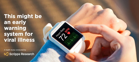
Examining data from the first six weeks of their landmark DETECT study, a team of scientists from the Scripps Research Translational Institute sees encouraging signs that wearable fitness devices can improve public health efforts to control COVID-19. The DETECT study, launched on March 25 2020, uses a mobile app to collect smartwatch and activity tracker data from consenting participants, and also gathers their self-reported symptoms and diagnostic test results. Any adult living in the United States is eligible to participate in the study by downloading the research app, MyDataHelps. In a study published October 29, 2020 in Nature Medicine, the Scripps Research team reports that wearable devices like Fitbit are capable of identifying cases of COVID-19 by evaluating changes in heart rate, sleep and activity levels, along with self-reported symptom data—and can identify cases with greater success than looking at symptoms alone. “What’s exciting here is that we now have a validated digital signal for COVID-19. The next step is to use this to prevent emerging outbreaks from spreading,” says Eric Topol, MD, director and founder of the Scripps Research Translational Institute and executive vice president of Scripps Research. “Roughly 100 million Americans already have a wearable tracker or smartwatch and can help us; all we need is a tiny fraction of them—just 1 percent or 2 percent—to use the app.” With data from the app, researchers can see when participants fall out of their normal range for sleep, activity level or resting heart rate; deviations from individual norms are a sign of viral illness or infection. But how do they know if the illness causing those changes is COVID-19? To answer that question, the team reviewed data from those who reported developing symptoms and were tested for the novel coronavirus. Knowing the test results enabled them to pinpoint specific changes indicative of COVID-19 versus other illnesses. “One of the greatest challenges in stopping COVID-19 from spreading is the ability to quickly identify, trace and isolate infected individuals,” says Giorgio Quer, PhD, director of artificial intelligence at Scripps Research Translational Institute and first author of the study. “Early identification of those who are pre-symptomatic or even asymptomatic would be especially valuable, as people may potentially be even more infectious during this period. That’s the ultimate goal.”
OmniVision OV6948 enters the Guinness Book of Records as the world's smallest camera. OmniVision OV6948 measures in super-small at just 0.575 x 0.575 x 0.232mm and is good for 40,000-pixel color images using an RGB Bayer back-side-illuminating chip. This new camera is ridiculosuly small, but it's for specific use cases in surgery. With the OmniVision OV6948 surgeons can have a camera so small it will fit into the smallest veins inside of the human body. This technology provides surgeons and doctors that have the OmniVision OV6948 with next-gen camera access for future surgeries. Until now, surgeons do this without any camera -- acting blind. The only cameras capable of anything close to this are very few, but they also have a much lower resolution fiber optic feed. The new OmniVision OV6948 captures images at 30FPS, and can have analog output at over 4mm away with minimal noise. The OmniVision OV6948 has a 120-degree super-side angle field of view, something that on a regular camera would come up as 14nm on a full-frame sensor. The depth of field for the OmniVision OV6948 spans between 3mm and 30mm.
Gut microbes affect human health, but there is still much to learn, in part because they're not easy to collect. But researchers now report in ACS Nano that they have developed an ingestible capsule that in rat studies captured bacteria and other biological samples while passing through the gastrointestinal (GI) tract. Currently, researchers obtain gut microbes by collecting stool samples or using techniques such as colonoscopy or endoscopy. However, stool samples can't capture all the microorganisms in the upper GI tract, and they can't keep microbes from different parts of the tract separate. Colonoscopy and endoscopy are invasive procedures, which deters some patients. Sarvesh Kumar Srivastava and colleagues wanted to avoid these drawbacks by designing a device that could be swallowed and then eliminated. The researchers developed a self-polymerizing reaction system of poly(ethylene glycol) diacrylate monomer, iron chloride and ascorbic acid -- all loaded into tiny hollow cylinders. The cylindrical microdevices were packaged in miniature gelatin capsules, which were coated with a protective layer to prevent digestion in the stomach's acidic environment. After they were fed to rats, the capsules remained protected in the stomach but disintegrated in the small intestine's more-neutral pH, releasing the microdevices. Exposure to intestinal fluid caused the cylinders' chemical cargo to polymerize, forming a hydrogel that trapped microbes and protein biomarkers in its surroundings, much like an instant snapshot of the intestine. The devices, which didn't cause inflammation or toxicity, were then surgically removed -- a step that the researchers say will be replaced by natural elimination in future. High-throughput sequencing studies showed that the bacterial population the devices captured closely resembled that of the gut. The researchers also demonstrated that these tiny cylinders could be triggered over a range of pH to deliver biologics, like insulin, to cells in a petri dish in the presence of intestinal mucus. This technology could advance understanding of host-microbiome interactions, providing insight into associated GI disease progression and paving the way for personalized gut therapies, the team says.
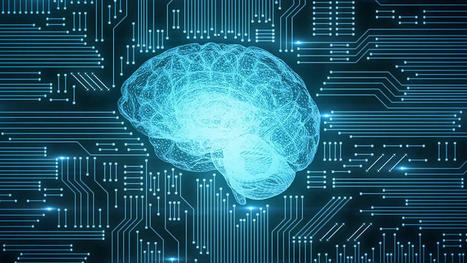
For most medical professionals, artificial intelligence (AI) will be an accelerant and enabler, not a threat. It would be good business for AI companies as well to help, rather than attempt to replace, medical professionals. It’s no secret that healthcare costs have risen faster than inflation for decades. Some experts estimate that healthcare will account for over 20% of the US GDP by 2025. Meanwhile, doctors are working harder than ever before to treat patients as the U.S. physician shortage continues to grow. Many medical professionals have their schedules packed so tightly that much of the human element which motivated their pursuit of medicine in the first place is reduced. In healthcare, artificial intelligence (AI) can seem intimidating. At the birthday party of a radiologist friend, she gently expressed how she felt her job would be threatened by AI in the coming decade. Yet, for most of the medical profession, AI will be an accelerant and enabler, not a threat. It would be good business for AI companies as well to help, rather than attempt to replace, medical professionals.
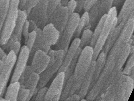
Unprecedented details of enamel structure may point to new ways to prevent or halt cavities. Scientists used a combination of advanced microscopy and chemical detection techniques to uncover the structural makeup of human tooth enamel at unprecedented atomic resolution, revealing lattice patterns and unexpected irregularities. The findings could lead to a better understanding of how tooth decay develops and might be prevented. The research was supported in part by the National Institute of Dental and Craniofacial Research (NIDCR) at the National Institutes of Health. The findings appear in Nature. “This work provides much more detailed information about the atomic makeup of enamel than we previously knew,” said Jason Wan, Ph.D., a program officer at NIDCR. “These findings can broaden our thinking and approach to strengthening teeth against mechanical forces, as well as repairing damage due to erosion and decay.” Your teeth are remarkably resilient, despite enduring the stress and strain of biting, chewing, and eating for a lifetime. Enamel — the hardest substance in the human body — is largely responsible for this endurance. Its high mineral content gives it strength. Enamel forms the outer covering of teeth and helps prevent tooth decay, or caries. Tooth decay is one of the most common chronic diseases, affecting up to 90% of children and the vast majority of adults worldwide, according to the World Health Organization. Left untreated, tooth decay can lead to painful abscesses, bone infection, and bone loss. Tooth decay starts when excess acid in the mouth erodes the enamel covering. Scientists have long sought a more complete picture of enamel’s chemical and mechanical properties at the atomic level to better understand—and potentially prevent or reverse—enamel loss. To survey enamel at the tiniest scales, researchers use microscopy methods such as scanning transmission electron microscopy (STEM), which directs a beam of electrons through a material to map its atomic makeup.
|