


Research and publish the best content.
Get Started for FREE
Sign up with Facebook Sign up with X
I don't have a Facebook or a X account
Already have an account: Login

Vectorology - GEG Tech top picks
21.7K views |
+0 today
 Your new post is loading...
 Your new post is loading...
Your new post is loading...
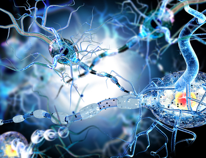
Gene expression is controlled in activated neurons in the mouse brain using a two-component optogenetic system.
BigField GEG Tech's insight:
Here, the scientists present a dual-protein switch system, Cal-Light, that translates neuronal-activity-mediated calcium signaling into gene expression in a light-dependent manner. In cultured neurons and brain slices, they show that Cal-Light drives expression of the reporter EGFP with high spatiotemporal resolution only in the presence of both blue light and calcium. Delivery of the Cal-Light components to the motor cortex of mice by viral vectors labels a subset of excitatory and inhibitory neurons related to learned lever-pressing behavior. By using Cal-Light to drive expression of the inhibitory receptor halorhodopsin (eNpHR), which responds to yellow light, we temporarily inhibit the lever-pressing behavior, confirming that the labeled neurons mediate the behavior. Thus, Cal-Light enables dissection of neural circuits underlying complex mammalian behaviors with high spatiotemporal precision. 
BigField GEG Tech's insight:
The Raf–MEK–ERK kinase pathway, which mediates cellular responses to a variety of extracellular signals, is regulated by positive and negative feedback mechanisms ranging from receptor degradation to transcriptional induction of inhibitory phosphatases. However, detecting these effects with temporal precision remains difficult. Zhou et al. engineered photodissociable dimeric variants of the Dronpa fluorescent protein (pdDronpa) that monomerize with cyan-light (500 nm) exposure and then attached pdDronpa domains to two locations on the Raf1 kinase domain. The interaction of the two pdDronpa domains under basal conditions prevents substrate access, while cyan-light exposure promotes dissociation, enabling kinase activity. This effect was reversible such that pdDronpa dimerization was restored with violet light (400 nm), inactivating kinase activity. Zhou et al. characterized a fast negative feedback mechanism on MEK, showing that a pulse of MEK activation by pdDronpa-modified Raf1 was followed by dephosphorylation attributable to ERK-mediated activation of protein phosphatases. Finally, the application of this technology to other kinases such as CDK5 provides additional support that pdDronpa may be a generally useful reagent for spatiotemporal control of kinase signaling.

Two neuronal pathways originating in the central amygdala coordinate distinct behaviors
BigField GEG Tech's insight:
Here, the scientists reveal a critical role for the central nucleus of the amygdala in predatory hunting. Both optogenetic and chemogenetic stimulation of central amygdala of mice elicited predatory-like attacks upon both insect and artificial prey.
BigField GEG Tech's insight:
Here, Tomas Bjorklund review design choices for enabling expression of two functional protein or RNA sequences from a single viral vector. These type of tools are very useful in neuroscience-related field of neuronal control and modulation, e.g., using optogenetics or DREADDs, but are also desirable in applications of CRISPR/Cas9 in situ genome editing and more refined therapeutic approaches.
BigField GEG Tech's insight:
The authors use a lentiviral vector expressing channelrhodopsin-2 (ChR2+) in catecholaminergic neurons (C1 and A5) to determine which of physiological responses are elicited when retrotrapezoid nucleus neurons (RTN) are selectively activated.They observed that RTN neurons activate breathing powerfully and, unlike the C1 cells, have minimal effects on blood pressure (BP) and have a weak arousal capability at best. A5 neuron stimulation produces little effect on breathing and BP, but does appear to facilitate arousal.
BigField GEG Tech's insight:
The authors stably introduced light-activated channelrhodopsin-2 (ChR2, a cation channel activated with 480 nm light) into undifferentiated hESC thanks to a lentiviral vector. They shown that a light sensitive protein can enable in vitro optical control of hESC-CM and that this activation occurs optimally above specific light stimulation intensity and pulse width thresholds.
|
BigField GEG Tech's insight:
Neurophysiological phenomena that underlie the symptoms of autism remain unclear. Genetics-based mouse models of autism have suggested that there is an increase in the neuronal excitation/inhibition (E:I) balance. An optogenetically driven increase in this E:I balance leads to social deficits in mice. Using mice lacking CNTNAP2 , a gene known to be associated with autism in humans, Selimbeyoglu and colleagues now show that real-time optogenetic modulation of the E:I balance rescued social behavior deficits and hyperactivity in these animals. This study highlights the potential for modulating neural circuits in the brain as a strategy for treating autism.

BigField GEG Tech's insight:
Researchers have identified two groups of neurons that can be turned on and off to alleviate the movement-related symptoms of Parkinson's disease. The activation of these cells in the basal ganglia relieves symptoms for much longer than current therapies, like deep brain stimulation and pharmaceuticals. The study, completed in a mouse model of Parkinson's, used optogenetics to better understand the neural circuitry involved in Parkinson's disease, and could provide the basis for new experimental treatment protocols.
BigField GEG Tech's insight:
Optogenetics is a new biological technique that allows reversible modulation of electrical function with unmatched spatiotemporal precision using light-gated ion channels. Here, the authors investigated optogenetic manipulation of anatomical re-entry in ventricular cardiac tissue.
BigField GEG Tech's insight:
Optogenetics is emerging in the cardiology field as a new strategy to explore biological functions through the use of light-sensitive proteins and dedicated light sources. For example, this technology allows modification of the electrophysiological properties of cardiac muscle cells with superb spatiotemporal resolution and quantitative control. In this chapter, the optogenetic modification of atrial cardiomyocytes (aCMCs) from 2-day-old Wistar rats using lentiviral vector (LV) technology and the subsequent activation of the light-sensitive proteins (i.e., ion channels) through light-emitting diodes (LEDs) are described.
BigField GEG Tech's insight:
To test long-term effects of chronic synaptic activation on Aβ pathology, the scientistss adopted optogenetics and transduced stabilized step-function opsin, a channelrhodopsin engineered to elicit a long-lasting neuronal hyperexcitability, into the hippocampal perforant pathway of APP transgenic mice. In vivo microdialysis revealed a ∼24% increase in the hippocampal interstitial fluid Aβ42 levels immediately after acute light activation and five months of chronic optogenetic stimulation increased Aβ burden specifically in the projection area of the perforant pathway(∼2.5-fold).
|


Optogenetics may enable mutation-independent, circuit-specific restoration of neuronal function in neurological diseases. Retinitis pigmentosa is a neurodegenerative eye disease where loss of photoreceptors can lead to complete blindness. In a blind patient, the researchers combined intraocular injection of an adeno-associated viral vector encoding ChrimsonR with light stimulation via engineered goggles.