healthcare technology
147.6K views | +20 today
Follow
healthcare technology
The ways in which technology benefits healthcare
Curated by nrip
Your new post is loading...
Scooped by nrip
Scoop.it!

Google and Harvard map brain connections in unprecedented detail

Google and Harvard map brain connections in unprecedented detail | healthcare technology | Scoop.it

The human brain is the most ridiculously complex computer that’s ever existed, and mapping this dense tangle of neurons, synapses and other cells is nigh on impossible. But engineers at Google and Harvard have given it the best shot yet, producing a browsable, searchable 3D map of a small section of human cerebral cortex.


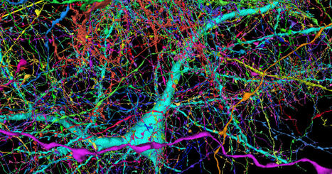
A browsable 3D map of just one millionth of the cerebral cortex has been created using 225 million images and a whopping 1.4 petabytes of data, illustrating the immense complexity of the human brain.


With about 86 billion neurons connecting via 100 trillion synapses, it’s a Herculean task to figure out exactly what each of them does and how those connections form the basis of thought, emotion, memory, behavior and consciousness. Daunting as it may be, though, teams of scientists around the world are rolling up their sleeves and trying to build a wiring diagram for the human brain – a so-called “connectome.”


he researchers started with a sample taken from the temporal lobe of a human cerebral cortex, measuring just 1 mm3. This was stained for visual clarity, coated in resin to preserve it, and then cut into about 5,300 slices each about 30 nanometers (nm) thick. These were then imaged using a scanning electron microscope, with a resolution down to 4 nm. That created 225 million two-dimensional images, which were then stitched back together into one 3D volume.


Machine learning algorithms scanned the sample to identify the different cells and structures within. After a few passes by different automated systems, human eyes “proofread” some of the cells to ensure the algorithms were correctly identifying them.


The end result, which Google calls the H01 dataset, is one of the most comprehensive maps of the human brain ever compiled. It contains 50,000 cells and 130 million synapses, as well as smaller segments of the cells such axons, dendrites, myelin and cilia. But perhaps the most stunning statistic is that the whole thing takes up 1.4 petabytes of data – that’s more than a million gigabytes.


read more at https://newatlas.com/biology/google-harvard-human-brain-connectome/


No comment yet.
Scooped by nrip
Scoop.it!

Where are memories of familiar places are stored in the brain?

Where are memories of familiar places are stored in the brain? | healthcare technology | Scoop.it

As we move through the world, what we see is seamlessly integrated with our memory of the broader spatial environment.


How does the brain accomplish this feat? A new study from Dartmouth College reveals that three regions of the brain in the posterior cerebral cortex, which the researchers call "place-memory areas," form a link between the brain's perceptual and memory systems. The findings are published in Nature Communications.


For the study, an innovative methodology was employed. Participants were asked to perceive and recall places that they had been to in the real world during functional magnetic resonance imaging (fMRI), which produced high-resolution, subject specific maps of brain activity. Past studies on scene perception and memory have often used stimuli that participants knew of but had never visited, like famous landmarks, and have pooled data across many subjects. By mapping the brain activity of individual participants using real-world places that they had been to, researchers were able to untangle the brain's fine-grained organization.


In one experiment, 14 participants provided a list of people that they knew personally and places that they have visited in real-life (e.g., their father or their childhood home). Then, while in the fMRI scanner, the participants imagined that they were seeing those people or visiting those places. Comparing the brain activity between people and places revealed the place-memory areas. Importantly, when the researchers compared these newly identified regions to the brain areas that process visual scenes, the new regions were overlapping but distinct.


 "Learning how the mind is organized is at the heart of the quest of understanding what makes us human.


The place-memory network provides a new framework for understanding the neural processes that drive memory-guided visual behaviors, including navigation," explains Robertson.


The research team is currently using virtual reality technology to explore how representations in the place-memory areas evolve as people become more familiar with new environments.


read the original unedited article at https://medicalxpress.com/news/2021-05-reveals-memories-familiar-brain.html


read the study paper "A network linking scene perception and spatial memory systems in posterior cerebral cortex"  at http://dx.doi.org/10.1038/s41467-021-22848-z


Nassima Chraibi's curator insight, January 9, 2023 12:30 PM
This article deals with a study describing the "place memory zones", highlighted by an MRI during a recollection of specific places. A map of the brain's functioning areas was established, with an evolution of stimulation according to the level of familiarity with the place.
Scooped by nrip
Scoop.it!

BrainNet, an interface to communicate between human brains, could soon make Telepathy real

BrainNet, an interface to communicate between human brains, could soon make Telepathy real | healthcare technology | Scoop.it

BrainNet provides the first multi-person brain-to-brain interface which allows a nonthreatening direct collaboration between human brains. It can help small teams collaborate to solve a range of tasks using direct brain-to-brain communication.

How does BrainNet operate?

The noninvasive interface combines electroencephalography (EEG) to record brain signals and transcranial magnetic stimulation (TMS) to deliver the required information to the brain. F


For now, the interface allows three human subjects to collaborate, handle and solve a task using direct brain-to-brain communication.


Two out of three human subjects are “Senders”.


The senders’ brain signals are decoded using real-time EEG data analysis. This technique allows extracting decisions which are vital in communicating in order to solve the required challenges.


Let’s take an example of a Tetris-like game–where you need quick decisions to decide whether to rotate a block or drop as it is in order to fill a line.


The senders’ signals (decisions) are transmitted to the third subject human brain via the Internet, the “Receiver” in this case.


The decisions are sent to the receiver brain via magnetic stimulation of the occipital cortex.


The receiver can’t see the game screen to decide if the rotation of the block is required.


The receiver integrates the decisions received and makes an informed call using an EEG interface regarding turning the position of the block or keeping it in the same position.


The second round of the game allows the senders to validate the previous move and provide the necessary feedback to the receiver’s action.


more at https://hub.packtpub.com/brainnet-an-interface-to-communicate-between-human-brains-could-soon-make-telepathy-real/


No comment yet.
Scooped by nrip
Scoop.it!

DARPA Wants to Fix Broken Brains, Restore Lost Memories

DARPA Wants to Fix Broken Brains, Restore Lost Memories | healthcare technology | Scoop.it

At the Society for Neuroscience meeting earlier this month in San Diego, California, Science sat down with Geoffrey Ling, deputy director of the Defense Sciences Office at the Defense Advanced Research Projects Agency (DARPA), to discuss the agency’s plans for the Brain Research through Advancing Innovative Neurotechnologies (BRAIN) Initiative, a neuroscience research effort put forth by President Barack Obama earlier this year.


So far, DARPA has released two calls for grant applications, with at least one more likely:


The first, calledSUBNETS (Systems-Based Neurotechnology for Emerging Therapies), asks researchers to develop novel, wireless devices, such as deep brain stimulators, that can cure neurological disorders such as posttraumatic stress (PTS), major depression, and chronic pain.


The second,RAM (Restoring Active Memory), calls for a separate wireless device that repairs brain damage and restores memory loss.


more at http://news.sciencemag.org/brain-behavior/2013/11/darpa-wants-fix-broken-brains-restore-lost-memories?rss=1

No comment yet.
Scooped by nrip
Scoop.it!

New tool activates deep brain neurons by combining ultrasound, genetics

New tool activates deep brain neurons by combining ultrasound, genetics | healthcare technology | Scoop.it

Neurological disorders such as Parkinson's disease and epilepsy have had some treatment success with deep brain stimulation, but those require surgical device implantation.


A multidisciplinary team at Washington University in St. Louis has developed a new brain stimulation technique using focused ultrasound that is able to turn specific types of neurons in the brain on and off and precisely control motor activity without surgical device implantation.


The team, led by Hong Chen, is the first to provide direct evidence showing noninvasive, cell-type-specific activation of neurons in the brain of mammal by combining ultrasound-induced heating effect and genetics, which they have named sonothermogenetics.


It is also the first work to show that the ultrasound- genetics combination can robustly control behavior by stimulating a specific target deep in the brain.


Results of the three years of research, which was funded in part by the National Institutes of Health's BRAIN Initiative, were published online in Brain Stimulation May 11, 2021.


"Our work provided evidence that sonothermogenetics evokes behavioral responses in freely moving mice while targeting a deep brain site," Chen said. "Sonothermogenetics has the potential to transform our approaches for neuroscience research and uncover new methods to understand and treat human brain disorders."


more at https://medicalxpress.com/news/2021-05-tool-deep-brain-neurons-combining.html


Nassima Chraibi's curator insight, January 9, 2023 12:07 PM
This article discusses a new non-invasive deep brain stimulation technique, called sonothermogenetics, which allows to precisely target certain types of neurons. This new technique could allow the development of new methods and research on brain disorders.
Scooped by nrip
Scoop.it!

New neuroelectronic system can read and modify brain circuits

New neuroelectronic system can read and modify brain circuits | healthcare technology | Scoop.it

As researchers learn more about the brain, it has become clear that responsive neurostimulation is becoming increasingly effective at probing neural circuit function and treating neuropsychiatric disorders, such as epilepsy and Parkinson's disease. But current approaches to designing a fully implantable and biocompatible device able to make such interventions have major limitations: their resolution isn't high enough and most require large, bulky components that make implantation difficult with risk of complications.


A Columbia Engineering team led by Dion Khodagholy has come up with a new approach that shows great promise to improve such devices. Building on their earlier work to develop smaller, more efficient conformable bioelectronic transistors and materials, the researchers orchestrated their devices to create high performance implantable circuits that allow reading and manipulation of brain circuits.


Their multiplex-then-amplify (MTA) system requires only one amplifier per multiplexer, in contrast to current approaches that need an equal number of amplifiers as number of channels.


The team built the MTA device and then confirmed its functionality by developing a fully implantable, responsive embedded system that can acquire—in real time—individual neural action potentials using conformable conducting polymer-based electrodes. It can accomplish this with low-latency arbitrary waveform stimulation and local data storage—all within a miniaturized (approximately the size of a quarter) physical footprint.


Khodagholy collaborated on the study, published today by Proceedings of the National Academy of Sciences (PNAS), with Jennifer N. Gelinas, Department of Neurology and the Institute for Genomic Medicine at Columbia University Irving Medical Center.


read more at https://medicalxpress.com/news/2021-05-neuroelectronic-brain-circuits.html


No comment yet.
Scooped by nrip
Scoop.it!

US Military developing brain implants to restore memory

US Military developing brain implants to restore memory | healthcare technology | Scoop.it

The U.S. military has chosen two universities to lead a program to develop brain implants to restore memory to veterans who have suffered brain injuries, officials said at a news conference Tuesday.


The Restoring Active Memory (RAM) program is a project of the Defense Advanced Research Projects Agency (DARPA), the branch of the U.S. Department of Defense charged with developing next-generation technologies for the military. The initiative aims to develop wireless, fully implantable "neuroprosthetics" for service members suffering from traumatic brain injury or illness, DARPA Program Manager Justin Sanchez said at the news conference.


DARPA has selected two teams of researchers to develop the implants: The University of California, Los Angeles (UCLA) and the University of Pennsylvania, in Philadelphia.

Currently, few treatments for TBI-related memory loss exist, but DARPA is trying to change that, Sanchez said. Deep brain stimulation, the use of implanted electrodes to deliver electrical signals to specific parts of the brain, has already demonstrated success in treating Parkinson's disease and other chronic brain conditions. Building on these advances, "we're developing new neuroprosthetics to bridge the gap in an injured brain to restore memory function," Sanchez said.


The UCLA team will focus on studying memory processes in the entorhinal cortex, an area of the brain known as the gateway of memory formation. Researchers will stimulate and record from neurons in patients with epilepsy who already have brain implants as part of their monitoring and treatment. The researchers will also develop computer models of how to stimulate the brain to re-establish memory function.


The University of Pennsylvania team will focus more on modeling how brain circuits work together more broadly, especially those in the brain's frontal cortex, an area involved in the formation of long-term memories. The university is collaborating with Minneapolis-based biomedical device company Medtronic to develop a memory prosthesis system.


No comment yet.