


 Your new post is loading...


Researchers have figured out a way to use images from a smartphone to identify potentially harmful bacteria on the skin and in the mouth.
A new method that uses smartphone-derived images can identify potentially harmful bacteria on the skin and in the mouth, research shows.
The approach can visually identify microbes on skin contributing to acne and slow wound healing, as well as bacteria in the oral cavity that can cause gingivitis and dental plaques.
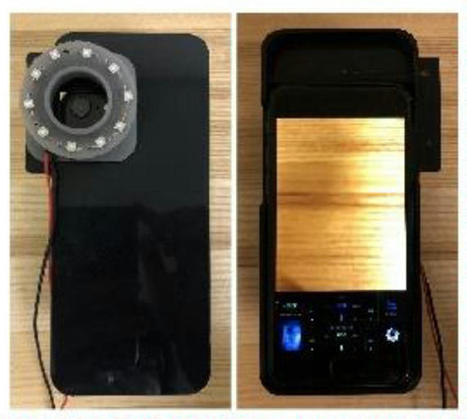
Researchers combined a smartphone-case modification with image-processing methods to illuminate bacteria on images taken by a conventional smartphone camera. This approach yielded a relatively low-cost and quick method that could be used at home.
The team augmented a smartphone camera’s capabilities by attaching a small 3D-printed ring containing 10 LED black lights around a smartphone case’s camera opening. The researchers used the LED-augmented smartphone to take images of the oral cavity and skin on the face of two research subjects.
The LED lights ‘excite’ a class of bacteria-derived molecules called porphyrins, causing them to emit a red fluorescent signal that the smartphone camera can then pick up
Other components in the image—such as proteins or oily molecules our bodies produce, as well as skin, teeth, and gums—won’t glow red under LED. They’ll fluoresce in other colors.
The LED illumination gave the team enough visual information to computationally “convert” the RGB colors from the smartphone-derived images into other wavelengths in the visual spectrum. This generates a “pseudo-multispectral” image consisting of 15 different sections of the visual spectrum—rather than the three in the original RGB image.
Obtaining this visual information up front would have required expensive and cumbersome lights, rather than using the relatively inexpensive LED black lights
With their greater degree of visual discrimination, the pseudo-multispectral images clearly resolved porphyrin clusters on the skin and within the oral cavity. In addition, though they tailored this method to show porphyrin, researchers could modify the image-analysis pipeline to detect other bacterial signatures that also fluoresce under LED.
read the study at https://doi.org/10.1016/j.optlaseng.2021.106546
read the original unedited article at https://www.futurity.org/smartphone-images-skin-mouth-bacteria-2581642/


Researchers have figured out a way to use images from a smartphone to identify potentially harmful bacteria on the skin and in the mouth. A new method using smartphone-derived images can identify potentially harmful bacteria on the skin and in the mouth, research shows. The approach visually identifies microbes on the skin contributing to acne and slow wound healing, as well as bacteria in the oral cavity that can cause gingivitis and dental plaques. Researchers combined a smartphone-case modification with image-processing methods to illuminate bacteria on images taken by a conventional smartphone camera. This approach yielded a relatively low-cost and quick method that could be used at home. Augmenting a smartphone camera’s capabilities by attaching a small 3D-printed ring containing 10 LED black lights around a smartphone case’s camera opening. The researchers used the LED-augmented smartphone to take images of the oral cavity and skin on the face of two research subjects. The LED lights ‘excite’ a class of bacteria-derived molecules called porphyrins, causing them to emit a red fluorescent signal that the smartphone camera can then pick up.
Other components in the image—such as proteins or oily molecules our bodies produce, as well as skin, teeth, and gums—won’t glow red under LED. They’ll fluoresce in other colors.