Your new post is loading...
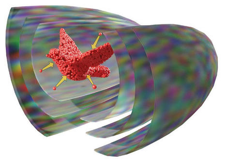
Scientists assemble matter in 3D using sound waves for 3D printing Scientists from the Max Planck Institute for Medical Research and the Heidelberg University have created a new technology to assemble matter in 3D. Their concept uses multiple acoustic holograms to generate pressure fields with which solid particles, gel beads and even biological cells can be printed. These results pave the way for novel 3D cell culture techniques with applications in biomedical engineering. Additive manufacturing or3D printing enables the fabrication of complex parts from functional or biological materials. Conventional 3D printing can be a slow process, where objects are constructed one line or one layer at a time. Researchers in Heidelberg and Tübingen now demonstrate how to form a 3D object from smaller building blocks in just a single step. "We were able to assemble microparticles into a three-dimensional object within a single shot using shaped ultrasound," says Kai Melde, postdoc in the group and first author of the study. "This can be very useful for bioprinting. The cells used there are particularly sensitive to the environment during the process," adds Peer Fischer, Professor at Heidelberg University. Sound waves exert forces on matter -- a fact that is known to any concert goer who experiences the pressure waves from a loudspeaker. Using high-frequency ultrasound, which is inaudible to the human ear, the wavelengths can be pushed below a millimeter into the microscopic realm, which is used by the researcher to manipulate very small building blocks, like biological cells. In their previous studies Peer Fischer and colleagues showed how to form ultrasound using acoustic holograms -- 3D-printed plates, which are made to encode a specific sound field. Those sound fields, they demonstrated, can be used to assemble materials into two-dimensional patterns. Based on this the scientists devised a fabrication concept. Acoustic field catches particles With their new study the team was able to take their concept a step further. They capture particles and cells freely floating in water and assemble them into three-dimensional shapes. On top of that, the new method works with a variety of materials including glass or hydrogel beads and biological cells. First author Kai Melde says that "the crucial idea was to use multiple acoustic holograms together and form a combined field that can catch the particles." Heiner Kremer, who wrote the algorithm to optimize the hologram fields, adds: "The digitization of an entire 3D object into ultrasound hologram fields is computationally very demanding and required us to come up with a new computation routine." The scientists believe that their technology is a promising platform for the formation of cell cultures and tissues in 3D. The advantage of ultrasound is that it is gentle for using biological cells and that it can travel deep into tissue. This way it can be used to remotely manipulate and push cells without harm.
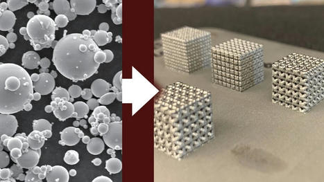
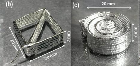
Laser powder bed fusion, a 3D-printing technique, offers potential in the manufacturing industry, particularly when fabricating nickel-titanium shape memory alloys with complex geometries. Although this manufacturing technique is attractive for applications in the biomedical and aerospace fields, it has rarely showcased the superelasticity required for specific applications using nickel-titanium shape memory alloys. Defects generated and changes imposed onto the material during the 3D-printing process prevented the superelasticity from appearing in 3D-printed nickel-titanium. Researchers from Texas A&M University recently showcased superior tensile superelasticity by fabricating a shape memory alloy through laser powder bed fusion, nearly doubling the maximum superelasticity reported in literature for 3D printing. This study was recently published in vol. 229 of the Acta Materialia journal. Nickel-titanium shape memory alloys have various applications due to their ability to return to their original shape upon heating or upon removal of the applied stress. Therefore, they can be used in biomedical and aerospace fields for stents, implants, surgical devices and aircraft wings. However, developing and properly fabricating these materials requires extensive research to characterize functional properties and examine the microstructure. "Shape memory alloys are smart materials that can remember their high-temperature shapes," said Dr. Lei Xue, a former doctoral student in the Department of Materials Science and Engineering and the first author of the publication. "Although they can be utilized in many ways, fabricating shape memory alloys into complex shapes requires fine-tuning to ensure the material exhibits the desired properties."
Following four years of planning and research, the world's first 3D printed footbridge recently opened to the public in Europe.
The almost 40-foot bridge, unveiled last month, was built by Dutch company MX3D and will serve as a "living laboratory" in Amsterdam's city center.
Researchers and engineers at Imperial College London were able to 3D-print the bridge — which now serves pedestrians and cyclists crossing Amsterdam's Oudezijds Achterburgwal canal.
From Guns To Chocolate: The Possibilities Of 3-D Printing
"A 3D-printed metal structure large and strong enough to handle pedestrian traffic has never been constructed before," said Imperial College London professor Leroy Gardner in a news release. A 12-meter 3D-printed pedestrian bridge designed by Joris Laarman and built by Dutch robotics company MX3D has been opened in Amsterdam six years after the project was launched.nAna Fernandez/SOPA Images/LightRocket via Gett Designers first created the concept for the bridge in 2015, with the goal of making an "exceptionally efficient structure," emphasizing both simplicity and safety, according to Popular Mechanics.
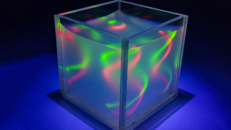
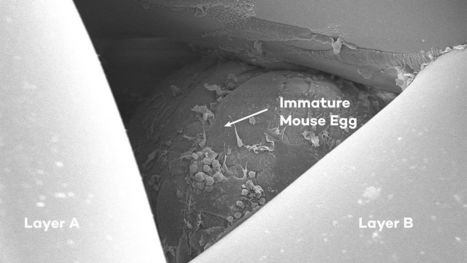
New technique makes it easier to build stable “tissues” 3D-printed tissues and organs could revolutionize transplants, drug screens, and lab models—but replicating complicated body parts such as gastric tracts, windpipes, and blood vessels is a major challenge. That’s because these vascularized tissues are hard to build up in traditional solid layer-by-layer 3D printing without constructing supporting scaffolding that can later prove impossible to remove. One potential solution is replacing these support structures with liquid—a specially designed fluid matrix into which liquid designs could be injected before the “ink” is set and the matrix is drained away. But past attempts to make such aqueous structures have literally collapsed, as their surfaces shrink and their structures crumple into useless blobs. So, researchers from China turned to water-loving, or hydrophilic, liquid polymers that create a stable membrane where they meet, thanks to the attraction of their hydrogen bonds. The researchers say various polymer combinations could work; they used a polyethylene oxide matrix and an ink made of a long carbohydrate molecule called dextran. They pumped their ink into the matrix with an injection nozzle that can move through the liquid and even suck up and rewrite lines that have already been drawn. The resulting liquid structures can hold their shape for as long as 10 days before they begin to merge, the team reported last month in Advanced Materials. Using their new method, the researchers printed an assortment of complex shapes—including tornadoesque whirls, single and double helices (above), branched treelike shapes, and even one that resembles a goldfish. Once printing is finished, the shapes are set by adding polyvinyl alcohol to the inky portion of the structure. That means, the scientists say, that complex 3D-printed tissues made by including living cells in the ink could soon be within our grasp.
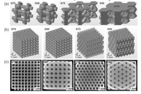
In ‘Application of high resolution DLP stereolithography for fabrication of tricalcium phosphate scaffolds for bone regeneration,’ researchers examine how to make complex, stable scaffolds based on β-tricalcium. Typically, there are obstacles to finding materials and techniques suitable for creating structures capable of sustaining cell life. The authors are aware of the necessities in tissue engineering: the material cannot be toxic, obviously, as that would cause further health issues in a patient, biodegradability is key, with the material being absorbed along with suitable bone growth, and porosity and density must be suitable too, balanced out with proper strength. DLP 3D printing has proven successful for creating scaffolds due to comprehensive irradiation over the whole cross-section, and shorter processing times in comparison to other processes. The researchers focused on DLP 3D printing for this study, in relation to the use of calcium phosphate structures that are not only complex and high resolution but also strong. The team assessed both rectilinear grid structure and hexagonal geometries (at 50 and 75 percent porosity) for mechanical properties, with complete chemical analyses performed before and after bioprinting.
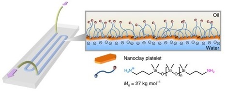
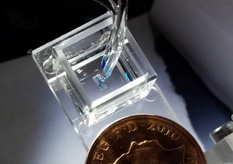
Researchers at Berkeley Lab have 3D-printed an all-liquid "lab on a chip" that can make battery materials or screen drug candidates. Researchers at DOE's Lawrence Berkeley National Laboratory (Berkeley Lab) have 3D-printed an all-liquid device that, with the click of a button, can be repeatedly reconfigured on demand to serve a wide range of applications -- from making battery materials to screening drug candidates. "What we demonstrated is remarkable. Our 3D-printed device can be programmed to carry out multistep, complex chemical reactions on demand," said Brett Helms, a staff scientist in Berkeley Lab's Materials Sciences Division and Molecular Foundry, who led the study. "What's even more amazing is that this versatile platform can be reconfigured to efficiently and precisely combine molecules to form very specific products, such as organic battery materials." The study's findings, which were reported in the journal Nature Communications, is the latest in a series of experiments at Berkeley Lab that fabricate all-liquid materials with a 3D printer. Last year, a study co-authored by Helms and Thomas Russell, a visiting researcher from the University of Massachusetts at Amherst who leads the Adaptive Interfacial Assemblies Toward Structured Liquids Program in Berkeley Lab's Materials Sciences Division, pioneered a new technique for printing various liquid structures -- from droplets to swirling threads of liquid -- within another liquid. "After that successful demonstration, a bunch of us got together to brainstorm on how we could use liquid printing to fabricate a functioning device," said Helms. "Then it occurred to us: If we can print liquids in defined channels and flow contents through them without destroying them, then we could make useful fluidic devices for a wide range of applications, from new types of miniaturized chemical laboratories to even batteries and electronic devices." To make the 3D-printable fluidic device, lead author Wenqian Feng, a postdoctoral researcher in Berkeley Lab's Materials Sciences Division, designed a specially patterned glass substrate. When two liquids -- one containing nanoscale clay particles, another containing polymer particles -- are printed onto the substrate, they come together at the interface of the two liquids and within milliseconds form a very thin channel or tube about 1 millimeter in diameter. Once the channels are formed, catalysts can be placed in different channels of the device. The user can then 3D-print bridges between channels, connecting them so that a chemical flowing through them encounters catalysts in a specific order, setting off a cascade of chemical reactions to make specific chemical compounds. And when controlled by a computer, this complex process can be automated "to execute tasks associated with catalyst placement, build liquid bridges within the device, and run reaction sequences needed to make molecules," said Russell.
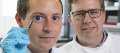
The first human corneas have been 3D printed by scientists at Newcastle University, UK. It means the technique could be used in the future to ensure an unlimited supply of corneas. As the outermost layer of the human eye, the cornea has an important role in focusing vision. Yet there is a significant shortage of corneas available to transplant, with 10 million people worldwide requiring surgery to prevent corneal blindness as a result of diseases such as trachoma, an infectious eye disorder. In addition, almost 5 million people suffer total blindness due to corneal scarring caused by burns, lacerations, abrasion or disease. The proof-of-concept research, published today in Experimental Eye Research, reports how stem cells (human corneal stromal cells) from a healthy donor cornea were mixed together with alginate and collagen to create a solution that could be printed, a 'bio-ink'. Using a simple low-cost 3D bio-printer, the bio-ink was successfully extruded in concentric circles to form the shape of a human cornea. It took less than 10 minutes to print. The stem cells were then shown to culture -- or grow. Che Connon, Professor of Tissue Engineering at Newcastle University, who led the work, said: "Many teams across the world have been chasing the ideal bio-ink to make this process feasible. "Our unique gel -- a combination of alginate and collagen -- keeps the stem cells alive whilst producing a material which is stiff enough to hold its shape but soft enough to be squeezed out the nozzle of a 3D printer. This builds upon our previous work in which we kept cells alive for weeks at room temperature within a similar hydrogel. Now we have a ready to use bio-ink containing stem cells allowing users to start printing tissues without having to worry about growing the cells separately." The scientists, including first author and PhD student Ms Abigail Isaacson from the Institute of Genetic Medicine, Newcastle University, also demonstrated that they could build a cornea to match a patient's unique specifications.
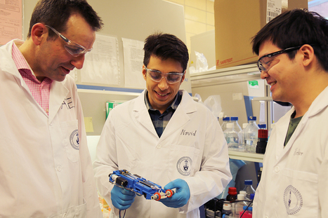
University of Toronto researchers have developed a handheld 3D skin printer that deposits even layers of skin tissue to cover and heal deep wounds. The team believes it to be the first device that forms tissue in situ, depositing and setting in place, within two minutes or less. The research, led by PhD student Navid Hakimi under the supervision of Associate Professor Axel Guenther of the Faculty of Applied Science & Engineering, and in collaboration with Dr. Marc Jeschke, director of the Ross Tilley Burn Centre at Sunnybrook Hospital and professor of immunology at the Faculty of Medicine, was recently published in the journal Lab on a Chip. For patients with deep skin wounds, all three skin layers – the epidermis, dermis and hypodermis – may be heavily damaged. The current preferred treatment is called split-thickness skin grafting, where healthy donor skin is grafted onto the surface epidermis and part of the underlying dermis. Split-thickness grafting on large wounds requires enough healthy donor skin to traverse all three layers, and sufficient graft skin is rarely available. This leaves a portion of the wounded area “ungrafted” or uncovered, leading to poor healing outcomes.
Edinburgh-based Skyrora, a company with partners in Ukraine, will launch a partially 3D printed suborbital vehicle from the north of Scotland later this year. Skyrora’s rocket engines run on hydrogen peroxide and kerosene. America, Russia, China, and… Scotland? Yes, the newest contender in the space race is that tiny northern country of the United Kingdom, Scotland, whose very own Skyrora has developed a suborbital launch vehicle that will take off from northern Scotland in the last three months of 2018. It’s a great achievement for the company, which also operates from Ukraine and which has used 3D printing to develop parts for its spacecraft. Potential launch locations include Shetland, where the Shetland Space Centre is bidding for a license from the UK Space Agency. If the company can secure a launch location, its partly 3D printed suborbital launch vehicle could be taking off within the year.
Via Aso Galicia
By printing custom bone prostheses, researchers hope they can better fix a certain kind of hearing loss. The auditory ossicles of the middle ear – the malleus, incus and stapes – are the tiniest bones in the human body. All three can fit on a dime, with room to spare. Their job is to transmit sounds from the ear drum to the liquid of the inner ear. Illnesses, accidents and tumors can damage these bones, causing what’s known as “conductive hearing loss.” The remedy is a delicate surgery, in which the bones are replaced with a tiny prosthesis. But the surgery has a relatively high failure rate, about 25 to 50 percent. Now, researchers at the University of Maryland Medical Center are using 3D printers to make custom-fitted ear bones. They hope these prostheses will improve on the current technology and raise the success rate of surgery. The team, made up of a radiologist and two ear, nose and throat doctors, took the ossicles from three human cadavers and removed the middle bones, or incuses. They then used a CT scanner to take images of the gaps left by the incuses, and designed tiny prostheses to fit those gaps. The prostheses varied by just fractions of millimeters, with ever so slightly different angles. The researchers then gave four different surgeons the three prostheses and had them guess which one went in which ear. Each surgeon independently matched the prostheses to the correct ears. “They said it wasn’t that hard to figure out,” says Jeffrey Hirsch, a radiology professor who led the research. “It was almost like a Goldilocks sort of thing – this prosthesis was too tight in this ear and too loose in this ear, but in this ear it’s just right.” The research was published recently in the journal 3D Printing in Medicine. The next step will be to test the prostheses for function using cadavers or animal models. They can run vibrations through a prosthesis to see how it transmits sound.
Imagine a bottle of laundry detergent that can sense when you're running low on soap—and automatically connect to the internet to place an order for more. University of Washington researchers are the first to make this a reality by 3-D printing plastic objects and sensors that can collect useful data and communicate with other WiFi-connected devices entirely on their own. With CAD models that the team is making available to the public, 3-D printing enthusiasts will be able to create objects out of commercially available plastics that can wirelessly communicate with other smart devices. That could include a battery-free slider that controls music volume, a button that automatically orders more cornflakes from Amazon or a water sensor that sends an alarm to your phone when it detects a leak. "Our goal was to create something that just comes out of your 3-D printer at home and can send useful information to other devices," said co-lead author and UW electrical engineering doctoral student Vikram Iyer. "But the big challenge is how do you communicate wirelessly with WiFi using only plastic? That's something that no one has been able to do before." The system is described in a paper presented Nov. 30 at the Association for Computing Machinery's SIGGRAPH Conference and Exhibition on Computer Graphics and Interactive Techniques in Asia.
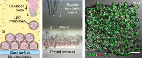
Scientists at the University of Oxford have developed a radical new method of 3D-printing laboratory-grown cells that can form complex living tissues and cartilage to potentially support, repair, or augment diseased and damaged areas of the body. Printing high-resolution living tissues is currently difficult because the cells often move within printed structures and can collapse on themselves. So the team devised a new way to produce tissues in protective nanoliter droplets wrapped in a lipid (oil-compatible) coating that is assembled, layer-by-layer, into living cellular structures. This new method improves the survival rate of the individual cells and allows for building each tissue one drop at a time to mimic the behaviors and functions of the human body. The patterned cellular constructs, once fully grown, can mimic or potentially enhance natural tissues. “We were aiming to fabricate three-dimensional living tissues that could display the basic behaviors and physiology found in natural organisms,” explained Alexander Graham, PhD, lead author and 3D Bioprinting Scientist at OxSyBio (Oxford Synthetic Biology). “To date, there are limited examples of printed tissues [that] have the complex cellular architecture of native tissues. Hence, we focused on designing a high-resolution cell printing platform, from relatively inexpensive components, that could be used to reproducibly produce artificial tissues with appropriate complexity from a range of cells, including stem cells.” The researchers hope that with further development, the materials could have a wide impact on healthcare worldwide and bypass clinical animal testing. The scientists plan to develop new complementary printing techniques that allow for a wider range of living and hybrid materials, producing tissues at industrial scale.
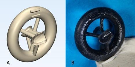
Fans of 3D printing say it has the potential to revolutionize medicine—think 3D-printed skin,ears, bone scaffolds, and heart valves. Now, prosthetic ovaries made of gelatin have allowed mice to conceive and give birth to healthy offspring. Such engineered ovaries could one day be used to help restore fertility in cancer survivors rendered sterile by radiation or chemotherapy. This “landmark study” is a “significant advance in the application of bioengineering to reproductive tissues,” says Mary Zelinski, a reproductive scientist at the Oregon National Primate Research Center in Beaverton who was not involved with the work. The researchers used a 3D printer with a nozzle that fired gelatin, derived from the collagen that’s naturally found in animal ovaries. The scientists built the ovaries by printing various patterns of overlapping gelatin filaments on glass slides—like building with Lincoln Logs, but on a miniature scale: Each scaffold measured just 15 by 15 millimeters. The team then carefully inserted mouse follicles—spherical structures containing a growing egg surrounded by hormone-producing cells—into these “scaffolds.” The scaffolds that were more tightly woven hosted a higher fraction of surviving follicles after 8 days, an effect the team attributes to the follicles having better physical support. The researchers then tested the more tightly woven scaffolds in live mice. The researchers punched out 2-millimeter circles through the scaffolds and implanted 40–50 follicles into each one, creating a “bioprosthetic” ovary. They then surgically removed the ovaries from seven mice and sutured the prosthetic ovaries in their place. The team showed that blood vessels from each mouse infiltrated the scaffolds. This vascularization is critical because it provides oxygen and nutrients to the follicles and allows hormones produced by the follicles to circulate in the blood stream. The researchers allowed the mice to mate, and three of the females gave birth to healthy litters, the team reports today in Nature Communications. The mice that gave birth also lactated naturally, which demonstrated that the follicles embedded in the scaffolds produced normal levels of hormones.
Via THE OFFICIAL ANDREASCY
|
Nature has an extraordinary knack for producing composite materials that are simultaneously light and strong, porous and rigid – like mollusk shells or bone. But producing such materials in a lab or factory – particularly using environmentally friendly materials and processes – is extremely challenging. Researchers in the Soft Materials Laboratory in the School of Engineering turned to nature for a solution. They have pioneered a 3D printable ink that contains Sporosarcina pasteurii: a bacterium which, when exposed to a urea-containing solution, triggers a mineralization process that produces calcium carbonate (CaCO3). The upshot is that the researchers can use their ink – dubbed BactoInk – to 3D-print virtually any shape, which will then gradually mineralize over the course of a few days. “3D printing is gaining increasing importance in general, but the number of materials that can be 3D printed is limited for the simple reason that inks must fulfil certain flow conditions,” explains lab head Esther Amstad. “For example, they must behave like a solid when at rest, but still be extrudable through a 3D printing nozzle – sort of like ketchup.” Amstad explains that 3D printing inks containing small mineral particles have previously been used to meet some of these flow criteria, but that the resulting structures tend to be soft, or to shrink upon drying, leading to cracking and loss of control over the shape of the final product. “So, we came up with a simple trick: instead of printing minerals, we printed a polymeric scaffold using our BactoInk, which is then mineralized in a second, separate step. After about four days, the mineralization process triggered by the bacteria in the scaffold leads to a final product with a mineral content of over 90%.” The result is a strong and resilient bio-composite, which can be produced using a standard 3D printer and natural materials, and without the extreme temperatures often required for manufacturing ceramics. Final products no longer contain living bacteria, as they are submerged in ethanol at the end of the mineralization process. The method, which describes the first 3D printing ink that uses bacteria to induce mineralization, has recently been published in the journal Materials Today.
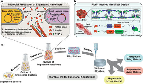
A team of researchers from Harvard University and Brigham and Women's Hospital, Harvard Medical School, has developed a type of living ink that can be used to print living materials. In their paper published in the journal Nature Communications, the group describes how they made their ink and possible uses for it. For several years, microbial engineers have been working to develop a means to create living materials for use in a wide variety of applications such as medical devices. But getting such materials to conform to desired 3D structures has proven to be a daunting task. In this new effort, the researchers have taken a new approach to tackling the problem—engineering E. coli to produce a product that can be used as the basis for an ink for use in a 3D printer. The work began by bioengineering the bacteria to produce living nanofibers. The researchers then bundled the fibers and added other ingredients to produce a type of living ink that could be used in a conventional 3D printer. Once they found the concept viable, the team bioengineered other microbes to produce other types of living fibers or materials and added them to the ink. They then used the ink to print 3D objects that had living components. One was a material that secreted azurin—an anticancer drug—when stimulated by certain chemicals. Another was a material that sequestered Bisphenol A (a toxin that has found its way into the environment) without assistance from other chemicals or devices. The researchers believe that their concept suggests that producing such inks could be a self-creating proposition. Engineering could be added to the microbes to push them to produce carbon copies of themselves—the ink could literally be grown in a jar. They also state that it appears possible that the technique could be used to print renewable building materials that would not only grow but could self heal—a possible approach to building self-sustaining homes here on Earth, or on the moon or on Mars.
Repairing and reusing plastics and delivering cancer drugs more effectively are only two of many of the potential applications a new 3D/4D printing technology offers, thanks to the pioneering work of a research collaboration between UNSW Sydney and The University of Auckland. The researchers of this project have revealed the successful merging of 3D/4D printing and photo-controlled/living polymerization - a chemical process to create polymers - in a paper published in Angewandte Chemie, International Edition. 4D printing is a subset of 3D printing, where the printed object can transform its shape in response to certain conditions. The new controlled polymerization method, where the researchers used visible light to create an environmentally friendly "living" plastic or polymer, opens up a new world of possibilities for the manufacture of advanced solid materials. Since this development, the technology has expanded and has proven useful for making well-controlled molecules for many different applications, including drug delivery and other biomaterials. Polymers can be synthetic, such as plastic, as well as biological, for example, DNA or RNA. Lead author Cyrille Boyer said his team's latest breakthrough was a world first in the development of a new 3D printing system using PET-RAFT polymerization, to allow 3D printed materials to be easily modified after printing. Controlled polymerization has never been used in 3D and 4D printing before, because the rates of typical controlled polymerization processes are too slow for 3D/4D printing, where the reaction must be fast for practical printing speeds. Such polymers can be reactivated for further growth, unlike traditional polymers which are "dead" after being made. After two years of research and hundreds of experiments, we developed a rapid process compatible with 3D printing. In contrast to conventional 3D printing, our new method of using visible light allows us to control the architecture of the polymers and tune the mechanical properties of the materials prepared by our process. This also gives us access to 4D printing and allows the material to be transformed or functionalized, which was not previously possible." UNSW's Nathaniel Corrigan, co-first author with UNSW PhD candidate Zhiheng Zhang, said a bonus advantage of their new system was the ability to finely control all molecules in the 3D-printed material could reversibly change its shape when it was exposed to water and then dried. For example, the 3D object starts as a flat plane and when exposed to certain conditions, it will start to fold - that's a 4D material. So, the fourth dimension is time.
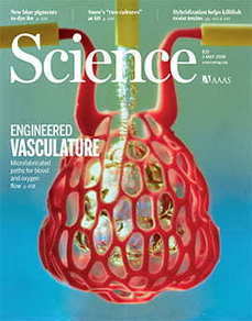
Bioengineers have cleared a major hurdle on the path to 3D printing replacement organs. It's a breakthrough technique for bioprinting tissues with exquisitely entangled vascular networks that mimic the body's natural passageways for blood, air, lymph and other vital fluids. The new innovation allows scientists to create exquisitely entangled vascular networks that mimic the body's natural passageways for blood, air, lymph and other vital fluids. The research is featured on the cover of this week's issue of Science. It includes a visually stunning proof-of-principle -- a hydrogel model of a lung-mimicking air sac in which airways deliver oxygen to surrounding blood vessels. Also reported are experiments to implant bioprinted constructs containing liver cells into mice. The work was led by bioengineers Jordan Miller of Rice University and Kelly Stevens of the University of Washington (UW) and included 15 collaborators from Rice, UW, Duke University, Rowan University and Nervous System, a design firm in Somerville, Massachusetts. "One of the biggest road blocks to generating functional tissue replacements has been our inability to print the complex vasculature that can supply nutrients to densely populated tissues," said Miller, assistant professor of bioengineering at Rice's Brown School of Engineering. "Further, our organs actually contain independent vascular networks -- like the airways and blood vessels of the lung or the bile ducts and blood vessels in the liver. These interpenetrating networks are physically and biochemically entangled, and the architecture itself is intimately related to tissue function. Ours is the first bioprinting technology that addresses the challenge of multivascularization in a direct and comprehensive way." Stevens, assistant professor of bioengineering in the UW College of Engineering, assistant professor of pathology in the UW School of Medicine, and an investigator at the UW Medicine Institute for Stem Cell and Regenerative Medicine, said multivascularization is important because form and function often go hand in hand. "Tissue engineering has struggled with this for a generation," Stevens said. "With this work we can now better ask, 'If we can print tissues that look and now even breathe more like the healthy tissues in our bodies, will they also then functionally behave more like those tissues?' This is an important question, because how well a bioprinted tissue functions will affect how successful it will be as a therapy."
The fund-raising event for the Metropolitan Museum of Art in New York is famous for the impressive, wild, and avant-garde looks worn by some of the biggest celebrities. This year, five of the outfits flaunted massive 3D-printed components, all created by designer Zac Posen in collaboration with GE Additive and Protolabs. The four gowns and one headdress, worn by the likes of Jourdan Dunn and Katie Holmes, took six months to put together. Each of the women wearing the 3D-printed outfits was scanned in advance to ensure that the pieces could be modeled with design software to perfectly fit her body. The pieces were primarily created using stereolithography (SLA) to get the high-quality finishes Posen was seeking. This method of 3D printing uses an ultraviolet laser to slowly cure a pool of resin. One layer at a time, the laser fires into the resin in a pattern dictated by a computer in which the 3D model has been loaded. The result is a hardened, smooth sculpture that is then painted or polished. The most intensive look, modeled after rose petals and worn by British model Jourdan Dunn, took more than 1,100 hours to create. “We never intended for these parts to be easy, and we wanted to push the limit a bit,” says Eric Utley, an applications engineer at Protolabs.
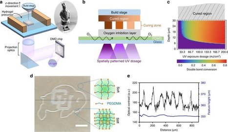
University of Colorado Boulder engineers have developed a 3-D printing technique that allows for localized control of an object's firmness, opening up new biomedical avenues that could one day include artificial arteries and organ tissue. The study, which was recently published in the journal Nature Communications, outlines a layer-by-layer printing method that features fine-grain, programmable control over rigidity, allowing researchers to mimic the complex geometry of blood vessels that are highly structured and yet must remain pliable. The findings could one day lead to better, more personalized treatments for those suffering from hypertension and other vascular diseases. "The idea was to add independent mechanical properties to 3-D structures that can mimic the body's natural tissue," said Xiaobo Yin, an associate professor in CU Boulder's Department of Mechanical Engineering and the senior author of the study. "This technology allows us to create microstructures that can be customized for disease models." Hardened blood vessels are associated with cardiovascular disease, but engineering a solution for viable artery and tissue replacement has historically proven challenging. To overcome these hurdles, the researchers found a unique way to take advantage of oxygen's role in setting the final form of a 3-D-printed structure. "Oxygen is usually a bad thing in that it causes incomplete curing," said Yonghui Ding, a postdoctoral researcher in Mechanical Engineering and the lead author of the study. "Here, we utilize a layer that allows a fixed rate of oxygen permeation." By keeping tight control over oxygen migration and its subsequent light exposure, Ding said, the researchers have the freedom to control which areas of an object are solidified to be harder or softer—all while keeping the overall geometry the same. "This is a profound development and an encouraging first step toward our goal of creating structures that function like a healthy cell should function," Ding said. As a demonstration, the researchers printed three versions of a simple structure: a top beam supported by two rods. The structures were identical in shape, size and materials, but had been printed with three variations in rod rigidity: soft/soft, hard/soft and hard/hard. The harder rods supported the top beam while the softer rods allowed it to fully or partially collapse. The researchers repeated the feat with a small Chinese warrior figure, printing it so that the outer layers remained hard while the interior remained soft, leaving the warrior with a tough exterior and a tender heart, so to speak. The tabletop-sized printer is currently capable of working with biomaterials down to a size of 10 microns, or about one-tenth the width of a human hair. The researchers are optimistic that future studies will help improve the capabilities even further.
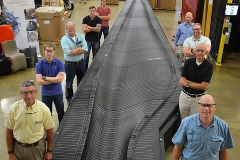
In collaboration with Oak Ridge National Laboratory's Manufacturing Demonstration Facility Team and turbine blade manufacturer TPI Composites, Sandia National Laboratories 3D printed a massive mold to produce wind turbine blades. Sandia researchers have been working on wind turbines for the better part of 40 years; it’s part of the lab's effort to make the renewable energy more affordable. However, building wind turbine prototypes takes a lot of time and effort, and each requires custom molds that take up to 16 months to complete before the blade can be developed and tested. Through the use of 3D printing, the team was able to cut mold development time by more than 80 percent, going from 16 months of development time down to 3 months. The work cut out more than a year of labor. The 13-meter blade mold is relatively small compared to other blades currently on the market and under development — for example, GE's Haliade-X blades will be 107 meters long. However, by cutting design and development time and cost, engineers could take greater risks during the prototype phase that could potentially accelerate innovation in the market.
Via Alan Charky
Researchers in Oregon State University’s College of Engineering have taken a key step toward the rapid manufacture of flexible computer screens and other stretchable electronic devices, including soft robots. The advance by a team within the college’s Collaborative Robotics and Intelligent Systems Institute paves the way toward the 3D printing of tall, complicated structures with a highly conductive gallium alloy. Researchers put nickel nanoparticles into the liquid metal, galinstan, to thicken it into a paste with a consistency suitable for additive manufacturing. “The runny alloy was impossible to layer into tall structures,” said Yiğit Mengüç, assistant professor of mechanical engineering and co-corresponding author on the study. “With the paste-like texture, it can be layered while maintaining its capacity to flow, and to stretch inside of rubber tubes. We demonstrated the potential of our discovery by 3D printing a very stretchy two-layered circuit whose layers weave in and out of each other without touching.” Findings were recently published in Advanced Materials Technologies. Gallium alloys are already being used as the conductive material in flexible electronics; the alloys have low toxicity and good conductivity, plus they’re inexpensive and “self-healing” – able to attach back together at break points. But prior to the modification developed at OSU, which used sonication – the energy of sound – to mix the nickel particles and the oxidized gallium into the liquid metal, the alloys’ printability was restricted to 2-dimensional. For this study, researchers printed structures up to 10 millimeters high and 20 millimeters wide. “Liquid metal printing is integral to the flexible electronics field,” said co-author Doğan Yirmibeşoğlu, a robotics Ph.D. student at OSU. “Additive manufacturing enables fast fabrication of intricate designs and circuitry.”
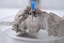
Have you ever taken your old compact discs and converted them to MP3 files so you could listen to your favorite music on your laptop, or through a portable MP3 device that’s much smaller than an unwieldy portable CD player? Now, researchers from the University of Glasgow are working on a very similar process, but instead of music files, they are using a chemical-to-digital converter to digitize the process of drug manufacturing; a chemical MP3 player, if you will, that can 3D print pharmaceuticals on demand. 3D printing in the pharmaceutical field is a fascinating concept, though not a new one. But this ‘Spotify for chemistry’ concept is new: it’s the first time we’ve seen an approach to manufacturing pharmaceuticals using digital code. According to Science, the University of Glasgow team “tailored a 3D printer to synthesize pharmaceuticals and other chemicals from simple, widely available starting compounds fed into a series of water bottle–size reactors.”
Via Mariaschnee
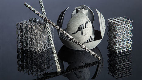
A new method by which to 3D print metals, involving an extensively used stainless steel, has been shown to realize exceptional levels of both ductility and strength, when compared to counterparts from more conventional processes. The research is opposing to the skepticism surrounding the ability to make robust and ductile metals using 3D printing, and as such the discovery is vital to moving the technology forward for the manufacturing of heavy duty components. 3D printing has long been accepted as a technology which can possibly transform the way of manufacturing, allowing one to quickly construct objects with intricate and tailored geometries. With the technology rapidly developing in recent years, 3D printing, particularly metal 3D printing, is swiftly progressing toward extensive industrial application. Indeed, the manufacturing leader General Electric (GE) has already been using metal 3D printing to create certain key parts, such as the fuel nozzles in their newest LEAP aircraft engine. The technology helps GE to minimize 900 separate parts into just 16, and make fuel nozzles 60% cheaper and 40% lighter. The worldwide revenue from the industry is predicted to be more than 20 billion USD per year by the year 2025. Regardless of the bright future, the quality of the products from metal 3D printing has been susceptible to skepticism. In majority of metal 3D printing processes, products are directly made from metal powders, which make it prone to defects, therefore causing weakening of mechanical properties. Dr. Leifeng Liu, who is the key participant of the project, lately moved to the University of Birmingham from Stockholm University as an AMCASH research fellow. He said, “Strength and ductility are natural enemies of one another, most methods developed to strengthen metals consequently reduce ductility.”
Via Alan Charky
Scientists at the University of Oxford have developed a new method to 3D-print laboratory-grown cells to form living structures. The approach could revolutionize regenerative medicine, enabling the production of complex tissues and cartilage that would potentially support, repair, or augment diseased and damaged areas of the body. Published in the journal Scientific Reports, an interdisciplinary team from the Department of Chemistry and the Department of Physiology, Anatomy, and Genetics at Oxford and the Centre for Molecular Medicine at Bristol, demonstrated how a range of human and animal cells can be printed into high-resolution tissue constructs. Interest in 3D printing living tissues has grown in recent years, but, developing an effective way to use the technology has been difficult, particularly since accurately controlling the position of cells in 3D is hard to do. They often move within printed structures and the soft scaffolding printed to support the cells can collapse on itself. As a result, it remains a challenge to print high-resolution living tissues. But, led by Hagan Bayley, professor of chemical biology in Oxford's Department of Chemistry, the team devised a way to produce tissues in self-contained cells that support the structures to keep their shape. The cells were contained within protective nanoliter droplets wrapped in a lipid coating that could be assembled, layer-by-layer, into living structures. Producing printed tissues in this way improves the survival rate of the individual cells, and allowed the team to improve on current techniques by building each tissue one drop at a time to a more favorable resolution. To be useful, artificial tissues need to be able to mimic the behaviors and functions of the human body. The method enables the fabrication of patterned cellular constructs, which, once fully grown, mimic or potentially enhance natural tissues. Dr. Alexander Graham, lead author and 3D bioprinting scientist at OxSyBio (Oxford Synthetic Biology), said: "We were aiming to fabricate three-dimensional living tissues that could display the basic behaviors and physiology found in natural organisms. To date, there are limited examples of printed tissues, which have the complex cellular architecture of native tissues. Hence, we focused on designing a high-resolution cell printing platform, from relatively inexpensive components, that could be used to reproducibly produce artificial tissues with appropriate complexity from a range of cells including stem cells."
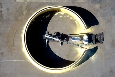
MIT researchers have developed a system that can 3-D print the basic structure of an entire building. MIT's system is a massive robotic arm attached to a track vehicle. The arm is fitted with nozzles that can spray foam insulation on the ground and fill the area in with concrete. It is operated electrically and can harvest its power from the sun using solar panels. A scoop attached to the robot lets it prepare the building surface and acquire local materials, such as dirt for a rammed-earth building, for the construction itself. To demonstrate the technology, the team constructed the walls of a 50-foot-diameter, 12-foot-high dome in 14 hours of 'printing' time. Although this technology could change how humans build homes on Earth, the team foresees this system being useful when we go to Mars, as it can create small dwellings in less than 24 hours.
|